Article

The First Non-LRV RNA Virus in Leishmania

1
Life Science Research Centre, Faculty of Science, University of Ostrava, 71000 Ostrava, Czech Republic
2
Central European Institute of Technology, Masaryk University, 60177 Brno, Czech Republic
3
Martsinovsky Institute of Medical Parasitology, Sechenov University, Moscow 119435, Russia
4
Division of Biomedical and Life Sciences, Faculty of Health and Medicine, Lancaster University, Lancaster LA1 4YE, UK
5
Laboratory of Electron Microscopy, Veterinary Research Institute, 62100 Brno, Czech Republic
6
Department of Parasitology, Faculty of Science, Charles University, 12844 Prague, Czech Republic
7
Laboratory of Cellular and Molecular Protistology, Zoological Institute of the Russian Academy of Sciences, St. Petersburg 199034, Russia
*
Authors to whom correspondence should be addressed.
These authors contributed equally to this work.
Viruses 2020, 12(2), 168; https://doi.org/10.3390/v12020168
Submission received: 6 January 2020 / Revised: 21 January 2020 / Accepted: 29 January 2020 / Published: 2 February 2020
(This article belongs to the Section Viruses of Plants, Fungi and Protozoa)

Abstract

:
In this work, we describe the first Leishmania-infecting leishbunyavirus—the first virus other than Leishmania RNA virus (LRV) found in trypanosomatid parasites. Its host is Leishmania martiniquensis, a human pathogen causing infections with a wide range of manifestations from asymptomatic to severe visceral disease. This virus (LmarLBV1) possesses many characteristic features of leishbunyaviruses, such as tripartite organization of its RNA genome, with ORFs encoding RNA-dependent RNA polymerase, surface glycoprotein, and nucleoprotein on L, M, and S segments, respectively. Our phylogenetic analyses suggest that LmarLBV1 originated from leishbunyaviruses of monoxenous trypanosomatids and, probably, is a result of genomic re-assortment. The LmarLBV1 facilitates parasites’ infectivity in vitro in primary murine macrophages model. The discovery of a virus in L. martiniquensis poses the question of whether it influences pathogenicity of this parasite in vivo, similarly to the LRV in other Leishmania species.

1. Introduction

Bunyavirales is an order of negative-sense single-stranded RNA (-ssRNA) viruses [1]. They typically have three genomic segments (large, L; medium, M; small, S) encoding a viral RNA-dependent RNA polymerase L (RDRP L), a surface glycoprotein precursor, and a nucleoprotein, respectively [2]. Additional ORFs, usually involved in counteracting the host antiviral response, may be present in S or M segments [3,4]. Each viral segment has terminal complementary sequences governing its interaction with the polymerase. Furthermore, multiple molecules of a nucleoprotein wrap around genomic RNA following helical symmetry [4]. Together, an RNA molecule, a polymerase, and the nucleoproteins form a functional viral ribonucleoprotein (vRNP) capable of transcription and replication [5]. Virions are usually 90–100 nm in diameter and consist of vRNPs of each genomic segment enclosed by a lipid membrane with incorporated viral glycoproteins [3]. Many bunyaviruses (a generic term for Bunyavirales) are causative agents of arthropod-borne diseases of vertebrates and plants [6].
Recent metatranscriptomic studies revealed a plethora of deep branching bunyaviruses from vertebrates and invertebrates, suggesting a long-term coevolution of these viruses with their hosts and vectors [7,8,9]. Of note, some bunyaviruses are capable of infecting distantly related eukaryotic cells. For example, Orthotospovirus (the tomato spotted wilt virus (Bunyavirales, Tospoviridae)) can replicate in both plant and insect cells [10,11].
The kinetoplastid flagellates of the family Trypanosomatidae are a eukaryotic group, whose viruses recently started attracting attention [12]. Trypanosomatids are obligate parasites of invertebrates, vertebrates, and plants [13]. They either have one or two hosts in their life cycle (monoxenous and dixenous species, respectively) [14,15,16]. Dixenous trypanosomatids originate from their monoxenous relatives and many of them are of medical or economic importance [17,18,19].
Members of the genus Leishmania infect vertebrates; they are transmitted by phlebotomine sand flies or, possibly, biting midges and cause a variety of diseases collectively named leishmaniases [20]. These diseases manifest with a wide spectrum of clinical symptoms from relatively harmless skin lesions to fatal cases involving failure of visceral organs. Currently, the genus Leishmania is subdivided into four subgenera: Leishmania (Leishmania), L. (Mundinia), L. (Sauroleishmania), and L. (Viannia) [21,22]. These groups are phylogenetically distinct and differ in host specificity or clinical symptoms. The recently established subgenus Mundinia is the most understudied one [23,24].
Thus far, only the representatives of the subgenera Viannia and Leishmania were extensively screened for viral presence, resulting in the discovery of Leishmania RNA viruses (LRVs). The first virus of this group was documented in L. (V.) guyanensis more than 30 years ago [25]. This double-stranded RNA (dsRNA) virus is classified as Leishmaniavirus within the family Totiviridae based on sequence similarity to the yeast L-A totivirus [26]. The genus Leishmaniavirus is subdivided into LRV1, infecting New World Leishmania (Viannia) [27,28], and LRV2 described from Old World Leishmania (Leishmania) [29,30,31]. Recently, new representatives of this viral genus were unexpectedly found in unrelated trypanosomatids, members of the monoxenous genus Blechomonas parasitizing fleas [32].
An increased interest in leishmaniaviruses was stimulated by the discovery that LRV1 presence may augment pathogenicity of some New World Leishmania species. It was shown that viral dsRNA interacts with Toll-like receptor 3 (TLR3) in the parasitophorous vacuole of a macrophage, initiating production of pro-inflammatory cytokines, including interferon-β [33] and subverts innate immunity via TLR3-mediated NLRP3 (NACHT, LRR and PYD domains-containing protein 3) inhibition of inflammasomes. This, in turn, leads to chronic inflammation that counteracts anti-leishmanial immune response and contributes to the metastatic potential of Leishmania [34,35]. It is argued that in this way the virus confers a selective advantage to Leishmania, resulting in its retention [36,37]. Only two strains of L. (Mundinia) enriettii were tested for LRV presence by PCR and both documented as negative [23].
No viruses other than LRVs were found in Leishmania spp [12]. At the same time, recent studies reveal numerous bunyaviruses infecting other trypanosomatids, including monoxenous relatives of Leishmania [38,39]. They all have a typical tripartite genome arrangement, although their M segment is markedly reduced in size and amino acid sequences of the M-encoded putative glycoprotein are extremely divergent. Sequences of their RDRPs and terminal complementary repeats are closest to those of Phenuiviridae. Leishbunyaviruses (LBVs, proposed family Leishbunyaviridae) form a single and well separated clade on a Bunyavirales tree, suggesting that they acquired the ability to infect trypanosomatids only once. Comparison of LBV and trypanosomatid phylogenies revealed cases of both co-evolution and horizontal viral transmissions [32,38].
In this work, we describe the first Leishmania-infecting leishbunyavirus as the first non-LRV virus in trypanosomatids of this genus.

2. Materials and Methods

2.1. Parasite Culture, DNA Isolation, and Verification of Species Identity

The following Leishmania (Mundinia) strains were used in this study: L. (M.) enriettii MCAV/BR/45/LV90, L. (M.) macropodum MMAC/AU/2004/AM-2004, L. (M.) orientalis MHOM/TH/2007/PCM2, and L. (M.) martiniquensis MHOM/MQ/92/MAR1. Promastigotes were cultured in modified M199 media supplemented with 1 mg/mL biotin, 0.5 mg/mL biopterin (both from Sigma-Aldrich, St. Louis, MO, USA), 2.5 µg/mL of hemin (Jena Bioscience GmbH, Jena, Germany), 1× MEM vitamin solution, 10% heat-inactivated fetal bovine serum, 500 units/mL of penicillin, and 0.5 µg/mL of streptomycin (all from Thermo Fisher Scientific, Waltham, MA, USA).
Total genomic DNA was isolated from 10 mL of log-phase trypanosomatid cultures with the DNeasy Blood & Tissue Kit (Qiagen, Hilden, Germany) according to the manufacturer’s instructions. Small subunit rRNA gene was amplified using primers S762 and S763 [40], following the previously described protocol [41]. The obtained PCR fragments were sequenced directly at Macrogen Europe (Amsterdam, The Netherlands) using the primers 883F, 907R, S757, and A757 [42]. The identity of the strains was confirmed by BLAST analysis [43].

2.2. DsRNA Isolation and Next-Generation Sequencing

Total RNA was extracted from 108 cells using TRIzol (Thermo Fisher Scientific), following the manufacturer’s guidelines. The dsRNA fraction was isolated from 200 µg of total RNA using the previously described DNase-S1 nuclease method [38] and visualized in 0.8% agarose gels. The abundance of fragments was analyzed using GeneTools v. 4.3.9 (Syngene, Cambridge, UK). RiboMinus libraries, prepared from the dsRNA sample, were sequenced on the Illumina HiSeq 2500 platform (Illumina, San Diego, CA, USA) at Macrogen Inc. (Seoul, South Korea).

2.3. Viral Sequence Assembly

Transcriptome assembly was carried out essentially as described earlier [32]. In brief, reads were trimmed with Trimmomatic v. 0.36 [44] and assembled de novo using Trinity v. 2.4.0 [45]. Minimal k-mer was set to 5, and other parameters were not changed. Read mapping was performed in Bowtie2 v. 2.3.4.1 [46] and SAMtools v. 1.8 [47], and the coverage was calculated using BEDTools v. 2.25 software [48]. Viral segments were identified by BLAST searches of the 100 most abundant transcripts. Borders of viral segments were determined based on coverage value (with 10 reads per base as the threshold) and presence of specific terminal sequences. To obtain the terminal complementary sequences, original reads were trimmed with BBduk and mapped with BBmap (https://jgi.doe.gov/data-and-tools/bbtools/) to viral contigs assembled previously. GenBank accession numbers for the L, M, and S segment sequences are MK356554, MK356555, and MK356556, respectively.

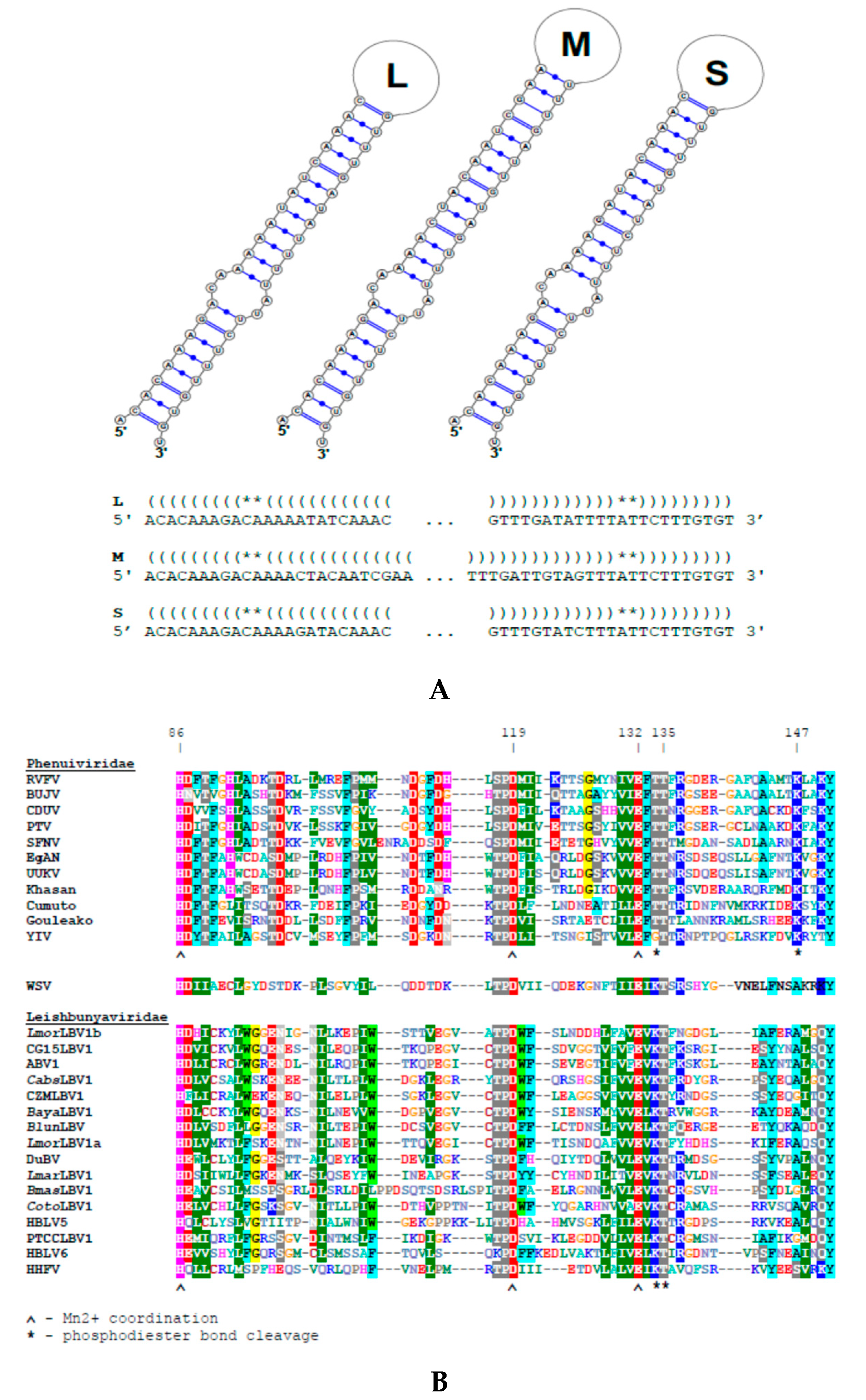
2.4. Prediction of Functional Elements

The search for ORFs in the viral contigs was performed using NCBI ORFfinder [49] with the minimal ORF length set to 150 nt. The identification of the RDRP domain was done using the NCBI Conserved Domain Search [50]. Predictions of the transmembrane domains and membrane-targeting signal peptides were made using the TMHMM v. 2.0 (www.cbs.dtu.dk/services/TMHMM/), TMPred [51], Phobius [52], MEMSTAT3 on PSIPRED server [53], and SignalP v. 4.1 [54] software packages. N-glycosylation sites were identified with NetNGlyc 1.0 Server (www.cbs.dtu.dk/services/NetNGlyc/).

2.5. Phylogenetic Analyses

Full-length amino acid sequences of Leishbunyaviridae and Phenuiviridae RDRPs were aligned using MAFFT v. 7.313 E-INS-i algorithm [55]. The alignment was trimmed in TrimAl v. 1.4 with “automated1” algorithm [56], producing a matrix with 1772 amino acid positions that was used for phylogenetic reconstructions. Maximum likelihood analysis was performed in IQ-TREE v. 1.6.1 [57]. The best amino acid substitution model, LG with rate heterogeneity across sites approximated using proportion of invariant sites and 4 categories of discrete Γ distribution (+ I + G4), as well as the empirical amino acid frequencies (+ F), was selected by both corrected Akaike information criterion and Bayesian information criterion in the built-in ModelFinder [58]. Statistical supports for the branches were generated by running 1000 thorough bootstrap replicates. Bayesian inference was accomplished in MrBayes v. 3.2.6 [59] with the same substitution model and estimated during the run using “mixed” prior (resulting in 1.0 posterior probability of LG) and other model parameters specified above. The analysis was run for 1,000,000 Monte-Carlo Markov chain generations with default settings. For the comparison of nucleoproteins and glycoproteins, the respective alignments were prepared and trimmed in the same way as described above resulting in 163 and 190 aa data matrices. Maximum likelihood analysis for the nucleoproteins was performed similarly to the RDRPs. For the glycoproteins, pairwise p-distances were estimated in MEGA X [60].

2.6. Negative-Stain Transmission Electron Microscopy

In brief, gradient-purified virus samples were applied to a carbon-coated copper grid, stained with molybdenum acetate, and examined under a Philips 201C transmission electron microscope as described previously [38].

2.7. Treatment with Ribavirin

Virus-positive L. (M.) martiniquensis culture was treated with 2 mM of ribavirin (Sigma-Aldrich) for 4 weeks. The cultures were passaged weekly and the viral loads were measured by RT-qPCR in the LightCycler480 (Roche Life Science, Penzberg, Germany) as described previously [61,62] using the SYBR Green Master mix (Roche Life Science) and the following primer pairs: LBV_RDRP_for 5’-ggatcagcaaacaggagtcag-3’, LBV_RDRP_rev 5’-acatccaaaggctggcataca-3’; and 18S_for 5’–ttatggagctgtgcgacaag-3’, 18S_rev 5’-agtacgttctcccccgaact-3’. The cDNA was synthesized with random hexamer primers using the Super Script III-First strand synthesis kit (Thermo Fisher Scientific) following the manufacturer’s instructions. Then, 18S rRNA expression was used for normalization. The anti-viral treatment was stopped after 4 weeks, but the viral load was followed for 2 more weeks to ensure stable depletion.

2.8. Macrophage Infection

Mouse bone-marrow derived macrophages were infected as described previously [63] with modifications [64]. In brief, differentiated macrophages were cultured in complete RPMI-1640 medium supplemented with 10% fetal bovine serum (FBS), 50 units/mL of penicillin, 50 μg/mL of streptomycin, 2 mM of L-glutamine, and 0.05 mM of 2-mercapto-ethanol (all from Sigma-Aldrich) at 37 °C with 5% CO2. These cells were plated into CellStar 24-wells (Greiner Bio-One GmbH, Kremsmünster, Austria) at 4 × 10⁵ cells/mL. The stationary-phase Leishmania cells were added at a parasite to macrophage ratio of 6 promastigotes to 1 macrophage. After 2 h, cells were left either in complete RPMI-1640 or in the media combined with 50 U/mL IFN-γ (Bio-Rad) and 0.5 µg/mL LPS (Sigma-Aldrich) (classically stimulated macrophages) or with 25 ng/mL IL-4 (eBioscience/Thermo Fisher Scientific) (alternatively stimulated macrophages). Then, 72 h post infection, macrophages were lysed and amastigotes were counted by a hemocytometer after resuspension in the complete RPMI medium. All experiments were performed in two independent biological replicates and samples were analyzed in triplicate. Statistical analysis was done with a generalized linear model of the negative binominal distribution.
Ethics statement: Animals were maintained and handled in the animal facility of Charles University in Prague in accordance with institutional guidelines and Czech legislation (Act No. 246/1992 and 359/2012 coll. on protection of animals against cruelty in present statutes at large), which complies with all relevant European Union guidelines. All the experiments were approved by the Committee on the Ethics of Laboratory Experiments of the Charles University and were performed under permission No. MSMT-31114/2015-13 of the Czech Ministry of the Environment. All efforts were made to minimize the number and suffering of experimental animals during the study.

3. Results

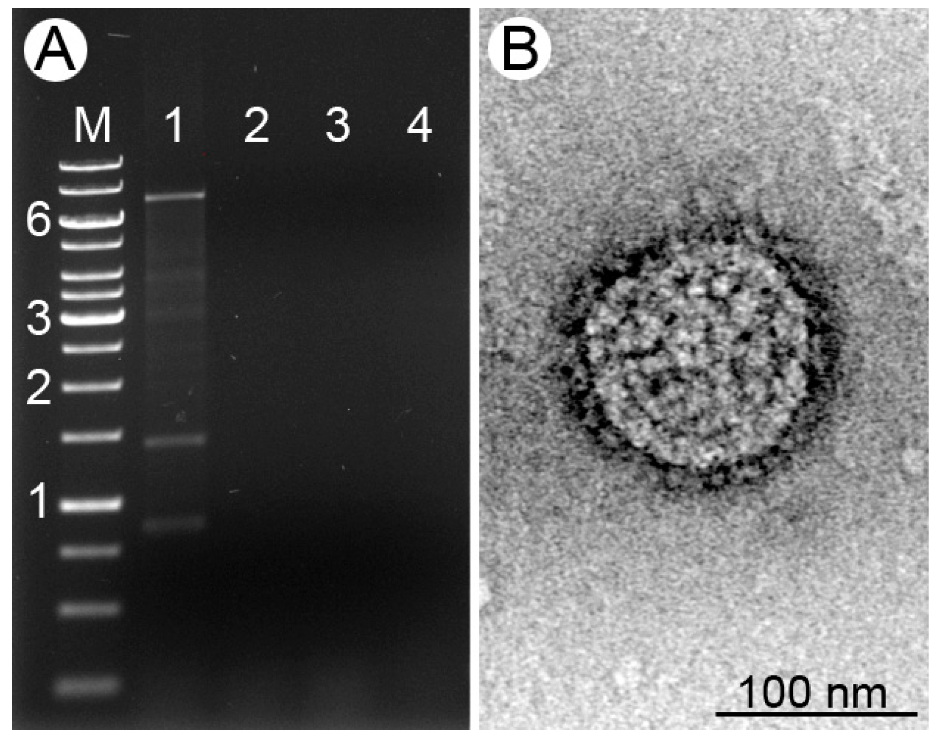
3.1. Viral dsRNA in Leishmania (M.) martiniquensis
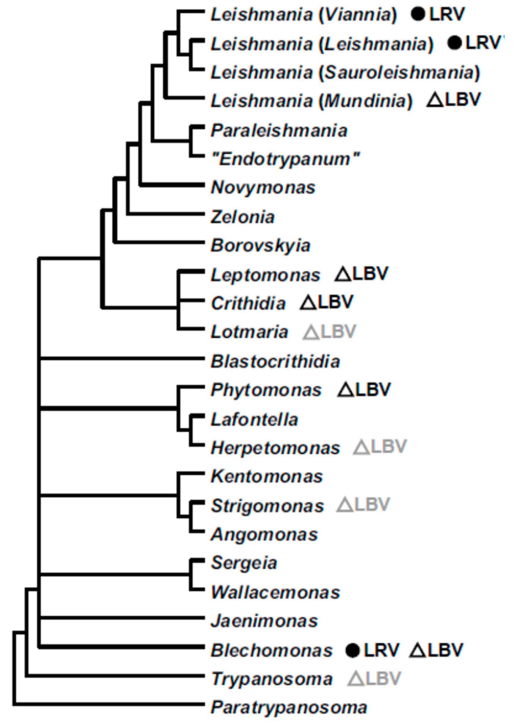
Four isolates of four different species of the leishmanial subgenus Mundinia were screened for the presence of dsRNA viruses. In one of these isolates, L. (M.) martiniquensis MHOM/MQ/92/MAR1, we documented the presence of three major dsRNA bands designated as L, M, and S for large, medium, and short, respectively (Figure 1A). This sample was sequenced using the Illumina HiSeq platform, yielding 5.4 Gbp of sequence data. The three viral contigs (6.1, 1.2, and 0.7 kb long) were highly abundant (60.1 to 354.2 fold above the average RPKM (Reads Per Kilobase per Million mapped reads) value), which facilitated their quick and reliable identification. Each contained a single ORF; 2012, 334, and 165 aa long in the L, M, and S fragments, respectively. As previously reported for other leishbunyaviruses [32], the proportions of particular viral segments were not even. As compared to the L RNA, the S segment was about six-fold more abundant (Table 1 and Table S1). This is in agreement with the higher demand for the S RNA-encoded nucleoprotein in vRNP formation. The sequences of all three viral segments were complete and included both 5′ and 3′ terminal "panhandle" inverted repeats (5′-acacaaaga tctttgtgt-3′, Figure 2) necessary for replication, transcription, and translation in bunyaviruses [3]. The sequences of the identified terminal repeats were identical to those of other known LBVs [32,38].
BLASTp searches demonstrate that the ORF sequences within the L and S segments are very similar to the RDRPs (up to 43% identity with 96% coverage) and nucleocapsid proteins (up to 51% identity with 96% coverage) of leishbunyaviruses. The Conserved Domain search identified a bunyaviral RDRP domain (pfam04196) between aa 588 and 1306 in the L segment ORF with an E-value = 2.24e−22. The region between the aa 86 and 151 of the same ORF displayed organization typical for the endonuclease domain of leishbunyaviruses (Figure 2).
Consistent with the previously published data on LBVs [38], the search for the homologs encoded in the viral M segment did not return any hits with BLASTp, Conserved Domain search, PHYRE2, and HHpred software. The analysis of the M segment-encoded glycoprotein with TMHMM, TMPred, and Phobius did not identify any transmembrane domains (TMDs) in the viruses under study. However, like in other LBVs and consistent with the glycoprotein annotation, SignalP detected the N-terminal membrane insertion peptide (with cleavage site between aa 21 and 22) and NetNGlyc predicted two N-glycosylation sites (at amino acid positions 34 and 237) in M segment sequences. Previously, similar results were obtained for LepmorLBV1, whereas in CabsLBV1, CotoLBV1, and the LBVs of Blechomonas spp., two TMDs were predicted in this segment [32,38]. We posit that such discrepancy could be explained by extreme sequence divergence preventing unambiguous identification of these elements. Application of a more sensitive algorithm, MEMSAT3, predicted one TMD in the virus investigated here, LepmorLBV1, as well as in Duke bunyavirus, which was not analyzed before. Similar to typical bunyaviruses, other LBVs have three TMDs in their glycoprotein ORFs. Analyses presented above suggest that the virus under investigation, as other bunyaviruses, can utilize host machinery for glycoprotein synthesis and virion assembly [65,66]. Indeed, the negatively stained transmission electron microscopy on purified virions from L. (M.) martiniquensis demonstrate the typical envelope with evenly spaced surface projections (Figure 1B).
In summary, we demonstrate that the new virus possesses many characteristic features of leishbunyaviruses and, therefore, we named it Leishmania martiniquensis leishbunyavirus 1 (LmarLBV1).

3.2. Phylogeny

The amino acid sequence of the RDRP was used in the phylogenetic inference of L. martiniquensis leishbunyavirus, using sequences of related Phenuiviridae as an outgroup (Figure 3). LmarLBV1 was nested within the clade Leishbunyaviridae with its closest relative being the Duke bunyavirus [67], which presumably infects a trypanosomatid from bees [38]. These two species proved to be sister to a big cluster of viruses from various monoxenous trypanosomatids. Judging by its phylogenetic position, we propose that L. martiniquensis acquired leishbunyavirus from a monoxenous trypanosomatid.
Although the glycoprotein sequences of LBVs are quite divergent, we perceived that the C-terminal part in some of them displayed conserved residues (Figure S1). Of note, all these viruses were those with one predicted TMD. Moreover, the sequence of this region in LmarLBV1 is more similar to that in LepmorLBV1s, than in DuBV, its closest relative according to the RDRP tree (Figure 3). Indels, rather than amino acid substitutions, distinguished these glycoprotein sequence fragments. LmarLBV1 had 28 and 77 indels compared to LepmorLBV1s and DuBV, respectively. The analysis of p-distances in trimmed alignments of the full glycoprotein sequences of all available species also showed markedly higher similarity between LmarLBV1 and LepmorLBV1s than between LmarLBV1 and DuBV (Table S3). Of interest, although nucleoprotein sequences were too short for reliable phylogenetic analysis, they grouped LmarLBV1 with DuBV, similarly to the RDRP-based tree (Figure S2).

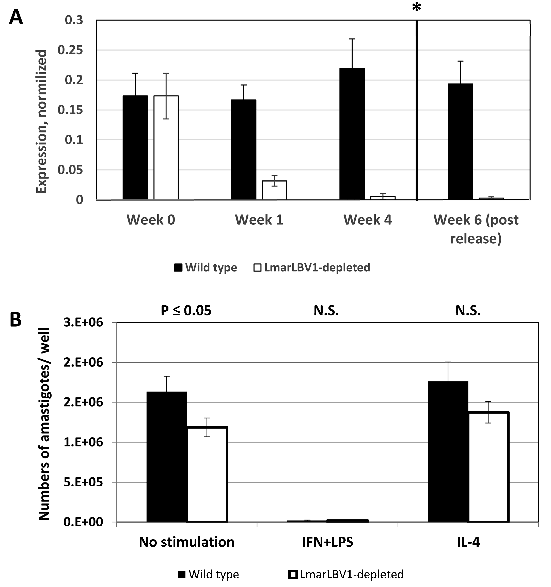
3.3. LmarLBV1 Has Minor Effect on Leishmania Infectivity In Vitro

To assess the role of LmarLBV1 in Leishmania biology, we first established an isogenic line of L. (M.) martiniquensis MHOM/MQ/92/MAR1 depleted of leishbunyavirus using ribavirin (Figure 4A). After four weeks of treatment, the viral load (as judged by RT-qPCR) was significantly diminished in the treated, compared to the untreated cells. Importantly, it stayed low even after the treatment was stopped (Figure 4A, asterisk), indicating that depletion was not transient.
Wild type and LmarLBV1-depleted L. (M.) martiniquensis were used to infect non-stimulated, classically (LPS/IFN-γ), or alternatively (IL4) stimulated primary murine macrophages to assess early stages of infection. As expected, the infection level in the classically stimulated macrophages was significantly lower compared to either non-stimulated or IL-4-treated cells. Importantly, parasites, which were depleted of virus, were less infective, compared to their wild type kin (Figure 4B). The effect of viral presence is minor, yet it is statistically significant and may point out the potential role of LmarLBV1 in L. (M.) martiniquensis biology.

4. Discussion

In this study, we describe the first leishbunyavirus of Leishmania. Previously, these viruses were discovered in monoxenous trypanosomatids of the subfamily Leishmaniinae (mainly in Crithidia spp.), genus Blechomonas (subfamily Blechomonadinae), as well as in one plant-infecting dixenous Phytomonas sp. (Figure 5) [32,38,39]. Leishbunyaviral sequences are also found in metatranscriptomes of insects infected by flagellates of other trypanosomatid genera, such as Strigomonas, Herpetomonas, and Trypanosoma (Figure 5, light grey) [38]. This is the most widespread and species-rich group of RNA viruses in trypanosomatids known to date. This fact, along with the discordance of viral and trypanosomatid phylogenies documented in the previous studies [32,38], strongly suggests that host-to-host transition is significantly facilitated in this group of viruses. It is explained when taking into account two facts: (i) LBVs are able to form membrane-bound viral particles [68] and (ii) the flagellar pocket of trypanosomatids is an organelle-governing intensive exchange with the milieu by endo- and exocytosis [69,70,71,72]. The documented particles of LBVs measure about 100 nm [38] corresponding to the typical size of clathrin-coated endocytic vesicles in trypanosomatids [73]. Interestingly, clathrin-mediated endocytosis is the general route for an uptake of bunyaviruses [74]. Bunyaviruses evolved to utilize the eukaryotic endomembrane system for virus assembly and spreading. Apparently, LBVs use the same strategy in trypanosomatids.
Infectivity and formation of viral particles in bunyaviruses depend on glycoproteins, type I transmembrane proteins that are proteolytically processed and glycosylated in the ER [3,8]. Their C-terminal cytoplasmic domains are thought to bind viral ribonucleoproteins and play a crucial role in genome packaging [75,76,77], whereas the N-terminal ectodomains are involved in receptor recognition and membrane fusion [65,78,79]. In leishbunyaviruses, the M segments and putative glycoproteins encoded within them are significantly reduced in size, extremely divergent, and sometimes contain a reduced number of transmembrane domains. We hypothesize that such a layout reflects a reduced functionality of these proteins and potential broad specificity of viral infection, which explains their facilitated host-to-host transition. It was demonstrated that extended deletions in the bunyaviral glycoprotein N-terminus (ectodomain) do not prevent cell fusion and transport to the Golgi, but lead to attenuation of viruses [80]. This illustrates the propensity of these proteins to undergo reduction. The opportunity to be inherited vertically probably removes the need for efficient proliferation and infection and may even make such properties undesirable.
Transfer of viruses between different species of trypanosomatids is possible because of coinfections, which are quite common in these parasites [81,82,83,84,85]. Coinfections were previously reported for Leptomonas moramango [39]. Here we did not observe viral coinfection but revealed a putative consequence of such an event—re-assortment of genomic segments. This assumption arises from discordance of phylogenies of proteins from the L and S segments on one hand, and the M segment on the other. The RDRP and nucleocapsid of LmarLBV1 are closely related to their counterparts in DukeBV, whereas its glycoprotein is more similar to the corresponding proteins of LepmorLBV1a and LepmorLBV1b.
LmarLBV1 is the first non-LRV virus discovered in Leishmania. It was found in one of the members of the most enigmatic subgenus of these flagellates—Mundinia. Although the first species was characterized over 70 years ago, the subgenus itself was established only recently [21]. For the moment, this taxon contains four described species: L. (M.) enriettii, L. (M.) macropodum, L. (M.) martiniquensis, and L. (M.) orientalis [86,87,88,89,90,91]. The first two infect guinea pigs and kangaroos, respectively, while the remaining two are isolated from humans. In contrast to other human-infecting Leishmania, which use sand flies as their vectors, these flagellates may be transmitted by biting midges [92,93]. Host switching may have shaped the genome evolution in these flagellates [94]. While the parasitofauna of biting midges is understudied, several species of monoxenous trypanosomatids are documented in these insects [95,96,97]. This is in agreement with our proposal that LmarLBV1 originates from LBVs of monoxenous trypanosomatids.
Leishmania martiniquensis is frequently found in skin lesions of immunocompromised patients indicating that it may be an opportunistic pathogen [98,99,100,101]. However, recent analysis of multiple records in Thailand and Myanmar reveals that neither the presence nor the severity of the infection is necessarily associated with HIV [87]. Notably, the clinical manifestations range from asymptomatic infection and various types of lesions to visceral disease. Previously, it was demonstrated that LRV1 boosts virulence of Leishmania guyanensis in humans [33,34,102,103]. The discovery of a virus in L. martiniquensis poses an important question on whether it also influences the pathogenicity of this parasite. We demonstrate that the presence of LmarLBV1 is slightly beneficial for Leishmania. The molecular mechanism of such facilitation may be non-specific, since it was recently shown that simultaneous inoculation of virus-negative L. guyanensis and Toscana virus (Bunyavirales, Phenuiviridae) increases footpad swelling and parasite burden in mice, reminiscent of the reaction to the LRV1-positive L. guyanensis [35]. Although it was not shown experimentally, the presence of the membrane-bound viral particles in LBVs suggest that they can be shed by trypanosomatid cells. This way, LmarLBV1 can interact with the immune system of a vertebrate host, increasing the severity of leishmanial infection. Our results signify the need for a systematic exploration of trypanosomatid viromes.

Supplementary Materials

The following are available online at https://www.mdpi.com/1999-4915/12/2/168/s1, Figure S1: Alignment of C-terminal part of the putative glycoproteins, possessing only one predicted transmembrane domain. Columns with ≥3 functionally similar amino acids are shaded, those with four functionally similar amino acids are marked with asterisk. Figure S2: Maximum likelihood phylogenetic tree of leishbunyaviral nucleoproteins. Numbers at branches indicate bootstrap support, values below 50 are not shown. The tree is rooted in agreement with RDRP-based reconstruction. Table S1: Summary statistics for RNA-seq data. Table S2: RDRP sequences of Bunyavirales with working abbreviations of viral names used in phylogenetic inferences. Table S3: Pairwise p-distances between glycoprotein sequences in Leishbunyaviridae.

Author Contributions

Conceptualization, V.Y. and A.Y.K.; methodology, D.G., T.L., P.K., N.K., and D.H.M.; validation, Y.K. and D.H.M.; formal analysis, D.G., N.K., T.L., and A.Y.K.; investigation, D.G., D.H.M., Y.K., P.K., and T.L.; resources, V.Y., A.N.L., P.A.B., and P.V.; data curation, A.N.L., P.V., and P.A.B.; writing—original draft preparation, D.G., D.H.M., A.Y.K., and V.Y..; writing—review and editing, V.Y., P.V., A.Y.K., T.L., N.K., A.N.L., and P.A.B.; visualization, D.G. and A.Y.K.; supervision, P.V. and V.Y.; funding acquisition, V.Y., P.V., A.Y.K., N.K., and D.H.M. All authors have read and agreed to the published version of the manuscript.

Funding

This research was funded by the Russian Science Foundation (grant 19-15-00054 (isolation, characterization, and bioinformatics analyses of LmarLBV1) to V.Y. and Y.K.), the ERD Funds (project OPVVV 16_019/0000759 to V.Y., A.Y.K., N.K., T.L., and P.V.), Grant Agency of Czech Republic (grant 20-22689S to V.Y.), Grant Agency of Charles University (UNCE 204072 to T.L.), University of Ostrava (SGS/PřF/2020 to V.Y. and D.H.M.), and the infrastructure grant “Přístroje IET” (CZ.1.05/2.1.00/19.0388 to V.Y.).

Acknowledgments

We thank Padet Siriyasatien (Chulalongkorn University, Bangkok, Thailand) for providing the strain of Leishmania (Mundinia) orientalis and Tatiana Spitzová (Charles University, Prague, Czech Republic) for her assistance with statistical analysis. We are grateful to the members of our laboratories for their stimulating discussions.

Conflicts of Interest

The authors declare no conflicts of interest. The funders had no role in the design of the study; in the collection, analyses, or interpretation of data; in the writing of the manuscript, or in the decision to publish the results.

References

  1. Lefkowitz, E.J.; Dempsey, D.M.; Hendrickson, R.C.; Orton, R.J.; Siddell, S.G.; Smith, D.B. Virus taxonomy: The database of the International Committee on Taxonomy of Viruses (ICTV). Nucleic Acids Res. 2018, 46, D708–D717. [Google Scholar] [CrossRef] [PubMed] [Green Version]
  2. Wichgers Schreur, P.J.; Kormelink, R.; Kortekaas, J. Genome packaging of the Bunyavirales. Curr. Opin. Virol. 2018, 33, 151–155. [Google Scholar] [CrossRef] [PubMed]
  3. Elliott, R.M. Molecular biology of the Bunyaviridae. J. Gen. Virol. 1990, 71, 501–522. [Google Scholar] [CrossRef] [PubMed]
  4. Sun, Y.; Li, J.; Gao, G.F.; Tien, P.; Liu, W. Bunyavirales ribonucleoproteins: The viral replication and transcription machinery. Crit. Rev. Microbiol. 2018, 44, 522–540. [Google Scholar] [CrossRef]
  5. Gerlach, P.; Malet, H.; Cusack, S.; Reguera, J. Structural insights into Bunyavirus replication and its regulation by the vRNA promoter. Cell 2015, 161, 1267–1279. [Google Scholar] [CrossRef] [Green Version]
  6. Junglen, S. Evolutionary origin of pathogenic arthropod-borne viruses - a case study in the family Bunyaviridae. Curr. Opin. Insect Sci. 2016, 16, 81–86. [Google Scholar] [CrossRef]
  7. Li, C.X.; Shi, M.; Tian, J.H.; Lin, X.D.; Kang, Y.J.; Chen, L.J.; Qin, X.C.; Xu, J.; Holmes, E.C.; Zhang, Y.Z. Unprecedented genomic diversity of RNA viruses in arthropods reveals the ancestry of negative-sense RNA viruses. Elife 2015, 4. [Google Scholar] [CrossRef]
  8. Shi, M.; Lin, X.D.; Tian, J.H.; Chen, L.J.; Chen, X.; Li, C.X.; Qin, X.C.; Li, J.; Cao, J.P.; Eden, J.S.; et al. Redefining the invertebrate RNA virosphere. Nature 2016, 540, 539–543. [Google Scholar] [CrossRef]
  9. Shi, M.; Lin, X.D.; Chen, X.; Tian, J.H.; Chen, L.J.; Li, K.; Wang, W.; Eden, J.S.; Shen, J.J.; Liu, L.; et al. The evolutionary history of vertebrate RNA viruses. Nature 2018, 556, 197–202. [Google Scholar] [CrossRef]
  10. Ullman, D.E.; German, T.L.; Sherwood, J.L.; Westcot, D.M.; Cantone, F.A. Tospovirus replication in insect vector cells - immunocytochemical evidence that the nonstructural protein encoded by the S RNA of Tomato spotted wilt tospovirus is present in thrips vector cells. Phytopathology 1993, 83, 456–463. [Google Scholar] [CrossRef]
  11. Whitfield, A.E.; Ullman, D.E.; German, T.L. Tospovirus-thrips interactions. Annu. Rev. Phytopathol. 2005, 43, 459–489. [Google Scholar] [CrossRef] [PubMed]
  12. Grybchuk, D.; Kostygov, A.Y.; Macedo, D.H.; d’Avila-Levy, C.M.; Yurchenko, V. RNA viruses in trypanosomatid parasites: A historical overview. Mem Inst. Oswaldo Cruz 2018, 113, e170487. [Google Scholar] [CrossRef] [PubMed] [Green Version]
  13. Maslov, D.A.; Opperdoes, F.R.; Kostygov, A.Y.; Hashimi, H.; Lukeš, J.; Yurchenko, V. Recent advances in trypanosomatid research: Genome organization, expression, metabolism, taxonomy and evolution. Parasitology 2019, 146, 1–27. [Google Scholar] [CrossRef] [PubMed] [Green Version]
  14. Maslov, D.A.; Votýpka, J.; Yurchenko, V.; Lukeš, J. Diversity and phylogeny of insect trypanosomatids: All that is hidden shall be revealed. Trends Parasitol. 2013, 29, 43–52. [Google Scholar] [CrossRef] [PubMed]
  15. McGhee, R.B.; Cosgrove, W.B. Biology and physiology of the lower Trypanosomatidae. Microbiol Rev. 1980, 44, 140–173. [Google Scholar] [CrossRef]
  16. Flegontov, P.; Butenko, A.; Firsov, S.; Kraeva, N.; Eliáš, M.; Field, M.C.; Filatov, D.; Flegontova, O.; Gerasimov, E.S.; Hlaváčová, J.; et al. Genome of Leptomonas pyrrhocoris: A high-quality reference for monoxenous trypanosomatids and new insights into evolution of Leishmania. Sci. Rep. 2016, 6, 23704. [Google Scholar] [CrossRef]
  17. Stevens, J.R.; Gibson, W.C. The evolution of pathogenic trypanosomes. Cad. Saude Publica 1999, 15, 673–684. [Google Scholar] [CrossRef] [Green Version]
  18. Camargo, E.P. Phytomonas and other trypanosomatid parasites of plants and fruit. Adv. Parasitol. 1999, 42, 29–112. [Google Scholar]
  19. Lukeš, J.; Skalický, T.; Týč, J.; Votýpka, J.; Yurchenko, V. Evolution of parasitism in kinetoplastid flagellates. Mol. Biochem. Parasitol. 2014, 195, 115–122. [Google Scholar] [CrossRef]
  20. Dvorák, V.; Shaw, J.J.; Volf, P. Parasite biology: The vectors. In The Leishmaniases: Old Neglected Tropical Diseases; Bruschi, F., Gradoni, L., Eds.; Springer: Cham, Switzerland, 2018; pp. 31–77. [Google Scholar]
  21. Espinosa, O.A.; Serrano, M.G.; Camargo, E.P.; Teixeira, M.M.; Shaw, J.J. An appraisal of the taxonomy and nomenclature of trypanosomatids presently classified as Leishmania and Endotrypanum. Parasitology 2018, 145, 430–442. [Google Scholar] [CrossRef]
  22. Kostygov, A.Y.; Yurchenko, V. Revised classification of the subfamily Leishmaniinae (Trypanosomatidae). Folia Parasitol 2017, 64, 020. [Google Scholar] [CrossRef] [PubMed] [Green Version]
  23. Paranaiba, L.F.; Pinheiro, L.J.; Macedo, D.H.; Menezes-Neto, A.; Torrecilhas, A.C.; Tafuri, W.L.; Soares, R.P. An overview on Leishmania (Mundinia) enriettii: Biology, immunopathology, LRV and extracellular vesicles during the host-parasite interaction. Parasitology 2018, 145, 1265–1273. [Google Scholar] [CrossRef] [PubMed]
  24. Paranaiba, L.F.; Pinheiro, L.J.; Torrecilhas, A.C.; Macedo, D.H.; Menezes-Neto, A.; Tafuri, W.L.; Soares, R.P. Leishmania enriettii (Muniz & Medina, 1948): A highly diverse parasite is here to stay. PLoS Pathog 2017, 13, e1006303. [Google Scholar] [CrossRef] [Green Version]
  25. Tarr, P.I.; Aline, R.F., Jr.; Smiley, B.L.; Scholler, J.; Keithly, J.; Stuart, K. LR1: A candidate RNA virus of Leishmania. Proc. Natl. Acad. Sci. USA 1988, 85, 9572–9575. [Google Scholar] [CrossRef] [Green Version]
  26. Stuart, K.D.; Weeks, R.; Guilbride, L.; Myler, P.J. Molecular organization of Leishmania RNA virus 1. Proc. Natl. Acad. Sci. USA 1992, 89, 8596–8600. [Google Scholar] [CrossRef] [Green Version]
  27. Widmer, G.; Comeau, A.M.; Furlong, D.B.; Wirth, D.F.; Patterson, J.L. Characterization of a RNA virus from the parasite Leishmania. Proc. Natl. Acad. Sci. USA 1989, 86, 5979–5982. [Google Scholar] [CrossRef] [Green Version]
  28. Guilbride, L.; Myler, P.J.; Stuart, K. Distribution and sequence divergence of LRV1 viruses among different Leishmania species. Mol. Biochem. Parasitol. 1992, 54, 101–104. [Google Scholar] [CrossRef]
  29. Scheffter, S.M.; Ro, Y.T.; Chung, I.K.; Patterson, J.L. The complete sequence of Leishmania RNA virus LRV2-1, a virus of an Old World parasite strain. Virology 1995, 212, 84–90. [Google Scholar] [CrossRef] [Green Version]
  30. Zangger, H.; Hailu, A.; Desponds, C.; Lye, L.F.; Akopyants, N.S.; Dobson, D.E.; Ronet, C.; Ghalib, H.; Beverley, S.M.; Fasel, N. Leishmania aethiopica field isolates bearing an endosymbiontic dsRNA virus induce pro-inflammatory cytokine response. PLoS Negl. Trop. Dis. 2014, 8, e2836. [Google Scholar] [CrossRef] [Green Version]
  31. Hajjaran, H.; Mahdi, M.; Mohebali, M.; Samimi-Rad, K.; Ataei-Pirkooh, A.; Kazemi-Rad, E.; Naddaf, S.R.; Raoofian, R. Detection and molecular identification of Leishmania RNA virus (LRV) in Iranian Leishmania species. Arch. Virol. 2016, 161, 3385–3390. [Google Scholar] [CrossRef]
  32. Grybchuk, D.; Kostygov, A.Y.; Macedo, D.H.; Votypka, J.; Lukes, J.; Yurchenko, V. RNA viruses in Blechomonas (Trypanosomatidae) and evolution of Leishmaniavirus. mBio 2018, 9, e01932-01918. [Google Scholar] [CrossRef] [PubMed] [Green Version]
  33. Ives, A.; Ronet, C.; Prevel, F.; Ruzzante, G.; Fuertes-Marraco, S.; Schutz, F.; Zangger, H.; Revaz-Breton, M.; Lye, L.F.; Hickerson, S.M.; et al. Leishmania RNA virus controls the severity of mucocutaneous leishmaniasis. Science 2011, 331, 775–778. [Google Scholar]
  34. De Carvalho, R.V.H.; Lima-Junior, D.S.; da Silva, M.V.G.; Dilucca, M.; Rodrigues, T.S.; Horta, C.V.; Silva, A.L.N.; da Silva, P.F.; Frantz, F.G.; Lorenzon, L.B.; et al. Leishmania RNA virus exacerbates leishmaniasis by subverting innate immunity via TLR3-mediated NLRP3 inflammasome inhibition. Nat. Commun. 2019, 10, 5273. [Google Scholar] [CrossRef] [Green Version]
  35. Hartley, M.A.; Bourreau, E.; Rossi, M.; Castiglioni, P.; Eren, R.O.; Prevel, F.; Couppie, P.; Hickerson, S.M.; Launois, P.; Beverley, S.M.; et al. Leishmaniavirus-dependent metastatic leishmaniasis is prevented by blocking IL-17A. PLoS Pathog 2016, 12, e1005852. [Google Scholar] [CrossRef] [Green Version]
  36. Rossi, M.; Castiglioni, P.; Hartley, M.A.; Eren, R.O.; Prevel, F.; Desponds, C.; Utzschneider, D.T.; Zehn, D.; Cusi, M.G.; Kuhlmann, F.M.; et al. Type I interferons induced by endogenous or exogenous viral infections promote metastasis and relapse of leishmaniasis. Proc. Natl. Acad. Sci. USA 2017, 114, 4987–4992. [Google Scholar] [CrossRef] [PubMed] [Green Version]
  37. Widmer, G.; Dooley, S. Phylogenetic analysis of Leishmania RNA virus and Leishmania suggests ancient virus-parasite association. Nucleic Acids Res. 1995, 23, 2300–2304. [Google Scholar] [CrossRef] [PubMed] [Green Version]
  38. Cantanhêde, L.M.; da Silva Junior, C.F.; Ito, M.M.; Felipin, K.P.; Nicolete, R.; Salcedo, J.M.; Porrozzi, R.; Cupolillo, E.; Ferreira Rde, G. Further evidence of an association between the presence of Leishmania RNA Virus 1 and the mucosal manifestations in tegumentary leishmaniasis patients. PLoS Negl. Trop. Dis. 2015, 9, e0004079. [Google Scholar] [CrossRef]
  39. Grybchuk, D.; Akopyants, N.S.; Kostygov, A.Y.; Konovalovas, A.; Lye, L.F.; Dobson, D.E.; Zangger, H.; Fasel, N.; Butenko, A.; Frolov, A.O.; et al. Viral discovery and diversity in trypanosomatid protozoa with a focus on relatives of the human parasite Leishmania. Proc. Natl. Acad. Sci. USA 2018, 115, E506–E515. [Google Scholar] [CrossRef] [Green Version]
  40. Akopyants, N.S.; Lye, L.F.; Dobson, D.E.; Lukeš, J.; Beverley, S.M. A novel bunyavirus-like virus of trypanosomatid protist parasites. Genome Announc 2016, 4, e00715-00716. [Google Scholar] [CrossRef] [Green Version]
  41. Maslov, D.A.; Lukeš, J.; Jirků, M.; Simpson, L. Phylogeny of trypanosomes as inferred from the small and large subunit rRNAs: Implications for the evolution of parasitism in the trypanosomatid protozoa. Mol. Biochem. Parasitol. 1996, 75, 197–205. [Google Scholar] [CrossRef]
  42. Yurchenko, V.; Kostygov, A.; Havlová, J.; Grybchuk-Ieremenko, A.; Ševčíková, T.; Lukeš, J.; Ševčík, J.; Votýpka, J. Diversity of trypanosomatids in cockroaches and the description of Herpetomonas tarakana sp. n. J. Eukaryot Microbiol 2016, 63, 198–209. [Google Scholar] [CrossRef] [PubMed]
  43. Gerasimov, E.S.; Kostygov, A.Y.; Yan, S.; Kolesnikov, A.A. From cryptogene to gene? ND8 editing domain reduction in insect trypanosomatids. Eur. J. Protistol. 2012, 48, 185–193. [Google Scholar] [CrossRef] [PubMed]
  44. Altschul, S.F.; Gish, W.; Miller, W.; Myers, E.W.; Lipman, D.J. Basic local alignment search tool. J. Mol. Biol 1990, 215, 403–410. [Google Scholar] [CrossRef]
  45. Bolger, A.M.; Lohse, M.; Usadel, B. Trimmomatic: A flexible trimmer for Illumina sequence data. Bioinformatics 2014, 30, 2114–2120. [Google Scholar] [CrossRef] [PubMed] [Green Version]
  46. Grabherr, M.G.; Haas, B.J.; Yassour, M.; Levin, J.Z.; Thompson, D.A.; Amit, I.; Adiconis, X.; Fan, L.; Raychowdhury, R.; Zeng, Q.; et al. Full-length transcriptome assembly from RNA-Seq data without a reference genome. Nat. Biotechnol. 2011, 29, 644–652. [Google Scholar] [CrossRef] [Green Version]
  47. Langmead, B.; Salzberg, S.L. Fast gapped-read alignment with Bowtie 2. Nat. Methods 2012, 9, 357–359. [Google Scholar] [CrossRef] [Green Version]
  48. Li, H.; Handsaker, B.; Wysoker, A.; Fennell, T.; Ruan, J.; Homer, N.; Marth, G.; Abecasis, G.; Durbin, R.; Genome Project Data Processing, S. The Sequence Alignment/Map format and SAMtools. Bioinformatics 2009, 25, 2078–2079. [Google Scholar] [CrossRef] [Green Version]
  49. Quinlan, A.R.; Hall, I.M. BEDTools: A flexible suite of utilities for comparing genomic features. Bioinformatics 2010, 26, 841–842. [Google Scholar] [CrossRef] [Green Version]
  50. Wheeler, D.L.; Church, D.M.; Federhen, S.; Lash, A.E.; Madden, T.L.; Pontius, J.U.; Schuler, G.D.; Schriml, L.M.; Sequeira, E.; Tatusova, T.A.; et al. Database resources of the National Center for Biotechnology. Nucleic Acids Res. 2003, 31, 28–33. [Google Scholar] [CrossRef]
  51. Marchler-Bauer, A.; Derbyshire, M.K.; Gonzales, N.R.; Lu, S.; Chitsaz, F.; Geer, L.Y.; Geer, R.C.; He, J.; Gwadz, M.; Hurwitz, D.I.; et al. CDD: NCBI’s conserved domain database. Nucleic Acids Res. 2015, 43, D222–D226. [Google Scholar] [CrossRef] [Green Version]
  52. Hofmann, K.; Stoffel, W. TMBase - a database of membrane spanning protein segments. Biol. Chem. Hoppe-Seyler 1993, 374, 166. [Google Scholar]
  53. Käll, L.; Krogh, A.; Sonnhammer, E.L. Advantages of combined transmembrane topology and signal peptide prediction--the Phobius web server. Nucleic Acids Res. 2007, 35, W429–W432. [Google Scholar] [CrossRef] [PubMed] [Green Version]
  54. McGuffin, L.J.; Bryson, K.; Jones, D.T. The PSIPRED protein structure prediction server. Bioinformatics 2000, 16, 404–405. [Google Scholar] [CrossRef] [PubMed]
  55. Petersen, T.N.; Brunak, S.; von Heijne, G.; Nielsen, H. SignalP 4.0: Discriminating signal peptides from transmembrane regions. Nat. Methods 2011, 8, 785–786. [Google Scholar] [CrossRef] [PubMed]
  56. Katoh, K.; Standley, D.M. MAFFT multiple sequence alignment software version 7: Improvements in performance and usability. Mol. Biol. Evol. 2013, 30, 772–780. [Google Scholar] [CrossRef] [PubMed] [Green Version]
  57. Capella-Gutiérrez, S.; Silla-Martinez, J.M.; Gabaldon, T. trimAl: A tool for automated alignment trimming in large-scale phylogenetic analyses. Bioinformatics 2009, 25, 1972–1973. [Google Scholar] [CrossRef]
  58. Nguyen, L.T.; Schmidt, H.A.; von Haeseler, A.; Minh, B.Q. IQ-TREE: A fast and effective stochastic algorithm for estimating maximum-likelihood phylogenies. Mol. Biol Evol 2015, 32, 268–274. [Google Scholar] [CrossRef]
  59. Kalyaanamoorthy, S.; Minh, B.Q.; Wong, T.K.F.; von Haeseler, A.; Jermiin, L.S. ModelFinder: Fast model selection for accurate phylogenetic estimates. Nat. Methods 2017, 14, 587–589. [Google Scholar] [CrossRef] [Green Version]
  60. Ronquist, F.; Teslenko, M.; van der Mark, P.; Ayres, D.L.; Darling, A.; Hohna, S.; Larget, B.; Liu, L.; Suchard, M.A.; Huelsenbeck, J.P. MrBayes 3.2: Efficient Bayesian phylogenetic inference and model choice across a large model space. Syst. Biol. 2012, 61, 539–542. [Google Scholar] [CrossRef] [Green Version]
  61. Kumar, S.; Stecher, G.; Li, M.; Knyaz, C.; Tamura, K. MEGA X: Molecular Evolutionary Genetics Analysis across computing platforms. Mol. Biol. Evol. 2018, 35, 1547–1549. [Google Scholar] [CrossRef]
  62. Ishemgulova, A.; Hlavacova, J.; Majerova, K.; Butenko, A.; Lukes, J.; Votypka, J.; Volf, P.; Yurchenko, V. CRISPR/Cas9 in Leishmania mexicana: A case study of LmxBTN1. PLoS ONE 2018, 13, e0192723. [Google Scholar] [CrossRef] [PubMed] [Green Version]
  63. Ishemgulova, A.; Kraeva, N.; Faktorová, D.; Podešvová, L.; Lukeš, J.; Yurchenko, V. T7 polymerase-driven transcription is downregulated in metacyclic promastigotes and amastigotes of Leishmania mexicana. Folia Parasitol. 2016, 63, 016. [Google Scholar] [CrossRef] [PubMed] [Green Version]
  64. Garin, Y.J.; Sulahian, A.; Meneceur, P.; Pratlong, F.; Prina, E.; Gangneux, J.; Dedet, J.P.; Derouin, F. Experimental pathogenicity of a presumed monoxenous trypanosomatid isolated from humans in a murine model. J. Eukaryot Microbiol 2001, 48, 170–176. [Google Scholar] [CrossRef] [PubMed]
  65. Giraud, E.; Leštinová, T.; Derrick, T.; Martin, O.; Dillon, R.J.; Volf, P.; Muller, I.; Bates, P.A.; Rogers, M.E. Leishmania proteophosphoglycans regurgitated from infected sand flies accelerate dermal wound repair and exacerbate leishmaniasis via insulin-like growth factor 1-dependent signalling. PLoS Pathog 2018, 14, e1006794. [Google Scholar] [CrossRef] [PubMed] [Green Version]
  66. Cifuentes-Muñoz, N.; Salazar-Quiroz, N.; Tischler, N.D. Hantavirus Gn and Gc envelope glycoproteins: Key structural units for virus cell entry and virus assembly. Viruses 2014, 6, 1801–1822. [Google Scholar] [CrossRef] [PubMed] [Green Version]
  67. Sanchez, A.J.; Vincent, M.J.; Nichol, S.T. Characterization of the glycoproteins of Crimean-Congo hemorrhagic fever virus. J. Virol. 2002, 76, 7263–7275. [Google Scholar] [CrossRef] [PubMed] [Green Version]
  68. Remnant, E.J.; Shi, M.; Buchmann, G.; Blacquiere, T.; Holmes, E.C.; Beekman, M.; Ashe, A. A diverse range of novel RNA viruses in geographically distinct honey bee populations. J. Virol. 2017, 91, e00158-00117. [Google Scholar] [CrossRef] [Green Version]
  69. Albornoz, A.; Hoffmann, A.B.; Lozach, P.Y.; Tischler, N.D. Early bunyavirus-host cell interactions. Viruses 2016, 8, 143. [Google Scholar] [CrossRef] [Green Version]
  70. Overath, P.; Stierhof, Y.D.; Wiese, M. Endocytosis and secretion in trypanosomatid parasites - tumultuous traffic in a pocket. Trends Cell Biol 1997, 7, 27–33. [Google Scholar] [CrossRef]
  71. Landfear, S.M.; Ignatushchenko, M. The flagellum and flagellar pocket of trypanosomatids. Mol. Biochem. Parasitol. 2001, 115, 1–17. [Google Scholar] [CrossRef]
  72. Szempruch, A.J.; Sykes, S.E.; Kieft, R.; Dennison, L.; Becker, A.C.; Gartrell, A.; Martin, W.J.; Nakayasu, E.S.; Almeida, I.C.; Hajduk, S.L.; et al. Extracellular vesicles from Trypanosoma brucei mediate virulence factor transfer and cause host anemia. Cell 2016, 164, 246–257. [Google Scholar] [CrossRef] [PubMed] [Green Version]
  73. Allen, C.L.; Goulding, D.; Field, M.C. Clathrin-mediated endocytosis is essential in Trypanosoma brucei. EMBO J. 2003, 22, 4991–5002. [Google Scholar] [CrossRef] [PubMed] [Green Version]
  74. Hung, C.H.; Qiao, X.; Lee, P.T.; Lee, M.G. Clathrin-dependent targeting of receptors to the flagellar pocket of procyclic-form Trypanosoma brucei. Eukaryot Cell 2004, 3, 1004–1014. [Google Scholar] [CrossRef] [PubMed] [Green Version]
  75. Léger, P.; Lozach, P.-Y. Bunyaviruses: From transmission by arthropods to virus entry into the mammalian host first-target cells. Future Virol. 2015, 10, 859–881. [Google Scholar] [CrossRef] [Green Version]
  76. Shi, X.; Elliott, R.M. Analysis of glycoproteins of viruses in the family Bunyaviridae. Methods Mol. Biol. 2007, 379, 137–148. [Google Scholar] [CrossRef] [PubMed]
  77. Överby, A.K.; Pettersson, R.F.; Neve, E.P. The glycoprotein cytoplasmic tail of Uukuniemi virus (Bunyaviridae) interacts with ribonucleoproteins and is critical for genome packaging. J. Virol. 2007, 81, 3198–3205. [Google Scholar] [CrossRef] [Green Version]
  78. Strandin, T.; Hepojoki, J.; Vaheri, A. Cytoplasmic tails of bunyavirus Gn glycoproteins – could they act as matrix protein surrogates? Virology 2013, 437, 73–80. [Google Scholar] [CrossRef] [Green Version]
  79. Wu, Y.; Zhu, Y.; Gao, F.; Jiao, Y.; Oladejo, B.O.; Chai, Y.; Bi, Y.; Lu, S.; Dong, M.; Zhang, C.; et al. Structures of phlebovirus glycoprotein Gn and identification of a neutralizing antibody epitope. Proc. Natl. Acad. Sci. USA 2017, 114, E7564–E7573. [Google Scholar] [CrossRef] [Green Version]
  80. Guardado-Calvo, P.; Bignon, E.A.; Stettner, E.; Jeffers, S.A.; Perez-Vargas, J.; Pehau-Arnaudet, G.; Tortorici, M.A.; Jestin, J.L.; England, P.; Tischler, N.D.; et al. Mechanistic insight into Bunyavirus-induced membrane fusion from structure-function analyses of the Hantavirus envelope glycoprotein Gc. PLoS Pathog 2016, 12, e1005813. [Google Scholar] [CrossRef] [Green Version]
  81. Shi, X.; Goli, J.; Clark, G.; Brauburger, K.; Elliott, R.M. Functional analysis of the Bunyamwera orthobunyavirus Gc glycoprotein. J. Gen. Virol. 2009, 90, 2483–2492. [Google Scholar] [CrossRef] [Green Version]
  82. Spodareva, V.V.; Grybchuk-Ieremenko, A.; Losev, A.; Votýpka, J.; Lukeš, J.; Yurchenko, V.; Kostygov, A.Y. Diversity and evolution of anuran trypanosomes: Insights from the study of European species. Parasit Vectors 2018, 11, 447. [Google Scholar] [CrossRef] [PubMed] [Green Version]
  83. Kozminsky, E.; Kraeva, N.; Ishemgulova, A.; Dobáková, E.; Lukeš, J.; Kment, P.; Yurchenko, V.; Votýpka, J.; Maslov, D.A. Host-specificity of monoxenous trypanosomatids: Statistical analysis of the distribution and transmission patterns of the parasites from Neotropical Heteroptera. Protist 2015, 166, 551–568. [Google Scholar] [CrossRef] [PubMed]
  84. Kraeva, N.; Butenko, A.; Hlaváčová, J.; Kostygov, A.; Myškova, J.; Grybchuk, D.; Leštinová, T.; Votýpka, J.; Volf, P.; Opperdoes, F.; et al. Leptomonas seymouri: Adaptations to the dixenous life cycle analyzed by genome sequencing, transcriptome profiling and co-infection with Leishmania donovani. PLoS Pathog 2015, 11, e1005127. [Google Scholar] [CrossRef] [PubMed] [Green Version]
  85. Votýpka, J.; d’Avila-Levy, C.M.; Grellier, P.; Maslov, D.A.; Lukeš, J.; Yurchenko, V. New approaches to systematics of Trypanosomatidae: Criteria for taxonomic (re)description. Trends Parasitol 2015, 31, 460–469. [Google Scholar] [CrossRef]
  86. Grybchuk-Ieremenko, A.; Losev, A.; Kostygov, A.Y.; Lukeš, J.; Yurchenko, V. High prevalence of trypanosome co-infections in freshwater fishes. Folia Parasitol 2014, 61, 495–504. [Google Scholar] [CrossRef] [Green Version]
  87. Muniz, J.; Medina, H. [Cutaneous leishmaniasis of the guinea pig, Leishmania enriettii n. sp]. Hospital (Rio J.) 1948, 33, 7–25. [Google Scholar]
  88. Jariyapan, N.; Daroontum, T.; Jaiwong, K.; Chanmol, W.; Intakhan, N.; Sor-Suwan, S.; Siriyasatien, P.; Somboon, P.; Bates, M.D.; Bates, P.A. Leishmania (Mundinia) orientalis n. sp. (Trypanosomatidae), a parasite from Thailand responsible for localised cutaneous leishmaniasis. Parasit Vectors 2018, 11, 351. [Google Scholar] [CrossRef]
  89. Dougall, A.; Shilton, C.; Low Choy, J.; Alexander, B.; Walton, S. New reports of Australian cutaneous leishmaniasis in Northern Australian macropods. Epidemiol Infect. 2009, 137, 1516–1520. [Google Scholar] [CrossRef] [Green Version]
  90. Rose, K.; Curtis, J.; Baldwin, T.; Mathis, A.; Kumar, B.; Sakthianandeswaren, A.; Spurck, T.; Low Choy, J.; Handman, E. Cutaneous leishmaniasis in red kangaroos: Isolation and characterisation of the causative organisms. Int. J. Parasitol. 2004, 34, 655–664. [Google Scholar] [CrossRef]
  91. Barratt, J.; Kaufer, A.; Peters, B.; Craig, D.; Lawrence, A.; Roberts, T.; Lee, R.; McAuliffe, G.; Stark, D.; Ellis, J. Isolation of novel trypanosomatid, Zelonia australiensis sp. nov. (Kinetoplastida: Trypanosomatidae) provides support for a Gondwanan origin of dixenous parasitism in the Leishmaniinae. PLoS Negl. Trop. Dis. 2017, 11, e0005215. [Google Scholar] [CrossRef] [Green Version]
  92. Desbois, N.; Pratlong, F.; Quist, D.; Dedet, J.P. Leishmania (Leishmania) martiniquensis n. sp. (Kinetoplastida: Trypanosomatidae), description of the parasite responsible for cutaneous leishmaniasis in Martinique Island (French West Indies). Parasite 2014, 21, 12. [Google Scholar] [CrossRef] [PubMed] [Green Version]
  93. Seblová, V.; Sádlová, J.; Vojtková, B.; Votýpka, J.; Carpenter, S.; Bates, P.A.; Volf, P. The biting midge Culicoides sonorensis (Diptera: Ceratopogonidae) is capable of developing late stage infections of Leishmania enriettii. PLoS Negl. Trop. Dis. 2015, 9, e0004060. [Google Scholar] [CrossRef] [Green Version]
  94. Dougall, A.M.; Alexander, B.; Holt, D.C.; Harris, T.; Sultan, A.H.; Bates, P.A.; Rose, K.; Walton, S.F. Evidence incriminating midges (Diptera: Ceratopogonidae) as potential vectors of Leishmania in Australia. Int. J. Parasitol. 2011, 41, 571–579. [Google Scholar] [CrossRef] [PubMed]
  95. Butenko, A.; Kostygov, A.Y.; Sádlová, J.; Kleschenko, Y.; Bečvář, T.; Podešvová, L.; Macedo, D.H.; Žihala, D.; Lukeš, J.; Bates, P.A.; et al. Comparative genomics of Leishmania (Mundinia). BMC Genomics 2019, 20, 726. [Google Scholar] [CrossRef] [PubMed] [Green Version]
  96. Zídková, L.; Čepička, I.; Votýpka, J.; Svobodová, M. Herpetomonas trimorpha sp. nov. (Trypanosomatidae, Kinetoplastida), a parasite of the biting midge Culicoides truncorum (Ceratopogonidae, Diptera). Int. J. Syst. Evol. Microbiol. 2010, 60, 2236–2246. [Google Scholar] [CrossRef] [PubMed] [Green Version]
  97. Svobodová, M.; Zídková, L.; Čepička, I.; Oborník, M.; Lukeš, J.; Votýpka, J. Sergeia podlipaevi gen. nov., sp. nov. (Trypanosomatidae, Kinetoplastida), a parasite of biting midges (Ceratopogonidae, Diptera). Int. J. Syst. Evol. Microbiol. 2007, 57, 423–432. [Google Scholar] [CrossRef] [PubMed]
  98. Podlipaev, S.; Votýpka, J.; Jirků, M.; Svobodová, M.; Lukeš, J. Herpetomonas ztiplika n. sp. (Kinetoplastida: Trypanosomatidae): A parasite of the blood-sucking biting midge Culicoides kibunensis Tokunaga, 1937 (Diptera: Ceratopogonidae). J. Parasitol. 2004, 90, 342–347. [Google Scholar] [CrossRef]
  99. Bualert, L.; Charungkiattikul, W.; Thongsuksai, P.; Mungthin, M.; Siripattanapipong, S.; Khositnithikul, R.; Naaglor, T.; Ravel, C.; El Baidouri, F.; Leelayoova, S. Autochthonous disseminated dermal and visceral leishmaniasis in an AIDS patient, southern Thailand, caused by Leishmania siamensis. Am. J. Trop Med. Hyg 2012, 86, 821–824. [Google Scholar] [CrossRef] [Green Version]
  100. Dedet, J.P.; Roche, B.; Pratlong, F.; Cales-Quist, D.; Jouannelle, J.; Benichou, J.C.; Huerre, M. Diffuse cutaneous infection caused by a presumed monoxenous trypanosomatid in a patient infected with HIV. Trans. R. Soc. Trop. Med. Hyg 1995, 89, 644–646. [Google Scholar] [CrossRef]
  101. Chicharro, C.; Alvar, J. Lower trypanosomatids in HIV/AIDS patients. Ann. Trop. Med. Parasitol. 2003, 97 Suppl. 1, 75–78. [Google Scholar] [CrossRef] [Green Version]
  102. Dedet, J.P.; Pratlong, F. Leishmania, Trypanosoma and monoxenous trypanosomatids as emerging opportunistic agents. J. Eukaryot Microbiol 2000, 47, 37–39. [Google Scholar] [CrossRef] [PubMed]
  103. Hartley, M.A.; Ronet, C.; Zangger, H.; Beverley, S.M.; Fasel, N. Leishmania RNA virus: When the host pays the toll. Front. Cell Infect. Microbiol. 2012, 2, 99. [Google Scholar] [CrossRef] [PubMed] [Green Version]
Figure 1. (A) Screening of double-stranded RNAs (dsRNAs) in Leishmania (Mundinia) spp. M, GeneRuler 1-kb DNA ladder. Indicated sizes are in kilobases: 1, L. (M.) martiniquensis MHOM/MQ/92/MAR1; 2, L. (M.) enriettii MCAV/BR/45/LV90; 3, L. (M.) macropodum MMAC/AU/2004/AM-2004; 4, L. (M.) orientalis MHOM/TH/2007/PCM2. (B) Negative-stain transmission electron micrographs of the virus particle isolated from L. (M.) martiniquensis. Scale bar is 100 nm.
Viruses 12 00168 g001
Figure 2. Structural features of LmarLBV1. (A) Secondary structures and complementary sequences on 5′ and 3′ ends of the LmarLBV1 L, M, and S RNA segments predicted by IPknot. (and) depicting complementary nucleotides forming the stem, *-non-complementary nucleotides forming a bulge. (B) Amino acid alignment of the N-terminal endonuclease domain of RDRP of Leishbunyaviridae and Phenuiviridae. Functionally important residues are marked with arrowheads. Numbering of positions in alignment are indicated as in LmarLBV1 polymerase protein. Shading: ≥80% identity within Phenui and Leishbunyaviruses + Wuhan Spider virus (LBV+WSV).
Viruses 12 00168 g002
Figure 3. RDRP-based maximum likelihood reconstruction of leishbunyaviruses’ phylogeny. Double-crossed branch is at 50% of its original lengths. Branch supports are Bayesian posterior probability and maximum likelihood bootstrap, respectively. Black circles indicate maximal (1/100) statistical supports. The scale bar indicates the number of substitutions per site. The tree was rooted with the sequences of Phenuiviridae. Lesihmania martiniquensis leishbunyavirus 1 (LmarLBV1) described here is highlighted in black. Abbreviations and GenBank accession numbers are in Table S2 [32].
Viruses 12 00168 g003
Figure 4. LmarLBV1 facilitates Leishmania infection in vitro. (A) Establishment of isogenic, virus-depleted line of L. (M.) martiniquensis MHOM/MQ/92/MAR1. The treatment with ribavirin was stopped after four weeks (asterisk), but the viral load remained low in LmarLBV1-depleted parasites. (B) Macrophage infection in vitro. The average number of parasite per well was calculated for the wild type and in LmarLBV1-depleted L. (M.) martiniquensis infecting non-stimulated, classically (LPS/IFN-γ) or alternatively (IL-4) stimulated primary murine macrophages. Data are summarized from two independent biological replicates (three technical replicates each). The error bars indicate standard deviations. N.S. = not statistically significant. p ≤ 0.05.
Viruses 12 00168 g004
Figure 5. A schematic phylogenetic tree of the family Trypanosomatidae (modified from [13]), demonstrating the distribution of leishbunyaviruses (triangles) and leishmaniaviruses (circles) over the genera of these flagellates. Viruses identified in metatranscriptomes of trypanosomatid-infected insects [38] are shown in grey.
Viruses 12 00168 g005
Table 1. Molecular data for the identified RNA sequences1.
Viral SequencesAccession Length, bpORF, AARPKM
LmarLBV1 SMK3565567211656,600.13
LmarLBV1 MMK3565551,2443341,368.42
LmarLBV1 LMK35655461022,0121,131.22
1 See also Table S1. ORF: Open Reading Frame, AA: Amino Acids, RPKM: Reads per kilobase per million mapped reads.

