Article

Shared Common Ancestry of Rodent Alphacoronaviruses Sampled Globally

by
Theocharis Tsoleridis
1,
Joseph G. Chappell
1,
Okechukwu Onianwa
1,
Denise A. Marston
3,
Anthony R. Fooks
3,
Elodie Monchatre-Leroy
4,
Gérald Umhang
4,
Marcel A. Müller
5,
Jan F. Drexler
5,
Christian Drosten
5,
Rachael E. Tarlinton
2,
Charles P. McClure
1,
Edward C. Holmes
6 and
Jonathan K. Ball
1,*
1
School of Life Sciences, University of Nottingham, Nottingham NG7 2UH, UK
2
School of Veterinary Medicine and Science, University of Nottingham, Sutton Bonington Campus, Loughborough LE12 5RD, UK
3
Wildlife Zoonoses and Vector-borne Diseases Research Group, Animal and Plant Health Agency (APHA), Weybridge-London KT15 3NB, UK
4
Anses, Laboratoire de la rage et de la faune sauvage, 54220 Malzéville, France
5
Charité-Universitätsmedizin Berlin, Corporate Member of Freie Universität Berlin, Humboldt-Universität zu Berlin, and Berlin Institute of Health, Institute of Virology, 10117 Berlin, Germany
6
Marie Bashir Institute for Infectious Diseases and Biosecurity, Charles Perkins Centre, School of Life and Environmental Sciences and Sydney Medical School, The University of Sydney, Sydney, NSW 2006, Australia
*
Author to whom correspondence should be addressed.
Viruses 2019, 11(2), 125; https://doi.org/10.3390/v11020125
Submission received: 14 January 2019 / Revised: 25 January 2019 / Accepted: 28 January 2019 / Published: 30 January 2019
(This article belongs to the Section Animal Viruses)

Abstract

:
The recent discovery of novel alphacoronaviruses (alpha-CoVs) in European and Asian rodents revealed that rodent coronaviruses (CoVs) sampled worldwide formed a discrete phylogenetic group within this genus. To determine the evolutionary history of rodent CoVs in more detail, particularly the relative frequencies of virus-host co-divergence and cross-species transmission, we recovered longer fragments of CoV genomes from previously discovered European rodent alpha-CoVs using a combination of PCR and high-throughput sequencing. Accordingly, the full genome sequence was retrieved from the UK rat coronavirus, along with partial genome sequences from the UK field vole and Poland-resident bank vole CoVs, and a short conserved ORF1b fragment from the French rabbit CoV. Genome and phylogenetic analysis showed that despite their diverse geographic origins, all rodent alpha-CoVs formed a single monophyletic group and shared similar features, such as the same gene constellations, a recombinant beta-CoV spike gene, and similar core transcriptional regulatory sequences (TRS). These data suggest that all rodent alpha CoVs sampled so far originate from a single common ancestor, and that there has likely been a long-term association between alpha CoVs and rodents. Despite this likely antiquity, the phylogenetic pattern of the alpha-CoVs was also suggestive of relatively frequent host-jumping among the different rodent species.

1. Introduction

Among the viruses that infect both humans and animals, coronaviruses are common and important pathogens. The first coronavirus (CoV) was discovered during the 1930s [1], and the first human coronavirus was characterized in the 1960s [2]. Since then, five novel human coronaviruses have been discovered. The coronavirus family gained particular notoriety with the emergence of the severe acute respiratory syndrome (SARS) in 2002/2003 and the Middle East respiratory syndrome (MERS) in 2013. In humans, coronaviruses are associated with respiratory disease, causing a range of symptoms from mild common colds to more severe lower respiratory tract infections that can be lethal. CoV infection in a range of non-human mammals and birds is associated with enteric and respiratory diseases as well as hepatitis and neurological disorders [3].
Rodents are an important source of emerging virus infections [4]. Rodentia is the single largest mammalian order and comprises ~40% of all mammals [5], including ~2200 species such as rats, mice, and voles [6]. Despite the key role played by rodents in virus emergence, until recently, only two rodent CoVs had been characterized—murine hepatitis virus (MHV) [7] and rat sialodacryoadenitis coronavirus (SADV) [8], both of which are from the same viral species and are betacoronaviruses.
We previously reported the discovery of novel alpha-CoVs in European rodents and shrews, encompassing several species such as field voles (Microtus agrestis), bank voles (Myodes glareolus), and rats (Rattus norvegicus) [9]. Since then, additional alpha-CoVs have been described infecting a variety of Chinese rodents, including field mice (Apodemus chevrieri), grey red-backed voles (Myodes rufocanus), and lesser rice field rats (Rattus losea) [10,11]. Interestingly, a recently discovered rabbit alphacoronavirus also clustered with rodent alpha-CoVs [12].
To gain a better understanding of the evolution of alphacoronaviruses in rodents, particularly how long these virus-host associations have been established, we recovered and characterized complete or partial genomes for European Norway rat, field vole, and bank vole CoVs, and compared these sequences to those available for other rodent CoVs obtained in other parts of the world. Our new phylogenetic analyses provide compelling evidence that the alpha-CoVs sampled from rodents to date have arisen from a single common ancestor, suggestive of a long-term virus-host association.

2. Materials and Methods

2.1. Samples

Samples previously reported to be positive for alpha-CoVs were used in this study [9,12]. These samples included a UK Rattus norvegicus (UKRn3), UK Microtus agrestis (UKMa1), Poland-resident Myodes glareolus (PLMg1), and the France-resident Oryctolagus cuniculus (L232) (Table 1).

2.2. Nucleic Acid Preparation

Total RNA was extracted from approximately one cubic millimetre sections of liver (UKRn3, PLMg1) or intestinal tissue samples (UKMa1) using the GenElute™ Mammalian Total RNA Miniprep Kit (Sigma Aldrich, Steinheim, Germany). Total RNA was extracted from intestinal fluid for the France resident L232 rabbit CoV using the QIAamp Viral RNA Mini Kit (Qiagen, Hilden, Germany). The RNA to cDNA EcoDry™ Premix - Random Hexamers kit (Clontech, Saint-Germain-en-Laye, France) was used for cDNA synthesis.

2.3. High-Throughput Sequencing

High-throughput sequencing was performed on total RNA extracted from UKMa1 and PLMg1 using Illumina MiSeq. Genomic DNA (gDNA) was depleted using DNaseI, and ribosomal RNA (rRNA) was removed using NEBNext® rRNA Depletion Kit (Human/Mouse/Rat) with RNA Sample Purification Beads (New England Biolabs, Ipswich, Massachusetts, USA). The samples were run on an Illumina MiSeq platform, generating a total of 8,045,758 paired reads for PLMg1 and 14,140,770 paired reads for UKMa1. Each read length was 2 × 150 bp, and the insert size was 400 bp on average. All the Next Generation Sequencing (NGS) data were analysed using Geneious 11.1.2 software. The reads were assembled into contigs with de novo assembly using Geneious. The assembled contigs were compared against the RefSeq sequence database of all the virus proteins downloaded from GenBank using blastX. The minimum e-value was 1 × 10⁻⁵ to maintain high sensitivity and low rates of false hits [13]. The contigs that matched known viruses were verified by PCR.

2.4. Genome Retrieval with PCR

The primers used for primer walking were either designed based on the Lucheng Rn coronavirus (Genbank Acc. No. KF294380) reference sequence or based on the known genome sequences recovered from UKRn3. The primers for contig confirmation and gap-filling between contigs for UKMa1 and PLMg1 were designed based on the sequences of the contigs. Geneious 11.1.2 software was used for genome mapping, annotating, and analysis. All the primers were evaluated in silico with the Primer3 online tool. For the retrieval of a short ORF1b fragment for L232, a previously described primer combination was used [9].
PCR reactions were carried out in a PCT-200 Peltier Thermal Cycler (MJ Research). The PCR reactions were performed with HotStarTaq Polymerase (Qiagen) according to manufacturer’s instructions.
The coronavirus sequences generated in this study were deposited in GenBank under the accession numbers MK163627 and MK249067-MK249069.

2.5. Phylogenetic Analysis

Reference sequences of α, β, and one γ-CoV were downloaded from GenBank for the ORF1b, S, and N genes and aligned using ClustalW [14] recognising codon positions. To provide evolutionary context, phylogenetic analyses were performed using the maximum likelihood (ML) method within the Molecular Evolutionary Genetics Analysis version 7 (MEGA7) [15] package. These analyses utilized a General Time Reversible (GTR) nucleotide substitution model with a gamma distribution of rate variation, a class of invariant sites (Γ+I) model of evolution, and complete deletion of gaps, with statistical robustness assessed using bootstrap resampling (1,000 pseudoreplicates). Equivalent ML phylogenies were also inferred using partial ORF1b, S, and N amino acid sequences and employing the Jones-Taylor-Thornton (JTT) substitution model with uniform rates, complete deletion of gaps, and statistical robustness of 1000 pseudoreplicates. Details regarding the number of sequences used for the phylogenetic analysis of each gene along with their length are given in Table 2, and the coronavirus reference sequences used in the phylogenetic analysis are given in Tables S1 and S2.
To determine the phylogenetic relationships among the host species analysed, the mitochondrial cytochrome b reference sequences of Rattus, Apodemus, Myodes, Microtus spp., and Oryctolagus cuniculus were downloaded from Genbank. In total, 44 full length (1136 nt) cytochrome b sequences were aligned as described above, and an ML tree was estimated using the same procedure as described above.

2.6. Analysis of the Virus-Host Co-Divergence

To compare the evolutionary history of rodents and their coronaviruses, particularly the relative frequencies of co-divergence versus cross-species transmission, a tanglegram was inferred by comparing the ML phylogenies of the rodent cytochrome b and the coronavirus ORF1b sequences, connecting hosts to their corresponding coronaviruses. The reconciliation between the alpha-CoV ORF1b and the host mtDNA cytochrome b phylogenies was performed using the Jane software with default parameters [16].

3. Results

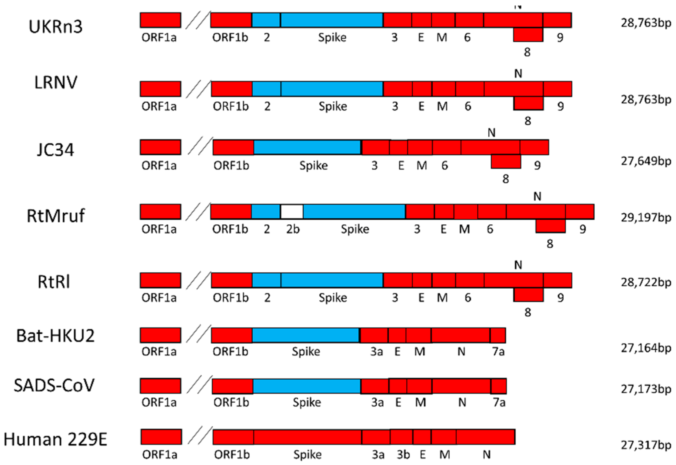
3.1. Genome Retrieval of Rodent and Rabbit Alphacoronaviruses

The near-full genome sequence of the UK rat CoV (UKRn3) was retrieved with primer-walking PCR. The 5′ and 3′ ends corresponded to the rat Lucheng CoV (KF294380)-derived primer sequences used for amplification of the genome termini and were not confirmed by the rapid amplification of cDNA ends (RACE) method. The genome of UKRn3 had a length of 28,763 nt and 40.2% GC content. Its pairwise distance to Lucheng CoV at the nucleotide level was 95.5%, indicating that they belong in the same species. Additionally, the rat UKRn3 was 90.2% similar to RtMruf (KY370045), 95.4% to RtRl-CoV (KY370050), and 75% to AcCoV-JC34 (NC034972) in the nucleotide level.
For the genome retrieval of the UK field vole CoV (UKMa1) and the Poland-resident bank vole (PLMg1), primer-walking PCR was not applicable due to the lack of a very close reference sequence. Thus, NGS performed on total RNA extracted from the Microtus agrestis gut and Myodes glareolus liver was used as an alternative method. Genome fragments representing 17,483 and 8603 nucleotides spanning various genes were retrieved for UKMa1 and PLMg1, respectively. Full-length N gene and partial ORF1ab, ORF2, S, ORF6, ORF8, and ORF9 genes were retrieved for UKMa1, whereas partial ORF1b and N genes were retrieved for PLMg1. For the L232 coronavirus, which was obtained from a rabbit living in France [12], additional sequences up to 621 bp of the ORF1b gene were retrieved by targeting conserved regions with PCR. Unfortunately, presumably due to the relatively poor quality of the recovered nucleic acid, we were unable to recover additional sequences.

3.2. Virus Genome Characterization

Genome analysis of all the rodent alpha-CoVs revealed a number of common features (Figure 1). All viruses, with the exception of the field mouse AcCoV-JC34 (NC034972), possessed a predicted ORF2 gene that was approximately 274 amino acids (aa) in length and was located between the ORF1b and S gene. Blast search revealed that this gene was not present in any other characterized alpha-CoVs and its closest match, at 44% amino acid similarity, was the NS2a gene of the Betacoronavirus1 Human CoV OC43. Interestingly, all the rodent α-CoVs possessed a β-CoV-like spike, which indicates recombination between α and β-CoV. All the rodent α-CoVs possessed a predicted 214 aa long ORF3 gene located between the S and the E gene. The closest match for ORF3 was the 3c-like protein of ferret alphacoronavirus with 47% similarity at the amino acid level. Another striking feature of rodent alphacoronavirus genomes was the presence of the predicted ORF6 (165 aa) gene between the M and N genes, which is observed in no other members of the genus Alphacoronavirus. Other common features of all the rodent α-CoV were the presence of a predicted ORF8 (~181 aa) within the N gene and ORF9 (~111 aa) downstream of the N gene. Finally, the grey red-backed vole RtMruf-CoV (KY370045) appeared to possess an additional 154 aa long predicted gene called ORF2b, located between ORF2 and S.
Further analysis revealed similarities in the core part of the transcription regulatory sequence (TRS) located upstream of the 5′ end of each ORF (Table 3). All the rodent α-CoVs possessed the same 5′-AACUAA-3′ core TRS sequence in the majority of their genes. However, some variations of the core TRS were observed; specifically, a 5′-AACUUUAA-3′ for ORF2, 5′-UACUAA-3′ for ORF3, 5′-CACUAA-3′, and 5′-GACUAA-3′ for ORF8.

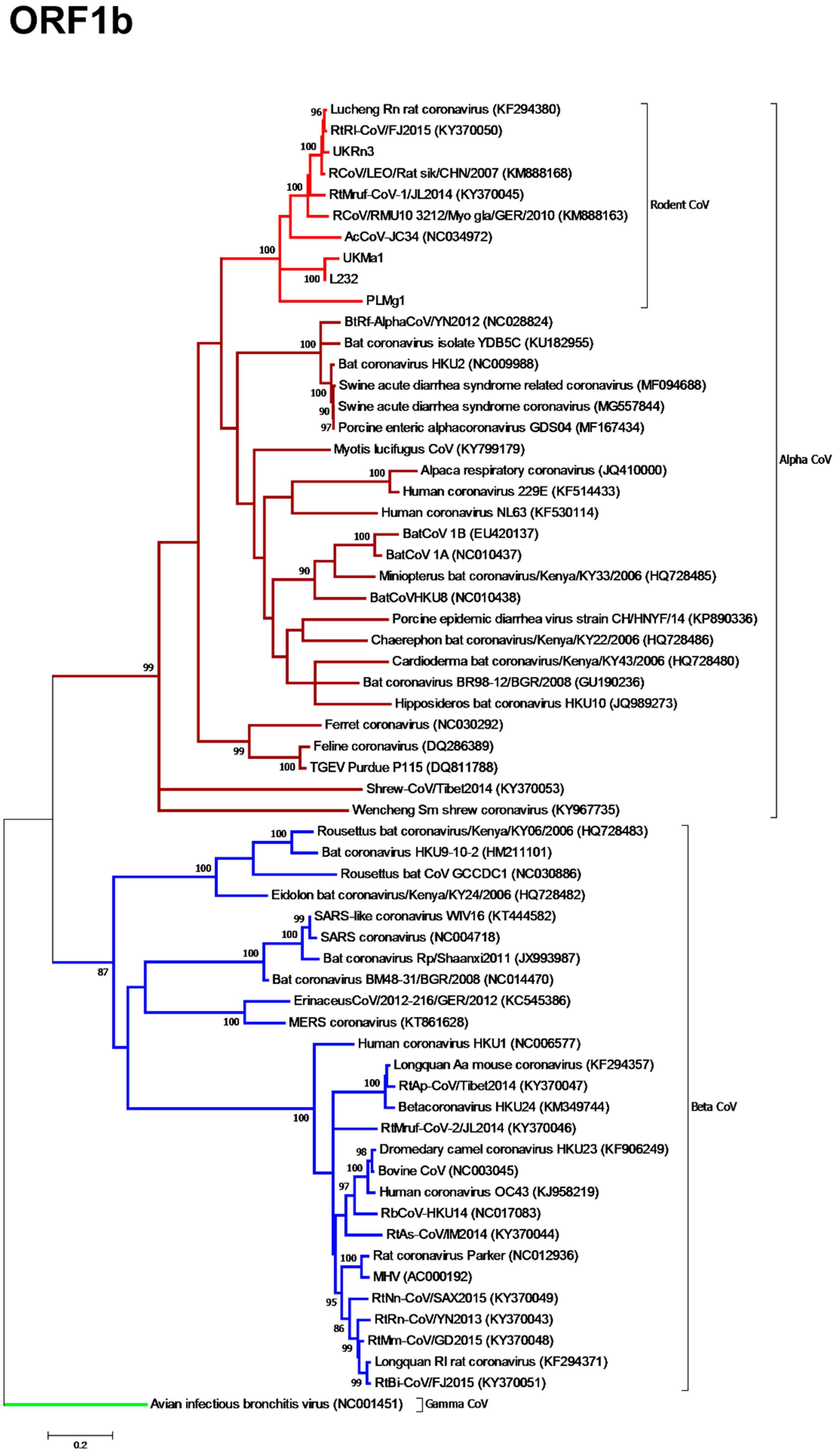
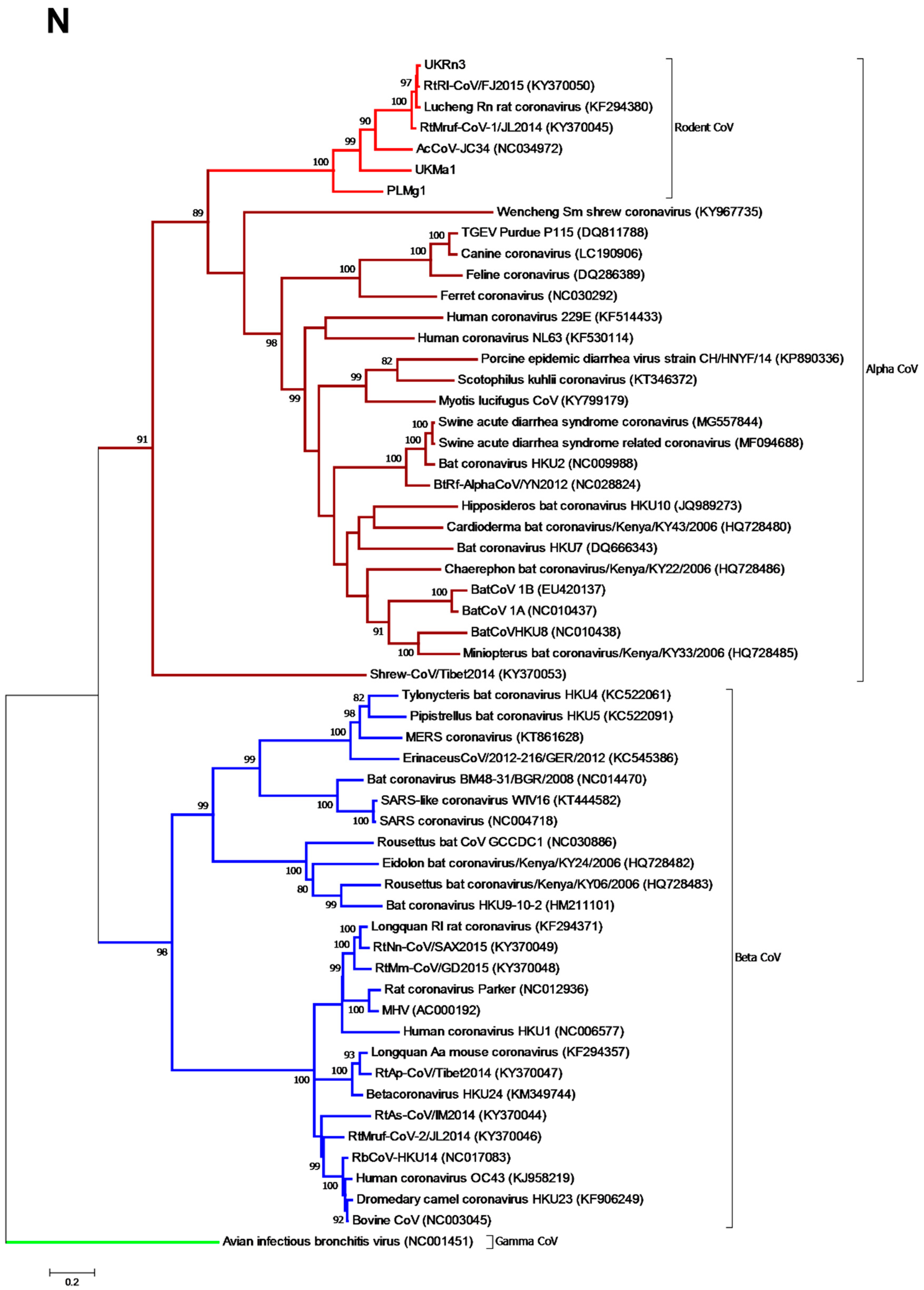
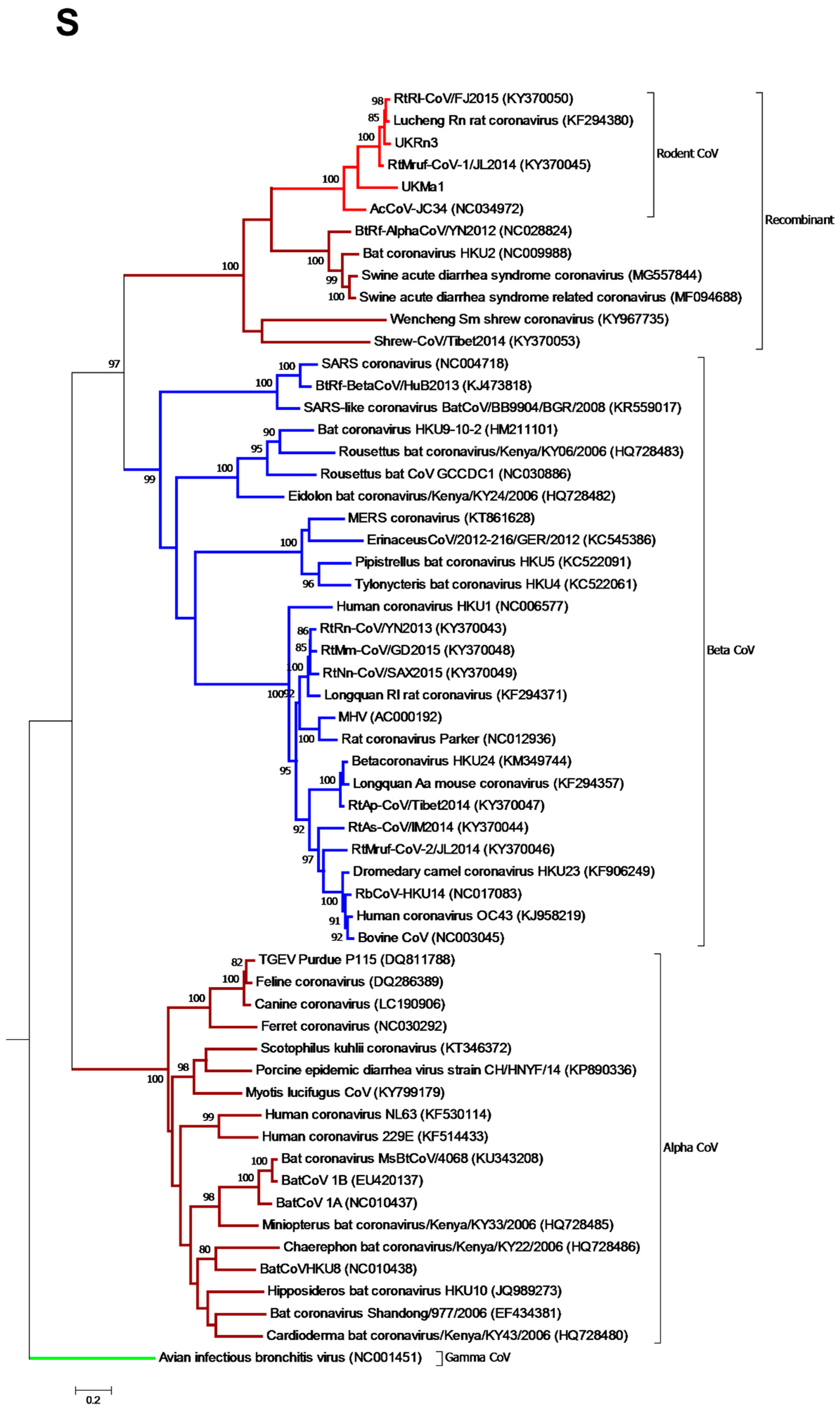
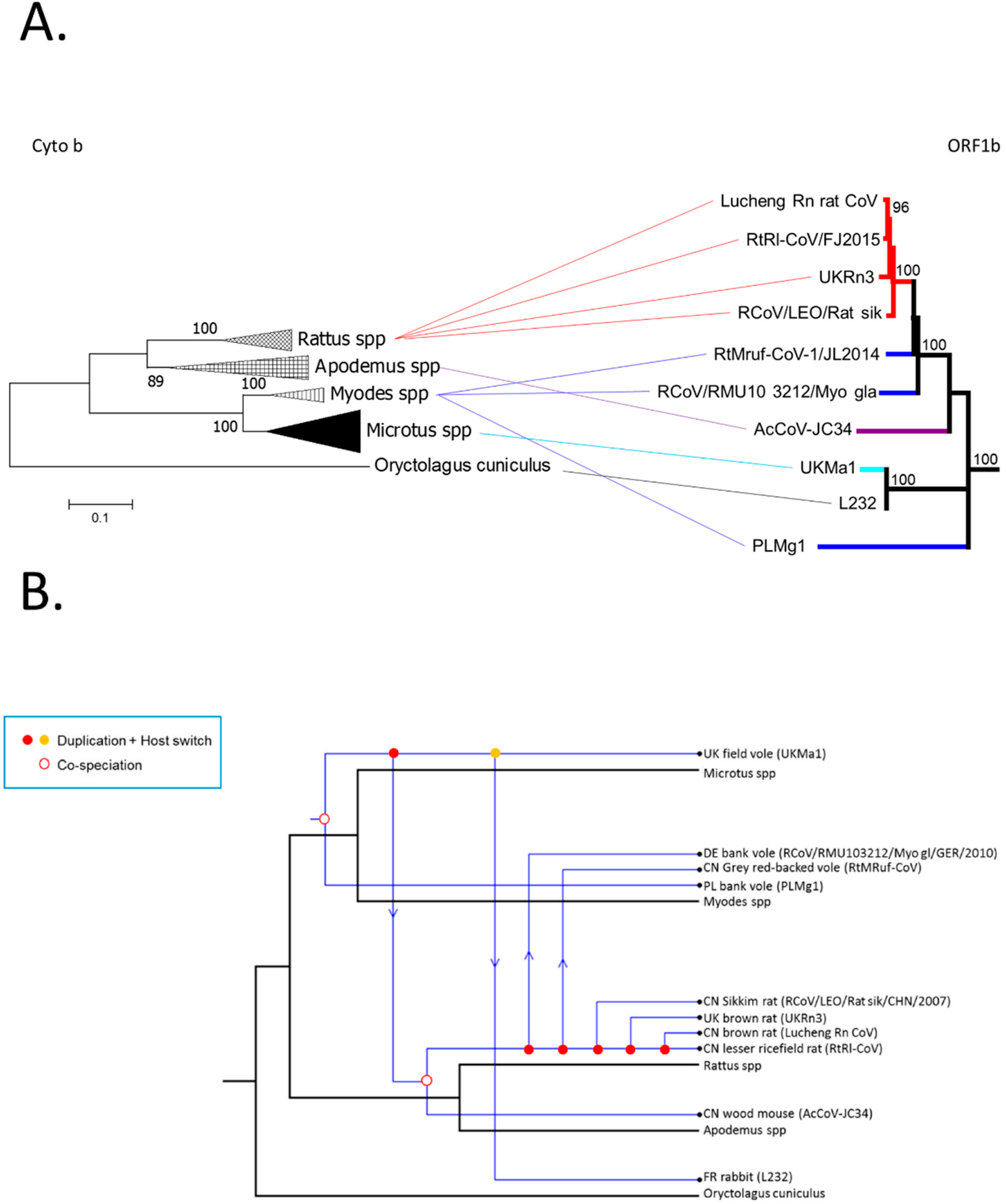
3.3. Phylogenetic Analysis of Rodent Alphacoronaviruses Suggests a Shared Common Ancestry

Having retrieved the whole genome for UKRn3 and partial genomes for UKMa1, PLMg1, and L232, we next evaluated the evolutionary relationships between the novel α-CoVs and the coronavirus sequences publicly available on GenBank. Accordingly, an analysis of a conserved 576 bp region of the ORF1b gene, which encodes the RNA-depended-RNA-polymerase (RdRp), revealed that all the rodent alpha-CoVs sampled from geographic localities as diverse as Western Europe and East Asia, as well as the rabbit L232 CoV, formed a distinct clade within the Alphacoronavirus genus with 100% bootstrap support (Figure 2). Within that cluster, viruses obtained from UK and Chinese rat species formed a single sub-clade with 100% bootstrap support. Other well supported subclades (i.e., all supported by >80% bootstrap replicates) contained (i) the Chinese grey red-backed vole (RtMruf), the German bank vole (RCoV/RMU10 3212/Myo gla/GER/2010), the Chinese field mouse (AcCoV-JC34), and (ii) the UK field vole (UKMa1) and French rabbit CoV (L232). The virus sampled from a Poland-resident bank vole (PLMg1) formed a distinct lineage.
Phylogenetic analysis of the N gene (Figure 3) revealed similar results to the ORF1b phylogeny, with all the available rodent coronaviruses forming a distinct clade within the genus Alphacoronavirus. The rat viruses contained within a single cluster supported by 100% of bootstrap trees, while PLMg1, UKMa1, and AcCoV-JC34 fell in strongly supported more divergent positions. The phylogenetic relationships within the rodent clade are therefore the same as those observed in the ORF1b phylogeny (rabbit L232 is not shown because it was not possible to retrieve the corresponding N gene sequence).
A markedly different evolutionary relationship was observed in the case of the partial S gene phylogeny, in which all the rodent-borne CoVs previously assigned as alpha-CoVs based on their ORF1 and N sequences clustered within the genus Betacoronavirus (Figure 4). Within this genome region, these viruses clustered with other known recombinant coronaviruses, including Wencheng Sm shrew CoV, Bat CoV HKU2, BtRf-AlphaCoV, Swine acute diarrhoea syndrome CoV, and swine acute diarrhoea syndrome related (bat) CoV. The rodent coronaviruses again formed a single branch with 100% bootstrap support, indicative of an ancient recombination event between the α- and β CoVs.
Importantly, amino acid-based ML trees were also inferred for the ORF1b, N, and S, and exhibited the same clustering of rodent alpha-CoVs with high bootstrap support (not shown, available on request).

3.4. Comparison of Virus and Host Phylogenies

To determine the extent to which the rodent CoVs may have co-diverged with their rodent hosts, indicative of a very long-term virus-host association, we examined the extent of congruence between the virus ORF1b (with the same topology observed in the N gene) and host mitochondrial (mt) cytochrome b phylogenies by inferring tanglegrams (Figure 5A). The tanglegram was generated by linking the phylogenies of rodent alpha-CoV ORF1b to their equivalent rodent species and assessing whether there were changes in the branching order in the two phylogenies, which would indicate cross-species transmission. The rabbit (Oryctolagus cuniculus; order Lagomorpha) sequence was used as outgroup as it has previously been shown to be closely related to the Rodentia order [5]. Notably, the topology of the cytochrome b tree did not fully match that observed for the rodent alpha-CoVs, in which PLMg1 (Myodes spp.) fell basal to all the other viruses, and RtMruf and RCoV/RMU10 3212/Myo gla/GER/2010 (Myodes spp.) were more closely related to Rattus spp. coronaviruses (UKRn3, Lucheng Rn, RtRl). Moreover, the rodent UKMa1 (Microtus spp.) clustered with L232 (Oryctolagus cuniculus) in the ORF1b tree, again indicative of some cross-species virus transmission.
In addition to the tanglegram, we conducted a co-phylogenetic analysis of the different types of evolutionary events that likely shaped the evolution of the rodent CoVs with their mammalian hosts (mtDNA cytochrome b) (Figure 5B). The co-phylogenetic JANE analysis between the rodent alpha-CoV ORF1b and the rodent cytochrome b phylogenies was performed to assess whether the two phylogenies matched, which would be indicative of co-evolution of virus and host. Under the most likely co-phylogenetic scenario, virus-host co-divergence explained 22% of all evolutionary events, with host-switching (i.e., cross-species transmission) more frequent at 45% (and duplications accounting for 33%), although the relative frequency of these events is likely to change with more extensive sampling. Hence, these data suggest that although the rodent alpha-CoV formed a monophyletic group indicative of a long virus-host association, the evolution of this group of viruses was likely characterized by relatively frequent cross-species transmission.

4. Discussion

We [9] and others [10,11,12,17,18] have previously shown the presence of novel CoVs in a range of rodent species. Here, we show that all rodent alpha-CoVs sampled to date—and from West Europe and East Asia—form a monophyletic group and have similar genome structure and common gene constellations and a recombinant spike gene. Hence, these data suggest that the rodent alpha-CoVs sampled to date originate from a single common ancestor and that there is a long association between rodents and CoVs. Moreover, our co-phylogenetic analysis revealed that the phylogenetic diversity in the rodent group was likely shaped by a combination of virus-host divergence and cross-species transmission, which appears to be a common mode of macroevolution in RNA viruses [19].
Also of note was the presence of a recombinant Betacoronavirus spike gene that clustered with a number of previously reported recombinant viruses that infect a diverse array of mammalian species, one of which was associated with a recent fatal outbreak in pigs in China [20,21,22,23]. Hence, the recombination event involving the S gene clearly occurred early on in the evolutionary history of the alpha- and betacoronaviruses.
In addition to the presence of a Betacoronavirus spike, the rodent CoVs also had similar, but not identical, gene constellations and genome features. Again, the most parsimonious explanation of this would be that all the rodent alpha-CoVs sampled to date originate from a single common ancestor, and the lack of an ORF2 (complete deletion) in AcCoV-JC34 likely reflects a secondary evolutionary loss.
Further analysis showed that the rodent alpha-CoVs shared the same predicted core TRS sequences upstream to each gene with a few exceptions. The predominant core TRS was 5′-AACUAA-3′. The same core sequence was also observed in Human NL63 (5′-AACUAAA-3′) [24] and Bat HKU2 CoV (5′-AACUAAA-3′) [20]. Although it was mentioned that no TRS was detected for ORF3 in Lucheng Rn CoV [17], we used the core TRS of AcCoV-JC34 (5′-UACUAAA-3′) [10] for ORF3 as a reference, and we were able to find the same sequence in Lucheng Rn CoV, UKRn3, and RtMruf. Other variations such as 5′-CACUAA-3′ and 5′-GACUAA-3′ were observed in ORF8 of UKMa1 and RtMruf, respectively. The variation 5′-CUAAAC-3′ has been observed in Feline infectious peritonitis virus (FIPV) and Transmissible gastroenteritis virus (TGEV) [25,26]. The conserved core TRS among rodent alpha-CoVs is another indicator that they originate from a common ancestor.
Comparison of rodent species’ cytochrome b and coronavirus ORF1b evolution showed incomplete congruence, such that despite forming a monophyletic group, these rodent CoVs also experienced cross-species transmission. In particular, according to the cytochrome b analysis, Rattus spp. formed a distinct clade with Apodemus spp., whereas Microtus spp. formed a separate clade with Myodes spp. However, according to ORF1b phylogeny, Rattus spp. CoVs clustered with two Myodes spp. CoV (RtMruf and RCoV/RMU10 3212/Myo gla/GER/2010), suggesting cross-species transmission (and this was confirmed in the co-phylogenetic analysis). Cross-species transmission is frequently observed in coronaviruses and representative examples are SARS-CoV, MERS-CoV, and SADS/SADSr-CoV [21,27].
That the rodent alpha-CoVs form a monophyletic group despite being sampled from multiple localities suggests that they have been associated with rodents for an extended period, although providing an exact chronological context is complex. Rattus norvegicus originated in Southeast Asia and spread to Northeast Asia approximately 200,000 years ago. It then spread from Southeast Asia to the Middle East ~3100 years ago, then to Africa ~2000 years ago, and finally to Europe ~1800 years ago [28]. Therefore, it is possible that even though the European UKRn3 coronavirus appears to be approximately as old as Lucheng Rn CoV, the global spread of the brown rat implies that the virus introduction into Europe occurred with that host and was a relatively recent event.
In sum, we show that all the rodent alpha-CoVs described to date share a single common ancestor and have likely evolved through a combination of virus-host co-divergence and cross-species transmission. Not only is this common ancestry evident in the phylogenetic relationships inferred from multiple genes, but also genomic signatures are the recombinant spike gene, same gene constellations (ORF2, ORF3, ORF6, ORF8, and ORF9), and similar core TRS sequences. Finally, although the current sequence data set of rodent alphacoronaviruses is relatively small, it is representative of a number of different rodent species living across a wide geographical area.

Supplementary Materials

The following are available online at https://www.mdpi.com/1999-4915/11/2/125/s1, Table S1 and Table S2.

Author Contributions

T.T., C.P.M. and J.K.B. conceived and designed the experiments; T.T., J.G.C. and O.O. performed the experiments; D.A.M. and A.R.F. performed NGS on our rodent alpha-CoV samples; E.M.-L., G.U. and R.E.T. provided samples; M.A.M., J.F.D. and C.D. provided training and contributed in genome retrieval of the viruses; T.T., E.C.H. and J.K.B. analysed the data and wrote the paper; all authors edited the final draft.

Funding

This study was supported by the Biotechnology and Biological Sciences Research Council [grant number BB/J014508/1]. ECH is supported by an ARC Australian Laureate Fellowship (FL170100022). JFD is supported by the European Union’s Horizon 2020 research and innovation program through the COMPARE project (grant agreement no. 643476).

Conflicts of Interest

The authors declare no conflict of interest.

References

  1. Hudson, C.; Beaudette, F.R. Infection of the cloaca with the virus of infectious bronchitis. Science 1932, 76, 34. [Google Scholar] [CrossRef]
  2. Hamre, D.; Procknow, J.J. A new virus isolated from the human respiratory tract. Proc. Soc. Exp. Biol. Med. 1966, 121, 190–193. [Google Scholar] [CrossRef]
  3. Weiss, S.; Navas-Martin, S. Coronavirus pathogenesis and the emerging pathogen severe acute respiratory syndrome coronavirus. Microbiol. Mol. Biol. Rev. 2005, 69, 635–664. [Google Scholar] [CrossRef]
  4. Luis, A.D.; Hayman, D.T.; O’Shea, T.J.; Cryan, P.M.; Gilbert, A.T.; Pulliam, J.R.; Mills, J.N.; Timonin, M.E.; Willis, C.K.; Cunningham, A.A.; et al. A comparison of bats and rodents as reservoirs of zoonotic viruses: Are bats special? Proc. Biol. Sci. 2013, 280. [Google Scholar] [CrossRef]
  5. Huchon, D.; Madsen, O.; Sibbald, M.J.; Ament, K.; Stanhope, M.J.; Catzeflis, F.; de Jong, W.W.; Douzery, E.J. Rodent phylogeny and a timescale for the evolution of Glires: Evidence from an extensive taxon sampling using three nuclear genes. Mol. Biol. Evol. 2002, 19, 1053–1065. [Google Scholar] [CrossRef]
  6. Phan, T.G.; Kapusinszky, B.; Wang, C.; Rose, R.K.; Lipton, H.L.; Delwart, E.L. The fecal viral flora of wild rodents. PLoS Pathog. 2011, 7, e1002218. [Google Scholar] [CrossRef]
  7. Cheever, F.S.; Daniels, J.B.; Pappenheimer, A.M.; Bailey, O.T. A murine virus (JHM) causing disseminated encephalomyelitis with extensive destruction of myelin. J. Exp. Med. 1949, 90, 181–194. [Google Scholar] [CrossRef]
  8. Parker, J.C.; Cross, S.S.; Rowe, W.P. Rat coronavirus (RCV): A prevalent, naturally occurring pneumotropic virus of rats. Arch. Gesamte Virusforsch. 1970, 31, 293–302. [Google Scholar] [CrossRef] [PubMed]
  9. Tsoleridis, T.; Onianwa, O.; Horncastle, E.; Dayman, E.; Zhu, M.; Danjittrong, T.; Wachtl, M.; Behnke, J.M.; Chapman, S.; Strong, V.; et al. Discovery of Novel Alphacoronaviruses in European Rodents and Shrews. Viruses 2016, 8, 84. [Google Scholar] [CrossRef] [PubMed]
  10. Ge, X.Y.; Yang, W.H.; Zhou, J.H.; Li, B.; Zhang, W.; Shi, Z.L.; Zhang, Y.Z. Detection of alpha- and betacoronaviruses in rodents from Yunnan, China. Virol. J. 2017, 14, 98. [Google Scholar] [CrossRef]
  11. Hu, D.; Zhu, C.; Wang, Y.; Ai, L.; Yang, L.; Ye, F.; Ding, C.; Chen, J.; He, B.; Zhu, J.; et al. Virome analysis for identification of novel mammalian viruses in bats from Southeast China. Sci. Rep. 2017, 7, 10917. [Google Scholar] [CrossRef] [Green Version]
  12. Monchatre-Leroy, E.; Boué, F.; Boucher, J.-M.; Renault, C.; Moutou, F.; Ar Gouilh, M.; Umhang, G. Identification of Alpha and Beta Coronavirus in Wildlife Species in France: Bats, Rodents, Rabbits, and Hedgehogs. Viruses 2017, 9, 364. [Google Scholar] [CrossRef]
  13. Shi, M.; Lin, X.D.; Tian, J.H.; Chen, L.J.; Chen, X.; Li, C.X.; Qin, X.C.; Li, J.; Cao, J.P.; Eden, J.S.; et al. Redefining the invertebrate RNA virosphere. Nature 2016. [Google Scholar] [CrossRef]
  14. Thompson, J.D.; Higgins, D.G.; Gibson, T.J. CLUSTAL W: Improving the sensitivity of progressive multiple sequence alignment through sequence weighting, position-specific gap penalties and weight matrix choice. Nucleic Acids Res. 1994, 22, 4673–4680. [Google Scholar] [CrossRef]
  15. Kumar, S.; Stecher, G.; Tamura, K. MEGA7: Molecular Evolutionary Genetics Analysis Version 7.0 for Bigger Datasets. Mol. Biol. Evol. 2016, 33, 1870–1874. [Google Scholar] [CrossRef]
  16. Conow, C.; Fielder, D.; Ovadia, Y.; Libeskind-Hadas, R. Jane: A new tool for the cophylogeny reconstruction problem. Algorithms for Mol. Biol. 2010, 5, 16. [Google Scholar] [CrossRef]
  17. Wang, W.; Lin, X.D.; Guo, W.P.; Zhou, R.H.; Wang, M.R.; Wang, C.Q.; Ge, S.; Mei, S.H.; Li, M.H.; Shi, M.; et al. Discovery, diversity and evolution of novel coronaviruses sampled from rodents in China. Virology 2015, 474, 19–27. [Google Scholar] [CrossRef] [Green Version]
  18. Lau, S.K.; Woo, P.C.; Li, K.S.; Tsang, A.K.; Fan, R.Y.; Luk, H.K.; Cai, J.P.; Chan, K.H.; Zheng, B.J.; Wang, M.; et al. Discovery of a novel coronavirus, China Rattus coronavirus HKU24, from Norway rats supports the murine origin of Betacoronavirus 1 and has implications for the ancestor of Betacoronavirus lineage A. J. Virol. 2015, 89, 3076–3092. [Google Scholar] [CrossRef]
  19. Geoghegan, J.L.; Duchene, S.; Holmes, E.C. Comparative analysis estimates the relative frequencies of co-divergence and cross-species transmission within viral families. PLoS Pathog. 2017, 13, e1006215. [Google Scholar] [CrossRef]
  20. Lau, S.K.; Woo, P.C.; Li, K.S.; Huang, Y.; Wang, M.; Lam, C.S.; Xu, H.; Guo, R.; Chan, K.H.; Zheng, B.J.; et al. Complete genome sequence of bat coronavirus HKU2 from Chinese horseshoe bats revealed a much smaller spike gene with a different evolutionary lineage from the rest of the genome. Virology 2007, 367, 428–439. [Google Scholar] [CrossRef] [Green Version]
  21. Zhou, P.; Fan, H.; Lan, T.; Yang, X.-L.; Shi, W.-F.; Zhang, W.; Zhu, Y.; Zhang, Y.-W.; Xie, Q.-M.; Mani, S.; et al. Fatal swine acute diarrhoea syndrome caused by an HKU2-related coronavirus of bat origin. Nature 2018, 556, 255–258. [Google Scholar] [CrossRef]
  22. Wang, W.; Lin, X.D.; Liao, Y.; Guan, X.Q.; Guo, W.P.; Xing, J.G.; Holmes, E.C.; Zhang, Y.Z. Discovery of a highly divergent coronavirus in the Asian house shrew from China illuminates the origin of the alphacoronaviruses. J. Virol. 2017. [Google Scholar] [CrossRef]
  23. Wu, Z.; Yang, L.; Ren, X.; He, G.; Zhang, J.; Yang, J.; Qian, Z.; Dong, J.; Sun, L.; Zhu, Y.; et al. Deciphering the bat virome catalog to better understand the ecological diversity of bat viruses and the bat origin of emerging infectious diseases. ISME J. 2016, 10, 609–620. [Google Scholar] [CrossRef]
  24. Pyrc, K.; Jebbink, M.F.; Berkhout, B.; van der Hoek, L. Genome structure and transcriptional regulation of human coronavirus NL63. Virol. J. 2004, 1, 7. [Google Scholar] [CrossRef]
  25. Dye, C.; Siddell, S.G. Genomic RNA sequence of Feline coronavirus strain FIPV WSU-79/1146. J. Gen. Virol. 2005, 86, 2249–2253. [Google Scholar] [CrossRef] [Green Version]
  26. Hiscox, J.A.; Mawditt, K.L.; Cavanagh, D.; Britton, P. Investigation of the control of coronavirus subgenomic mRNA transcription by using T7-generated negative-sense RNA transcripts. J. Virol. 1995, 69, 6219–6227. [Google Scholar]
  27. De Wit, E.; van Doremalen, N.; Falzarano, D.; Munster, V.J. SARS and MERS: Recent insights into emerging coronaviruses. Nat. Rev. Microbiol 2016, 14, 523–534. [Google Scholar] [CrossRef]
  28. Zeng, L.; Ming, C.; Li, Y.; Su, L.-Y.; Su, Y.-H.; Otecko, N.O.; Dalecky, A.; Donnellan, S.; Aplin, K.; Liu, X.-H.; et al. Out of Southern East Asia of the Brown Rat Revealed by Large-Scale Genome Sequencing. Mol. Biol. Evol. 2018, 35, 149–158. [Google Scholar] [CrossRef]
Figure 1. Genome organisation of rodent alphacoronaviruses. Schematic representation of the UKRn3 genome organisation along with those of other rodent alphacoronaviruses (α-CoVs): Lucheng Rn CoV (LRNV), AcCoV-JC34, RtRl-CoV, RtMruf-CoV, and the related Bat-HKU2 and Swine acute diarrhoea syndrome coronaviruses (CoV) (SADS-CoV). Its genome organisation was similar to those of members of the genus Alphacoronavirus, with the characteristic 5′- ORF1ab - spike (S) - envelope (E)membrane (M) - nucleocapsid (N) - 3′ gene order. The red colour represents genes that belong to the genus Alphacoronavirus, whereas light blue denotes genes that belong to the genus Betacoronavirus, indicative of recombination. The Human 229E genome represents a typical alpha-CoV genome organisation.
Viruses 11 00125 g001
Figure 2. Phylogenetic relationships of the rodent alphacoronaviruses based on the ORF1b gene. Maximum likelihood phylogenetic analysis of coronavirus partial 576 nt ORF1b gene sequences, revealing that the rodent alphaviruses formed a single clade with the novel rabbit L232 CoV within the genus alphacoronavirus. The novel coronavirus sequence obtained from a French rabbit L232 CoV was analysed alongside reference sequences representing three different coronavirus genera (Alphacoronavirus, Betacoronavirus, and Gammacoronavirus as an outgroup). Reference sequences are indicated by their GenBank accession numbers. Branch lengths are drawn to a scale of nucleotide substitutions per site. Numbers above individual branches indicate the percentage that that branch was found in 1000 bootstrap replicates; only values >80% are shown. Alpha-CoVs are highlighted in brown, betacoronaviruses in blue, gammacoronaviruses in green, and the rodent coronaviruses in light red.
Viruses 11 00125 g002
Figure 3. Phylogenetic relationships of the rodent alphacoronaviruses based on the N gene. Maximum likelihood phylogenetic analysis of coronavirus partial N gene sequences corresponding to positions 10–1173 of the Lucheng Rn CoV (KF294380). Novel coronavirus sequences obtained from PL-resident Myodes glareolus (PLMg1) and the UK Microtus agrestis (UKMa1) and the UK Rattus norvegicus (UKRn3) were analysed alongside reference sequences representing three different coronavirus genera (Alphacoronavirus, Betacoronavirus, and Gammacoronavirus as an outgroup). Reference sequences are indicated by their GenBank accession numbers. Branch lengths are drawn to a scale of nucleotide substitutions per site. Numbers above individual branches indicate the percentage that that branch was found in 1000 bootstrap replicates; only values >80% are shown. Alpha-CoVs are highlighted in brown, betacoronaviruses in blue, gammacoronaviruses in green, and the rodent coronaviruses in light red.
Viruses 11 00125 g003
Figure 4. Phylogenetic relationships of the rodent alphacoronaviruses based on the S gene. Maximum likelihood phylogenetic analysis of coronavirus partial S gene sequences corresponding to positions 1831–3228 of the Lucheng Rn CoV (KF294380), indicating that the rodent alpha-CoVs formed a distinct clade within the genus Betacoronavirus. The novel coronavirus sequence obtained from a UK-resident Microtus agrestis was analysed alongside reference sequences representing three different coronavirus genera (Alphacoronavirus, Betacoronavirus, and Gammacoronavirus as an outgroup). Reference sequences are indicated by their GenBank accession numbers. Branch lengths are drawn to a scale of nucleotide substitutions per site. Numbers above individual branches indicate the percentage that the branch was found in 1000 bootstrap replicates; only values >80% are shown. Alpha-CoVs are highlighted in brown, betacoronaviruses in blue, gammacoronaviruses in green, and the rodent coronaviruses in light red.
Viruses 11 00125 g004
Figure 5. (A) Tanglegram of rodent cytochrome b and CoV ORF1b evolution. Maximum likelihood phylogenetic analysis of full-length rodent cytochrome b nucleotide sequences compared to rodent alpha-CoV ORF1b phylogeny. Cytochrome b sequences were obtained from GenBank for the rodent species Microtus, Myodes, Rattus, and Apodemus, and the rabbit Oryctolagus cuniculus, which was used as an outgroup. Branch lengths are drawn to scale: the bar indicates 0.1 nucleotide substitutions per site. Numbers above individual branches indicate the percentage that that branch was found in 1000 bootstrap replicates; only percentages >80 are shown. Blue indicates the Myodes spp., cyan the Microtus spp., purple the Apodemus spp., red Rattus spp., and black indicates Oryctolagus cuniculus. (B) Reconciliation of the sampled rodent alpha-CoV phylogeny with that of their mammalian hosts, utilizing the co-phylogenetic method implemented in the Jane package [16]. This figure shows the maximum possible co-divergence, duplication, and host switching events.
Viruses 11 00125 g005
Table 1. List of sample name, species, country of origin, organ, and original publication.
NameSpeciesCountryOrganReference
UKRn3Rattus norvegicusUnited KingdomLiver[9]
UKMa1Microtus agrestisUnited KingdomGut[9]
PLMg1Myodes glareolusPolandLiver[9]
L232Oryctolagus cuniculusFranceIntestinal fluid[12]
Table 2. List of the different genes used for phylogenetic analyses, number of sequences, sequence length, and corresponding positions on the Lucheng CoV genome.
GeneNumber of SequencesLength (nt)Corresponding Positions on Lucheng CoV Genome
ORF1b4657614,140–14,715
S31139823,242–24,639
N32117026,977–28,146
Table 3. List of all the predicted core transcription regulatory sequences (TRS) for all the genes of the rodent alpha-CoVs. Core TRS could not be predicted for ORF1ab, S, ORF3, E, and M for UKMa1 as there was no available sequence for those genes. AcCoV-JC34 did not possess an ORF2 such that there was no core TRS for that gene.
VirusGenes
ORF1abORF2SORF3EMORF6NORF8ORF9
UKRn3AACUAAAACUUUAAAACUAAUACUAAAACUAAAACUAAAACUAAAACUAAAACUAAAACUAA
LRNVAACUAAAACUAAAACUAAUACUAAAACUAAAACUAAAACUAAAACUAAAACUAAAACUAA
UKMa1N/AAACUUUAAN/AN/AN/AN/AAACUAAAACUAACACUAAAACUAA
RtRlAACUAAAACUUUAAAACUAAAACUAAAACUAAAACUAAAACUAAAACUAAAACUAAAACUAA
RtMrufAACUAAAACUUUAAAACUAAUACUAAAACUAAAACUAAAACUAAAACUAAGACUAAAACUAA
JC34AACUAAN/AAACUUAUACUAAAAACUAAAACUAAAACUAAAACUAACACUAAAACUAA

