Article

New Method for Differentiation of Granuloviruses (Betabaculoviruses) Based on Real-Time Polymerase Chain Reaction (Real-Time PCR)

by
Martyna Krejmer-Rabalska
1,
Lukasz Rabalski
1,*,
Michael D. Jukes
2,3,
Marlinda Lobo de Souza
4,
Sean D. Moore
3,5 and
Boguslaw Szewczyk
1
1
Laboratory of Recombinant Vaccines, Intercollegiate Faculty of Biotechnology of University of Gdansk and Medical University of Gdansk, University of Gdansk, 80-307 Gdansk, Poland
2
Department of Biochemistry and Microbiology, Rhodes University, P.O. Box 94, Grahamstown 6140, South Africa
3
Centre for Biological Control, Department of Zoology and Entomology, Rhodes University, P.O. Box 94, Grahamstown 6140, South Africa
4
Embrapa Recursos Genéticos e Biotecnologia, Parque Estacao Biológica, Brasilia 70770-900, Brazil
5
Citrus Research International (CRI), P.O. Box 5095, Walmer 6065, Port Elizabeth, South Africa
*
Author to whom correspondence should be addressed.
Viruses 2019, 11(2), 115; https://doi.org/10.3390/v11020115
Submission received: 23 December 2018 / Revised: 24 January 2019 / Accepted: 24 January 2019 / Published: 29 January 2019
(This article belongs to the Special Issue Insect Viruses and Pest Management)

Abstract

:
Baculoviridae is a highly diverse family of rod-shaped viruses with double-stranded DNA. To date, almost 100 species have had their complete genomic sequences deposited in the GenBank database, a quarter of which comprises granuloviruses (GVs). Many of the genomes are sequenced using next-generation sequencing, which is currently considered the best method for characterizing new species, but it is time-consuming and expensive. Baculoviruses form a safe alternative to overused chemical pesticides and therefore there is a constant need for identifying new species that can be active components of novel biological insecticides. In this study, we have described a fast and reliable method for the detection of new and differentiation of previously analyzed granulovirus species based on a real-time polymerase chain reaction (PCR) technique with melting point curve analysis. The sequences of highly conserved baculovirus genes, such as granulin and late expression factors 8 and 9 (lef-8 and lef-9), derived from GVs available to date have been analyzed and used for degenerate primer design. The developed method was tested on a representative group of eight betabaculoviruses with comparisons of melting temperatures to allow for quick and preliminary granulovirus detection. The proposed real-time PCR procedure may be a very useful tool as an easily accessible screening method in a majority of laboratories.

1. Introduction

Insect pests pose a threat for crops and forests all over the world. To manage them, chemical control agents are usually applied, and are very often overused, because an immediate effect is sought. To reduce chemicals in the environment, there is a constant need for finding novel biologically safe alternatives. Some very promising candidates for biopesticides belong to the Baculoviridae family, which is currently the biggest insect virus family, comprising around 1000 described species of insect viruses [1,2,3]. Baculoviruses are known to naturally control many insect populations. They not only possess a very narrow host range but also do not accumulate in the environment and, being selective, are safe for humans and animals. They have been applied in the field with greater or lesser success since the 1940s, but in the last two decades, they have once again gained popularity [3,4].
The Baculoviridae family consists of viruses with double-stranded DNA, covalently closed, circular genomes [5] that range in size from 82 kilo-base pair (kbp) (Neodiprion lecontei nucleopolyhedrovirus (NeleNPV) [6]) to 179 kbp (Xestia c-nigrum granulovirus (XecnGV) [7]) and encode between 89 and 181 open reading frames (orfs). Based on common structural features, phylogeny, and host taxonomy, baculoviruses are divided into four genera. Of these, the genus Betabaculovirus contains all known lepidopteran-specific granuloviruses. Its representatives differ from members of the remaining three genera—Alphabaculovirus, Deltabaculovirus, and Gammabaculovirus—because of smaller, ovo-cylindrical occlusion bodies consisting of the major occlusion protein, granulin, which is a highly conserved structural protein [8,9,10]. The life cycle of granuloviruses (GVs) is similar to that of alphabaculoviruses, while little detail is known about the remaining two genera [11]. Betabaculoviruses are reported to have different tissue tropism and, on this basis, are divided into three groups [12]. To the first group belong the slow-killing GVs that infect the insect midgut epithelium and fat tissue. Representatives include XcGV [7] or Helicoverpa armigera granulovirus (HearGV) [13]. The second group consists of fast-killing GVs, including Cydia pomonella granulovirus (CpGV) [14], Cryptophlebia leucotreta granulovirus (CrleGV) [15,16] and Epinotia aporema granulovirus (EpapGV) [17]. In this group, the infection spreads in most tissues of the caterpillar in a similar manner to alphabaculovirus infections, resulting in the fast death of an infected larvae. The third group is known only for Harrisina brillians granulovirus (HabrGV) [18] and is restricted to the midgut epithelium. This group is highly infectious, causing diarrhea in infected larvae, resulting in the quick spread of infection in larval populations [19]. The complete genome of HabrGV remains unknown [20] (GenBank, 2018).
All members of the Baculoviridae family retain a set of 38 core genes conserved in all genomic sequences, with this set of common genes playing an important role in the baculovirus life cycle. These genes are involved in viral functions such as transcription, DNA replication, virion structure, and impairing cellular metabolism during infection [21,22,23]. For example, the late expression factor 8 and 9 (lef-8 and lef-9) core genes code for viral RNA polymerase subunits, which regulate transcription of late and very late viral genes [11,24].
Nearly 100 genomes of the Baculoviridae family species have been sequenced and deposited in GenBank to date, of which 24 are granuloviruses (GenBank, 2018). At present, the most reliable technique for detection and confirmation of new species is Sanger sequencing of fragments of gran, lef-8 and lef-9 genes [25]. This method is based on generating (during specific PCR conditions for every gene) amplicons and further sequencing. Its main drawback is an obligatory nucleotide sequence analysis which cannot be performed in a majority of laboratories and also generates extra costs. Another technique that can be used for new species detection is a whole genome sequencing (no need for specific primers/PCR). Unfortunately, Next Generation Sequencing (NGS) technology has some disadvantages. Although it becomes less expensive each consecutive year, the cost is still very high, and the procedure includes time-consuming sample preparation and data processing. Furthermore, it cannot be used routinely in screening samples from the field because there is a need for long and complicated library preparations [26].
It was reported that one insect host species may be infected with a few different baculoviruses [27,28] or, conversely, one virus can have different hosts [29], indicating that an efficient screening method for mixed populations of multiple baculoviruses may be useful. Methods based on PCR utilizing granulin or polyhedrin sequences with specific primers enables simple detection of known NPVs or GVs in a sample and has been commonly used in many laboratories. PCR amplification followed by restriction fragment length polymorphism (PCR-RFLP), with degenerate primers for the polyhedrin fragment, was also used for the identification of a few NPVs [30]. Subsequently, a PCR-based method for the detection and identification of different species was established for NPVs and GVs, but the sequencing of products was necessary [31]. All these methods rely on the identification of different species at the end-point and not during the reaction. Fast differentiation of a few (or completely new) species in the environmental sample may be quite complex. In the past, our team has developed a method for the differentiation of nucleopolyhedroviruses [32] and, more recently, for granuloviruses [33], both based on the Multi-temperature Single-Stranded Conformational Polymorphism (MSSCP) technique [34]. However, in the current study, we report a method based on real-time polymerase chain reaction (real-time PCR) that allows for differentiation between betabaculoviruses. The method can be complimentary to the MSSCP method or it can be used to screen large sample sets for a variety of granuloviruses without requiring sequencing of each amplicon generated.

2. Materials and Methods

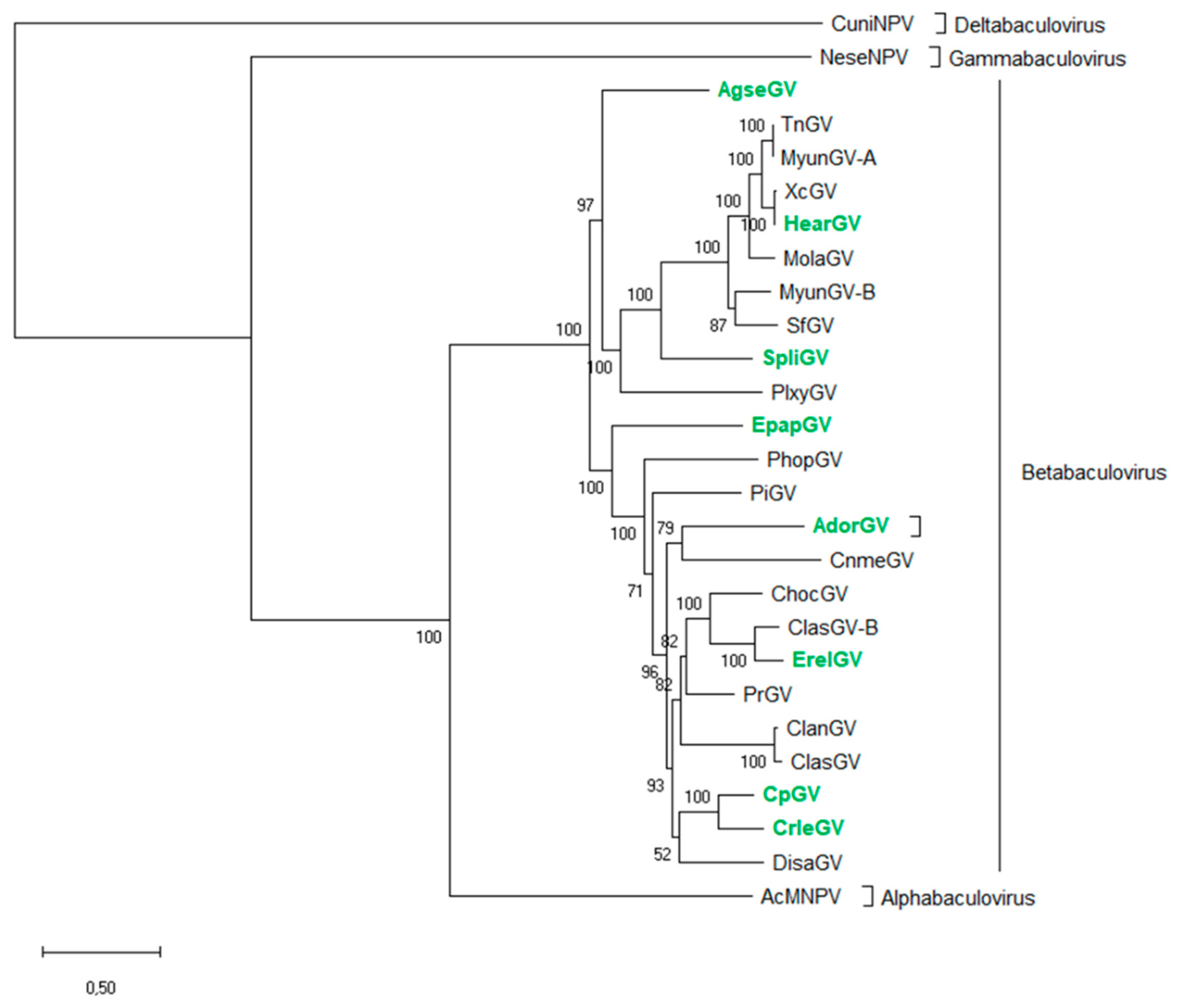
2.1. Phylogenetic Analysis of Granuloviruses

The 38 core genes were extracted from nucleotide sequences of genomes of each betabaculovirus available in the GenBank database and were translated into amino acid sequences using Geneious Pro 7.1 (Biomatters, Auckland, New Zealand). Then, the MAFFT alignment was performed for each gene from all the known species. Subsequently, alignments were concatenated and the maximum-likelihood (ML) method applied with 1000 bootstrap replicates (percentage of replicates in which the associated Baculoviridae members clustered together is presented next to the branches) used to construct a phylogenetic tree using MEGA 7 software. GenBank accession numbers of available betabaculovirus species are presented in Table 1. The alphabaculovirus Autographa californica multiple nucleopolyhedrovirus (AcMNPV; L22858), deltabaculovirus Culex nigripalpus nucleopolyhedrovirus (CuniNPV; AF403738), and gammabaculovirus Neodiprion sertifer nucleopolyhedrovirus (NeseNPV; AY430810) were added to demonstrate the general relationship in the Baculoviridae family.

2.2. Determination of Betabaculovirus Representative Group

A representative group of eight betabaculoviruses were chosen for further studies on the basis of the phylogenetic tree (Section 2.1) and meeting the following criteria: (i) different grannulin, lef-8 and lef-9 sequences, (ii) different clades among the genera, (iii) distant from each other in the phylogenetic tree, (iv) a pair of closely related species included, and (v) DNA availability.

2.3. Virus Purification and DNA Isolation

Viruses were purified from crude larval homogenates through filtration and then centrifugation at 5000× g for 10 min at room temperature. The supernatant was discarded, and a 200 µL of ddH2O was added to the pellet and centrifuged again as before. The supernatant was discarded, and the pellet with granules was re-suspended in 0.5% SDS and centrifuged as before. In the next step, the pellet was re-suspended in 0.5 M NaCl and the centrifugation was repeated. Thereafter, the granules in the pellet were re-suspended in a 200 µL of ddH2O. Such samples were then dissolved in an alkaline solution (0.1 M Na2CO3, pH 10.0) for 30 min. The solution was then neutralized with an equal amount of Tris-HCl buffer (pH 6.4). The DNA extraction was performed with the use of the MagAttract HMW DNA Kit (Qiagen, Venlo, The Netherlands) according to the manufacturer’s protocol. The DNA concentration was determined using a Quantus Fluorometer (Promega, Madison, WI, USA) and its dedicated Qubit dsDNA HS (High Sensitivity) Assay Kit (Thermo Fisher Scientific, Waltham, MA, USA).

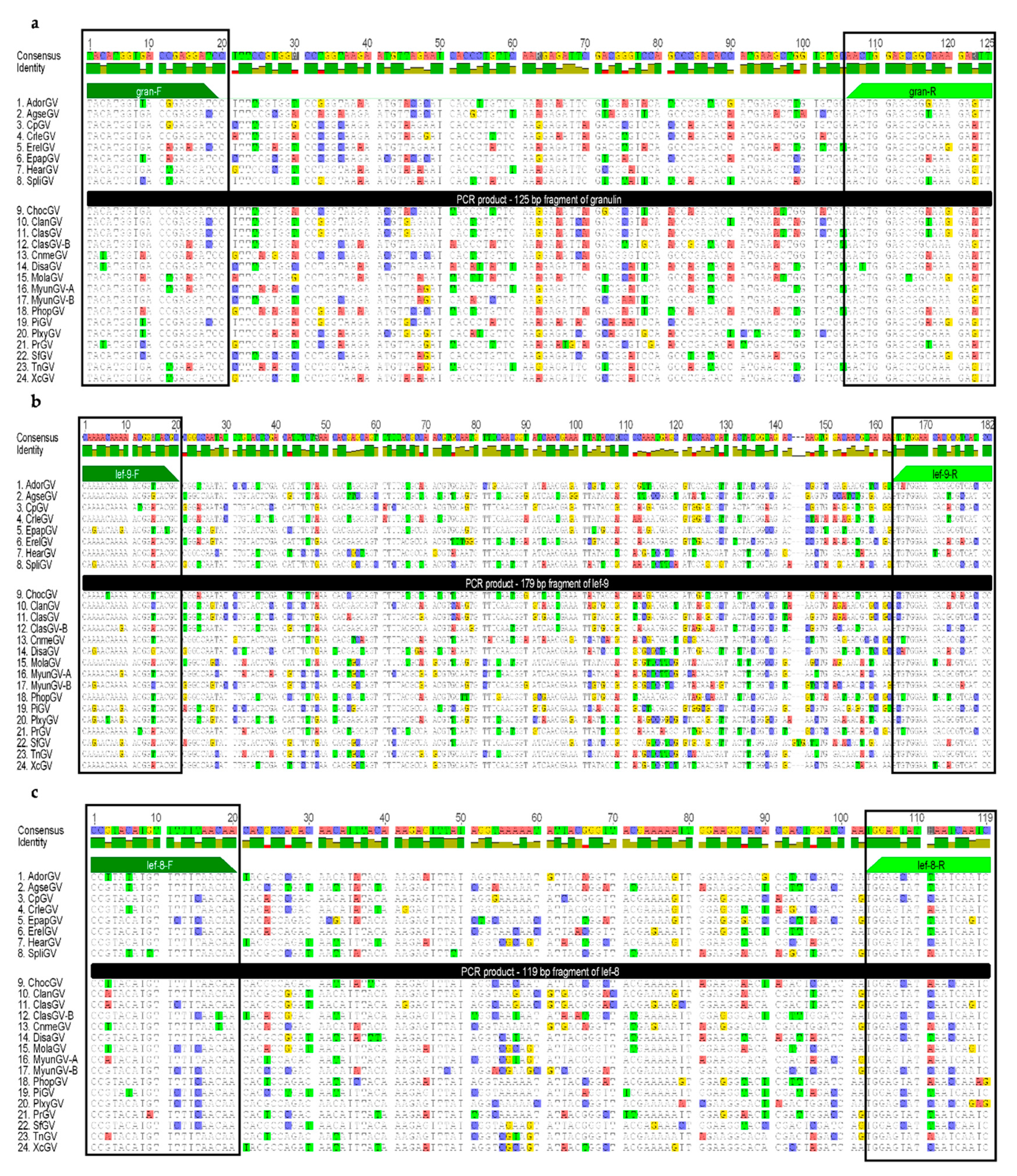
2.4. Granulin, Late Expression Factor-9 and Late Expression Factor-8 Nucleotide Sequence Alignments and Degenerate Primer Design; Amino Acid Alignment of the PCR Products.

The nucleotide sequence alignments based on whole genes, granulin, late expression factor-9 (lef-9), and late expression factor-8 (lef-8), were extracted from the genome sequences of betabaculoviruses (24 species) available in the GenBank database (Table 1) using Geneious Pro 7.1 (Biomatters, Auckland, New Zealand) with default settings. Prior to primer design for real-time PCR, the alignments were manually analyzed to choose the most variable region occurring between the two most conserved sequences while also being around 100–250 bp in length. Although gran, lef-9 and lef-8 share high levels of similarity between GVs, there are some differences in the nucleotide sequences, and to therefore enable the detection of different species, it is necessary to design degenerate universal primers. The primers used for short fragments of granulin (125 bp) and lef-9 (179 bp) previously published by Krejmer-Rabalska et al. [33] (gran-F–5′TAC ATG GTB ACN GAR GA3′, Tm = 43.4–52.2 °C, gran-R-5′AAY TCY TTN CCG CTC CAG TT3′, Tm = 51.9–59.4 °C; lef-9-F-5′CAR AAC AAR AAY GGR TAY GC3′, Tm = 45.9–56.1 °C, lef-9-R-5′GGR TGN CGH GTG TTC CAY AC3′, Tm = 52.7–64.3 °C) were re-evaluated against a broader group of betabaculovirus species (all 24 available, Figure 3a,b) for use in real-time PCR reactions. Degenerate primers were designed to amplify a short fragment of lef-8 (119 bp)—lef-8-F-5′CCK TAY ATK TTY TTY AAC AA3′, Tm = 39–50.5 °C, lef-8-R-5′GAT TGA TTD ATR CTC CA3′, Tm = 39.2–46.4 °C (Figure 3c; IUPAC genetic code: B–C or G or T, N–A or T or C or G; R–A or G, Y–C or T, H–A or C or T, K–G or T, D–A or G or T).
The nucleotide sequences of the predicted PCR products were extracted from the various genomic sequences along with one or two additional nucleotides (to maintain the correct open reading frame), translated into amino acid sequences, and aligned using Geneious Pro 7.1 (Biomatters, Newark, NJ, USA), with forward and reverse primers shown (Figure S1).

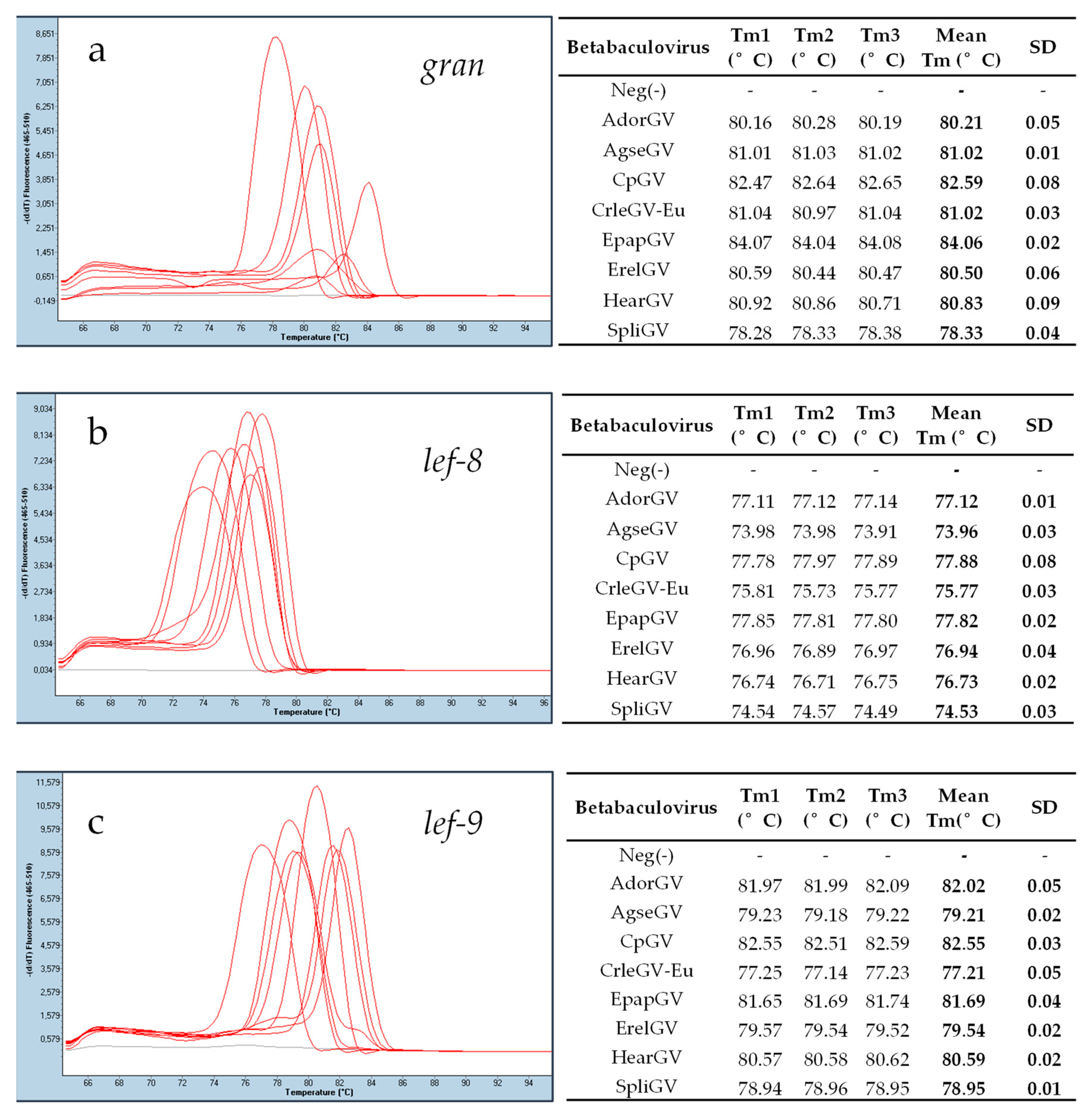
2.5. Real-Time PCR Reaction

Real-time PCR was performed using a LightCycler 480 Instrument II (Roche, Germany). DNA templates from eight granuloviruses and one additional CrleGV isolate were subjected to real-time PCR with the use of SG qPCR Master Mix (2×) (EURX, Poland) for each of the three pairs of primers to amplify the granulin, lef-9, and lef-8 short fragments. The annealing temperature was optimized to be the same for all three primer pairs (gran, lef-9, and lef-8).
The final mixture of 25 µL (prepared on a 96-well plate) contained 12.5 µL of Master Mix (2×) (Perpetual Taq DNA Polymerase, reaction buffer with 2.5 mM MgCl2, dNTPs and SYBR Green I dye), 0.3 μM of each primer, and 0.6 μl DNA template (0.5 ng in total). A negative control (with alphabaculovirus Lymantria dispar nucleopolyhedrovirus (LdMNPV) DNA) was prepared as well. The samples were initially stored at 50 °C for 2 min to activate the hot start polymerase, then denatured at 95 °C for 10 min, followed by 45 cycles: 94 °C (denaturation) for 15 s, 50 °C (annealing) for 30 s, and 72 °C (extension) for 30 s. To confirm the specificity of the amplified product, a melting curve analysis step was included. The internal temperature of the Light Cycler was increased rapidly to 95 °C, then decreased to 65 °C, and then the sample was incubated for 1 min. The fluorescence at 530 nm was measured continuously. The melting peaks were generated using Light Cycler software by plotting the first negative derivative of the fluorescence over the temperature versus the temperature (−dF/dT). To confirm the presence of specific products, agarose electrophoresis (2%) was performed. All the reactions were repeated three times with the mean Tm calculated from all the repetitions of the experiment, and standard deviation (SD) was also calculated.
Additionally, serial dilutions from 10−1–10−10 of HearGV DNA were prepared to check the detection level of the method. Following real-time PCR amplification, DNA concentrations for each reaction were measured using the fluorometric method (Qubit ssDNA HS Assay Kit, Thermo Fisher Scientific, USA using Quantus fluorometer, Promega, USA). The software from http://scienceprimer.com/copy-number-calculator-for-realtime-pcr was employed to count DNA copies.

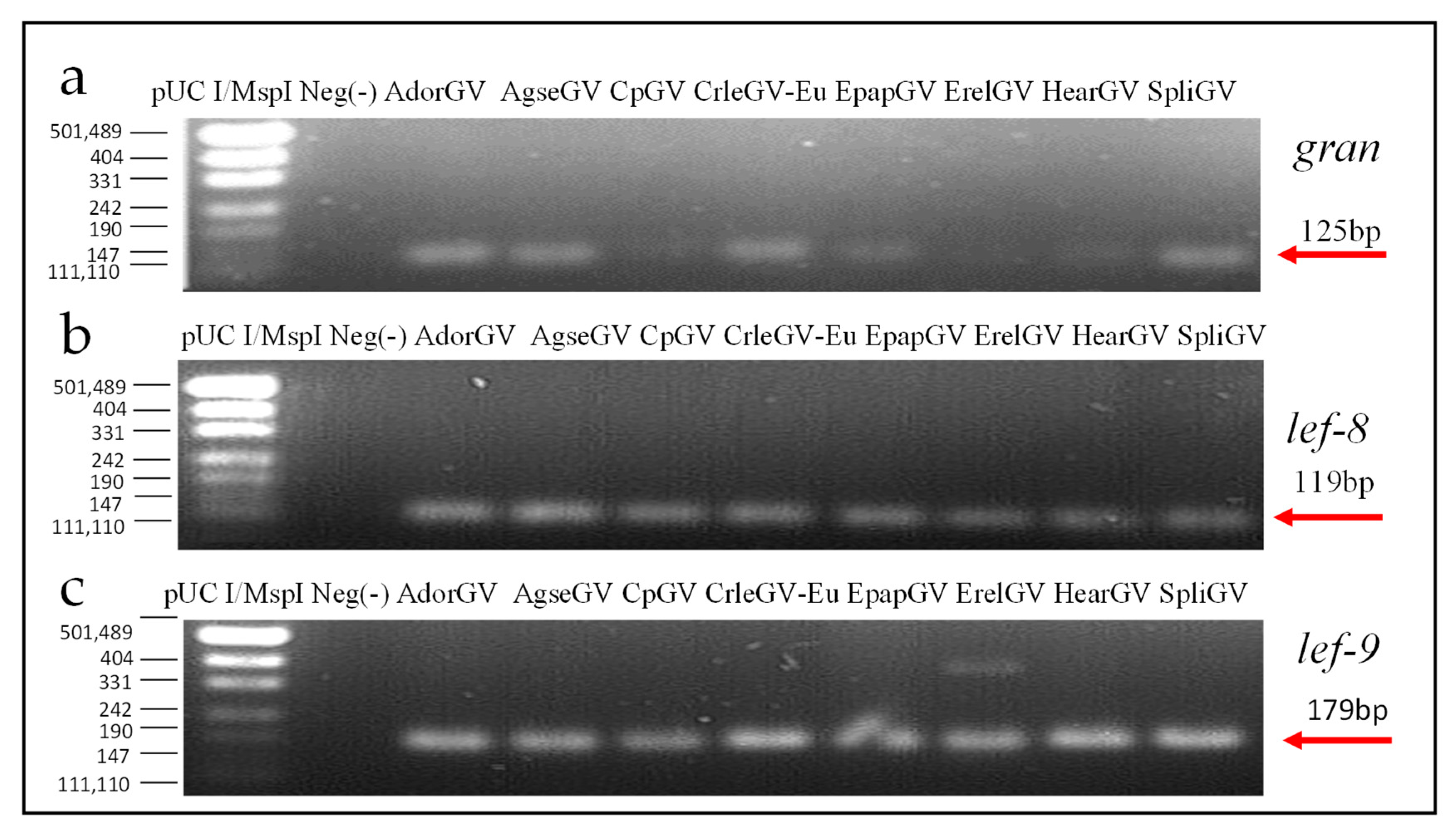
2.6. Agarose Gel Electrophoresis

The real-time PCR products were subjected to electrophoretic analysis in 2% agarose gel in TAE buffer (20 mM sodium acetate, 1 mM EDTA, and 40 mM TRIS, pH adjusted to 7.2) using SimplySafe (Eurx, Poland). Electrophoresis was carried out at 80 V for 30 min.

3. Results

3.1. Phylogenetic Analysis and the Representative group of Betabaculovirus

The phylogenetic tree based on 38 amino acid sequences of baculovirus core genes represents the general phylogeny of the Baculoviridae family (Figure 1), with special emphasis on the Betabaculovirus genus. All species included in the analysis are listed in Table 1 with the addition of host family and genome length. There are 24 betabaculovirus genomes deposited in the GenBank database, all of which differ in the nucleotide sequence (Geneious Pro 7.1 (Biomatters, Auckland, New Zealand)-MAFFT alignment. Two species, Trichoplusia ni granulovirus (175,360 bp, 172 orfs) and Mythimna (previous Pseudaletia) unipuncta granulovirus-A (176,677 bp, 183 orfs), share 96% of genome similarity and have almost all core genes sequences being identical (only five of them, i.e., lef-2 (98%), DNA polymerase (99%), p49 (99%), p18 (98%), and pif-3 (99%) share lower than 100% homology). Furthermore, these viruses cluster together on the phylogenetic tree.
Based on this molecular phylogenetic tree and the alignments of short nucleotide sequences generated from granulin, lef-8, and lef-9 of GVs, the representative group previously described by us [32] was selected again. The selected species (Figure 1, labelled in green) differ in genomic nucleotide sequences and belong to different clades in the phylogenetic tree. Two closely related and similar betabaculoviruses, CpGV and CrleGV, were also included in the study to prove that the presented method is also able to discriminate between similar genetic sequences. The eight species (all recognized by the International Committee on Taxonomy of Viruses, ICTV) and with criteria as defined in Section 2.2, which were chosen as the demonstrative group are Adoxophyes orana granulovirus (AdorGV), Agrotis segetum granulovirus (AgseGV), CpGV, CrleGV, EpapGV, Erinnyis ello granulovirus (ErelGV), HearGV, and Spodoptera litura granulovirus (SpliGV) (bold font in Table 1 and labelled in green in Figure 1).

3.2. Granulin, Late Expression Factor-9, and Late Expression Factor-8 Nucleotide and Amino Acid Sequence Alignment

The complete nucleotide sequence of granulin is 747 bp long in all GVs (except for SpliGV, 750 bp) and the identity is between 70–100%. The lef-9 gene is more variable in length, ranging from 1476 to 1512 bp with one exception: SfGV lef-9, which is the longest at 1632 bp. The sequence identity ranges from 54–100%. The lef-8 codes for the biggest protein among the analyzed genes. The shortest nucleotide sequence belongs to PhopGV (2484 bp), while ClasGV-B possesses the longest lef-8 gene of 2655 bp and the sequence identity is between 56–100%. The 100% identity between sequences is observed for TnGV and MyunGV-A. All 24 sequences (from all betabaculovirus species with complete genomic sequences in the GenBank database) were aligned and analyzed for highly conserved DNA regions that could be used as targets for degenerate primers. Highly similar sequences flank the most variable region (the localization and the size of fragments are presented in Figure 2 and fragments of the alignments are presented in Figure 3).
To show that the gran, lef-9, and lef-8 regions chosen are conserved among sequenced betabaculoviruses and suitable for real-time PCR, amino acid (aa) alignments of the predicted products were performed (Figure S1). Every forward primer is coherent with codons in the orf and starts from the first nucleotide in the codon. Figure S1 shows high conservation of aa sequences in the annealing primer regions. For both granulin primers, lef-9-F and lef-8-R, the alignments are 100% similar in the aa sequence. For lef-8-F, only one aa change has occurred—the third aa, which was methionine (M) in most species and was exchanged for isoleucine (I) in two baculoviruses, SpliGV (included in the representative group) and PrGV (Figure S1b). In lef-9-R in the representative group, there are similar aa sequences in all eight tested species, but in 6 out of the remaining 16, a difference of an alanine (A) instead of valine (V) was determined (Figure S1c).
The selected, highly conserved regions were used to design a set of universal primers to amplify short fragments that are suitable in size for real-time PCR and will allow for detection of the majority of granuloviruses in less than two hours post DNA isolation. The two pairs of primers developed in the previous study [32] remain unchanged and they are universal for the larger group of granuloviruses (before—18 species, now—24 species). Therefore, they can be used in real time-PCR (Figure 3a,b). The lef-8 alignment (Figure 3c) called for the design of a third completely new pair of degenerate primers. Other primer pairs for each gene were checked, and the results presented here represent the best (lowest degeneration and the highest specificity).

3.3. Real-Time PCR Assay

The DNA templates from eight betabaculoviruses, AdorGV, AgseGV, CpGV, CrleGV, EpapGV, ErelGV, HearGV, and SpliGV and the three pairs of universal primers, gran-F and gran-R, lef-9-F and lef-9-R, lef-8-F and lef-8-R were used for real-time PCR reactions. The results are presented in Figure 4. Sharp peaks in melting curves correspond to specific products for all three tested genes: granulin, lef-9, and lef-8. Each product for a single pair of primers exhibited different melting temperatures Tm in a single run (Figure 4), which indicates that real-time PCR products have different nucleotide sequences, and the method can be used for differentiation of granuloviruses. When analyzing mean Tm (Table S1), there is one exception: The granulin products for AgseGV and CrleGV-Eu give the same Tm (Figure 4a). All SD values range from 0.01 to 0.09.
After real-time PCR, the samples were loaded on an agarose gel (2%) to confirm the presence of specific products. All the bands were of expected sizes: 125 bp for gran, 179 bp for lef-9, and 119 bp for lef-8 (Figure 5). The negative controls (with DNA from alphabaculovirus LdMNPV) are presented with grey curves in Figure 4 and in the second well of each panel on agarose gel in Figure 5.
To estimate the level of detection, the DNA of one betabaculovirus, HearGV, of known concentration was serially diluted 10−1–10−10, and the real-time PCR assay was repeated with the set of tested universal primers for granuloviruses. DNA concentration was assessed fluorometrically. For gran, the level of detection was 104 copies of DNA molecules per reaction, and for lef-9 and lef-8, it was 103 copies of DNA molecules per reaction.
The real-time PCR analysis was also performed for two closely related CrleGV isolates that are commercially produced in Europe (CrleGV-Eu) and South Africa (CrleGV-SA) as components of plant protection agents (Table 2) (both originated from South Africa).
For both CrleGV isolates tested, the European and the South African, the melting temperatures differed for all three of the primer pairs used (Table 2). The largest difference in Tm was observed between the gran and lef-8 amplicons at 0.95 and 1.09 °C. The difference in Tm between the lef-9 amplicons was not as large; however, it was well within the sensitivity of the real-time PCR thermocycler used. Polymorphism sites that exist in sequences may or may not introduce a detectable change in Tm, which will be a cumulative effect of nucleotide type and position in the whole sequence. Therefore, as expected, no correlation between the number of single nucleotide polymorphism sites (SNPs) and the |Δ Mean Tm| was observed. When multiple SNPs exist, they can increase (here for lef-8) or decrease (here for lef-9) ΔTm in the compared sequences.

4. Discussion

The primary objective of the presented study was to develop a fast and easily accessible method for granulovirus detection and differentiation. For this purpose, a real-time PCR technique was chosen. Real-time PCR is commonly used for the detection of many different viruses, from various families, that infect humans, animals, or bacteria [26,35,36]. In the case of insect pathogens, the method based on real-time PCR was developed for sensitive detection and quantitative analysis of larvae infected with one granulovirus: EpapGV [37]. The method proposed in this study allows for the discrimination of different GVs and gives comparable results with the one based on the MSSCP technique described by us previously [33] (Figure 6).
The comparison of Tm from three amplified fragments between each other for all tested species has been done and presented in Table S1 as absolute values from the Tm difference between each species (e.g., AdorGV Tmgran compared with AgseGV Tmgran). For granulovirus species from this study, the Tm in a single run for each of three genes differs. In addition, when the Tm values for each gene in one species are compared with the Tm values for each gene in another species, there is always at least one high Tm difference (a ≥ 0.3 °C difference between Tm was assigned as “high”, and “low” values (<0.3 °C) are marked in red in Table S1). It is therefore possible, when differentiation is not based on one gene alone but on all three genes, to differentiate between the granulovirus species tested using the method described. Except for AdorGV and EpapGV (gran and lef-8), all the other absolute value Tm differences are higher than 0.3 °C for at least two out of three genes. For two species, AgseGV and CrleGV, while there was no difference between the mean Tm for gran fragments, the absolute values of Tm difference for the remaining two genes are well above the value of sensitivity of detection, which is 0.01 °C, with lef-8 at 1.81 °C and lef-9 at 2 °C. Hence, the proposed procedure may be very useful as a screening method, as the high differentiation levels provide a greater margin, which allows for easy application of this method in numerous real-time thermocyclers from different vendors.
Granulin/polyhedrin, late expression factor-8, and late expression factor-9 belong to the most conserved group of genes among baculoviruses. Lef-8 and lef-9 are core genes present in every species, while granulin, though not coded by a core gene, has highly conserved structure and resembles another major occlusion body protein—polyhedrin (30 kDa)—found in nucleopolyhedroviruses. Only the Deltabaculovirus group, which, according to phylogeny, is older and more distant from lepidopteran-specific baculoviruses (therefore closer to a common ancestor) possesses a gene coding for a trimer built of polypeptides of 30 kDa which is non-homologous towards the polyhedrin or granulin genes [22,38,39]. The lepidopteran-specific baculovirus demarcation criterion that is commonly used for species designation includes Kimura-two parameter (K2P) nucleotide distance comparisons based on partial sequences of polh/gran, lef-8, and lef-9 [25]. Recently, it has been extended for larger group of genes, i.e., all core genes, and the grouping of baculoviruses has not changed based on the larger number of genes. In general, the criterion mentioned was confirmed by the distance of 38 common gene nucleotide sequences [40], which means that species defined based on fragments of these three genes remain unchanged for known members of the Baculoviridae family. Therefore, gran, lef-8, and lef-9 genes selected for this study are an ideal choice for betabaculovirus determination.
The number of completely sequenced granulovirus species (24) is still low in comparison to nucleopolyhedroviruses (73) (GenBank, August 2018). The possibility that the primers will not match sequences of some of the newly discovered isolates/species cannot be excluded. The nucleotide and amino acid (Figure S1) sequences of available species were analyzed. Although the representative group consists of eight members of Betabaculovirus, which equals one-third part of all completely sequenced species, we hypothesize that it represents the majority of known GVs, with the main limitation encountered being DNA availability. However, the regions selected for degenerate primer targets have highly conserved protein structure, so in future, the method may find application for the detection and characterization of new granuloviruses for samples originating from the environment.
It is common knowledge that many pathogens of humans and animals, including viruses infecting insects, possess high levels of genotypic variation [41,42]. Many variants of baculoviruses circulate in one host or can be isolated from the same species from distinct geographic localizations. The genotype sequences may be characterized with single nucleotide polymorphisms (SNPs), substitutions, and indels (insertion/deletion) that do or do not cause orf changes [43,44,45,46,47]. Baculovirus variability in genomes should be carefully investigated because of selection pressure due to biopesticide application in the field. Using standard PCR with bulk products and sequencing may not be necessary to detect minor variants that may affect virus stability and biopesticide efficacy. Over a decade ago, after around 20 years of field application of commercial insecticide based on CpGV, resistance development in caterpillars has appeared in orchards in France and Germany [48]. To date, three mechanisms of resistance have been described [48,49,50,51,52,53]. Taking such a possibility into account for other viral biopesticides, the monitoring of isolates and variants occurring in caterpillars should be implemented routinely in biopesticide production. The proposed real-time PCR-based method shows that it is possible to distinguish two CrleGV isolates originated from South Africa on the basis of three genes. The method could likely be used to detect minor variants in the virus population (even another closely related granulovirus species).
We have previously developed a method based on the Multi-temperature Single Stranded Conformational Polymorphism (MSSCP) technique, which has advantages similar to the one described in this study [33]. However, the MSSCP-based method has some drawbacks: It requires dedicated equipment, dealing with polyacrylamide gel, and DNA silver staining. In the current work, we developed a method for discriminating between the representative groups of betabaculoviruses using a real-time thermocycler, which may be more accessible to many laboratories in contrast to the MSSCP technique. Real-time thermocyclers are increasingly more affordable (e.g., a single channel costs ~4299 USD) and are starting to become standardized equipment in many laboratories, with methods that make use of them therefore becoming considerably more implementable. Obviously, NGS-based methods supply a wealth of data, allowing for the detection and differentiation of baculoviruses, but the costs of sequencing runs and the amount of data needed to be processed is still too high for routine monitoring of infected insects. Furthermore, this method is particularly advantageous when screening many samples, allowing detection and differentiation without the need for amplicon purification, sequencing, and bioinformatic analysis. Therefore, the method presented in this study may find extensive application in laboratories screening large batch samples of current or new betabaculoviruses, as we estimate a cost per sample around 2 USD.

Supplementary Materials

The following are available online at https://www.mdpi.com/1999-4915/11/2/115/s1. Table S1: The absolute values from the Tm difference between each species (e.g., AdorGV Tm gran compared with AgseGV Tm gran). Figure S1: The amino acid sequence alignments of predicted PCR products with primers: (a) gran, (b) lef-8, and (c) lef-9. 1–8 are the representative group in this study, 9–24 are all the remaining granuloviruses.

Author Contributions

Conceptualization: M.L.d.S., S.D.M. and B.S.; data curation, L.R. and M.D.J.; funding acquisition: M.K.-R. and B.S.; methodology: M.K.-R. and L.R.; resources: M.L.d.S, S.D.M. and B.S.; supervision: B.S.; validation: L.R.; visualization, M.K.-R. and M.D.J.; writing—original draft: M.K.-R.; writing—review & editing: M.K.-R., L.R., M.D.J., M.L.d.S., S.D.M. and B.S.

Funding

The research was funded by NATIONAL CENTRE FOR RESEARCH DEVELOPMENT (POLAND), grant number BIOCIT PL-RPA/BIOCIT/04/2016 and NATIONAL SCIENCE CENTRE, grant number PRELUDIUM 9 UMO-2015/17/N/NZ2/02092.

Conflicts of Interest

The authors declare no conflict of interest.

References

  1. Lacey, L.A.; Grzywacz, D.; Shapiro-Ilan, D.I.; Frutos, R.; Brownbridge, M.; Goettel, M.S. Insect pathogens as biological control agents: Back to the future. J. Invertebr. Pathol. 2015, 132, 1–41. [Google Scholar] [CrossRef] [PubMed] [Green Version]
  2. Martignioni, M.; Iwai, P.J. A catalogue of viral diseases of insects, mites and thicks. In Microbial Control of Pests and Plant Diseases; Burges, H.D., Ed.; Arcade: London, UK, 1987; pp. 897–911. [Google Scholar]
  3. Szewczyk, B.; Souza, M.L.; Castro, M.E.B.; Moscardi, M.L.; Moscardi, F. Baculovirus Biopesticides. In Pesticides–Formulation, Effects, Fate; Stoytcheva, M., Ed.; Intech Open: London, UK, 2011; pp. 25–36. [Google Scholar]
  4. Haase, S.; Sciocco-Cap, A.; Romanowski, V. Baculovirus insecticides in Latin America: Historical overview, current status and future perspectives. Viruses 2015, 7, 2230–2267. [Google Scholar] [CrossRef] [PubMed]
  5. Blissard, G.W.; Rohrmann, G.F. Baculovirus Diversity and Molecular Biology. Annu. Rev. Entomol. 1990, 35, 127–155. [Google Scholar] [CrossRef]
  6. Lauzon, H.A.M.; Lucarotti, C.J.; Krell, P.J.; Feng, Q.; Retnakaran, A.; Arif, B.M. Sequence and organization of the Neodiprion lecontei nucleopolyhedrovirus genome. J. Virol. 2004, 78, 7023–7035. [Google Scholar] [CrossRef] [PubMed]
  7. Hayakawa, T.; Ko, R.; Okano, K.; Seong, S.I.; Goto, C.; Maeda, S. Sequence analysis of the Xestia c-nigrum granulovirus genome. Virology 1999, 262, 277–297. [Google Scholar] [CrossRef] [PubMed]
  8. Jehle, J.A.; Blissard, G.W.; Bonning, B.C.; Cory, J.S.; Herniou, E.A.; Rohrmann, G.F.; Theilmann, D.A.; Thiem, S.M.; Vlak, J.M. On the classification and nomenclature of baculoviruses: A proposal for revision. Arch. Virol. 2006, 151, 1257–1266. [Google Scholar] [CrossRef] [Green Version]
  9. Herniou, E.A.; Jehle, J.A. Baculovirus phylogeny and evolution. Curr. Drug Targets 2007, 8, 1043–1050. [Google Scholar] [CrossRef]
  10. Rohrmann, G.F. Polyhedrin structure. J. Gen. Virol. 1986, 67, 1499–1513. [Google Scholar] [CrossRef]
  11. Rohrmann, G.F. Baculovirus Molecular Biology, 3rd ed.; National Center for Biotechnology Information (US): Bethesda, MD, USA, 2013. [Google Scholar]
  12. Federici, B.A. Baculovirus pathogenesis. In The Baculoviruses; Miller, L.K., Ed.; Springer Science & Business Media: New York, NY, USA, 1997; pp. 33–59. [Google Scholar]
  13. Harrison, R.L.; Popham, H.J.R. Genomic sequence analysis of a granulovirus isolated from the Old World bollworm, Helicoverpa armigera. Virus Genes 2008, 36, 565–581. [Google Scholar] [CrossRef]
  14. Luque, T.; Finch, R.; Crook, N.; O’Reilly, D.R.; Winstanley, D. The complete sequence of the Cydia pomonella granulovirus genome. J. Gen. Virol. 2001, 82, 2531–2547. [Google Scholar] [CrossRef] [PubMed]
  15. Moore, S.; Kirkman, W.; Richards, G.; Stephen, P.; Moore, S.D.; Kirkman, W.; Richards, G.I.; Stephen, P.R. The Cryptophlebia Leucotreta Granulovirus—10 Years of Commercial Field Use. Viruses 2015, 7, 1284–1312. [Google Scholar] [CrossRef] [Green Version]
  16. Tanada, Y.; Kaya, H. Insect Pathology, 1st ed.; Academic Press: Cambridge, MA, USA, 1993; ISBN 978-0-08-092625-4. [Google Scholar]
  17. Ferrelli, M.L.; Salvador, R.; Biedma, M.E.; Berretta, M.F.; Haase, S.; Sciocco-Cap, A.; Ghiringhelli, P.D.; Romanowski, V. Genome of Epinotia aporema granulovirus (EpapGV), a polyorganotropic fast killing betabaculovirus with a novel thymidylate kinase gene. BMC Genom. 2012, 13, 548. [Google Scholar] [CrossRef]
  18. Federici, B.A.; Stern, V.M. Replication and occlusion of a granulosis virus in larval and adult midgut epithelium of the western grapeleaf skeletonizer, Harrisina brillians. J. Invertebr. Pathol. 1990, 56, 401–414. [Google Scholar] [CrossRef]
  19. Bideshi, D.K.; Bigot, Y.; Federici, B.A. Molecular characterization and phylogenetic analysis of the Harrisina brillians granulovirus granulin gene. Arch. Virol. 2000, 145, 1933–1945. [Google Scholar] [CrossRef] [PubMed]
  20. Yin, F.; Zhu, Z.; Liu, X.; Hou, D.; Wang, J.; Zhang, L.; Wang, M.; Kou, Z.; Wang, H.; Deng, F.; et al. The Complete Genome of a New Betabaculovirus from Clostera anastomosis. PLoS ONE 2015, 10, e0132792. [Google Scholar] [CrossRef] [PubMed]
  21. Miele, S.A.B.; Garavaglia, M.J.; Belaich, M.N.; Ghiringhelli, P.D. Baculovirus: Molecular Insights on Their Diversity and Conservation. Int. J. Evol. Biol. 2011. [Google Scholar] [CrossRef] [PubMed]
  22. Garavaglia, M.J.; Miele, S.A.B.; Iserte, J.A.; Belaich, M.N.; Ghiringhelli, P.D. The ac53, ac78, ac101, and ac103 Genes Are Newly Discovered Core Genes in the Family Baculoviridae. J. Virol. 2012, 86, 12069–12079. [Google Scholar] [CrossRef] [Green Version]
  23. Javed, M.A.; Biswas, S.; Willis, L.G.; Harris, S.; Pritchard, C.; Oers, M.M.; van Donly, B.C.; Erlandson, M.A.; Hegedus, D.D.; Theilmann, D.A. Autographa californica Multiple Nucleopolyhedrovirus AC83 is a per os infectivity factor (PIF) protein required for occlusion-derived virus (ODV) and budded virus nucleocapsid assembly as well as assembly of the PIF complex in ODV envelopes. J. Virol. 2017, 91. [Google Scholar] [CrossRef]
  24. Guarino, L.A.; Xu, B.; Jin, J.; Dong, W. A virus-encoded RNA polymerase purified from baculovirus-infected cells. J. Virol. 1998, 72, 7985–7991. [Google Scholar]
  25. Jehle, J.A.; Lange, M.; Wang, H.; Hu, Z.; Wang, Y.; Hauschild, R. Molecular identification and phylogenetic analysis of baculoviruses from Lepidoptera. Virology 2006, 346, 180–193. [Google Scholar] [CrossRef] [Green Version]
  26. Liu, H.; Niu, Y.D.; Li, J.; Stanford, K.; McAllister, T.A. Rapid and accurate detection of bacteriophage activity against Escherichia coli O157:H7 by propidium monoazide real-time PCR. Biomed. Res. Int. 2014, 2014, 319351. [Google Scholar] [CrossRef] [PubMed]
  27. Groner, A. Specificity and safety of baculoviruses. In The Biology of Baculoviruses; Granados, R.R., Federici, B.A., Eds.; CRC Press: Boca Raton, FL, USA, 1986; Volume I, pp. 177–201. [Google Scholar]
  28. Harrison, R.L.; Mowery, J.D.; Rowley, D.L.; Bauchan, G.R.; Theilmann, D.A.; Rohrmann, G.F.; Erlandson, M.A. The complete genome sequence of a third distinct baculovirus isolated from the true armyworm, Mythimna unipuncta, contains two copies of the lef-7 gene. Virus Genes 2018, 54, 297–310. [Google Scholar] [PubMed]
  29. Harrison, R.L.; Bonning, B.C. The nucleopolyhedroviruses of Rachiplusia ou and Anagrapha falcifera are isolates of the same virus. J. Gen. Virol. 1999, 80, 2793–2798. [Google Scholar] [CrossRef] [PubMed]
  30. de Moraes, R.R.; Maruniak, J.E. Detection and identification of multiple baculoviruses using the polymerase chain reaction (PCR) and restriction endonuclease analysis. J. Virol. Methods 1997, 63, 209–217. [Google Scholar] [CrossRef]
  31. Lange, M.; Wang, H.; Zhihong, H.; Jehle, J.A. Towards a molecular identification and classification system of lepidopteran-specific baculoviruses. Virology 2004, 325, 36–47. [Google Scholar] [CrossRef] [PubMed] [Green Version]
  32. Szewczyk, B.; Barski, P.; Sihler, W.; Rabalski, L.; Skrzecz, I.; Hoyos-Carvajal, L.; de Souza, M.L. Detection and identification of baculovirus pesticides by multitemperature single-strand conformational polymorphism. J. Environ. Sci. Health B 2008, 43, 539–545. [Google Scholar] [CrossRef]
  33. Krejmer-Rabalska, M.; Rabalski, L.; Lobo de Souza, M.; Moore, S.D.; Szewczyk, B. New method for differentiation of granuloviruses (Betabaculoviruses) based on multitemperature single stranded conformational polymorphism. Int. J. Mol. Sci. 2017, 19. [Google Scholar] [CrossRef]
  34. Kaczanowski, R.; Trzeciak, L.; Kucharczyk, K. Multitemperature single-strand conformation polymorphism. Electrophoresis 2001, 22, 3539–3545. [Google Scholar] [CrossRef]
  35. Moreau, F.; Fetouchi, R.; Micalessi, I.; Brejeon, V.; Bacon, N.; Jannes, G.; Le Pendeven, C.; Lekbaby, B.; Kremsdorf, D.; Lacau Saint Guily, J.; et al. Detection and genotyping of human papillomavirus by real-time PCR assay. J. Clin. Virol. 2013, 56, 244–249. [Google Scholar] [CrossRef]
  36. Günes, A.; Marek, A.; Grafl, B.; Berger, E.; Hess, M. Real-time PCR assay for universal detection and quantitation of all five species of fowl adenoviruses (FAdV-A to FAdV-E). J. Virol. Methods 2012, 183, 147–153. [Google Scholar] [CrossRef]
  37. Arneodo, J.D.; König, G.A.; Berretta, M.F.; Rienzo, J.A.D.; Taboga, O.; Sciocco-Cap, A. Detection and kinetic analysis of Epinotia aporema granulovirus in its lepidopteran host by real-time PCR. Arch. Virol. 2012, 157, 1149–1153. [Google Scholar] [CrossRef]
  38. Afonso, C.L.; Tulman, E.R.; Lu, Z.; Balinsky, C.A.; Moser, B.A.; Becnel, J.J.; Rock, D.L.; Kutish, G.F. Genome sequence of a baculovirus pathogenic for Culex nigripalpus. J. Virol. 2001, 75, 11157–11165. [Google Scholar] [CrossRef] [PubMed]
  39. Herniou, E.A.; Olszewski, J.A.; O’Reilly, D.R.; Cory, J.S. Ancient coevolution of baculoviruses and their insect hosts. J. Virol. 2004, 78, 3244–3251. [Google Scholar] [CrossRef] [PubMed]
  40. Wennmann, J.T.; Keilwagen, J.; Jehle, J.A. Baculovirus Kimura two-parameter species demarcation criterion is confirmed by the distances of 38 core gene nucleotide sequences. J. Gen. Virol. 2018, 99, 1307–1320. [Google Scholar] [CrossRef]
  41. Brito, A.F.; de Braconi, C.T.; Weidmann, M.; Dilcher, M.; Alves, J.M.P.; Gruber, A.; de Zanotto, P.M.A. The Pangenome of the Anticarsia gemmatalis Multiple Nucleopolyhedrovirus (AgMNPV). Genome Biol. Evol. 2015, 8, 94–108. [Google Scholar] [CrossRef]
  42. Bonsall, D.; Black, S.; Howe, A.Y.; Chase, R.; Ingravallo, P.; Pak, I.; Brown, A.; Smith, D.A.; Bowden, R.; Barnes, E. Characterization of hepatitis C virus resistance to grazoprevir reveals complex patterns of mutations following on-treatment breakthrough that are not observed at relapse. Infect. Drug Resist. 2018, 11, 1119–1135. [Google Scholar] [CrossRef] [PubMed]
  43. Cory, J.S.; Green, B.M.; Paul, R.K.; Hunter-Fujita, F. Genotypic and phenotypic diversity of a baculovirus population within an individual insect host. J. Invertebr. Pathol. 2005, 89, 101–111. [Google Scholar] [CrossRef]
  44. Chateigner, A.; Bézier, A.; Labrousse, C.; Jiolle, D.; Barbe, V.; Herniou, E.A. Ultra Deep Sequencing of a Baculovirus Population Reveals Widespread Genomic Variations. Viruses 2015, 7, 3625–3646. [Google Scholar] [CrossRef] [Green Version]
  45. Wennmann, J.T.; Radtke, P.; Eberle, K.E.; Gueli Alletti, G.; Jehle, J.A. Deciphering Single Nucleotide Polymorphisms and Evolutionary Trends in Isolates of the Cydia pomonella granulovirus. Viruses 2017, 9. [Google Scholar] [CrossRef]
  46. Van der Merwe, M.; Jukes, M.D.; Rabalski, L.; Knox, C.; Opoku-Debrah, J.K.; Moore, S.D.; Krejmer-Rabalska, M.; Szewczyk, B.; Hill, M.P. Genome Analysis and Genetic Stability of the Cryptophlebia leucotreta Granulovirus (CrleGV-SA) after 15 Years of Commercial Use as a Biopesticide. Int. J. Mol. Sci. 2017, 18. [Google Scholar] [CrossRef]
  47. Harrison, R.L.; Rowley, D.L.; Mowery, J.; Bauchan, G.R.; Theilmann, D.A.; Rohrmann, G.F.; Erlandson, M.A. The Complete Genome Sequence of a Second Distinct Betabaculovirus from the True Armyworm, Mythimna unipuncta. PLoS ONE 2017, 12, e0170510. [Google Scholar] [CrossRef] [PubMed]
  48. Asser-Kaiser, S.; Fritsch, E.; Undorf-Spahn, K.; Kienzle, J.; Eberle, K.E.; Gund, N.A.; Reineke, A.; Zebitz, C.P.W.; Heckel, D.G.; Huber, J.; et al. Rapid emergence of baculovirus resistance in codling moth due to dominant, sex-linked inheritance. Science 2007, 317, 1916–1918. [Google Scholar] [CrossRef] [PubMed]
  49. Alletti, G.G.; Sauer, A.J.; Weihrauch, B.; Fritsch, E.; Undorf-Spahn, K.; Wennmann, J.T.; Jehle, J.A. Using next generation sequencing to identify and quantify the genetic composition of resistance-breaking commercial isolates of Cydia pomonella granulovirus. Viruses 2017, 9. [Google Scholar] [CrossRef] [PubMed]
  50. Asser-Kaiser, S.; Heckel, D.G.; Jehle, J.A. Sex linkage of CpGV resistance in a heterogeneous field strain of the codling moth Cydia pomonella (L.). J. Invertebr. Pathol. 2010, 103, 59–64. [Google Scholar] [CrossRef]
  51. Asser-Kaiser, S.; Radtke, P.; El-Salamouny, S.; Winstanley, D.; Jehle, J.A. Baculovirus resistance in codling moth (Cydia pomonella L.) caused by early block of virus replication. Virology 2011, 410, 360–367. [Google Scholar] [CrossRef]
  52. Jehle, J.A.; Schulze-Bopp, S.; Undorf-Spahn, K.; Fritsch, E. Evidence for a second type of resistance against Cydia pomonella granulovirus in field populations of codling moths. Appl. Environ. Microbiol. 2017, 83. [Google Scholar] [CrossRef]
  53. Sauer, A.J.; Schulze-Bopp, S.; Fritsch, E.; Undorf-Spahn, K.; Jehle, J.A. A third type of resistance to Cydia pomonella granulovirus in codling moths shows a mixed Z-linked and autosomal inheritance pattern. Appl. Environ. Microbiol. 2017, 83. [Google Scholar] [CrossRef]
Figure 1. Maximum likelihood (ML) molecular phylogenetic tree based on amino acid sequences of 38 core genes of 24 baculovirus species from the Betabaculovirus genus and one of each of Alpha-, Delta-, and Gammabaculovirus. Species marked in green indicate betabaculoviruses chosen as a representative group in this study.
Viruses 11 00115 g001
Figure 2. The localization of short fragments amplified in the presented real time PCR method within the granulin, lef-9, and lef-8 genes, using AdorGV as an example.
Viruses 11 00115 g002
Figure 3. The alignment of nucleotide sequences of (a) granulin (125 bp), (b) lef-9 (179 bp), and (c) lef-8 (119 bp) fragments from betabaculovirus complete genomic sequences available in GenBank. 1–8 are representative groups in this study, and 9–24 are all the remaining granuloviruses. Dark and light green annotations show primers: (a) forward gran-F and reverse gran-R, (b) forward lef-9-F and reverse lef-9-R; and (c) forward lef-8-F and reverse lef-8-R. The histogram on the top shows the identity in every column of the alignment.
Viruses 11 00115 g003
Figure 4. Real-time PCR results (melting curves) using universal pairs of primers that allow for the amplification of short fragments of three betabaculovirus genes: (a) granulin, (b) late expression factor-8, and (c) late expression factor-9. On the left side of (a), (b), and (c), melting curve analyses with red peaks from a single run (Tm1) are shown; the grey line represents a negative control [Neg(-)] with alphabaculovirus Lymantria dispar nucleopolyhedrovirus (LdMNPV) DNA; the X-axis represents temperatures; and the Y-axis represents –(d/dT) fluorescence (465–510 nm). On the right side of (a), (b), and (c), the melting temperatures (Tm) for each PCR product from three repetitions and mean Tm with SD calculated are shown.
Viruses 11 00115 g004
Figure 5. Agarose gel electrophoresis (2%) of real-time PCR products of short fragments of (a) gran—125 bp long, (b) lef-8—119 bp long, and (c) lef-9—179 bp long, after real-time PCR reactions are presented. Lane 1: pUC I/MspI DNA Ladder (Thermo Fisher Scientific, Waltham, MA, USA); lane 2: negative control with alphabaculovirus Lymantria dispar nucleopolyhedrovirus (LdMNPV) DNA; lanes 3–10: granuloviruses from this study.
Viruses 11 00115 g005
Figure 6. Schematic representation of the results of the described real-time PCR-based method for differentiation of betabaculovirus species based on three genes: gran, lef-8, and lef-9. Each line represents Tm for real-time PCR products (gran—blue, lef-8—green, and lef-9—black) with SD values.
Viruses 11 00115 g006
Table 1. List of betabaculoviruses (24 species) which have complete genomic sequences available in GenBank (August 2018). Granuloviruses (GVs) chosen as the representative group in this study are shown in bold.
AbrreviationBetabaculovirus NameHost FamilyGenBank Accession NumberGenome Size (bp)
AdorGVAdoxophyes orana granulovirusTortricidaeAF54798499,657
AgseGVAgrotis segetum granulovirusNoctuidaeAY522332131,680
ChocGVChoristoneura occidentalis granulovirusTortricidaeDQ333351104,710
ClanGVClostera anachoreta granulovirusNotodontidaeHQ116624101,487
ClasGVClostera anastomosis granulovirusNotodontidaeKC179784101,818
ClasGV-BClostera anastomosis granulovirus BNotodontidaeKR091910107,439
CnmeGVCnaphalocrocis medinalis granulovirusCrambidaeKU593505111,246
CpGVCydia pomonella granulovirusTortricidaeU53466123,500
CrleGVCryptophlebia leucotreta granulovirusTortricidaeAY229987110,907
DisaGVDiatraea saccharalis granulovirusCrambidaeKP29618698,392
EpapGVEpinotia aporema granulovirusTortricidaeJN408834119,082
ErelGVErinnyis ello granulovirusSphingidaeKJ406702102,759
HearGVHelicoverpa armigera granulovirusNoctuidaeEU255577169,794
MolaGVMocis latipes granulovírusNoctuidaeKR011718134,272
MyunGV-AMythimna unipuncta granulovirus ANoctuidaeEU678671176,677
MyunGV-BMythimna unipuncta granulovírus BNoctuidaeKX855660144,673
PhopGVPhthorimaea operculella granulovirusGelechiidaeAF499596119,217
PiGVPlodia interpunctella granulovirusPyralidaeKX151395112,536
PlxyGVPlutella xylostella granulovirusPlutellidaeAF270937100,999
PrGVPieris rapae granulovirus isolatePieridaeGQ884143108,592
SfGVSpodoptera frugiperda granulovirusNoctuidaeKM371112140,913
SpliGVSpodoptera litura granulovirusNoctuidaeDQ288858124,121
TnGVTrichoplusia ni granulovirusNoctuidaeKU752557175,360
XcGVXestia c-nigrum granulovirusNoctuidaeAF162221178,733
Table 2. Melting temperatures from real-time PCR analysis using universal pairs of primers that allow for amplification of short fragments of gran, lef-8, and lef-9 on the DNA of CrleGV isolates—European (Eu) and South African (SA). Tm from three repetitions with mean Tm and SD are shown. The negative control (Neg(-)) included alphabaculovirus LdMNPV DNA. The absolute values from the Tm difference between both isolates are shown. SNPs—single nucleotide polymorphism sites.
IsolateTm1
(°C)
Tm2
(°C)
Tm3
(°C)
Mean
Tm (°C)
SD|ΔMean Tm|
(°C)
SNPs
granNeg(-)------
CrleGV-Eu80.9781.0481.0481.020.030.951
CrleGV-SA82.0581.9281.9581.970.06
lef-8Neg(-)------
CrleGV-Eu 75.7375.7775.8175.770.031.092
CrleGV-SA76.8976.8576.8476.860.02
lef-9Neg(-)------
CrleGV-Eu 77.1477.2377.2577.210.050.293
CrleGV-SA77.5377.4577.5377.500.04

