Article

Prevalence and Association of Trypanosomes and Sodalis glossinidius in Tsetse Flies from the Kafue National Park in Zambia

1
Department of Disease Control, School of Veterinary Medicine, University of Zambia, Lusaka P.O. Box 32379, Zambia
2
College of Veterinary Medicine, Haramaya University, Dire Dawa P.O. Box 138, Ethiopia
3
Department of Virology-I, National Institute of Infectious Diseases, Toyama 1-23-1, Shinjuku, Tokyo 162-8640, Japan
4
Management Department of Biosafety, Laboratory Animal, and Pathogen Bank, National Institute of Infectious Diseases, Toyama 1-23-1, Shinjuku, Tokyo 162-8640, Japan
5
Laboratory of Parasitology, Department of Disease Control, Faculty of Veterinary Medicine, Hokkaido University, N18 W9, Kitaku, Sapporo 060-0818, Japan
6
Africa Centre of Excellence for Infectious Diseases of Humans and Animals, University of Zambia, Lusaka P.O. Box 32379, Zambia
*
Author to whom correspondence should be addressed.
Trop. Med. Infect. Dis. 2023, 8(2), 80; https://doi.org/10.3390/tropicalmed8020080
Submission received: 24 November 2022 / Revised: 13 January 2023 / Accepted: 14 January 2023 / Published: 21 January 2023
(This article belongs to the Section Infectious Diseases)

Abstract

:
Tsetse flies are obligate hematophagous vectors of animal and human African trypanosomosis. They cyclically transmit pathogenic Trypanosoma species. The endosymbiont Sodalis glossinidius is suggested to play a role in facilitating the susceptibility of tsetse flies to trypanosome infections. Therefore, this study was aimed at determining the prevalence of S. glossinidius and trypanosomes circulating in tsetse flies and checking whether an association exists between trypanosomes and Sodalis infections in tsetse flies from Kafue National Park in Zambia. A total of 326 tsetse flies were sampled from the Chunga and Ngoma areas of the national park. After DNA extraction was conducted, the presence of S. glossinidius and trypanosome DNA was checked using PCR. The Chi-square test was carried out to determine whether there was an association between the presence of S. glossinidius and trypanosome infections. Out of the total tsetse flies collected, the prevalence of S. glossinidius and trypanosomes was 21.8% and 19.3%, respectively. The prevalence of S. glossinidius was 22.2% in Glossina morsitans and 19.6% in Glossina pallidipes. In relation to sampling sites, the prevalence of S. glossinidius was 26.0% in Chunga and 21.0% in Ngoma. DNA of trypanosomes was detected in 18.9% of G. morsitans and 21.4% of G. pallidipes. The prevalence of trypanosomes was 21.7% and 6.0% for Ngoma and Chunga, respectively. The prevalences of trypanosome species detected in this study were 6.4%, 4.6%, 4.0%, 3.7%, 3.1%, and 2.5% for T. vivax, T. simiae, T. congolense, T. godfreyi, T. simiae Tsavo, and T. b. brucei, respectively. Out of 63 trypanosome infected tsetse flies, 47.6% of the flies also carried S. glossinidius, and the remaining flies were devoid of S. glossinidius. A statistically significant association was found between S. glossinidius and trypanosomes (p < 0.001) infections in tsetse flies. Our findings indicated that presence of S. glossinidius increases the susceptibility of tsetse flies to trypanosome infections and S. glossinidius could be a potential candidate for symbiont-mediated vector control in these tsetse species.

1. Introduction

Tsetse flies (Glossina) are biological vectors of African trypanosomes which cause animal African trypanosomosis (AAT) and human African trypanosomosis (HAT) [1]. Tsetse flies occupy the ‘tsetse belt’ which covers an area of 10 million km2 that is about one third of the total land of the continent in 38 sub-Saharan African countries [2]. The density of the vector and the prevalence of trypanosome infections in the host is ascribed to complex interactions between and among humans, domestic livestock, wildlife, tsetse flies, trypanosomes and different economic and ecological factors [3].
AAT represents a group of vector-borne (Glossina) parasitic ailments in ruminants, camels, equines and carnivores which induce dramatic economic losses to animal producers as a result of mortality, morbidity and inefficient productivity [4]. Within the tsetse-infested areas, trypanosomosis reduces the offtake of meat and milk by a minimum of 50% [5]. The total loss of AAT to livestock productivity was estimated to be about USD 4.5 billion per year [6]. AAT is caused by several tsetse fly-transmitted trypanosome species including Trypanosoma brucei brucei, Trypanosoma congolense, Trypanosoma simiae, and Trypanosoma vivax [3,7,8].
Two trypanosome sub-species are responsible for HAT: Trypanosoma brucei gambiense and Trypanosoma brucei rhodesiense [9]. These cause high mortality in infected human population if left untreated. Trypanosoma brucei gambiense is found in 24 countries in west and central Africa [10]. This form of the disease currently accounts for over 98% of reported cases of sleeping sickness, transmitted through human-tsetse contact and causes a chronic infection. Trypanosoma brucei rhodesiense is found in 13 countries in eastern and southern Africa, representing less than two percent of reported cases [10]. This parasite has a complex transmission cycle involving a wide range of wildlife and livestock reservoirs and causes an acute infection in humans [10]. Even though 260 million people live in tsetse infested areas, only about 60 million are considered to be at risk of contracting the disease. The distribution of sleeping sickness areas has a focal nature and the localization of the actual areas fluctuates over the course of time [11,12].
Tsetse flies have established symbiotic relationship with maternally transmitted bacteria [13]. Many endosymbionts have been reported in various tissues of tsetse flies, but Wigglesworthia glossinidia, Sodalis glossinidius and Wolbachia species are the three major bacterial species that they harbor [14]. Sodalis glossinidius is found in the midgut, haemolymph, muscles, fat bodies, salivary glands, milk glands and reproductive system and so could interact with multiple species of trypanosomes that are harbored in different tissues [15]. Sodalis lacks a clearly defined functional role within its tsetse host [13]. However, it is suggested to play a role in facilitating susceptibility to trypanosome infection in tsetse by inhibiting the efficacy of the tsetse immune system [16]. N-acetyl glucosamine specific trypanocidal lectin is secreted during feeding, and trypanosomes need to successfully evade this lectin activity to establish in the midgut of the tsetse fly [17].
Because of their distinct reproductive biology, tsetse flies are recalcitrant to germ-line transformation [18]. Sodalis glossinidius is the only gamma proteobacterial tsetse endosymbiont to be cultured and is thus amenable to genetic modification [19]. A paratransgenic approach using S. glossinidius as a delivery system for trypanocidal components is currently of considerable interest to generate a trypanosome resistant tsetse fly [20]. Hence, investigation of the interactions between trypanosomes and S. glossinidius and, therefore, their influence on tsetse can provide new insights to design new vector control strategies.
In previous studies analysing natural tsetse fly populations, the relationship between S. glossinidius and trypanosomes varies with respect to tsetse fly species and trypanosome species/subspecies. For instance, in tsetse flies from Maasai Mara National Reserve, Kenya, there was a statistically significant relationship in G. pallidipes tsetse flies but not in G. swynnertoni [21]. There were no significant relationships found between Sodalis and trypanosomes in G. brevipalpis, G. morsitans morsitans and G. pallidipes tsetse flies from Luambe National Park, Zambia [22], but significant associations were found in G. m. morsitans from western Zambia [23]. Sodalis glossinidius has been shown to be positively associated with T. congolense and T. b. rhodesiense in G. m. morsitans [17], T. b. gambiense and T. b. brucei in G. p. gambiensis [24] and T. congolense Forest, T. brucei s. l. and T. b. gambiensis in G. p. palpalis [25]. These results show that the relationship between Sodalis and the presence of trypanosomes varies depending on geographic areas, and there is lack of information about the tripartite relationship between Sodalis, trypanosomes and wild caught tsetse flies in Kafue National Park (KNP) in Zambia. As such, we conducted a tsetse fly survey to determine the prevalence and identify trypanosome species circulating in tsetse flies and to assess the associations between Sodalis and trypanosomes in this area.

2. Materials and Methods

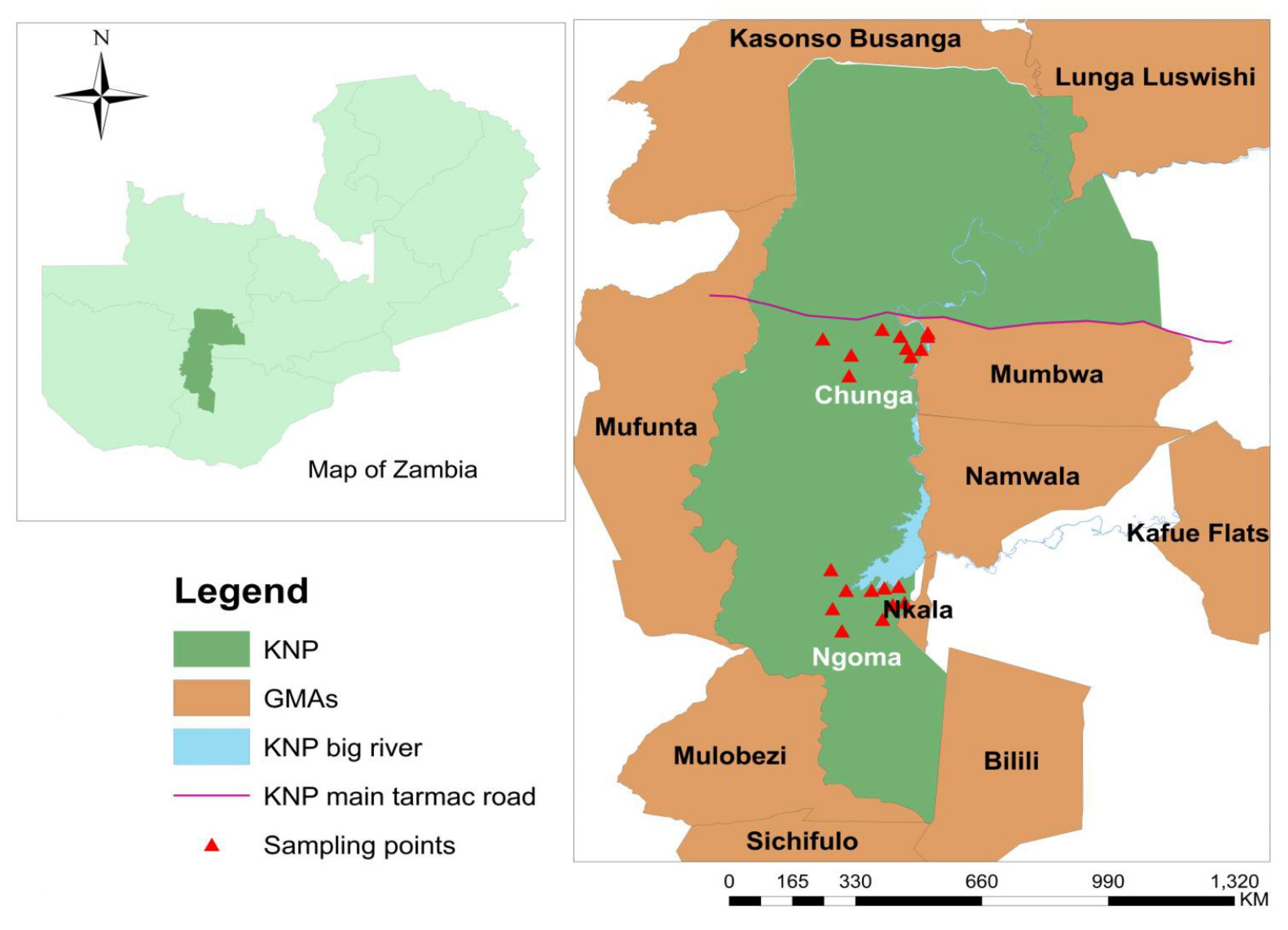
2.1. Study Area and Tsetse Fly Collection

Tsetse fly samples were collected from the KNP ecosystem which is situated between 14°03″ S and 16°43″ S and 25°13″ E and 26°46″ E (Figure 1). The KNP ecosystem is the largest conservation area in Zambia and covers approximately 68,000 km2 of the country. It is the oldest and the largest national park (22,480 km2) in Zambia and is surrounded by 45,406 km2 of game management areas, which stretch over four provinces [26]. The park is rich in animal and natural diversity and forms one of the main important terrestrial ecosystems in Africa [27]. The study was carried out at the Chunga and Ngoma sampling locations within the KNP ecosystem and 150 km apart. The Chunga sampling site is located approximately 150 km from Mumbwa town and it is situated on the Kafue River. The sampling points in this area are covered by thicket vegetation. The Ngoma sampling site is located 26 km from Itezhi tezhi on the Kafue River and close to Itezhi tezhi Dam. The vegetation type at the Ngoma sampling points were thicket and miombo woodlands.
The number of tsetse flies required for analysis of trypanosomes was estimated using previous findings of 26.85% prevalence in the same study area [28]. Based on this, the minimum number of tsetse flies was 301, and we caught 326 tsetse flies during the tsetse survey which were used for the analysis.
Six Epsilon traps containing 3-n-propylphenol, octanol and 4-methylphenol at a ratio of 1:6:12 and an open 300 mL bottle containing acetone at the entrance to the trap were deployed under the tree sheds between September and December 2021. All the traps were deployed between 17:00 and 18:00, and the captured tsetse flies were collected by visiting traps at about 12 h intervals daily between 6:00 and 7:00 and 17:00 and 18:00. The geographic coordinates were recorded for each trap. ArcMap in ArcGIS was used to record the spatial locations of the sampling points on the map (Figure 1). The trapped tsetse flies were counted and grouped into teneral and non-teneral flies, as previously described elsewhere [29]. The teneral flies were discarded. The non-teneral flies were stored individually in a 1.5 mL Eppendorf tubes with silica beads and were transported to the University of Zambia, School of Veterinary Medicine Laboratory for DNA extraction and further analysis.

2.2. Sex and Species Determination of Tsetse Flies

Morphological characterization was used to sort out the sex and species of the captured tsetse flies. The species and sex were identified using a stereomicroscope based on standard published keys [29].

2.3. DNA Extraction

Each tsetse fly was transferred to a new 2 mL microcentrifuge tube and smashed for 70 s at 3500 rpm in a Micro SmashTM MS-100R bead cell disrupter (TOMY MEDICO. Ltd., Tokyo, JAPAN) using five 3-mm diameter zirconium beads. DNA was extracted from the homogenate of each tsetse fly using QIAGEN DNeasy Blood and Tissue Kit following the manufacturer’s instructions (Qiagen Sciences, Hilden, Germany). The DNA was quantified using a NanoDropTM 1000 Spectrophotometer (Thermo Scientific, Waltham, MA, USA), and DNA samples were stored at −80 °C until PCR analysis.

2.4. PCR for the Identification of African Trypanosomes and Sodalis glossinidius

All PCR reactions were conducted using OneTaq ®Quick-Load® 2X Master Mix with Standard Buffer (20 mM Tris-HCI (pH 8.9 @ 25 °C), 1.8 mM MgCl2, 22 mM NH4Cl, 22 mM KCl, 0.2 mM dNTPs, 5% glycerol, 0.06% IGEPAL® CA-630, 0.05% Tween® 20, 25 units/mL OneTaq DNA Polymerase) (NEW ENGLAND BioLabs Inc., Ipswich, MA, USA). PCR reactions for both trypanosomes and S. glossinidius were carried out in a 10 μL reaction volume containing 5 μL of One Taq ®Quick-Load® 2X Master Mix, 3.2 μL of nuclease free water, 0.4 μL from 10 μM of each forward and reverse primer and 1 μL of template DNA. Initial screening for the presence of trypanosome parasites was conducted using ITS1 CF and BR primers that target the internal transcribed spacer 1 (ITS1) (Table 1) [30]. The PCR conditions were an initial step at 94 °C for 5 min, followed by 35 cycles of 94 °C for 40 s, 58 °C for 40 s, 72 °C for 90 s, and final extension at 72 °C for 5 min. However, ITS1 primer has low sensitivity against T. vivax species [31]. To solve this problem, T. vivax specific primers (TVIV 1 and TVIV 2) were used with the same PCR conditions except the annealing temperature, which was 60 °C [32].
When the ITS1 PCR generated a PCR product of between 500 bp and 800 bp band sizes, T. congolense subgroup-specific PCR were conducted to differentiate the subgroup Kilifi, Forest and Savannah using subgroup-specific primers (Table 1) with PCR conditions of an initial step at 94 °C for 5 min, followed by 35 cycles of 94 °C for 30 s, 55 °C for 30 s, 72 °C for 90 s, and final extension step at 72 °C for 5 min. All tsetse flies which showed band sizes between 250 and 500 bp were subjected to PCR that differentiate pathogenic trypanosome species (T. b. rhodesiense, T. b. brucei, T. simiae, T. simiae Tsavo and T. godfreyi) using species-specific primers (Table 1). To check the human infective trypanosome species (T. b. rhodesiense), the serum resistance associated (SRA) gene PCR was performed using an amplification program with an initial denaturation step at 95 °C for 15 min followed by 35 cycles of 94 °C for 1 min, 68 °C for 1 min, 72 °C for 1 min and a final extension step of 72 °C for 10 min.
The presence of S. glossinidius in all tsetse flies was determined using the primer pair GPO1F and GPO1R, which amplifies the 1200 bp product of the extrachromosomal plasmid, GPO1, of Sodalis [14]. The amplification program was initiated with an initial step at 94 °C for 5 min, followed by 35 amplification cycles with denaturation step of each cycle at 94 °C for 1 min, an annealing step at 55 °C for 1 min, and an extension step of 72 °C for 1 min followed by a final extension step at 72 °C for 10 min.
All PCR reactions included appropriate positive and negative controls. The PCR products were size-separated by electrophoresis in 1x TAE buffer (40 mM Tris, 20 mM acetic acid, 1 mM EDTA, pH 8.0) (BioConcept Ltd., Allschwil, Switzerland) on 1.5% agarose gel (CSL-AG100, Cleaver Scientific Ltd., Rugby, UK), stained with Ethidium bromide and visualized under UV light. Amplicon sizes were determined relative to a 100 bp DNA ladder.

2.5. Data Analysis

Data were entered into MS-Excel ® and analysed using R software version 4.1.0 [37]. The prevalence of S. glossinidius and trypanosomes were estimated using frequencies. The Chi-square test or Fisher’s Exact test where appropriate were used to compare the prevalence of trypanosomes and S. glossinidius with sex, species and collection site of tsetse flies. They were also used to assess whether the presence of S. glossinidius was associated with trypanosome infections. All the statistics were considered significant at p ≤ 0.050.

3. Results

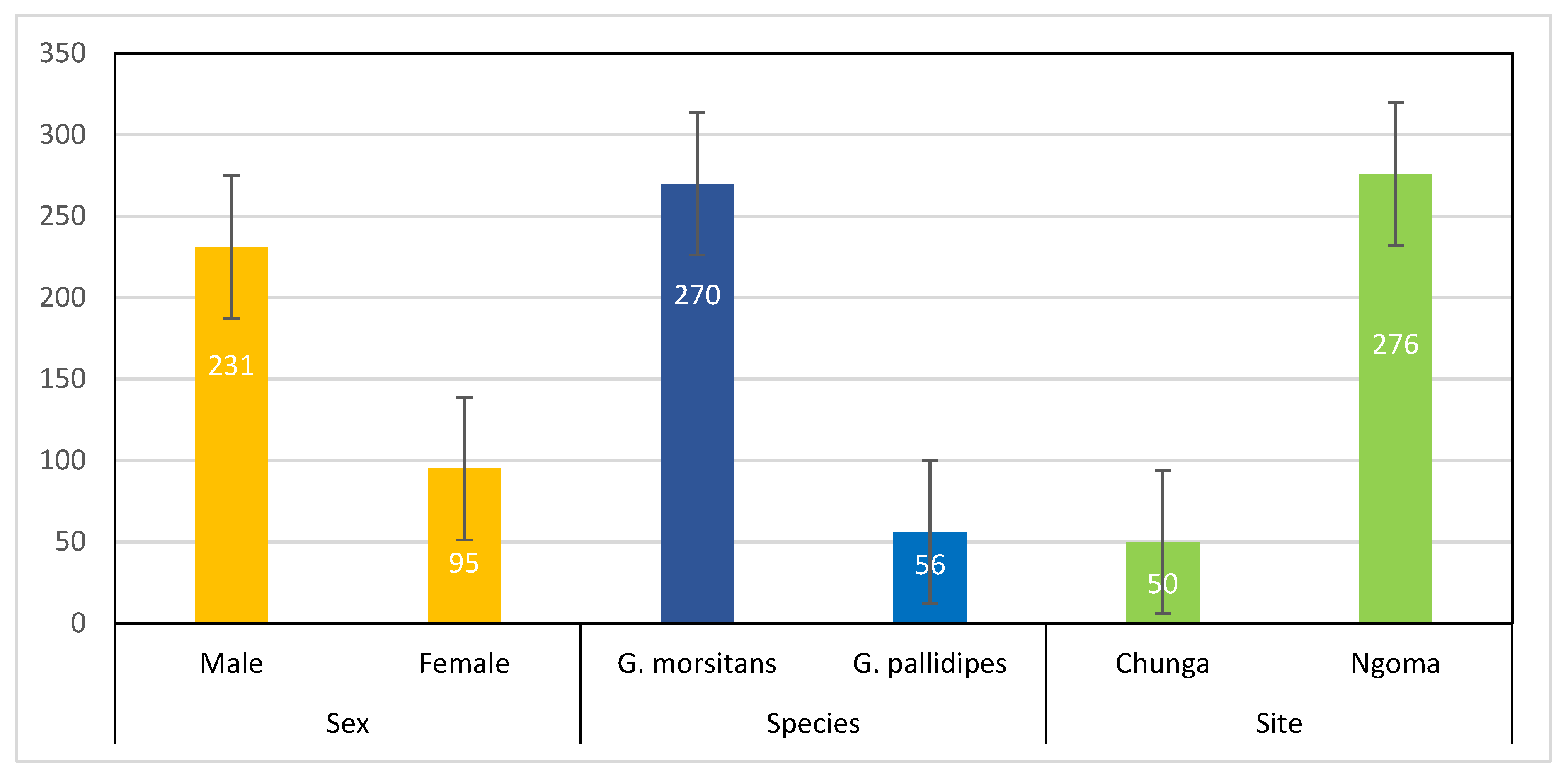
3.1. Tsetse Fly Survey

A total of 326 tsetse flies were trapped: 231 (70.9%, 95% CI: 65.7–75.5%) were male and 95 (29.1%, 95% CI: 24.5–34.3%) were female tsetse flies. Out of the total tsetse samples collected, 270 (82.8%, 95% CI: 78.35–86.53) were G. morsitans and 56 (17.2%, 95% CI: 13.47–21.65) were G. pallidipes, and 50 (15.3%, 95% CI: 11.83–19.85) were from Chunga and 276 (84.7%, 95% CI: 80.35–88.17) were from Ngoma sampling locations (Figure 2).

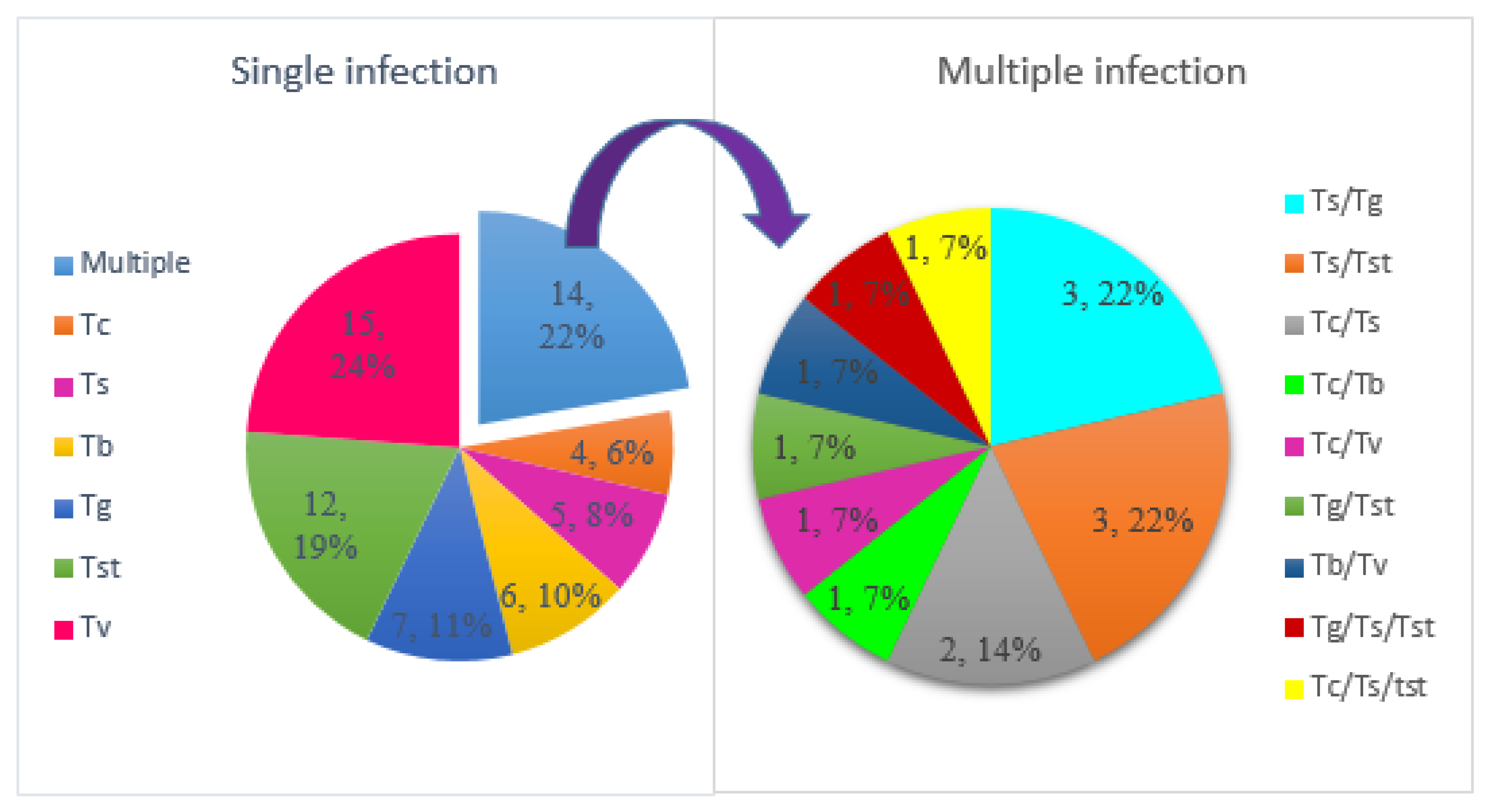
3.2. Prevalence of Trypanosomes in Tsetse Flies

Of 326 tsetse fly samples subjected to PCR using general ITS1 primers, 63 (19.3%) were found with DNA of at least one trypanosome species, indicating an overall prevalence of 19.3% (95% CI: 15.41–23.96). The differences in prevalence of trypanosomes between G. morsitans and G. pallidipes, Chunga and Ngoma, and male and female tsetse flies are shown in Table 2. The prevalence of trypanosomes was significantly higher in Ngoma than in Chunga (χ 2 = 6.73, p = 0.009) (Table 2). There were no statistically significant differences in the prevalence of trypanosomes between male and female (χ2 = 0.53, p = 0.467) and between G. morsitans and G. pallidipes2 = 0.19, p = 0.661) tsetse flies (Table 2).
Six trypanosome species were detected in all tsetse flies. These were T. vivax, T. simiae, T. congolense, T. godfreyi, T. simiae Tsavo, and T. brucei brucei. Trypanosoma vivax (6.4%, 95% CI = 4.25–9.65) was the most prevalent, and T. b. brucei (2.5%, 1.25–4.77) was the least. Table 3 and Table 4 summarise the prevalence of each trypanosome species that was identified with respect to species, sex and sampling site of tsetse flies. There were no significant differences in the prevalence of trypanosome species detected between species, sex and sampling sites (Table 3 and Table 4).
Among 13 tsetse flies which were positive for T. congolense, eight had the T. congolense Kilifi subgroup, two had the T. congolense Forest subgroup, and one had T. congolense Savannah. Two tsetse flies had mixed T. congolense subgroups. Of these, one had a mixed infection of T. congolense Kilifi and T. congolense Forest. The other had T. congolense Forest mixed with T. congolense Savannah.
No human infective T. b. rhodesiense was detected in either species of tsetse flies.
Most tsetse flies were infected with a single trypanosome species (49/63, 77.8%), followed by tsetse flies infected by two trypanosome species (12/63, 19.0%) and tsetse flies that had three trypanosome species (2/63, 3.2%) (Figure 3). Multiple infections with two trypanosome species included three tsetse flies that had a mixture of T. simiae and T. godfreyi, three tsetse flies that had T. simiae and T. simiae Tsavo, two tsetse flies infected with T. congolense and T. simiae, one tsetse fly with T. congolense and T. vivax, one tsetse fly contained T. congolense mixed with T. b. brucei, one tsetse fly with T. b. brucei and T. vivax and one tsetse fly with T. simiae Tsavo and T. godfreyi. Triple infections were found in two tsetse flies which had T. simiae/T. godfreyi/T. simiae Tsavo and T. simiae/T. congolense/T. simiae Tsavo.

3.3. Prevalence of S. glossinidius in Tsetse Flies

The overall prevalence of S. glossinidius from the 326 tsetse flies was estimated to be 21.8% (95% CI: 17.64–26.57). The prevalence was higher in female (24.2%; 95% CI: 16.71–33.72) than in male tsetse flies (20.8%; 95% CI: 16.05–26.47), although this was not statistically significant (χ2 = 0.46, p = 0.670). It was also slightly higher in G. morsitans (22.2%; 95% CI: 17.67–27.55) than in G. pallidipes (19.6%; 95% CI: 11.34–31.84) but not statistically significantly different (χ2 = 0. 0.18, p = 0.495). The prevalence between the two sampling sites was not significantly different (χ2 = 0.62, p = 0.432), although it was slightly higher in Chunga (26.0%: 95% CI: 15.87–39.55) than in Ngoma (21.0%; 95% CI: 16.62–26.20).
The prevalence of S. glossinidius in each tsetse species was examined based on sampling location and the sex of tsetse flies (Table 5). In G. morsitans, there were no statistically significant differences in the prevalence of S. glossinidius between Chunga and Ngoma (χ2 = 0.97, p = 0. 324) or between male and female flies (χ2 = 0.01, p = 0.943) (Table 5). There were also no significant differences in the prevalence of S. glossinidius between Chunga and Ngoma (p = 1.000) or between male and female (p = 0.142) G. pallidipes flies (Table 5).

3.4. Association between S. glossinidius and Presence of African Trypanosomes

Out of 63 trypanosome-infected flies, 47.6% of the flies were also co-infected with S. glossinidius, while the remaining flies were devoid of S. glossinidius. The analysis performed on the overall dataset indicated that there was a significant association between tsetse flies harboring S. glossinidius and tsetse flies infected with trypanosomes (χ2 = 30.61, p < 0.001). The association varied between sampling sites, with tsetse flies from Ngoma showing a statistically significant association (χ2 = 30.39, p < 0.001), whereas tsetse flies from Chunga showed no statistically significant association (p = 0.162). In G. morsitans, twenty-five out of sixty tsetse flies with S. glossinidius were infected with trypanosomes, and there was a statistically significant association (χ2 = 26.12, p < 0.001) between the two pathogens. From the eleven S. glossinidius positive G. pallidipes tsetse flies, five had trypanosome co-infections, and the association was statistically significant (p = 0.045). A statistically significant association was also observed between S. glossinidius and trypanosome prevalence in male tsetse flies (χ2 = 28.42, p < 0.001), but no such association was observed in female tsetse flies (p = 0.058) (Table 6).
Among the tsetse flies infected with T. simiae Tsavo, T. simiae, and T. vivax, the co-infection rates with S. glossinidius were 60.0%, 46.7%, and 47.6%, respectively. Among the tsetse flies infected by T. congolense, co-infection with S. glossinidius was 30.8%, and for T. b. brucei and T. godfreyi, the co-infection rate was 50.0% for both (Table 7). There was a statistically significant association between S. glossinidius and T. vivax (p = 0.006), T. simiae (p = 0.025), T. simiae Tsavo (p = 0.009), and T. godfreyi (p = 0.027), but no such association was detected between S. glossinidius and T. congolense (p = 0.491) and T. b. brucei (p = 0.072) (Table 7).

4. Discussion

4.1. Prevalence of Trypanosome Species

The aim of this study was to determine the prevalence of trypanosome and S. glossinidius in tsetse flies collected from the Chunga and Ngoma areas of the Kafue National Park. The study also intended to determine whether an association between trypanosomes and S. glossinidius existed in these tsetse flies. Both Sodalis and trypanosomes were prevalent in tsetse flies obtained from the study area. The prevalence of trypanosomes found in this study was similar to the 17.4% prevalence reported from Ghana [38] but lower than previously reported results from national park and wildlife reserve areas such as Nkhotakota Wildlife Reserve and Liwonde Wildlife Reserve, Malawi [39,40], Luambe National Park, Luangwa Valley, and the Kafue ecosystem, Zambia [22,28,41], Nech Sar National Park, Ethiopia [42], Shimba Hills National Reserve, Kenya [43], and Santchou Wildlife Reserve, Cameroon [44]. However, the prevalence obtained from the current study was higher compared with the 0.8% reported from Ghana [28], 2.40% from Kenya [21], 3.4% from Tanzania [45], 6.31% from Zimbabwe [46], 10.7% from Uganda [47], and 11.4% from Kenyan coastal forests and South Africa [48]. These differences could be explained by differences in geographic location, the availability of potential tsetse species and presence of appropriate vertebrate hosts.
In the current work, six trypanosome species were detected with T. vivax being the most prevalent. This is in agreement with another study that reported a high prevalence of T. vivax in tsetse flies and cattle in the same study area [28]. The result is also in agreement with other studies in Zambia [23] and other parts of Africa [21,45]. Other trypanosome species detected were T. simiae, T. congolense, T. godfreyi, T. simiae Tsavo, and T. b. brucei. The higher prevalence of T. vivax in tsetse flies compared with other trypanosome species may be due to the differences in development cycles as T. vivax completes its entire development only in proboscis, whereas T. congolense, T. simiae, T. godfreyi, and T. simiae Tsavo in complete it in the proboscis and midgut and T. b. brucei in the midgut and salivary gland, which can be affected by low pH, protease activity and lectins [49,50].
In this study, there were no significant differences in the levels of trypanosome infections between male and female tsetse flies. This result is similar to a finding from Chad [51] and other experimental studies [52] but in disagreement with previous studies from Nigeria [53] and Côte d’Ivoire [54] where researchers reported higher prevalences of trypanosomes in females than male tsetse flies. It is also in contrast to other experimental studies in the same tsetse species [55] in which males had higher trypanosome infectivity than their female counterparts.
Despite a recent report of a HAT case in an adult male [56] and the presence of T. b. rhodesiense in vervet monkey, sable antelope, buffalo [57] and in cattle [28] from the KNP ecosystem, no human infective trypanosome species were detected in this study. Although T. b. rhodesiense was not detected in the current study, the presence of the most competent tsetse fly vectors of T. b. rhodesiense (G. morsitans and G. pallidipes) in KNP and the high prevalence of T. b. rhodesiense previously reported in wildlife [57] and cattle [28] in the area indicate an existing risk of emergence of HAT, so coordinated surveillance and control efforts are required in the study area.

4.2. Prevalence of S. glossinidius

The overall prevalence of S. glossinidius estimated from this study was lower than the 31.3% prevalence reported from southwest Nigeria [58] and the 34.0% prevalence reported from the Shimba Hills and Nguruman regions in Kenya [43]. However, the prevalence in the current study was higher than that in the Maasai Mara National Reserve (6.6%), a wildlife–human–livestock interface in Kenya [21] and that reported in the Shimba Hills National Reserve (15.9%), a wildlife–human–livestock interface on Kenya’s south coast [48].
The difference in the prevalence of S. glossinidius in relation to species of tsetse flies was not statistically significant. In G. morsitans, the prevalence of S. glossinidius was higher than the 17.5% reported in Luambe National Park, in eastern Zambia [22] and the 15.9% obtained from Western Zambia [23]. However, this value is lower than the prevalence of 29.6% that was reported from Zimbabwe [59] and the 28.6% reported from Adamawa region of Cameroon [60]. In this study, the prevalence of S. glossinidius in G. pallidipes was higher than the 1.4%, 6.5%, 15.9%, and 16% recorded in Luambe National Park, Zambia [22], Maasai Mara National Reserve, Kenya [21], Shimba Hills National Reserve, Kenya [48], and tsetse flies collected from Zimbabwe [59], respectively. This prevalence was, however, lower than the 83.3% in G. pallidipes collected from Tanzania [59]. These differences may be linked to environmental and ecological variations between sampling areas which can highly affect the biology of tsetse flies and the presence of different S. glossinidius genotypes and trypanosomes [61].
In this study, no significant difference in the prevalence of S. glossinidius between male and female tsetse flies was detected. This finding is in agreement with other studies by Dennis et al. [22] and Mathew [59] which reported similar results. Data analysed for individual tsetse species also indicated no significant difference in the prevalence of S. glossinidius between sexes of G. morsitans and G. pallidipes.

4.3. Association between S. glossinidius and Trypanosome Infections in Tsetse Flies

From the overall data analysed, the co-infection rate between S. glossinidius and trypanosomes in this study were lower than the 37% rate reported from the “Faro and Déo” division of the Adamawa region of Cameroon [60] and the rate of 32.2% in two historical HAT foci in Cameroon [25], but higher than 2% co-infection rate in Kenyan coastal forests [48]. This result indicates that the presence of S. glossinidius is not absolutely necessary for tsetse flies to be infected by trypanosomes, but the presence of S. glossinidius would highly favor such infections.
In the current study, significant associations were found between the presence of S. glossinidius and the presence of trypanosomes in tsetse flies. This maybe an indication that presence of S. glossinidius favors trypanosome infections in tsetse flies. This is in agreement with other studies from Cameroon [25], western Zambia [23] and Kenya [21], where significant associations were reported between S. glossinidius and trypanosome infections in different tsetse fly species. However, there was variation in the association of Sodalis and trypanosomes between tsetse fly species, sex and sampling locations.
There were no large differences in the proportions of co-infected tsetse flies with Sodalis and trypanosomes in G. morsitans and G. pallidipes (9.3% and 8.9%, respectively). Significant associations were found between Sodalis and trypanosome infections in G. morsitans and G. pallidipes tsetse fly species. These findings are in line with other studies conducted in G. m. centralis [23], G. pallidipes [48], G. pallidipes and G. swynnertoni [21] and G. p. palpalis [25] where significant associations between S. glossinidius and trypanosome infections were reported in the respective tsetse fly species. These findings support the hypothesis that presence of S. glossinidius increases the susceptibility to and establishment of trypanosome infections in G. morsitans and G. pallidipes tsetse flies. However, this is in contrast to the findings of a study of G. morsitans and G. pallidipes in tsetse flies from Luambe National Park, Zambia [22] where no association was found. In addition to the presence or absence of Sodalis, this difference may be due to a difference in S. glossinidius genotype which may affect the association between S. glossinidius and trypanosome infections, as described by Geiger et al. [62]. Based on the sex of tsetse flies, there was a significant association between S. glossinidius and trypanosome infections in males, but no significant association was observed between the endosymbiont and trypanosome infections in females. These differences may be due to the small number of female tsetse flies collected for the endosymbiont and trypanosome infections. Separate analyses of the data for each sampling site indicate there are differences in the statistical association between the endosymbiont and trypanosome infections, where a statistically significant association was observed for the Ngoma sampling site, but not for the Chunga sampling site. This difference could be due to the low trypanosome infection rate and small number of tsetse flies captured at the Chunga sampling site.
The associations between S. glossinidius and each trypanosome species infection were also examined. The result of this analysis clearly indicates that significant associations were found between S. glossinidius and T. simiae, T. vivax, T. simiae Tsavo, and T. godfreyi. However, there were no significant associations between S. glossinidius and T. congolense and T. b. brucei. This difference is probably due to S. glossinidius affecting the establishment of trypanosomes depending on the trypanosome genotype [24].
One limitation of this study was that we could not discriminate between established infections and residual bloodmeal contamination as PCR detects trypanosome DNA in the fly bloodmeal, which can remain in the tsetse tissues after the death of the parasite. This could lead to higher prevalence estimates of trypanosomes than the true prevalence and further affect the association between S. glossinidius and trypanosomes.

5. Conclusions

Investigation of S. glossinidius confirmed the presence of the endosymbiont in G. morsitans and G. pallidipes tsetse flies. The study confirmed the circulation of pathogenic trypanosome species in G. morsitans and G. pallidipes in the study area. The results also show that some tsetse flies were infected by both the endosymbiont and trypanosome, whereas others were infected by either the endosymbiont or trypanosome only, or had no infection at all. The association between S. glossinidius and trypanosome infections is complex and seems to vary according to tsetse fly sex and trypanosome species, with T. simiae, T. simiae Tsavo, T. vivax, and T. godfreyi being significantly associated with S. glossinidius. To increase understanding about the tripartite association and to use S. glossinidius as a potential target for genetic transformation to control vectors of trypanosomes, further research on genetic comparisons between S. glossinidius detected in tsetse flies co-infected with trypanosomes and S. glossinidius detected without trypanosome infections is required.

Author Contributions

Conceptualization, S.A.K. and M.C.S.; methodology, S.A.K. and M.C.S.; software, S.A.K. and M.C.S.; validation, S.A.K. and M.C.S.; formal analysis, S.A.K. and M.C.S.; investigation, S.A.K. and J.N.; resources, M.C.S. and Y.Q.; data curation, S.A.K. and J.N.; writing—original draft preparation, S.A.K.; writing—review and editing, S.A.K., M.C.S.,Y.Q. and R.N.; visualization, S.A.K. and M.C.S.; supervision, M.C.S.; project administration, M.C.S., S.A.K. and J.N.; funding acquisition, M.C.S. and S.A.K. All authors have read and agreed to the published version of the manuscript.

Funding

This research was supported by the Africa Centre of Excellence for Infectious Diseases of Humans and Animals (ACEIDHA), grant number P151847, funded by the World Bank to the Government of the Republic of Zambia, and the APC was funded by ACEIDHA.

Institutional Review Board Statement

The study protocol was approved by Biomedical Research Ethics Committee of The University of Zambia (protocol code 1865-2021, 20 October 2021).

Informed Consent Statement

Not applicable.

Data Availability Statement

All the datasets used and/or analysed in this study are available from the corresponding author on reasonable request.

Acknowledgments

We would like to thank the Africa Centre of Excellence for Infectious Diseases of Humans and Animals (ACEIDHA) for financial and logistical support for this study. We would like to acknowledge the staff of the Department of National Parks and Wildlife, Zambia for their full support during tsetse fly collection. We are thankful to Amos Chota for the assistance with the identification of the tsetse flies to species level.

Conflicts of Interest

The authors declare no conflict of interest. The funders had no role in the design of the study; in the collection, analyses, or interpretation of data; in the writing of the manuscript; or in the decision to publish the results.

References

  1. Geiger, A.; Ponton, F.; Simo, G. Adult blood-feeding tsetse flies, trypanosomes, microbiota and the fluctuating environment in sub-Saharan Africa. ISME J. 2015, 9, 1496–1507. [Google Scholar] [CrossRef] [PubMed] [Green Version]
  2. Kristjanson, P.M.; Swallow, B.M.; Rowlands, G.J.; Kruska, R.L.; de Leeuw, P.N. Measuring the costs of African animal trypanosomosis, the potential benefits of control and returns to research. Agric. Syst. 1999, 59, 79–98. [Google Scholar] [CrossRef]
  3. Steverding, D. The history of African trypanosomiasis. Parasites Vectors 2008, 1, 3. [Google Scholar] [CrossRef] [Green Version]
  4. Shaw, A.P.M.; Cecchi, G.; Wint, G.R.W.; Mattioli, R.C.; Robinson, T.P. Mapping the economic benefits to livestock keepers from intervening against bovine trypanosomosis in Eastern Africa. Prev. Vet. Med. 2014, 113, 197–210. [Google Scholar] [CrossRef] [PubMed]
  5. Swallow, B.S. Impacts of Trypanosomiasis on African Agriculture; ILRI/FAO: Rome, Italy, 2000. [Google Scholar]
  6. Holmes, P. Tsetse-transmitted trypanosomes—Their biology, disease impact and control. J. Invertebr. Pathol. 2013, 112, S11–S14. [Google Scholar] [CrossRef] [PubMed]
  7. Adams, E.R.; Hamilton, P.B.; Gibson, W.C. African trypanosomes: Celebrating diversity. Trends Parasitol. 2010, 26, 324–328. [Google Scholar] [CrossRef]
  8. Cecchi, G.; Paone, M.; Feldmann, U.; Vreysen, M.J.B.; Diall, O.; Mattioli, R.C. Assembling a geospatial database of tsetse-transmitted animal trypanosomosis for Africa. Parasit Vectors 2014, 7, 39. [Google Scholar] [CrossRef] [Green Version]
  9. Büscher, P.; Cecchi, G.; Jamonneau, V.; Priotto, G. Human African trypanosomiasis. Lancet 2017, 390, 2397–2409. [Google Scholar] [CrossRef]
  10. WHO. Trypanosomiasis, Human African (Sleeping Sickness). 2020. Available online: https://www.who.int/news-room/fact-sheets/detail/trypanosomiasis-human-african-(sleeping-sickness) (accessed on 8 February 2021).
  11. Rock, K.S.; Stone, C.M.; Hastings, I.M.; Keeling, M.J.; Torr, S.J.; Chitnis, N. Mathematical models of human African trypanosomiasis epidemiology. Adv. Parasitol. 2015, 87, 53–133. [Google Scholar] [CrossRef]
  12. WHO. Control and Surveillance of Human African Trypanosomiasis: Report of a WHO Expert Committee; World Health Organization: Geneva, Switzerland, 2013. Available online: https://apps.who.int/iris/handle/10665/95732 (accessed on 8 February 2021).
  13. Wang, J.; Weiss, B.L.; Aksoy, S. Tsetse fly microbiota: Form and function. Front. Cell. Infect. Microbiol. 2013, 3, 69. [Google Scholar] [CrossRef]
  14. O’Neill, S.L.; Gooding, R.H.; Aksoy, S. Phylogenetically distant symbiotic microorganisms reside in Glossina midgut and ovary tissues. Med. Vet. Entomol. 1993, 7, 377–383. [Google Scholar] [CrossRef]
  15. Cheng, Q.; Aksoy, S. Tissue tropism, transmission and expression of foreign genes in vivo in midgut symbionts of tsetse flies. Insect Mol. Biol. 1999, 8, 125–132. [Google Scholar] [CrossRef] [PubMed]
  16. Welburn, S.C.; Maudlin, I. Tsetse-trypanosome interactions: Rites of passage. Parasitol. Today 1999, 15, 399–403. [Google Scholar] [CrossRef] [PubMed]
  17. Maudlin, I.; Welburn, S.C. Lectin mediated establishment of midgut infections of Trypanosoma congolense and Trypanosoma brucei in Glossina morsitans. Trop. Med. Parasitol. 1987, 38, 167–170. [Google Scholar] [PubMed]
  18. Attardo, G.M.; Guz, N.; Strickler-Dinglasan, P.; Aksoy, S. Molecular aspects of viviparous reproductive biology of the tsetse fly (Glossina morsitans morsitans): Regulation of yolk and milk gland protein synthesis. J. Insect Physiol. 2006, 52, 1128–1136. [Google Scholar] [CrossRef] [PubMed] [Green Version]
  19. Beard, C.B.; O’Neill, S.L.; Mason, P.; Mandelco, L.; Woese, C.R.; Tesh, R.B.; Richards, F.F.; Aksoy, S. Genetic transformation and phylogeny of bacterial symbionts from tsetse. Insect Mol. Biol. 1993, 1, 123–131. [Google Scholar] [CrossRef]
  20. De Vooght, L.; Van Keer, S.; Van Den Abbeele, J. Towards improving tsetse fly paratransgenesis: Stable colonization of Glossina morsitans morsitans with genetically modified Sodalis. BMC Microbiol. 2018, 18, 31–38. [Google Scholar] [CrossRef]
  21. Makhulu, E.E.; Villinger, J.; Adunga, V.O.; Jeneby, M.M.; Kimathi, E.M.; Mararo, E.; Oundo, J.W.; Musa, A.A.; Wambua, L. Tsetse blood-meal sources, endosymbionts and trypanosome-associations in the Maasai Mara National Reserve, a wildlife-human-livestock interface. PLoS Negl. Trop. Dis. 2021, 15, e0008267. [Google Scholar] [CrossRef]
  22. Dennis, J.W.; Durkin, S.M.; Horsley Downie, J.E.; Hamill, L.C.; Anderson, N.E.; MacLeod, E.T. Sodalis glossinidius prevalence and trypanosome presence in tsetse from Luambe National Park, Zambia. Parasit Vectors 2014, 7, 378. [Google Scholar] [CrossRef] [Green Version]
  23. Mbewe, N.J.; Mweempwa, C.; Guya, S.; Wamwiri, F.N. Microbiome frequency and their association with trypanosome infection in male Glossina morsitans centralis of Western Zambia. Vet. Parasitol. 2015, 211, 93–98. [Google Scholar] [CrossRef]
  24. Geiger, A.; Ravel, S.; Mateille, T.; Janelle, J.; Patrel, D.; Cuny, G.; Frutos, R. Vector competence of Glossina palpalis gambiensis for Trypanosoma brucei s.l. and genetic diversity of the symbiont Sodalis glossinidius. Mol. Biol. Evol. 2007, 24, 102–109. [Google Scholar] [CrossRef] [PubMed] [Green Version]
  25. Farikou, O.; Njiokou, F.; Mbida Mbida, J.A.; Njitchouang, G.R.; Djeunga, H.N.; Asonganyi, T.; Simarro, P.P.; Cuny, G.; Geiger, A. Tripartite interactions between tsetse flies, Sodalis glossinidius and trypanosomes--an epidemiological approach in two historical human African trypanosomiasis foci in Cameroon. Infect. Genet. Evol. 2010, 10, 115–121. [Google Scholar] [CrossRef] [PubMed]
  26. Thapa, B.; Child, B.; Parent, G.; Mupeta, P. Tourism Demand Assessment-Kafue National Park, Zambia. Center for African Studies Research Report; University of Florida: Gainesville, FL, USA, 2011; pp. 58–59. [Google Scholar]
  27. Mwima, H. A Brief history of Kafue National Park, Zambia. Koedoe Afr. Prot. Area Conserv. Sci. 2001, 44, 57–72. [Google Scholar] [CrossRef] [Green Version]
  28. Nakamura, Y.; Hayashida, K.; Delesalle, V.; Qiu, Y.; Omori, R.; Simuunza, M.; Sugimoto, C.; Namangala, B.; Yamagishi, J. Genetic diversity of African trypanosomes in tsetse flies and cattle from the Kafue ecosystem. Front. Vet. Sci. 2021, 8, 599815. [Google Scholar] [CrossRef] [PubMed]
  29. Pollock, J.N. Training Manual for Tsetse Control Personnel: Tsetse Biology, Systematics and Distribution; Techniques; FAO: Rome, Italy, 1982; Volume 1. [Google Scholar]
  30. Njiru, Z.K.; Constantine, C.C.; Guya, S.; Crowther, J.; Kiragu, J.M.; Thompson, R.C.A.; Dávila, A.M.R. The use of ITS1 rDNA PCR in detecting pathogenic African trypanosomes. Parasitol. Res. 2005, 95, 186–192. [Google Scholar] [CrossRef] [PubMed]
  31. Desquesnes, M.; McLaughlin, G.; Zoungrana, A.; Dávila, A.M. Detection and identification of Trypanosoma of African livestock through a single PCR based on internal transcribed spacer 1 of rDNA. Int. J. Parasitol. 2001, 31, 610–614. [Google Scholar] [CrossRef]
  32. Adams, E.R.; Malele, I.I.; Msangi, A.R.; Gibson, W.C. Trypanosome identification in wild tsetse populations in Tanzania using generic primers to amplify the ribosomal RNA ITS-1 region. Acta Trop. 2006, 100, 103–109. [Google Scholar] [CrossRef]
  33. Masiga, D.K.; Smyth, A.J.; Hayes, P.; Bromidge, T.J.; Gibson, W.C. Sensitive detection of trypanosomes in tsetse flies by DNA amplification. Int. J. Parasitol. 1992, 22, 909–918. [Google Scholar] [CrossRef]
  34. Radwanska, M.; Chamekh, M.; Vanhamme, L.; Claes, F.; Magez, S.; Magnus, E.; de Baetselier, P.; Büscher, P.; Pays, E. The serum resistance-associated gene as a diagnostic tool for the detection of Trypanosoma brucei rhodesiense. Am. J. Trop. Med. Hyg. 2002, 67, 684–690. [Google Scholar] [CrossRef] [Green Version]
  35. Majiwa, P.A.; Thatthi, R.; Moloo, S.K.; Nyeko, J.H.; Otieno, L.H.; Maloo, S. Detection of trypanosome infections in the saliva of tsetse flies and buffy-coat samples from antigenaemic but aparasitaemic cattle. Parasitology 1994, 108 Pt 3, 313–322. [Google Scholar] [CrossRef]
  36. Masiga, D.K.; McNamara, J.J.; Laveissière, C.; Truc, P.; Gibson, W.C. A high prevalence of mixed trypanosome infections in tsetse flies in Sinfra, Côte d’Ivoire, detected by DNA amplification. Parasitology 1996, 112 Pt 1, 75–80. [Google Scholar] [CrossRef]
  37. R Core Team. R: A Language and Environment for Statistical Computing; R Foundation for Statistical Computing: Vienna, Austria, 2021; Available online: http://www.R-project.org/ (accessed on 7 November 2021).
  38. Nakayima, J.; Nakao, R.; Alhassan, A.; Mahama, C.; Afakye, K.; Sugimoto, C. Molecular epidemiological studies on animal trypanosomiases in Ghana. Parasit Vectors 2012, 5, 217. [Google Scholar] [CrossRef] [PubMed] [Green Version]
  39. Musaya, J.; Chisi, J.; Senga, E.; Nambala, P.; Maganga, E.; Matovu, E.; Enyaru, J. Polymerase chain reaction identification of Trypanosoma brucei rhodesiense in wild tsetse flies from Nkhotakota Wildlife Reserve, Malawi. Malawi Med. J. 2017, 29, 5–9. [Google Scholar] [CrossRef] [Green Version]
  40. Nayupe, S.F.; Simwela, N.V.; Kamanga, P.M.; Chisi, J.E.; Senga, E.; Musaya, J.; Maganga, E. The use of molecular technology to investigate trypanosome infections in tsetse flies at Liwonde Wild Life Reserve. Malawi Med. J. 2019, 31, 233–237. [Google Scholar] [CrossRef] [Green Version]
  41. Laohasinnarong, D.; Goto, Y.; Goto, Y.; Asada, M.; Nakao, R.; Hayashida, K.; Kajino, K.; Kawazu, S.; Sugimoto, C.; Inoue, N.; et al. Studies of trypanosomiasis in the Luangwa valley, north-eastern Zambia. Parasit Vectors 2015, 8, 497. [Google Scholar] [CrossRef] [PubMed] [Green Version]
  42. Rodrigues, C.M.F.; Garcia, H.A.; Sheferaw, D.; Rodrigues, A.C.; Pereira, C.L.; Camargo, E.P.; Teixeira, M.M.G. Genetic diversity of trypanosomes pathogenic to livestock in tsetse flies from the Nech Sar National Park in Ethiopia: A concern for tsetse suppressed area in Southern Rift Valley? Infect. Genet. Evol. 2019, 69, 38–47. [Google Scholar] [CrossRef]
  43. Channumsin, M.; Ciosi, M.; Masiga, D.; Turner, C.M.R.; Mable, B.K. Sodalis glossinidius presence in wild tsetse is only associated with presence of trypanosomes in complex interactions with other tsetse-specific factors. BMC Microbiol. 2018, 18, 163. [Google Scholar] [CrossRef] [Green Version]
  44. Kamdem, C.N.; Tiofack, A.A.Z.; Mewamba, E.M.; Ofon, E.A.; Gomseu, E.B.D.; Simo, G. Molecular identification of different trypanosome species in tsetse flies caught in the wildlife reserve of Santchou in the western region of Cameroon. Parasitol. Res. 2020, 119, 805–813. [Google Scholar] [CrossRef]
  45. Simwango, M.; Ngonyoka, A.; Nnko, H.J.; Salekwa, L.P.; Ole-Neselle, M.; Kimera, S.I.; Gwakisa, P.S. Molecular prevalence of trypanosome infections in cattle and tsetse flies in the Maasai Steppe, northern Tanzania. Parasit Vectors 2017, 10, 507. [Google Scholar] [CrossRef] [Green Version]
  46. Shereni, W.; Anderson, N.E.; Nyakupinda, L.; Cecchi, G. Spatial distribution and trypanosome infection of tsetse flies in the sleeping sickness focus of Zimbabwe in Hurungwe District. Parasit Vectors 2016, 9, 605. [Google Scholar] [CrossRef]
  47. Opiro, R.; Opoke, R.; Angwech, H.; Nakafu, E.; Oloya, F.A.; Openy, G.; Njahira, M.; Macharia, M.; Echodu, R.; Malinga, G.M.; et al. Apparent density, trypanosome infection rates and host preference of tsetse flies in the sleeping sickness endemic focus of northwestern Uganda. BMC Vet. Res. 2021, 17, 365. [Google Scholar] [CrossRef]
  48. Wamwiri, F.N.; Alam, U.; Thande, P.C.; Aksoy, E.; Ngure, R.M.; Aksoy, S.; Ouma, J.O.; Murilla, G.A. Wolbachia, Sodalis and trypanosome co-infections in natural populations of Glossina austeni and Glossina pallidipes. Parasit Vectors 2013, 6, 232. [Google Scholar] [CrossRef] [PubMed] [Green Version]
  49. Dyer, N.A.; Rose, C.; Ejeh, N.O.; Acosta-Serrano, A. Flying tryps: Survival and maturation of trypanosomes in tsetse flies. Trends Parasitol. 2013, 29, 188–196. [Google Scholar] [CrossRef] [PubMed]
  50. Rotureau, B.; Van Den Abbeele, J. Through the dark continent: African trypanosome development in the tsetse fly. Front. Cell. Infect. Microbiol. 2013, 3, 53. [Google Scholar] [CrossRef] [Green Version]
  51. Signaboubo, D.; Payne, V.K.; Moussa, I.M.A.; Hassane, H.M.; Berger, P.; Kelm, S.; Simo, G. Diversity of tsetse flies and trypanosome species circulating in the area of Lake Iro in southeastern Chad. Parasites Vectors 2021, 14, 293. [Google Scholar] [CrossRef]
  52. Welburn, S.C.; Maudlin, I. The nature of the teneral state in Glossina and its role in the acquisition of trypanosome infection in tsetse. Ann. Trop. Med. Parasitol. 1992, 86, 529–536. [Google Scholar] [CrossRef]
  53. Isaac, C.; Ciosi, M.; Hamilton, A.; Scullion, K.M.; Dede, P.; Igbinosa, I.B.; Nmorsi, O.P.G.; Masiga, D.; Turner, C.M.R. Molecular identification of different trypanosome species and subspecies in tsetse flies of northern Nigeria. Parasit Vectors 2016, 9, 301. [Google Scholar] [CrossRef] [Green Version]
  54. Jamonneau, V.; Ravel, S.; Koffi, M.; Kaba, D.; Zeze, D.G.; Ndri, L.; Sane, B.; Coulibaly, B.; Cuny, G.; Solano, P. Mixed infections of trypanosomes in tsetse and pigs and their epidemiological significance in a sleeping sickness focus of Côte d’Ivoire. Parasitology 2004, 129 Pt 6, 693–702. [Google Scholar] [CrossRef] [PubMed]
  55. Moloo, S.K.; Sabwa, C.L.; Kabata, J.M. Vector competence of Glossina pallidipes and G. morsitans centralis for Trypanosoma vivax, T. congolense and T. b. brucei. Acta Trop. 1992, 51, 271–280. [Google Scholar] [CrossRef]
  56. Squarre, D.; Kabongo, I.; Munyeme, M.; Mumba, C.; Mwasinga, W.; Hachaambwa, L.; Sugimoto, C.; Namangala, B. Human African trypanosomiasis in the Kafue National Park, Zambia. PLoS Negl. Trop. Dis. 2016, 10, e0004567. [Google Scholar] [CrossRef]
  57. Squarre, D.; Hayashida, K.; Gaithuma, A.; Chambaro, H.; Kawai, N.; Moonga, L.; Namangala, B.; Sugimoto, C.; Yamagishi, J. Diversity of trypanosomes in wildlife of the Kafue ecosystem, Zambia. Int. J. Parasitol. Parasites Wildl. 2020, 12, 34–41. [Google Scholar] [CrossRef] [PubMed]
  58. Odeniran, P.O.; Macleod, E.T.; Ademola, I.O.; Welburn, S.C. Endosymbionts interaction with trypanosomes in Palpalis group of Glossina captured in southwest Nigeria. Parasitol. Int. 2019, 70, 64–69. [Google Scholar] [CrossRef] [PubMed]
  59. Mathew, C.Z. Biological and Molecular Aspects of Sodalis Glossinidius. Ph.D. Thesis, The University of Edinburgh, College of Medicine and Veterinary Medicine, Edinburgh, UK, 2007. [Google Scholar]
  60. Kame-Ngasse, G.I.; Njiokou, F.; Melachio-Tanekou, T.T.; Farikou, O.; Simo, G.; Geiger, A. Prevalence of symbionts and trypanosome infections in tsetse flies of two villages of the “Faro and Déo” division of the Adamawa region of Cameroon. BMC Microbiol. 2018, 18 (Suppl. 1), 159. [Google Scholar] [CrossRef] [PubMed] [Green Version]
  61. Farikou, O.; Njiokou, F.; Cuny, G.; Geiger, A. Microsatellite genotyping reveals diversity within populations of Sodalis glossinidius, the secondary symbiont of tsetse flies. Vet. Microbiol. 2011, 150, 207–210. [Google Scholar] [CrossRef] [PubMed] [Green Version]
  62. Geiger, A.; Ravel, S.; Frutos, R.; Cuny, G. Sodalis glossinidius (Enterobacteriaceae) and vectorial competence of Glossina palpalis gambiensis and Glossina morsitans morsitans for Trypanosoma congolense savannah type. Curr. Microbiol. 2005, 51, 35–40. [Google Scholar] [CrossRef]
Figure 1. Map of the study area. Note: KNP = Kafue National Park, GMA = Game Management Area.
Tropicalmed 08 00080 g001
Figure 2. Tsetse fly population structure according to sex, species and sampling site. Note: Error bars correspond to 95% confidence interval.
Tropicalmed 08 00080 g002
Figure 3. Distribution of single and multiple infections of trypanosome in tsetse flies. Note: Tc_T. congolense, Tb_T. b. brucei, Tg_T. godfreyi, Ts_T. simiae, Tst_T. simiae Tsavo, and Tv_T. vivax.
Tropicalmed 08 00080 g003
Table 1. Primers used.
OrganismTarget GenePrimer NamePrimer Sequence (5’ to 3’)Amplicon Size (bp)Annealing Temperature (°C)Reference
Trypanosoma spp.ITS1 rDNAITS1 CFCCGGAAGTTCACCGATATTGVariable58[30]
ITS1 BRTTGCTGCGTTCTTCAACGAA
T. congolense KilifiSatellite DNA
monomer
TCK 1GTGCCCAAATTTGAAGTGAT29455[33]
TCK 2ACTCAAAATCGTGCACCTCG
T. congolense ForestSatellite DNA
monomer
TCF 1GGACACGCCAGAAGGTACTT35055[33]
TCF 2GTTCTCGCACCAAATCCAAC
T. congolense
Savannah
Satellite DNA
monomer
TCS 1CGAGAACGGGCACTTTGCGA31655[33]
TCS 2GGACAAAGAAATCCCGCACA
T.b. rhodesienseSRA geneSRA284 FATAGTGACAAGATGCGTACCAACGC28468[34]
SRA284 RAATGTGTTCGAGTACTTCGGTCACCT
T. vivax TVIV-FCTGAGTGCTCCATGTCCCAC14260[32]
TVIV-RCCACCAGAACACCAACCTGA
T. brucei s. l. TBR 1GAATATTAAACAATGCGCAG16458[33]
TBR 2CCATTTATTAGCTTTGTTGC
T. simiae TSM1CCGGTCAAAAACGCATT43758[33]
TSM2AGTCGCCCGGAGTCGAT
T. simiae Tsavo TST1GTCCTGCCACCGAGTATGC45058[35]
TST2CGAGCATGCAGGATGGCCG
T. godfreyi DGG1CTGAGGCTGAACAGCGACTC14958[36]
DGG2GGCGTATTGGCATAGCGTAC
S. glossinidiusGPO1GPO1FTGAGAGGTTCGTCAATGA120055[14]
GPO1RACGCTGCGTGACCATTC
Table 2. Prevalence of African trypanosomes in relation to sex, species and sampling site of tsetse flies.
VariableCategoriesnPrevalence (%)95% CIStatistical Analysis
Sampling siteChunga506.02.06–16.22χ2 = 6.73, p = 0.009 *
Ngoma27621.717.28–26.98
SexMale23120.415.66–26.00χ2 = 0.53, p = 0.467
Female9516.810.64–25.62
SpeciesG. morsitans27018.914.67–23.98χ2 = 0.19, p = 0.661
G. pallidipes5621.412.71–33.82
G. morsitansMale19020.014.93–26.26χ2 = 0.30, p = 0.583
Female8016.39.75–25.84
G. pallidipesMale4122.012.00–36.71p = 1.000 a
Female1520.07.05–45.19
ChungaG. morsitans476.42.19–17.16p = 1.000 a
G. pallidipes30.00.00–56.15
NgomaG. morsitans22321.516.64–27.38χ2 = 0.03, p = 0.859
G. pallidipes5322.613.45–35.53
n: sample size; 95% CI: confidence interval; a Fisher’s exact, * statistically significant.
Table 3. Prevalence of trypanosomes species identified according to tsetse fly species.
Trypanosome Species Overall (n = 326) G. morsitans (n = 270) G. pallidipes (n = 56)
T. congolense 4.0% (2.35–6.70) 4.4% (2.56–7.61) 1.8% (0.32–9.45)
p = 0.705 a
T. vivax 6.4% (4.25–9.65) 6.3% (3.97–9.85) 7.1% (2.81–16.98)
p = 0.768 a
T. b. brucei 2.5% (1.25–4.77) 1.9% (0.79–4.26) 5.4% (1.84–14.61)
p = 0.142 a
T. simiae 4.6% (2.81–7.45) 4.8% (2.84–8.06) 3.6% (0.98–12.12)
p = 1.000 a
T. godfreyi 3.7% (2.12–6.32) 3.3% (1.76–6.21) 5.4% (1.84–14.61)
p = 0.440 a
T. s. Tsavo 3.1% (1.67–5.55) 3.7% (2.02–6.68) 0.0% (0.0–6.42)
p = 0.221 a
n = number of tsetse flies; a Fisher’s exact.
Table 4. Prevalence of trypanosome species according sex and sampling site of tsetse flies.
Trypanosome SpeciesSexSampling Site
Male (n = 231)Female (n = 95)Chunga (n = 50)Ngoma (n = 276)
T. congolense4.8% (2.68–8.32)2.1% (0.58–7.35)2.0% (0.35–10.50)4.4% (2.50–7.44)
p = 0.360 ap = 0.700 a
T. vivax6.5% (3.97–10.44) 6.3% (2.93–13.10) 2.0% (0.35–10.50) 7.3% (4.74–10.93)
X2 = 0.004, p = 0.953p = 0.220 a
T. b. brucei2.6% (1.20–5.55)2.1% (0.58–7.35)2.0% (0.35–10.50)2.5% (1.23–5.14)
p = 1.000 ap = 1.000 a
T. simiae4.3% (2.37–7.78)5.3% (2.27–11.73)2.0% (0.35–10.50)5.1% (3.05–8.33)
p = 0.773 ap = 0.483 a
T. godfreyi3.9% (2.06–7.24)3.2% (1.08–8.88)2.0% (0.35–10.50)4.0% (2.24–6.99)
p = 1.000 ap = 0.700 a
T. s. Tsavo3.0% (1.48–6.12)3.2% (1.08–8.88)0.0% (0.0–7.13)3.6% (1.98–6.54)
p = 1.000 ap = 0.371 a
n = number of tsetse flies; a Fisher’s exact.
Table 5. Prevalence of S. glossinidius in G. morsitans and G. pallidipes based on sex and sampling site.
SpeciesLocationnPrevalence (95% CI)
MFTotalMFOverallp-Value
G. morsitansChunga22254727.3% (13.15–48.15)28.0% (14.28–47.58)27.7% (16.94–41.76)χ2 = 0.97, p = 0.324
Ngoma1685522321.4% (15.90–28.24)20.0% (11.55–32.36)21.1% (16.24–26.90)
Total1908027025.3% (19.62–31.89)22.5% (14.73–32.79)22.2% (17.67–27.55)
χ2 = 0.01, p = 0.943
G. pallidipesChunga2130.0 0.0 0.0 p = 1.000 a
Ngoma39145315.4% (7.25–29.73)35.7% (16.34–61.24)20.8% (12.00–33.46)
Total41155614.6% (6.88–28.44)33.3% (15.18–58.29)19.6% (11.34–31.84)
p = 0.142 a
n = number of tsetse flies checked, M = Male, F = Female, p = p-value, a Fisher’s exact.
Table 6. Association between S. glossinidius and the presence of trypanosomes.
OverallG. morsitansG. pallidipesMaleFemaleChungaNgoma
T+T−T+T−T+T−T+T−T+T−T+T−T+T−
S+304125355623267162112830
S−33222261847382415996313632190
χ2 = 30.61, p < 0.001χ2 = 26.12, p < 0.001 p = 0.045 aχ2 = 28.42, p < 0.001p = 0.058 ap = 0.162 aχ2 = 30.39, p < 0.001
T+ = Trypanosome positive, T− = Trypanosome negative, S+ = Sodalis positive, S− = Sodalis negative, p = p-value, a Fisher’s exact.
Table 7. Association between S. glossinidius and Trypanosoma species detected in tsetse flies.
T. congolenseT. vivaxT. b. bruceiT. simiaeT. simiae TsavoT. godfreyi
Tc+Tc−Tv+Tv−Tbb+Tbb−Ts+Ts−Tst+Tst−Tg+Tg−
S+4671061467764665666
S−9247112444251824742516249
p = 0.491 ap = 0.006 ap = 0.072 ap = 0.025 ap = 0.009 ap = 0.027 a
S+ = Sodalis positive, S− = Sodalis negative, Tc+ = T. congolense positive, Tc− = T. congolense negative, Tv+ = T. vivax positive, Tv− = T. vivax negative, Tbb+ = T. b. brucei positive, Tbb− = T. b. brucei negative, Ts+ = T. simiae positive, Ts− = T. simiae negative, Tst+ = T. simiae Tsavo positive, Tst− = T. simiae Tsavo negative, Tg+ = T. godfreyi positive, Tg− = T. godfreyi negative; a Fisher’s exact, p = p-value.
Disclaimer/Publisher’s Note: The statements, opinions and data contained in all publications are solely those of the individual author(s) and contributor(s) and not of MDPI and/or the editor(s). MDPI and/or the editor(s) disclaim responsibility for any injury to people or property resulting from any ideas, methods, instructions or products referred to in the content.

