
Instrumented Timed Up and Go Test (iTUG)—More Than Assessing Time to Predict Falls: A Systematic Review
 , , , and
Abstract
:1. Introduction
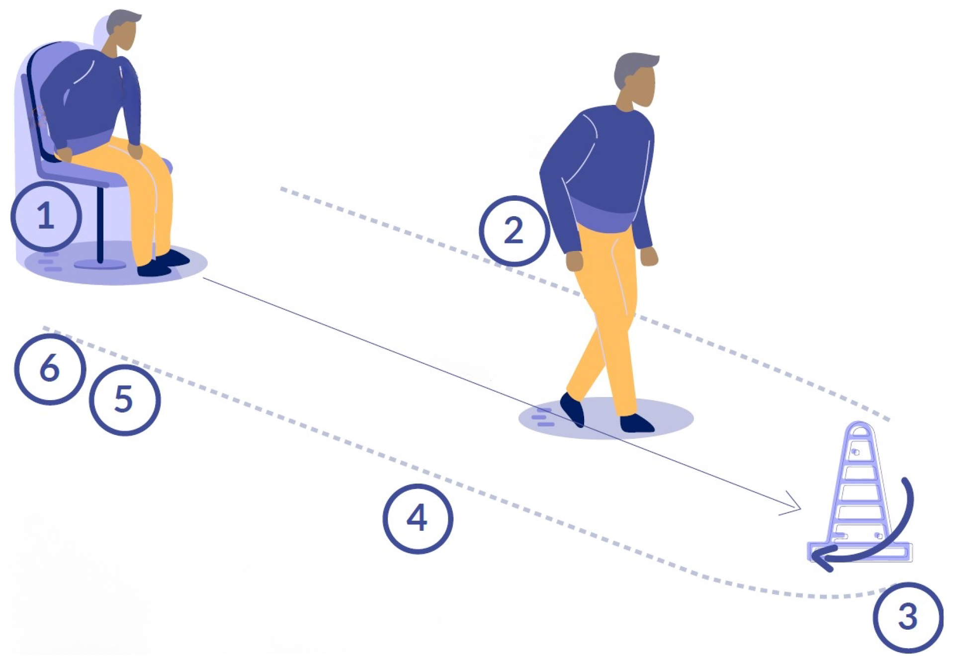
2. Methods
- Authors;
- Year/Country;
- Study methodology;
- Institutions;
- Inclusion criteria;
- Exclusion criteria;
- Participants;
- Age of participants;
- Number of participants;
- Number of analyzed participants;
- Gender distribution;
- Technology/Sensors used;
- iTUG implementation;
- TUG implementation;
- Raw data;
- Index, parameters, and variables extracted;
- Segmentation Algorithm;
- Main outcomes;
- Main results.
3. Results
3.1. Characteristics of the Participants
3.2. Methodological Design of the Selected Studies
3.3. Types of Technology, Procedure and Instrumentation Used in the Timed Up and Go Tests
3.4. Algorithmic Procedures for Segmentation and Extraction of iTUG Features
3.5. Features Extracted from iTUG
3.6. Main Clinical Results from the Selected Studies
4. Discussion
5. Conclusions
Author Contributions
Funding
Informed Consent Statement
Data Availability Statement
Conflicts of Interest
References
- Sharif, S.I.; Al-Harbi, A.B.; Al-Shihabi, A.M.; Al-Daour, D.S.; Sharif, R.S. Falls in the elderly: Assessment of prevalence and risk factors. Pharm. Pract. (Granada) 2018, 16, 1206. [Google Scholar] [CrossRef] [PubMed] [Green Version]
- Mak, T.C.; Wong, T.W.; Ng, S.S. Visual-related training to improve balance and walking ability in older adults: A systematic review. Exp. Gerontol. 2021, 156, 111612. [Google Scholar] [CrossRef]
- Phelan, E.A.; Ritchey, K. Fall prevention in community-dwelling older adults. Ann. Intern. Med. 2018, 169, ITC81–ITC96. [Google Scholar] [CrossRef]
- Ambrose, A.F.; Paul, G.; Hausdorff, J.M. Risk factors for falls among older adults: A review of the literature. Maturitas 2013, 75, 51–61. [Google Scholar] [CrossRef] [PubMed]
- Park, S.H. Tools for assessing fall risk in the elderly: A systematic review and meta-analysis. Aging Clin. Exp. Res. 2018, 30, 1–16. [Google Scholar] [CrossRef]
- Marques, N.R.; Spinoso, D.H.; Cardoso, B.C.; Moreno, V.C.; Kuroda, M.H.; Navega, M.T. Is it possible to predict falls in older adults using gait kinematics? Clin. Biomech. 2018, 59, 15–18. [Google Scholar] [CrossRef] [Green Version]
- Gschwind, Y.J.; Kressig, R.W.; Lacroix, A.; Muehlbauer, T.; Pfenninger, B.; Granacher, U. A best practice fall prevention exercise program to improve balance, strength/power, and psychosocial health in older adults: Study protocol for a randomized controlled trial. BMC Geriatr. 2013, 13, 1–13. [Google Scholar] [CrossRef] [PubMed] [Green Version]
- Barry, E.; Galvin, R.; Keogh, C.; Horgan, F.; Fahey, T. Is the Timed Up and Go test a useful predictor of risk of falls in community dwelling older adults: A systematic review and meta-analysis. BMC Geriatr. 2014, 14, 1–14. [Google Scholar] [CrossRef] [Green Version]
- Zasadzka, E.; Borowicz, A.M.; Roszak, M.; Pawlaczyk, M. Assessment of the risk of falling with the use of timed up and go test in the elderly with lower extremity osteoarthritis. Clin. Interv. Aging 2015, 10, 1289. [Google Scholar] [CrossRef] [Green Version]
- Persson, C.U.; Danielsson, A.; Sunnerhagen, K.S.; Grimby-Ekman, A.; Hansson, P.O. Timed Up & Go as a measure for longitudinal change in mobility after stroke–Postural Stroke Study in Gothenburg (POSTGOT). J. Neuroeng. Rehabil. 2014, 11, 1–7. [Google Scholar]
- Nguyen, H.; Lebel, K.; Boissy, P.; Bogard, S.; Goubault, E.; Duval, C. Auto detection and segmentation of daily living activities during a Timed Up and Go task in people with Parkinson’s disease using multiple inertial sensors. J. Neuroeng. Rehabil. 2017, 14, 1–13. [Google Scholar] [CrossRef] [Green Version]
- Podsiadlo, D.; Richardson, S. The timed “Up & Go”: A test of basic functional mobility for frail elderly persons. J. Am. Geriatr. Soc. 1991, 39, 142–148. [Google Scholar] [PubMed]
- Vervoort, D.; Vuillerme, N.; Kosse, N.; Hortobágyi, T.; Lamoth, C.J. Multivariate analyses and classification of inertial sensor data to identify aging effects on the Timed-Up-and-Go Test. PLoS ONE 2016, 11, e0155984. [Google Scholar] [CrossRef] [PubMed] [Green Version]
- Mancilla, E.; Valenzuela, J.; Escobar, M. Rendimiento en las pruebas “Timed Up and Go” y “Estación Unipodal” en adultos mayores chilenos entre 60 y 89 años. Rev. Méd. Chile 2015, 143, 39–46. [Google Scholar] [CrossRef] [Green Version]
- Flansbjer, U.B.; Holmbäck, A.M.; Downham, D.; Patten, C.; Lexell, J. Reliability of gait performance tests in men and women with hemiparesis after stroke. J. Rehabil. Med. 2005, 37, 75–82. [Google Scholar] [PubMed] [Green Version]
- Hafsteinsdottir, T.B.; Rensink, M.; Schuurmans, M. Clinimetric properties of the Timed Up and Go Test for patients with stroke: A systematic review. Top. Stroke Rehabil. 2014, 21, 197–210. [Google Scholar] [CrossRef] [PubMed] [Green Version]
- Dal Bello-Haas, V.; Klassen, L.; Sheppard, M.S.; Metcalfe, A. Psychometric properties of activity, self-efficacy, and quality-of-life measures in individuals with Parkinson disease. Physiother. Can. 2011, 63, 47–57. [Google Scholar] [CrossRef] [Green Version]
- Van Lummel, R.C.; Walgaard, S.; Hobert, M.A.; Maetzler, W.; Van Dieën, J.H.; Galindo-Garre, F.; Terwee, C.B. Intra-rater, inter-rater and test-retest reliability of an instrumented timed up and go (iTUG) test in patients with Parkinson’s disease. PLoS ONE 2016, 11, e0151881. [Google Scholar] [CrossRef] [Green Version]
- Salarian, A.; Horak, F.B.; Zampieri, C.; Carlson-Kuhta, P.; Nutt, J.G.; Aminian, K. iTUG, a sensitive and reliable measure of mobility. IEEE Trans. Neural Syst. Rehabil. Eng. 2010, 18, 303–310. [Google Scholar] [CrossRef] [Green Version]
- Caronni, A.; Sterpi, I.; Antoniotti, P.; Aristidou, E.; Nicolaci, F.; Picardi, M.; Pintavalle, G.; Redaelli, V.; Achille, G.; Sciumè, L.; et al. Criterion validity of the instrumented Timed Up and Go test: A partial least square regression study. Gait Posture 2018, 61, 287–293. [Google Scholar] [CrossRef]
- Williams, J.M.; Nyman, S.R. Age moderates differences in performance on the instrumented timed up and go test between people with dementia and their informal caregivers. J. Geriatr. Phys. Ther. 2021, 44, E150–E157. [Google Scholar] [CrossRef] [PubMed]
- Ayena, J.C.; Otis, M.J.D. Dimensional reduction of balance parameters in risk of falling evaluation using a minimal number of force-sensitive resistors. Int. J. Occup. Saf. Ergon. 2022, 28, 507–518. [Google Scholar] [CrossRef] [PubMed]
- Gasparutto, X.; Gueugnon, M.; Laroche, D.; Martz, P.; Hannouche, D.; Armand, S. Which functional tasks present the largest deficits for patients with total hip arthroplasty before and six months after surgery? A study of the timed up-and-go test phases. PLoS ONE 2021, 16, e0255037. [Google Scholar] [CrossRef] [PubMed]
- Frenken, T.; Brell, M.; Gövercin, M.; Wegel, S.; Hein, A. aTUG: Technical apparatus for gait and balance analysis within component-based Timed Up & Go using mutual ambient sensors. J. Ambient Intell. Humaniz. Comput. 2013, 4, 759–778. [Google Scholar]
- Mollinedo, I.; Cancela, J.M. Evaluation of the psychometric properties and clinical applications of the Timed Up and Go test in Parkinson disease: A systematic review. J. Exerc. Rehabil. 2020, 16, 302. [Google Scholar] [CrossRef]
- Gafner, S.C.; Allet, L.; Hilfiker, R.; Bastiaenen, C.H.G. Reliability and diagnostic accuracy of commonly used performance tests relative to fall history in older persons: A systematic review. Clin. Interv. Aging 2021, 16, 1591. [Google Scholar] [CrossRef]
- Christopher, A.; Kraft, E.; Olenick, H.; Kiesling, R.; Doty, A. The reliability and validity of the Timed Up and Go as a clinical tool in individuals with and without disabilities across a lifespan: A systematic review: Psychometric properties of the Timed Up and Go. Disabil. Rehabil. 2021, 43, 1799–1813. [Google Scholar] [CrossRef]
- Wiener, J.; McIntyre, A.; Janssen, S.; Chow, J.T.; Batey, C.; Teasell, R. Effectiveness of high-intensity interval training for fitness and mobility post stroke: A systematic review. PM&R 2019, 11, 868–878. [Google Scholar]
- Page, M.J.; Moher, D.; Bossuyt, P.M.; Boutron, I.; Hoffmann, T.C.; Mulrow, C.D.; Shamseer, L.; Tetzlaff, J.M.; Akl, E.A.; Brennan, S.E.; et al. PRISMA 2020 explanation and elaboration: Updated guidance and exemplars for reporting systematic reviews. BMJ 2021, 372, n160. [Google Scholar] [CrossRef]
- Page, M.J.; McKenzie, J.E.; Bossuyt, P.M.; Boutron, I.; Hoffmann, T.C.; Mulrow, C.D.; Shamseer, L.; Tetzlaff, J.M.; Akl, E.A.; Brennan, S.E.; et al. The PRISMA 2020 statement: An updated guideline for reporting systematic reviews. Syst. Rev. 2021, 10, 1–11. [Google Scholar] [CrossRef]
- Ayena, J.C.; Otis, M.J.D. Validation of minimal number of force sensitive resistors to predict risk of falling during a timed up and go test. J. Med. Biol. Eng. 2020, 40, 348–355. [Google Scholar] [CrossRef]
- Bergquist, R.; Nerz, C.; Taraldsen, K.; Mellone, S.; Ihlen, E.A.; Vereijken, B.; Helbostad, J.L.; Becker, C.; Mikolaizak, A.S. Predicting advanced balance ability and mobility with an instrumented timed up and go test. Sensors 2020, 20, 4987. [Google Scholar] [CrossRef] [PubMed]
- Weiss, A.; Mirelman, A.; Buchman, A.S.; Bennett, D.A.; Hausdorff, J.M. Using a body-fixed sensor to identify subclinical gait difficulties in older adults with IADL disability: Maximizing the output of the timed up and go. PLoS ONE 2013, 8, e68885. [Google Scholar] [CrossRef] [PubMed]
- Guzmán, J.C.; Silva, R.G.; Guzmán-Venegas, R. Reproducibilidad de los tiempos de ejecución de la prueba de Timed Up and Go, medidos con acelerómetros de smartphones en personas mayores residentes en la comunidad. Rev. Esp. Geriatr. Gerontol. 2017, 52, 249–252. [Google Scholar] [CrossRef]
- Mellone, S.; Tacconi, C.; Chiari, L. Validity of a Smartphone-based instrumented Timed Up and Go. Gait Posture 2012, 36, 163–165. [Google Scholar] [CrossRef] [PubMed]
- Coni, A.; Mellone, S.; Colpo, M.; Bandinelli, S.; Chiari, L. Influence of age and gender on sensor-based functional measures: A factor analysis approach. In Proceedings of the 2015 37th Annual International Conference of the IEEE Engineering in Medicine and Biology Society (EMBC), Milan, Italy, 25–29 August 2015; pp. 5054–5057. [Google Scholar]
- David, V.; Forjan, M.; Martinek, J.; Kotzian, S.; Jagos, H.; Rafolt, D. Evaluating wearable multimodal sensor insoles for motion-pattern measurements in stroke rehabilitation—A pilot study. In Proceedings of the 2017 International Conference on Rehabilitation Robotics (ICORR), London, UK, 17–20 July 2017; pp. 1543–1548. [Google Scholar]
- Holzreiter, S.; Jennings, S. Programming language for motion analysis. Hum. Mov. Sci. 1996, 15, 497–508. [Google Scholar] [CrossRef]
- Delbroek, T.; Vermeylen, W.; Spildooren, J. The effect of cognitive-motor dual task training with the biorescue force platform on cognition, balance and dual task performance in institutionalized older adults: A randomized controlled trial. J. Phys. Ther. Sci. 2017, 29, 1137–1143. [Google Scholar] [CrossRef] [Green Version]
- Dibilio, V.; Nicoletti, A.; Mostile, G.; Toscano, S.; Luca, A.; Raciti, L.; Sciacca, G.; Vasta, R.; Cicero, C.E.; Contrafatto, D.; et al. Dopaminergic and non-dopaminergic gait components assessed by instrumented timed up and go test in Parkinson’s disease. J. Neural Transm. 2017, 124, 1539–1546. [Google Scholar] [CrossRef]
- Fastame, M.C.; Mulas, I.; Putzu, V.; Asoni, G.; Viale, D.; Mameli, I.; Pau, M. Motor proficiency as a correlate of coping in late adult lifespan. An exploratory study. Anxiety Stress Coping 2022, 35, 687–700. [Google Scholar] [CrossRef] [PubMed]
- Figueiredo, A.I.; Balbinot, G.; Brauner, F.O.; Schiavo, A.; Baptista, R.R.; Pagnussat, A.S.; Hollands, K.; Mestriner, R.G. SPARC metrics provide mobility smoothness assessment in oldest-old with and without a history of falls: A case control study. Front. Physiol. 2020, 11, 540. [Google Scholar] [CrossRef]
- Mangano, G.R.; Valle, M.S.; Casabona, A.; Vagnini, A.; Cioni, M. Age-related changes in mobility evaluated by the timed up and go test instrumented through a single sensor. Sensors 2020, 20, 719. [Google Scholar] [CrossRef] [PubMed] [Green Version]
- Mancini, M.; Priest, K.C.; Nutt, J.G.; Horak, F.B. Quantifying freezing of gait in Parkinson’s disease during the instrumented timed up and go test. In Proceedings of the 2012 Annual International Conference of the IEEE Engineering in Medicine and Biology Society, San Diego, CA, USA, 28 August–1 September 2012; pp. 1198–1201. [Google Scholar]
- King, L.A.; Mancini, M.; Priest, K.; Salarian, A.; Rodrigues-de Paula, F.; Horak, F. Do clinical scales of balance reflect turning abnormalities in people with Parkinson’s disease? J. Neurol. Phys. Ther. 2012, 36, 25. [Google Scholar] [CrossRef] [Green Version]
- Ishikawa, M.; Yamada, S.; Yamamoto, K.; Aoyagi, Y. Gait analysis in a component timed-up-and-go test using a smartphone application. J. Neurol. Sci. 2019, 398, 45–49. [Google Scholar] [CrossRef]
- Salarian, A.; Zampieri, C.; Horak, F.B.; Carlson-Kuhta, P.; Nutt, J.G.; Aminian, K. Analyzing 180° turns using an inertial system reveals early signs of progression of parkinson’s disease. In Proceedings of the 2009 Annual International Conference of the IEEE Engineering in Medicine and Biology Society, Minneapolis/St. Paul, MN, USA, 3–6 September 2009; pp. 224–227. [Google Scholar]
- Silva, J.; Sousa, I. Instrumented timed up and go: Fall risk assessment based on inertial wearable sensors. In Proceedings of the 2016 IEEE International Symposium on Medical Measurements and Applications (MeMeA), Benevento, Italy, 15–18 May 2016; pp. 1–6. [Google Scholar]
- Hoskovcová, M.; Dušek, P.; Sieger, T.; Brožová, H.; Zárubová, K.; Bezdíček, O.; Šprdlík, O.; Jech, R.; Štochl, J.; Roth, J.; et al. Predicting falls in Parkinson disease: What is the value of instrumented testing in off medication state? PLoS ONE 2015, 10, e0139849. [Google Scholar] [CrossRef] [PubMed]
- Hellmers, S.; Izadpanah, B.; Dasenbrock, L.; Diekmann, R.; Bauer, J.M.; Hein, A.; Fudickar, S. Towards an automated unsupervised mobility assessment for older people based on inertial TUG measurements. Sensors 2018, 18, 3310. [Google Scholar] [CrossRef] [Green Version]
- Beyea, J.; McGibbon, C.A.; Sexton, A.; Noble, J.; O’Connell, C. Convergent validity of a wearable sensor system for measuring sub-task performance during the timed up-and-go test. Sensors 2017, 17, 934. [Google Scholar] [CrossRef] [Green Version]
- Galan-Mercant, A.; Cuesta-Vargas, A. Clinical frailty syndrome assessment using inertial sensors embedded in smartphones. Physiol. Meas. 2015, 36, 1929. [Google Scholar] [CrossRef] [PubMed]
- Galán-Mercant, A.; Cuesta-Vargas, A.I. Differences in trunk kinematic between frail and nonfrail elderly persons during turn transition based on a smartphone inertial sensor. BioMed Res. Int. 2013, 2013, 279197. [Google Scholar] [CrossRef] [Green Version]
- Galán-Mercant, A.; Cuesta-Vargas, A.I. Differences in trunk accelerometry between frail and non-frail elderly persons in functional tasks. BMC Res. Notes 2014, 7, 1–9. [Google Scholar] [CrossRef] [Green Version]
- Van Uem, J.M.; Walgaard, S.; Ainsworth, E.; Hasmann, S.E.; Heger, T.; Nussbaum, S.; Hobert, M.A.; Mico-Amigo, E.M.; Van Lummel, R.C.; Berg, D.; et al. Quantitative timed-up-and-go parameters in relation to cognitive parameters and health-related quality of life in mild-to-moderate Parkinson’s disease. PLoS ONE 2016, 11, e0151997. [Google Scholar] [CrossRef] [Green Version]
- Walgaard, S.; Faber, G.S.; van Lummel, R.C.; van Dieën, J.H.; Kingma, I. The validity of assessing temporal events, sub-phases and trunk kinematics of the sit-to-walk movement in older adults using a single inertial sensor. J. Biomech. 2016, 49, 1933–1937. [Google Scholar] [CrossRef] [PubMed] [Green Version]
- Tan, D.; Pua, Y.H.; Balakrishnan, S.; Scully, A.; Bower, K.J.; Prakash, K.M.; Tan, E.K.; Chew, J.S.; Poh, E.; Tan, S.B.; et al. Automated analysis of gait and modified timed up and go using the Microsoft Kinect in people with Parkinson’s disease: Associations with physical outcome measures. Med. Biol. Eng. Comput. 2019, 57, 369–377. [Google Scholar] [CrossRef]
- Sheehan, K.; Greene, B.; Cunningham, C.; Crosby, L.; Kenny, R. Early identification of declining balance in higher functioning older adults, an inertial sensor based method. Gait Posture 2014, 39, 1034–1039. [Google Scholar] [CrossRef] [PubMed]
- Seo, J.; Kim, T.; Lee, J.; Kim, J.; Choi, J.; Tack, G. Fall prediction of the elderly with a logistic regression model based on instrumented timed up & go. J. Mech. Sci. Technol. 2019, 33, 3813–3818. [Google Scholar]
- Picardi, M.; Redaelli, V.; Antoniotti, P.; Pintavalle, G.; Aristidou, E.; Sterpi, I.; Meloni, M.; Corbo, M.; Caronni, A. Turning and sit-to-walk measures from the instrumented Timed Up and Go test return valid and responsive measures of dynamic balance in Parkinson’s disease. Clin. Biomech. 2020, 80, 105177. [Google Scholar] [CrossRef] [PubMed]
- Nguyen, H.P.; Ayachi, F.; Lavigne-Pelletier, C.; Blamoutier, M.; Rahimi, F.; Boissy, P.; Jog, M.; Duval, C. Auto detection and segmentation of physical activities during a Timed-Up-and-Go (TUG) task in healthy older adults using multiple inertial sensors. J. Neuroeng. Rehabil. 2015, 12, 1–12. [Google Scholar] [CrossRef] [PubMed] [Green Version]
- Najafi, B.; Armstrong, D.G.; Mohler, J. Novel wearable technology for assessing spontaneous daily physical activity and risk of falling in older adults with diabetes. J. Diabetes Sci. Technol. 2013, 7, 1147–1160. [Google Scholar] [CrossRef] [Green Version]
- Mirelman, A.; Weiss, A.; Buchman, A.S.; Bennett, D.A.; Giladi, N.; Hausdorff, J.M. Association between performance on Timed Up and Go subtasks and mild cognitive impairment: Further insights into the links between cognitive and motor function. J. Am. Geriatr. Soc. 2014, 62, 673–678. [Google Scholar] [CrossRef] [PubMed] [Green Version]
- Mariani, B.; Jiménez, M.C.; Vingerhoets, F.J.; Aminian, K. On-shoe wearable sensors for gait and turning assessment of patients with Parkinson’s disease. IEEE Trans. Biomed. Eng. 2012, 60, 155–158. [Google Scholar] [CrossRef]
- Gerhardy, T.; Gordt, K.; Jansen, C.P.; Schwenk, M. Towards using the instrumented timed up-and-go test for screening of sensory system performance for balance control in older adults. Sensors 2019, 19, 622. [Google Scholar] [CrossRef] [Green Version]
- Weiss, A.; Herman, T.; Plotnik, M.; Brozgol, M.; Maidan, I.; Giladi, N.; Gurevich, T.; Hausdorff, J.M. Can an accelerometer enhance the utility of the Timed Up & Go Test when evaluating patients with Parkinson’s disease? Med. Eng. Phys. 2010, 32, 119–125. [Google Scholar] [PubMed]
- Weiss, A.; Mirelman, A.; Giladi, N.; Barnes, L.L.; Bennett, D.A.; Buchman, A.S.; Hausdorff, J.M. Transition between the Timed Up and Go turn to sit subtasks: Is timing everything? J. Am. Med. Dir. Assoc. 2016, 17, 864.e9–864.e15. [Google Scholar] [CrossRef] [Green Version]
- Yu, L.; Zhao, Y.; Wang, H.; Sun, T.L.; Murphy, T.E.; Tsui, K.L. Assessing elderly’s functional balance and mobility via analyzing data from waist-mounted tri-axial wearable accelerometers in timed up and go tests. BMC Med. Inform. Decis. Mak. 2021, 21, 1–14. [Google Scholar] [CrossRef]
- Zarzeczny, R.; Nawrat-Szołtysik, A.; Polak, A.; Maliszewski, J.; Kiełtyka, A.; Matyja, B.; Dudek, M.; Zborowska, J.; Wajdman, A. Aging effect on the instrumented Timed-Up-and-Go test variables in nursing home women aged 80–93 years. Biogerontology 2017, 18, 651–663. [Google Scholar] [CrossRef] [PubMed] [Green Version]
- Toosizadeh, N.; Mohler, J.; Lei, H.; Parvaneh, S.; Sherman, S.; Najafi, B. Motor performance assessment in Parkinson’s disease: Association between objective in-clinic, objective in-home, and subjective/semi-objective measures. PLoS ONE 2015, 10, e0124763. [Google Scholar] [CrossRef] [Green Version]
- Montesinos, L.; Castaldo, R.; Pecchia, L. Wearable inertial sensors for fall risk assessment and prediction in older adults: A systematic review and meta-analysis. IEEE Trans. Neural Syst. Rehabil. Eng. 2018, 26, 573–582. [Google Scholar] [CrossRef] [Green Version]
- Kose, A.; Peruzzi, A.; Cereatti, A.; Laudani, L.; Della Croce, U. Detection of heel strikes and toe-offs during gait using a single inertial measurement unit attached to the waist. In Proceedings of the Second National Congress of Biongineering, Turin, Italy, 8–10 July 2010; Volume 233. [Google Scholar]
- Weinberg, H. Using the ADXL202 in pedometer and personal navigation applications. Analog Devices AN-602 Appl. Note 2002, 2, 1–6. [Google Scholar]
- Sejdić, E.; Lowry, K.A.; Bellanca, J.; Perera, S.; Redfern, M.S.; Brach, J.S. Extraction of stride events from gait accelerometry during treadmill walking. IEEE J. Transl. Eng. Health Med. 2015, 4, 1–11. [Google Scholar] [CrossRef]
- Wüest, S.; Massé, F.; Aminian, K.; Gonzenbach, R.; De Bruin, E.D. Reliability and validity of the inertial sensor-based Timed “Up and Go” test in individuals affected by stroke. J. Rehabil. Res. Dev. 2016, 53, 599–610. [Google Scholar] [CrossRef]
- Pelicioni, P.H.; Menant, J.C.; Latt, M.D.; Lord, S.R. Falls in Parkinson’s disease subtypes: Risk factors, locations and circumstances. Int. J. Environ. Res. Public Health 2019, 16, 2216. [Google Scholar] [CrossRef] [Green Version]
- Janssen, W.G.; Bussmann, H.B.; Stam, H.J. Determinants of the sit-to-stand movement: A review. Phys. Ther. 2002, 82, 866–879. [Google Scholar] [CrossRef] [PubMed]


| Criteria | Description |
|---|---|
| iTUG | Studies describe iTUG using technological support to enrich the test (cameras, inertial sensors, environmental sensors, pressure sensors, optoelectronic systems). |
| Index, parameters or variables | Proposals that, in addition to the total duration traditionally obtained in TUG, provide other variables or measurements that allow the identification of motor alterations in the participants. |
| Context | Proposals evaluated in community, clinical or academic settings. |
| Study methodology | Descriptive, experimental, quasi-experimental and proof-of-concept clinical studies were included that used validated commercial technology or new technologies whose applications were applied in the elderly population. No methodological limitations were applied to carry out the screening by the level of evidence. |
| Participants | Elderlies with a mean age equal or greater than 65 years, with or without associated pathology. |
| Language | Studies published in English or Spanish. |
| Study year | Studies published between 2012 and 2022. |
| Criteria | Description |
|---|---|
| Index, parameters or variables | Proposals that only provided the total value of TUG without demonstrating any other characteristic extracted, independent of declaring the use of any technology, were excluded. |
| Study methodology | Other narratives, bibliographic, systematic or scoping reviews were excluded, as well as “one-page” conference articles, abstracts, posters, letters to the editor and studies of iTUG psychometric properties validation. |
| Author | Methods | iTUG Implementation | Segmentation | Features | Main Results |
|---|---|---|---|---|---|
| Ayena et al., 2022 [22] | Experimental clinical study | Traditional 3-meter TUG procedure in people with PD. Segmentation in four phases: Standing, Go and Return Walking and Sitting. Insole with 4 FSR, one in the heel, the other in the medial and lateral foot, one in the first and the other in the fifth metatarsal. The tri-axial accelerometer is in the instep of the foot. | Automatic segmentation based on patterns of plantar pressures and accelerations. The research team developed the algorithm. | Gait index variability. | A reduced set of FSR allows the measurement of gait parameters for measuring the RoF in young elderly and PD. |
| Ayena et al., 2020 [31] | Experimental clinical study | Traditional 3-meter TUG procedure in people with PD. Segmentation in four phases: Standing, Go and Return Walking and Sitting. Insole with 4 FSR, one in the heel, the other in the medial and lateral foot, one in the first and the other in the fifth metatarsal. The tri-axial accelerometer is in the instep of the foot. | Automatic segmentation based on patterns of plantar pressures and accelerations. The research team developed the algorithm. | 12 spatiotemporal gait parameters. | The risk of falls index calculated with a minimum number of sensors is a promising tool for real-time analysis, as it provides similar statistical information in risk analysis with a larger number of sensors. |
| Bergquist et al., 2020 [32] | Cross-sectional study | Traditional procedure of the 3-meter TUG procedure in communitary elderlies performing five repetitions. Segmentation into five phases: Standing, Go walking, 3-meter turning, Return walking and Sitting. Smartphone is located on the lower back. | Segmentation using acceleration and angular velocity signals with the algorithm by Weiss et al. [33] | 78 features: Total iTUG duration. iTUG sub-phase duration. Spatial gait parameters in turning and walking sub-phases. | The iTUG model has a predictive ability similar to the scores of the CBMS and the battery of clinical tests with a significantly lower prediction error. |
| Campillay et al., 2017 [34] | Descriptive study. | Traditional procedure of the 3-meter TUG procedure in elderlies, selecting the best of three repetitions. Segmentation into five phases: Standing, Go walking, 3-meter turning, Return walking and Sitting. Smartphone is located on the lower back. | Segmentation using acceleration and angular velocity signals with the algorithm by Mellone et al. [35] | Total iTUG duration. iTUG sub-phase duration. | Duration of iTUG sub-phases measured through the smartphone IMU are highly reproducible. |
| Coni et al., 2015 [36] | Cohort study. | Traditional procedure of the 3-meter TUG procedure in communitary elderlies. Segmentation into five phases: Standing, Go walking, 3-meter turning, Return walking and Sitting. Smartphone located on the waist. | Segmentation using acceleration and angular velocity signals with the algorithm by Weiss et al. [33] | 38 features: Total duration of iTUG; iTUG sub-phase duration. Statistical descriptors of angular velocity in the turning sub-phase: peak, mean, Root Mean Square Value (RMS). Spatial gait parameters: number of steps on walking and turning sub-phases. | A significant number of features can be derived from the sensor signals, which can be grouped into factors with clear clinical value, allowing the measurement of many mobility skills from one, and not just total time. |
| David et al., 2017 [37] | Pilot study. | Traditional procedure of the 3-meter TUG procedure in people with stroke. Segmentation into three phases: Standing, Walking and Sitting. | Segmentation using the spatial position of markers with Machine Learning (Neural Networks) [38] | Spatiotemporal gait parameters. | The system has proven to be capable of measuring the level of walking and TUG, providing details in the analysis of the movement and parameters compared with the current data, which is the total time measured manually. |
| Delbroek et al., 2017 [39] | Randomized clinical study. | Traditional procedure of the 3-meter TUG procedure in elderlies with visual tasks. IMU on ankles, wrists and chest. Segmentation into six phases: Standing, Go walking, 3-meter turning, Return walking, Pre-sitting turning, Sitting. | APDM Mobility Lab proprietary algorithm. | Total duration of iTUG. Spatiotemporal gait parameters. Statistical descriptors of angular velocity and accelerations. | All iTUG measurements improve with l-dopa, except sit-to-stand transition (duration and AP acceleration) and stand-to-sit transition (all parameters). |
| Dibilio et al., 2017 [40] | Does not specify. | Traditional procedure of the 3-meter TUG procedure in people with PD with visual tasks. The sensor is on the lower back at L4–L5. Segmentation into six phases: Standing, Go walking, 3-meter turning, Return walking, Pre-sitting turning, Sitting. | BTS G-Studio proprietary algorithm. | Freezing of gait. Total duration of iTUG. Duration per sub-phase of iTUG. Spatiotemporal gait parameters. | Low but significant correlations were found between motor scores (iTUG parameters) and global cognitive function. |
| Fastame et al., 2022 [41] | Exploratory study. | Traditional procedure of the 3-meter TUG procedure in communitary elderlies with visual tasks. The sensor is on the lower back at L5–S1. Segmentation into six phases: Standing, Go walking, 3-meter turning, Return walking, Pre-sitting turning, Sitting. | BTS G-Studio proprietary algorithm. | Duration and average speed of each sub-phase of iTUG. | It is a robust, stable and sensitive measurement tool for “movement smoothness” independent of the speed and duration of the movement, capable of identifying RoF independent of the day’s speed. |
| Figuiredo et al., 2020 [42] | case-control study. | Traditional procedure of the 3-meter TUG procedure in elderlies with visual tasks. The sensor is on the lower back at L5–S1. Segmentation into six phases: Standing, Go walking, 3-meter turning, Return walking, Pre-sitting turning, Sitting. | BTS G-Studio proprietary algorithm. | Total duration of iTUG. Duration per sub-phase of iTUG. Statistical descriptors of angular velocity by sub-phase. Statistical descriptors of degrees of orientation and inclination by sub-phase. | It is possible to identify significant differences in the temporal variables of the sub-phases and their speed, mainly in the elderly population. |
| Mangano et al., 2020 [43] | Transversal study. | Traditional procedure of the 3-meter TUG procedure and an extended 7-meter version in elderlies. The sensor is on the lower back at L1. Segmentation into six phases: Standing, Go walking, 3-meter turning, Return walking, Pre-sitting turning, Sitting. | BTS G-Studio proprietary algorithm. | Duration and average speed of each sub-phase of iTUG. | No differences were found between Parkinson’s patients with RoF and without RoF in stride length and speed parameters. Conversely, there was a difference in the time of double support. In addition, good correlations were found between the ABC Scale and iTUG scales. |
| Mancini et al., 2012 [44] | Cross-sectional study. | Extended 7-meter TUG in people with PD. The sensor is on the lower back and on the leg shank segment. Segmentation into six phases: Standing, Go walking, 3-meter turning, Return walking, Pre-sitting turning, Sitting. | APDM Mobility Lab proprietary algorithm. | 52 features: Spatiotemporal gait parameters. Statistical descriptors of acceleration of the walking sub-phase. Acceleration frequency analysis of the gait sub-phase. | An instrumented scale can reveal deficits in the turns of patients with Parkinson’s (severe and mild), even though they have a normal score and walk correctly. |
| King et al., 2012 [45] | Exploratory study. | Extended 7-meter TUG in people with PD. The sensor is on the lower back, leg shank segment, arms and chest. Segmentation into six phases: Standing, Go walking, 3-meter turning, Return walking, Pre-sitting turning, Sitting. | APDM Mobility Lab proprietary algorithm. | Total duration of iTUG; Turning sub-phase duration. Spatial gait parameters: Step number during turning. Statistical descriptors of angular velocity during turning. | The system is useful for analyzing the gait patterns during TUG, applicable to healthy subjects or with different gait disorders by analyzing the correlations of the go and return signals. |
| Ishikawa et al., 2019 [46] | Exploratory study. | Traditional procedure of the 3-meter TUG procedure in elderlies with iNPH. Smartphone on the abdomen. Segmentation into six phases: Standing, Go walking, 3-meter turning, Return walking, Pre-sitting turning, Sitting. | Segmentation using acceleration and angular velocity signals with the algorithm by Salarian et al. [47]. | Spatiotemporal gait parameters: Gait speed, cadence, stride time variability. | It could be correctly identified and with an error under the test transition elements in the predetermined order and in any other sequence of the movements or activities measured during TUG. |
| Silva et al., 2016 [48] | Exploratory study. | Traditional procedure of the 3-meter TUG procedure in elderlies. The sensor is in a pocket or attached to the thigh. Segmentation into three phases: Standing, Go walking, 3-meter turning. | Automatic segmentation algorithm using the integral of the angular velocity signal developed by the same research team. | Total duration of iTUG; Duration per sub-phase of iTUG; Spatial gait parameters: Step number during walking. Statistical descriptors of acceleration by sub-phase. Acceleration frequency analysis by sub-phase. | A strategy implemented in stages with and without medication in patients with Parkinson’s makes it possible to predict falls. |
| Hoskovcova et al., 2015 [49] | Prospective study. | Traditional 3-meter TUG and extended 7-meter TUG in people with PD. Use of five inertial sensors located in the leg, wrist and sternum areas. | Does not report segmentation strategy. | Spatiotemporal gait parameters: gait speed, cadence and stride time variability. | Using principal component analysis, it was observed that the characteristics that contributed the most to the discrimination with respect to the control group were in the phase of getting up, the chest flexion, the maximum obliquity of the chest and the maximum vertical velocity of the chest. |
| Hellmers et al., 2018 [50] | Exploratory study. | Traditional 3-meter TUG in elderlies. A belt with sensors in L4–L5 and an instrumented chair with four pressure sensors and a laser are used. Segmentation into six phases: Standing, Go walking, 3-meter turning, Return walking, Pre-sitting turning, Sitting. | Segmentation with the algorithm of Nguyen et al., 2015 [11]. | Total duration of iTUG; Turning sub-phase duration. Statistical descriptors of acceleration by sub-phase. | There are some differences in the acceleration signals between groups of frail adults compared to non-frail adults by means of a smartphone placed on the chest. |
| Gasparutto et al., 2021 [23] | Prospective study. | Traditional procedure of the 3-meter TUG procedure in elderlies with hip arthroplasty. In total, 35 reflective markers were used according to the conventional gait model. Segmentation into five phases: Standing, Go walking, 3-meter turning, Return walking and Sitting. | Automatic segmentation with the algorithm by Bayea et al. [51]. | 33 features: joint mobility range descriptors by iTUG sub-phase: spine at thorax level and c7. The base of support by sub-phase. Spatial gait parameters: number of steps during walking and turning. Statistical descriptors of angular velocity by sub-phase of the thorax and pelvis. | This simple test could be appropriate for quantifying patient-specific deficits in function, and hence, guiding and monitoring post-operative rehabilitation in clinical settings. |
| Galán-Mercant et al., 2015 [52] | Cross-sectional study. | Extended 10-meter TUG in elderlies with the frail syndrome. Smartphone on thorax. Segmentation into five phases: Standing, Go walking, 3-meter turning, Return walking and Sitting. | Segmentation using acceleration and angular velocity signals with algorithms from Weiss et al. [33] and Salarian et al. [47]. | Duration per sub-phase of iTUG. Statistical descriptors of acceleration by sub-phase. Statistical descriptors of angular velocity by sub-phase. Statistical descriptors of degrees of orientation and inclination by sub-phase. | An inertial sensor from the iPhone 4 can study and analyze the kinematics of the different sub-phases of the Extended TUG test in frail and non-frail elderly people. |
| Galán-Mercant et al., 2013 [53] | Cross-sectional study. | Extended 10-meter TUG in elderlies with frail syndrome. Smartphone on thorax. Segmentation into five phases: Standing, Go walking, 3-meter turning, Return walking and Sitting. | Segmentation using acceleration and angular velocity signals with algorithms from Weiss et al. [33] and Salarian et al. [47]. | Statistical descriptors of acceleration by sub-phase. Statistical descriptors of angular velocity by sub-phase. Statistical descriptors of degrees of orientation and inclination by sub-phase. Total duration of iTUG. Duration per sub-phase of iTUG. | The inertial sensor found in the iPhone 4 is able to study and analyze the kinematics of the turning transitions in frail and physically active elderly persons. The accelerometry values for the frail elderly are lower than the physically active elderly, while variability in the readings for the frail elderly is also lower than the control group. |
| Galán-Mercant et al., 2014 [54] | Cross-sectional study. | Extended 10-meter TUG in elderlies with frail syndrome. Smartphone on thorax. Segmentation into five phases: Standing, Go walking, 3-meter turning, Return walking and Sitting. | Segmentation using acceleration and angular velocity signals with algorithms from Weiss et al. [33] and Salarian et al. [47]. | Statistical descriptors of acceleration by sub-phase. Statistical descriptors of angular velocity by sub-phase. Total duration of iTUG. Duration per sub-phase of iTUG. Joint mobility ranges by sub-phase of iTUG (Thorax). | For the Extended TUG test, this device allows more sensitive differentiation between population groups than the traditionally used variable, namely time. |
| Van Uem et al., 2016 [55] | Cross-sectional study. | Traditional 3-meter TUG in people with PD. A belt with sensors in L4–L5. Segmentation into six phases: Standing, Go walking, 3-meter turning, Return walking, Pre-sitting turning, Sitting. | Segmentation using angular velocity signals and accelerations with the algorithm of Walgaard et al. [56]. | Statistical descriptors of acceleration by sub-phase. Statistical descriptors of angular velocity by sub-phase. Total duration of iTUG. Duration per sub-phase of iTUG. Joint mobility ranges by sub-phase of iTUG (Lumbar). | Spontaneous physical activity at home and instrumented assessments in the clinic demonstrated strong discriminatory power in detecting impaired motor function in Parkinson’s disease. |
| Van Lummel et al., 2016 [18] | Exploratory study. | Traditional 3-meter TUG in people with PD. A belt with sensors in L4–L5. Segmentation into six phases: Standing, Go walking, 3-meter turning, Return walking, Pre-sitting turning, Sitting. | Segmentation using angular velocity signals and accelerations with the algorithm of Walgaard et al. [56]. | Total duration of iTUG. Duration per sub-phase of iTUG. | Intra-rater, inter-rater and test-retest reliability of the individual components of iTUG was excellent to good for total duration and turning durations and good to poor for sitting durations and kinematics. |
| Toosizadeh et al., 2015 [53] | Cohort study. | Traditional 3-meter TUG in people with PD. Five units of measurement, one on each leg, one on each thigh, and one on the lower back. Segmentation into five phases: Standing, Go walking, 3-meter turning, Return walking, Sitting. | Segmentation using acceleration and angular velocity signals with the algorithm by Salarian et al. [47]. | Statistical descriptors of acceleration by sub-phase. Statistical descriptors of angular velocity by sub-phase. Total duration of iTUG. Duration per sub-phase of iTUG. Joint mobility ranges by sub-phase of iTUG. | iTUG measurements obtained from trunk angular velocity during the turning and standing phases are adequate measures of dynamic balance in Parkinson’s disease. These measurements respond to rehabilitation, being able to detect improvement in patients after treatment. |
| Tan et al., 2018 [57] | Does not specify. | Reduced version at 2-meter TUG in people with PD. The Kinect camera is located 1.4 m from the test circuit. | Algorithm developed by the same research team. | Spatial gait parameters: Length of the first step. Total duration of iTUG. Turning sub-phase duration. | The only Kinect-derived variable significantly associated with the motor analysis scale was first step length. |
| Sheehan et al., 2014 [58] | Longitudinal study. | Traditional 3-meter TUG in elderlies. Sensors are located on each leg. | Does not specify a segmentation algorithm. | Spatiotemporal gait parameters. Spatial gait parameters. Statistical descriptors of angular velocity by sub-phase. | The quantitative values of iTUG and linear regression models allow us to identify decreases in the balance of highly functional older adults. |
| Seo et al., 2019 [59] | Transversal study. | Traditional procedure of the 3-meter TUG procedure in elderlies with visual tasks. IMU on ankles, wrists, lower back and chest. Segmentation into five phases: Standing, Go walking, 3-meter turning, Return walking, Sitting. | APDM Mobility Lab proprietary algorithm. | 36 features: Spatiotemporal gait parameters. Statistical descriptors of angular velocity by sub-phase. Total duration of iTUG. Duration per sub-phase of iTUG. Joint mobility ranges by sub-phase of iTUG (thorax). | It was possible to predict the falls with 70.2% accuracy using the characteristics of the total duration of the test, standing characteristics, the maximum speed of the trunk in the sagittal plane and its range of movement in the horizontal plane during walking and the speed angular maxima during sitting. |
| Picardi et al., 2020 [60] | Longitudinal study. | Traditional 3-meter TUG in people with PD. The sensor on the lower back. Segmentation into five phases: Standing, Go walking, 3-meter turning, Return walking and Sitting. | Segmentation using the morphology of the acceleration signal with the algorithm of Mellone et al. [35]. | Spatiotemporal gait parameters. Statistical descriptors of angular velocity by sub-phase. Total duration of iTUG. Duration per sub-phase of iTUG. | iTUG measurements obtained from trunk angular velocity during the turning and standing phases are adequate measures of dynamic balance in Parkinson’s disease, and these measurements respond to rehabilitation, being able to detect improvement in patients after treatment. |
| Nguyen et al., 2015 [61] | Exploratory study. | Extended 5-meter TUG and 10-meter TUG in elderlies. 17 IMUs in each body segment. Segmentation into six phases: Standing, Go walking, 3-meter turning, Return walking, Pre-sitting turning, Sitting. | Algorithm developed by the same research team. | 36 features: Spatiotemporal gait parameters. Statistical descriptors of angular velocity by sub-phase. Total duration of iTUG. Duration per sub-phase of iTUG. Joint mobility ranges by iTUG sub-phase. | The proposed algorithm allows detecting and segmenting typical TUG activities using inertial sensors. |
| Nguyen et al., 2017 [11] | Exploratory study. | Extended 5-meter TUG and 10-meter TUG in eldelries. 17 IMUs in each body segment. Segmentation into six phases: Standing, Go walking, 3-meter turning, Return walking, Pre-sitting turning, Sitting. | Algorithm developed by the same research team. | Spatial gait parameters: number of steps during walking and turning. Statistical descriptors of acceleration by sub-phase. Statistical descriptors of angular velocity by sub-phase. Joint mobility ranges by sub-phase of iTUG. | The transferability of the segmentation methodology to Parkinson’s patients is demonstrated, using only 4 of the 17 initially predisposed sensors. |
| Najafi et al., 2013 [62] | Exploratory study. | Traditional procedure of the 3-meter TUG procedure in elderlies with peripheral neuropathy and diabetes with visual tasks. Sensor on thorax. Segmentation into five phases: Standing, Go walking, 3-meter turning, Return walking and Sitting. | Evaluation of postural transitions through accelerometer analysis. The algorithm was developed by the same research team. | Duration per sub-phase of iTUG. | The proposed system can identify and monitor postural transitions, accurately identifying subjects with a high RoF with potential use in monitoring older adults with diabetes. |
| Mirelman et al., 2014 [63] | Cross-sectional study. | Traditional 3-meter TUG in elderlies. Sensor on the lower back. Segmentation into five phases: Standing, Go walking, 3-meter turning, Return walking, Sitting. | Segmentation using the morphology of the acceleration and angular velocity signal with the algorithm of Weiss et al. [33]. | Duration per sub-phase of iTUG. | Using a single sensor on the back during TUG can quantify mobility, facilitating the understanding of problems related to cognitive decline. |
| Mariani et al., 2013 [64] | Exploratory study. | Traditional 3-meter TUG in people with PD. IMU on the instep of the foot. Segmentation into three phases: Go walking, 3-meter turning, Return walking. | Segmentation using angular velocity signals and accelerations. The algorithm was developed by the same research team. | Duration per sub-phase of iTUG. Spatiotemporal gait parameters: step number and gait velocity. | All the sub-phases of the test could be detected in the control groups, with medication and without medication. Variations of the measured gait characteristics are observed, where the temporal variables are the most important within the TUG test. |
| Frenken et al., 2013 [24] | Experimental study. | Traditional 3-meter TUG in elderlies. No sensors on the participants. aTUG (ambient TUG) chair integrated with environmental sensors, four force sensors and an optical laser. Segmentation into fivex phases: Standing, Go walking, 3-meter turning, Return walking, Sitting. | Algorithm developed by the same research team. | Statistical descriptors of degrees of orientation and inclination by sub-phase. Duration per sub-phase of iTUG. | The proposed method is able to accurately compute the duration of the TUG components using only the force sensors and the laser range scanner. |
| Gerhardy et al., 2019 [65] | Cross-sectional study. | Traditional procedure of the 3-meter TUG procedure in elderlies. The sensor is on lower back. Segmentation into five phases: Standing, Go walking, 3-meter turning, Return walking, Sitting. | Segmentation using the morphology of the acceleration signal with the algorithm of Mellone et al. [35]. | Total duration of iTUG. Duration per sub-phase of iTUG. | Total TUG time was strongly associated with vestibular and somatosensory system performance. |
| Weiss et al., 2013 [33] | Cohort study. | Traditional 3-meter TUG in elderlies. Sensor on the lower back. Segmentation into five phases: Standing, Go walking, 3-meter turning, Return walking, Sitting. | Segmentation using acceleration and angular velocity signals with algorithms developed by the same team [66]. | Spatio temporal gait parameters: Stride speed, stride length, swing width, stride length. Statistical descriptors of degrees of orientation and inclination by sub-phase of rotation. Duration per sub-phase of iTUG. | People with mobility impairments have problems in the five sub-phases of TUG. |
| Weiss et al., 2016 [67] | Cohort study. | Traditional 3-meter TUG in elderlies with dementia. The sensor on the lower back. Segmentation into six phases: Standing, Go walking, 3-meter turning, Return walking, Pre-sitting turning, Sitting. | Segmentation using acceleration and angular velocity signals with algorithms developed by the same team [66]. | Total duration of iTUG. Duration per sub-phase of iTUG. | Longer separations between the movements of the sub-phases and a longer overlap between turning and the stand-to-sit sub-phase are related to poorer cognitive and motor function and greater disability. |
| Williams et al., 2021 [21] | Cross-sectional study. | Traditional 3-meter TUG in elderlies with dementia. The sensor is on the lower back. Do not specify level of segmentation. | Segmentation using angular velocity signals and accelerations with the algorithm of Walgaard et al. [56]. | Statistical descriptors of angular velocity by sub-phase. | The sensor and its algorithms were able to quantify the sub-phases of iTUG and demonstrate that age moderates differences in the performance of iTUG and its informal caregivers. |
| Yu et al., 2021 [68] | Descriptive study. | Traditional procedure of the 3-meter TUG procedure in communitary elderlies. Sensor on the lower back. Segmentation into five phases: Standing, Go walking, 3-meter turning, Return walking, Sitting. | Segmentation using acceleration and angular velocity signals with algorithms from Weiss et al. [33]. | Statistical descriptors of acceleration by sub-phase. Statistical descriptors of angular velocity by sub-phase. Statistical descriptors of degrees of orientation and inclination by sub-phase. | The proposal is a reliable option for objective, unsupervised and unobtrusive balance measurement in a clinical or home setting. |
| Zarzeczny et al., 2017 [69] | Cross-sectional study. | Traditional procedure of the 3-meter TUG procedure in elderlies with visual tasks. Sensor on the lower back at L4–L5. Segmentation into six phases: Standing, Go walking, 3-meter turning, Return walking, Pre-sitting turning, Sitting. | BTS G-Studio proprietary algorithm. | Statistical descriptors of acceleration by sub-phase. Statistical descriptors of angular velocity by sub-phase. Statistical descriptors of degrees of orientation and inclination by sub-phase. | The performance of the functional tests is more dependent on the range of force developed than the maximum isometric force of the muscles of the lower extremities during acceleration when sitting is measured. |
| Subjects | Studies | Participants |
|---|---|---|
| Elderlies [24,33,34,39,42,43,48,50,58,59,61,63,65,67] | 14 | 2448 |
| Community elderlies [32,36,41,68] | 4 | 521 |
| Residence elderlies [69] | 1 | 26 |
| PD [11,18,31,40,44,45,49,55,57,60,64,70] | 13 | 426 |
| People with stroke [13] | 1 | 35 |
| Elderlies with other pathology [21,23,46,52,53,54,62] | 7 | 376 |
| Total | 40 | 3822 |
| Type of Study | Studies |
|---|---|
| Cross-sectional [21,32,44,52,53,54,55,63,65,69] | 10 |
| Exploratory [11,18,41,45,46,48,50,61,62,64] | 10 |
| Cohort [33,36,67,70] | 4 |
| Clinical-Experimental [22,24,31] | 3 |
| Descriptive [34,68] | 2 |
| Transversal [43,59] | 2 |
| Prospective [23,49] | 2 |
| Longitudinal [58,60] | 2 |
| case-control [42] | 1 |
| Clinical-Randomized [39] | 1 |
| Pilot [37] | 1 |
| Not mentioned [40,57] | 2 |
| Total | 40 |
| Technology | Studies |
|---|---|
| Insoles [22,31,37] | 3 |
| Smartphone [32,34,36,46,48,52,53,54,65] | 9 |
| Inertial Sensors [11,18,21,33,39,40,41,42,43,44,45,49,55,58,59,60,61,62,63,64,67,68,69,70] | 24 |
| Opto–electronic System [23,55] | 2 |
| Xbox Kinect [57] | 1 |
| Instrumented Chair [24,50] | 2 |
| Author | Proposal | Inputs | Transitions | Studies |
|---|---|---|---|---|
| Ayena et al. [31] | Rule-based. | Tri-axial foot accelerations. Plantar pressures. | S2W, W2S. | 2 |
| Weiss et al. [33] | Rule-based. | Tri-axial lower back accelerations and tri-axial lower back angular velocities | St, S2W, W2T, T2W, W2T, T2S, Si. | 7 |
| Mellone et al. [35] | Rule-based. | Tri-axial chest accelerations. | St, S2W, W2T, T2W, W2T, Si. | 3 |
| Holzreiter et al. [38] | Machine Learning. | On-body reflective infrared marker’s coordinates. | St, S2W, W2T, T2W, W2T, Si. | 1 |
| APDM Mobility Lab. | Propietary. | Tri-axial accelerations and tri-axial angular velocities from both wrists, shanks, lower back and/or chest. | St, S2W, W2T, T2W, W2T, T2S, Si. | 4 |
| BTS G-Studio. | Propietary. | Tri-axial lower back accelerations and tri-axial lower back angular velocities. | St, S2W, W2T, T2W, W2T, T2S, Si. | 5 |
| Silva et al. [48] | Rule-based. | Tri-axial thigh angular velocities. | St, S2W, W2T. | 1 |
| Salarian et al. [19] | Rule-based. | Tri-axial waist accelerations and tri-axial waist angular velocities. | St, S2W, W2T, T2W, W2T, T2S, Si. | 3 |
| Nguyen et al. [11] | Rule-based. | Tri-axial lower back accelerations and tri-axial lower back angular velocities. | St, S2W, W2T, T2W, W2T, T2S, Si. | 3 |
| Bayea et al. [51] | Rule-based. | Tri-axial upper body accelerations. Tri-axial upper body angular velocities. | St, S2W, W2T, T2W, W2T, T2S, Si. | 1 |
| Walgaard et al. [56] | Rule-based. | Tri-axial lower back accelerations. Tri-axial lower back angular velocities. Displacements and angles of lower back and feet. | St, S2W, W2T, T2W, W2T, T2S, Si. | 3 |
| Tan et al. [57] | Rule-based. | Kinect video. | St, S2W, W2T, T2W, W2T, T2S, Si. | 1 |
| Najafi et al. [62] | Rule-based. | Tri-axial chest accelerations. Tri-axial chest angular velocities. | St, S2W, W2T, T2W, W2T, T2S, Si. | 1 |
| Mariani et al. [64] | Rule-based. | Tri-axial foot accelerations. Tri-axial foot angular velocities. | W2T, T2W. | 1 |
| Frenken et al. [24] | Rule-based. | Pressure sensors on chair. Light laser distance. | St, S2W, W2T, T2W, W2T, Si. | 1 |
| Feature | Relevant Parameters | Studies |
|---|---|---|
| Total iTUG duration. | Clinical Score. | 19 |
| Duration per sub-phase of iTUG. | Standing, walking (go and return), turning (3-meter and pre-sitting), sitting. | 23 |
| Spatiotemporal gait parameters. | Cadence, stride time, step time, stride length, step length, gait velocity, number of steps, double support time, stride time variability, gait index variability. | 20 |
| Statistical descriptors in the acceleration time domain by sub-phase. | Maximum value, minimum value, mean, standard deviation, kurtosis, root mean square value, entropy. | 9 |
| Statistical descriptors in the time domain of angular velocity by sub-phase. | Maximum value, minimum value, mean, standard deviation, kurtosis, root mean square value, entropy. | 17 |
| Statistical descriptors in time domain of mobility ranges in degrees per sub-phase. | Main joints measured: thorax (chest or sternum), cervical (C7), lumbar (L4–L5), pelvis. | 14 |
| Acceleration frequency domain descriptors per sub-phase. | Fast Fourier transform. | 2 |
Disclaimer/Publisher’s Note: The statements, opinions and data contained in all publications are solely those of the individual author(s) and contributor(s) and not of MDPI and/or the editor(s). MDPI and/or the editor(s) disclaim responsibility for any injury to people or property resulting from any ideas, methods, instructions or products referred to in the content. |
© 2023 by the authors. Licensee MDPI, Basel, Switzerland. This article is an open access article distributed under the terms and conditions of the Creative Commons Attribution (CC BY) license (https://creativecommons.org/licenses/by/4.0/).
Share and Cite
Ortega-Bastidas, P.; Gómez, B.; Aqueveque, P.; Luarte-Martínez, S.; Cano-de-la-Cuerda, R. Instrumented Timed Up and Go Test (iTUG)—More Than Assessing Time to Predict Falls: A Systematic Review. Sensors 2023, 23, 3426. https://doi.org/10.3390/s23073426
Ortega-Bastidas P, Gómez B, Aqueveque P, Luarte-Martínez S, Cano-de-la-Cuerda R. Instrumented Timed Up and Go Test (iTUG)—More Than Assessing Time to Predict Falls: A Systematic Review. Sensors. 2023; 23(7):3426. https://doi.org/10.3390/s23073426
Chicago/Turabian StyleOrtega-Bastidas, Paulina, Britam Gómez, Pablo Aqueveque, Soledad Luarte-Martínez, and Roberto Cano-de-la-Cuerda. 2023. "Instrumented Timed Up and Go Test (iTUG)—More Than Assessing Time to Predict Falls: A Systematic Review" Sensors 23, no. 7: 3426. https://doi.org/10.3390/s23073426