Article

An Efficient Electrochemical Sensor Driven by Hierarchical Hetero-Nanostructures Consisting of RuO2 Nanorods on WO3 Nanofibers for Detecting Biologically Relevant Molecules

Department of Chemistry & Nanoscience, Ewha Womans University, Seoul 03760, Korea
*
Authors to whom correspondence should be addressed.
Authors contribute this work equally.
Sensors 2019, 19(15), 3295; https://doi.org/10.3390/s19153295
Submission received: 26 June 2019 / Revised: 17 July 2019 / Accepted: 23 July 2019 / Published: 26 July 2019
(This article belongs to the Special Issue Sensors for Hazardous Material Detection)

Abstract

:
By means of electrospinning with the thermal annealing process, we investigate a highly efficient sensing platform driven by a hierarchical hetero-nanostructure for the sensitive detection of biologically relevant molecules, consisting of single crystalline ruthenium dioxide nanorods (RuO2 NRs) directly grown on the surface of electrospun tungsten trioxide nanofibers (WO3 NFs). Electrochemical measurements reveal the enhanced electron transfer kinetics at the prepared RuO2 NRs-WO3 NFs hetero-nanostructures due to the incorporation of conductive RuO2 NRs nanostructures with a high surface area, resulting in improved relevant electrochemical sensing performances for detecting H2O2 and L-ascorbic acid with high sensitivity.

Graphical Abstract

1. Introduction

Recently, a variety of metal oxide nanostructures have been extensively utilized as efficient electrode substances owing to their outstanding electrocatalytic properties. Among them, ruthenium dioxide (RuO2) has been well described as one of the best electrocatalysts for diverse energy related applications, such as the hydrogen evolution reaction (HER), oxygen evolution reaction (OER), and supercapacitors because of its high electric conductivity, catalytic activity, and thermal stability [1,2,3]. Especially, RuO2 has been used as an efficient electrode system for supercapacitors owing to its excellent charging-discharging behavior [1,4,5,6,7,8]. Generally, RuO2 as a promising catalytic material is often used in the forms of hybrid structures or alloys with other abundant transition metals in consideration of the relatively high cost of RuO2. Thus, there have been previous reports regarding the use of RuO2 nanostructures with other metal oxides as supercapacitors [9,10,11,12], and biosensing applications [1,2,3,13,14].
Tungsten trioxide (WO3) nanostructures have been also extensively studied in various applications due to its earth-abundance, high durability, and chemical stabilities in aqueous acid media, as well as good electrochemical conductivity [15,16,17,18]. Thereby it has been developed as a catalyst for the hydrogen evolution reaction (HER) and supercapacitors in an acidic solution [19,20,21,22]. WO3 also constitutes composites with other novel metals like Pt [23,24,25,26], Ir [17,23,27], and Ru [16,28,29,30], or supporting materials.
Nanostructured catalysts are applied to nonenzymatic electrochemical biosensors. Electrochemical properties can be enhanced from the increase of active surfaces. The detection of hydrogen peroxide (H2O2) is important in not only biomedical and environmental applications, but also in the enzymatic system [31]. While ascorbic acid (AA) has an important role in the physiological function of organisms, a deficiency of AA causes several diseases [32,33]. Therefore, the detection and accurate quantification of target material with selectivity is highly required.
In this study, we introduce a facile fabrication of hybrid nanostructures consisting of single crystalline RuO2 nanorods on eletrospun WO3 nanofibers by utilizing electrospinning and thermal annealing processes. In addition, the fundamental electrochemical performances of RuO2 nanorods-WO3 nanofibers (RuO2 NRs-WO3 NFs) are carefully investigated, which confirm their characteristics of fast electron-transfer reactions and possibility as a catalytic sensing platform for detecting l-ascorbic acid (AA) and hydrogen peroxide (H2O2) in phosphate buffered solution (PBS).

2. Materials and Methods

Tungsten chloride (WCl6, ≥ 99.9% true metal basis), ruthenium chloride hydrate (RuCl3xH2O, 99.98% trace metal basis), poly(vinyl pyrrolidone) (PVP, MW = 1,300,000), N,N-dimethylformamide (DMF), potassium ferricyanide (K3[Fe(CN)6]), l-ascorbic acid (AA), 4-acetamidophenol (AP), dopamine hydrochloride (DA), uric acid (UA), d-(+)-glucose, hydrogen peroxide (H2O2, 35 wt% solution in water), sodium phosphate monobasic, and sodium phosphate dibasic were supplied by Sigma Aldrich (St. Louis, MO). Commercial Pt/C and Ir/C (both of them were 20 wt% each metal loading on Vulcan XC-72) were obtained from E-TEK Company. Sulfuric acid (H2SO4) and acetic acid were provided by Ducksan (Korea). Sodium hydroxide (NaOH) was purchased from Daejung (Korea). Deionized water with resistivity ≥ 18 MΩ∙cm was used in all processes.
First, WO3 nanofibers were synthesized by electrospinning and thermal annealing process according to the reported method [23]. To prepare electrospinning solution, 1.5 g WCl6 were dissolved in 10.549 mL DMF with 1.25 g PVP and 0.191 mL acetic acid. After being stirred overnight, the solution was loaded into syringe and applied to the needle of the electrospinning system (Nano NC ESR 200R2). The needle was connected to a voltage power supply (applied voltage = 17.5 kV) at a flow rate of 5 μL/min, and the distance from needle tip to aluminum plate to collect as spun NFs was 15 cm. The collected electrospun NFs were calcinated at 500 °C for 1 h under a mixed gas atmosphere of 80 sccm of He and 10 sccm of O2 with ramping rate of 1 °C/min.
Ruthenium hydroxide (Ru(OH)3) precursor was prepared by a precipitation process via the acid-base reaction with controlling pH of aqueous solution. The pH of the final precursor solution at about pH 10 was carefully achieved by slowly dropping 0.1 M NaOH dilute solution into 5 mM RuCl3xH2O aqueous solution [2,13]. After precipitation, the precursor solution was washed five times with deionized water, and then re-dispersed in 2~3 mL pure deionized water again. To grow RuO2 NRs on WO3 NFs, 2 mg of WO3 NFs was dispersed into 1 mL deionized water and then mixed with 1 mL Ru(OH)3 precursor solution. After sonication for 30 min, the mixed solution was directly dropped on the center of Si wafer. WO3 nanofibers containing Ru(OH)3 precursors loaded on the Si wafer was placed into the center of a furnace and calcined at 300 ℃ for 5 h in air. The furnace was then allowed to cool to room temperature.
The surface morphology of as-grown products was examined by field emission scanning electron microscopy (FE-SEM; JEOL JSM-6700F). The detailed crystal structures were also investigated by a high-resolution transmission electron microscopy (HRTEM, Cs-corrected STEM, JEOL JEM-2100F) instrument equipped with selected area electron diffraction (SAED) micrographs and elemental EDX mapping with a Tecnai-F20 system operated at 200 kV. Additionally, high resolution X-ray diffraction measurement (XRD; Bruker D8 DISCOVER, Cu Kα radiation), and X-ray photoelectron spectroscopy (XPS; Theta Probe AR-XPS System. Al Kα radiation) were performed to investigate the crystal structure and surface binding energies of as-grown RuO2 NR-WO3 NFs.
For electrochemical measurements, a three-electrode system was used with a modified glassy carbon (GC) electrode (3 mm in diameter), a saturated calomel electrode (S.C.E.), and a coiled Pt wire (1 mm in diameter, length immersed in a solution ~ 10 cm) as the working electrode, the reference electrode, and the counter electrode, respectively. All electrochemical experiments conducted with CHI 650E workstation (CH Instruments) and BAS100B (BAS Inc.). To modify the surface of a GC electrode with synthesized nanomaterials, 2 mg of RuO2 NR-WO3 NFs was suspended in 1.0 mL deionized water. Subsequently, 10 μL of the solution were dropped onto the GC electrode surface three times. Then, 10 μL of 0.05 wt% Nafion solution were loaded onto the modified GC electrode surface. Cyclic voltammetry (CV) measurements was used for analyze the capacitive behavior in 1 M H2SO4. For sensing experiments, linear sweep voltammetry (LSV) was also used with rotating disk electrode (RDE) at a scan rate of 5 mV s−1 with rotating speed of 1600 rpm, and amperometry measurements were used in 0.1 M phosphate buffered saline (PBS) at physiological condition pH (7.4).

3. Results and Discussion

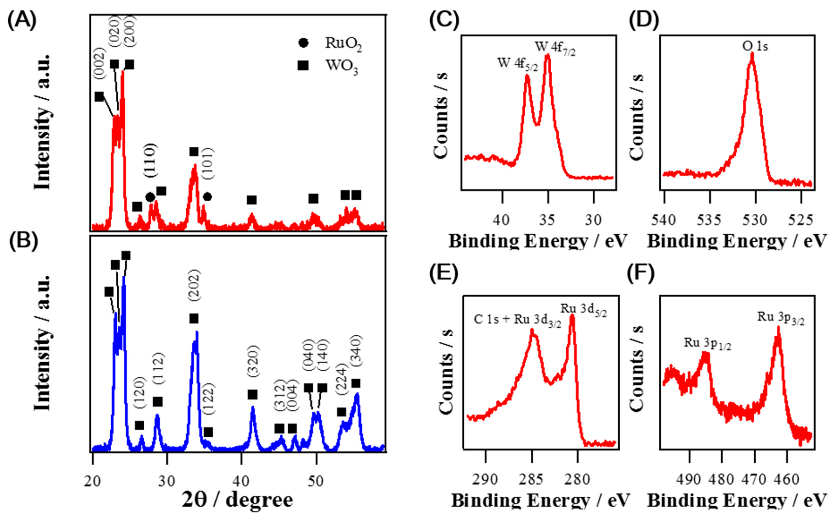
3.1. Synthesis of Hybrid Nanostructures of RuO2 Nanorods on Electrospun WO3 Nanofibers

Figure 1A,B show FE-SEM images of WO3 NFs annealed at 500 °C. The calcined WO3 NFs revealed a very fine structure and the diameter of the fibers was around 200 nm. On the other hand, after the heat treatment of the mixed solution composed of Ru(OH)3 precursors and WO3 NFs at 300 ℃ for 5 h, it is readily identified that RuO2 NRs were directly grown on the electrospun WO3 NFs as shown in Figure 1C,D. Figure 1D represents the as grown RuO2 NRs covering the entire surface of WO3 NFs. The lateral dimension of RuO2 NRs is estimated to be about 40 nm and the length up to 300 nm. Careful EDS measurements indicate that the atomic ratio of Ru to W is confirmed as 45:55. According to our previous real-time study by in situ synchrotron XRD, a simple recrystallization process by thermal annealing might be responsible for the growth mechanism of RuO2 NRs. It was carefully suggested that Ru diffusion to the amorphous nanoparticles followed by diffusion to the growing surface of the nanorod plays an essential role in the growth of RuO2 NRs in oxygen ambient, which is supported by the nucleation theory [34].
Figure 2 represents XRD spectra and high resolution XPS spectra of composite RuO2 NRs-WO3 NFs and pure WO3 NFs. XRD spectrum of pure WO3 NFs in Figure 2B demonstrates that all peaks are closely matched with the monoclinic phase of WO3 [19,35]. On the other hand, XRD spectrum of composite RuO2 NR-WO3 NFs confirms the same monoclinic phase WO3 peaks including two major peaks at 27.1° and 34.8° corresponding to (110) and (101) crystallographic planes of tetragonal RuO2 structure as displayed in Figure 2A [2,13]. To investigate the oxidation states of Ru, W, and O atoms, XPS measurements were performed. In Figure 2C, two separated binding energies at 35.1 eV and 37.3 eV are clearly identified as two spin-orbit states of W 4f5/2 and W 4f7/2, respectively, which indicates the oxidation state of +6 for W in WO3 NFs [16,36]. Both high resolution Ru 3d and Ru 3p spectra were shown in Figure 2E,F. Although the peak position of Ru 3d3/2 is overlapped with C 1s [16,37], the oxidation state of Ru species is readily identified to Ru4+ based on the binding energies of 280.7 eV and 462.8 eV, indexed to Ru 3d5/2 and Ru 3p3/2, respectively [37,38]. In addition, the peak at 530.5 eV of O 1s is associated with O2− in RuO2 and WO3 metal oxides as shown in Figure 2D.
Figure 3 indicates TEM images and SAED pattern for a single WO3 nanofiber covered with RuO2 nanorods. As shown in Figure 3A,B, low-magnification TEM images show the high density of RuO2 nanorods directly grown on the porous surface of WO3 nanofiber. The SAED pattern shown in Figure 3E reveals the existence of many different crystalline phases in a WO3 nanofiber which confirms the polycrystalline nature of a WO3 nanofiber. On the contrary, the fast Fourier transform (FFT) of the lattice-resolved image for a RuO2 nanorod in Figure 3F represents highly ordered lattice fringes with a single crystal nature. The values of lattice spacing of adjacent planes are estimated by about 0.318 nm and 0.263 nm, corresponding to those of between the (110) planes and (101) for the tetragonal RuO2, respectively. Furthermore, TEM-EDS element mapping analysis from the high-angle annular dark field (HAADF) STEM image shown in Figure S1 confirms the homogenous distribution of Ru, W, and O in distinct regions in the hierarchical nanostructure. W atoms exist on the backbone of the nanofibers, whereas Ru atoms exclusively exist on the branched nanorods. Oxygen atoms exist both on the backbone of the nanofibers and branched nanorods. Thus, we successfully fabricate the high density of single-crystalline RuO2 nanorods on WO3 nanofibers by using a combination of an electrospinning process and a thermal annealing process. Our growth process thus provides a simple methodology for the fabrication of highly efficient electrocatalysts.

3.2. Electrochemical Properties for Capacitive Behaviors of RuO2 NRs-WO3 NFs

The general electrochemical activities of RuO2 NRs-WO3 NFs and WO3 NFs were examined by CV in 10 mM [Fe(CN)6]3− aqueous solution containing 1 M KCl. Figure S2 displays CV curves of RuO2 NRs-WO3 NFs and WO3 NFs at a scan rate 100 mV s−1. Voltammetric current peaks at RuO2 NRs-WO3 NFs are reversible, while those of WO3 NFs are quasi-reversible. It seems to be ascribed to that RuO2 NRs-WO3 NFs allow very facile heterogeneous electron transfer kinetics with high electric conductivities in contrast to WO3 NFs. Moreover, RuO2 NRs-WO3 NFs show a much larger charging current in CV than WO3 NFs.
To characterize the charging behavior of the synthesized materials, CV was measured for a potential range from 0.1 V to 0.9 V (vs. S.C.E.) in 1 M H2SO4 as seen in Figure 4. Figure 4A shows CV results comparing RuO2 NRs-WO3 NFs and WO3 NFs at a scan rate 100 mV s−1. It supports the enhanced capacity of RuO2 NRs-WO3 NFs as the RuO2 NRs were grown on WO3 NFs. To examine the charging performance, the average specific capacitance values (Csp, F g−1) were calculated with the following Equation (1) using CV curves shown in Figure 4B.
C sp = 1 2 × v × m × V   I d V
where v is the scan rate (V s−1), m is the weight of electrode materials, V is the potential range, and I d V is the area under CV curve [39]. At the scan rate of 10 mV s−1, the Csp values of the synthesized materials, RuO2 NRs-WO3 NFs and WO3 NFs, are 98.15 F g−1 and 0.95 F g−1, respectively. The Csp of RuO2 NRs-WO3 NFs is obviously 103-fold higher than that of WO3 NFs as shown in Figure 4C. As the scan rate increases, Csp becomes smaller and the Csp of RuO2 NRs-WO3 NFs and WO3 NFs decreased down to 57% and 42%, respectively, while increasing the scan rate from 10 mV s−1 to 200 mV s−1. This additionally indicates the successful decoration of WO3 NFs with RuO2 NRs forming the hierarchical hetero-nanostructures.
Electrochemical impedance spectroscopy (EIS) was also employed to examine the electrochemical behavior of RuO2 NRs-WO3 NFs and WO3 NFs. EIS measurement was carried out at 0.5 V (vs. S.C.E.) under the same condition of CV experiments with a frequency range of 0.1 Hz–1000 kHz as shown in Figure S3. The Nyquist plot of RuO2 NRs-WO3 NFs was closer to a vertical line than that of WO3 NFs, exhibiting nearly pure capacitive behavior of RuO2 NR-WO3 NFs [1,40]. The stability of RuO2 NRs-WO3 NFs for capacitance was demonstrated by monitoring the change of Csp during repeated CV cycles as depicted in Figure 4D. RuO2 NRs-WO3 NFs excellently maintained about 96% of its original Csp for the 1000 CV cycles at a scan rate of 100 mV s−1.

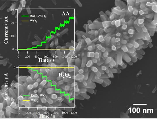
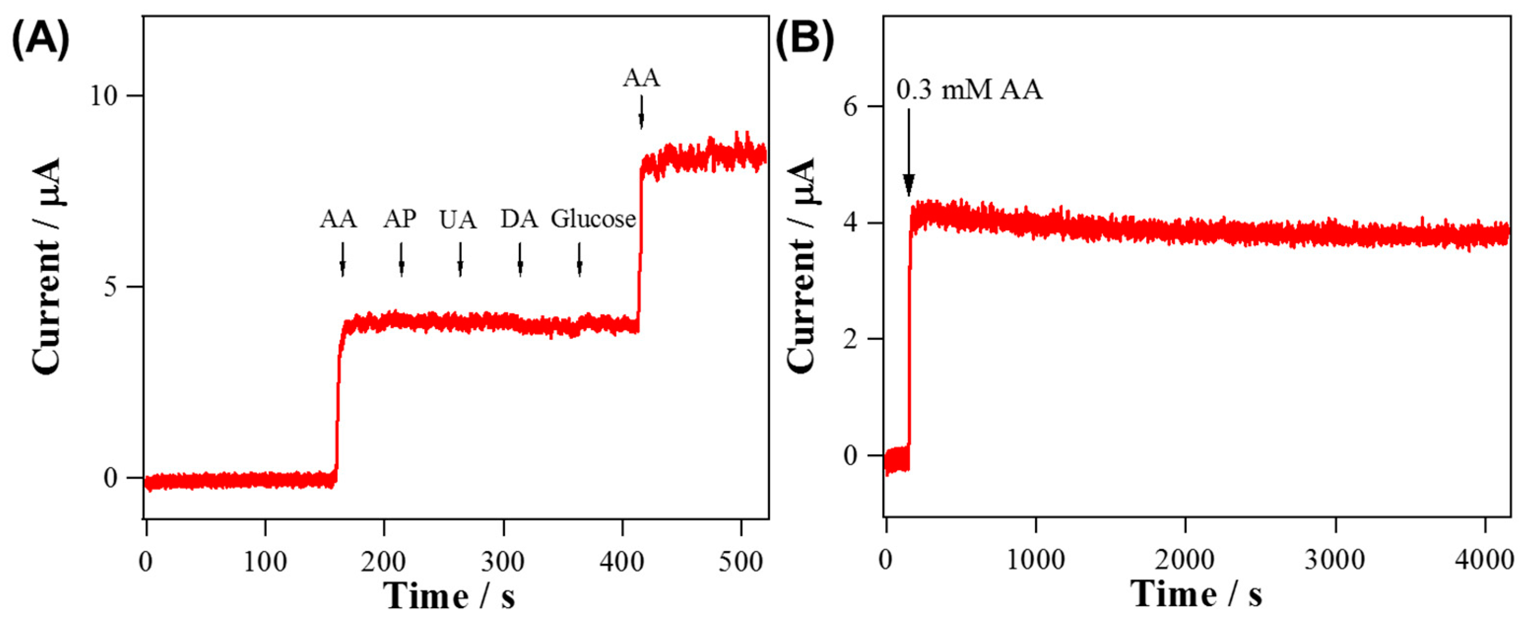
3.3. Applications to Electrochemical Sensing of AA and H2O2

The electrochemical properties of RuO2 NRs-WO3 NFs for AA oxidation were also studied. LSV measurements in 0.1 M PBS were used for examining the oxidations of various biomaterials such as AA, DA, UA, AP, and glucose. The chosen concentrations are slightly above the physiological concentrations. As shown in Figure 5A, AA oxidation started to occur from the most negative potential compared with other biomaterials. Amperometric measurements of RuO2 NRs-WO3 NFs and WO3 NFs were conducted at 0 V (vs. S.C.E.) which possibly allow for the oxidation of AA only, excepting for the other tested biomolecules as seen in the LSV results of Figure 5A.
As observed in Figure 5B, the anodic currents of both electrodes were increased linearly with the concentration of AA increased from 5 μM to 2 mM. Also, the calibration curves based on the amperometric data were depicted in inset of Figure 5B. The sensitivity of RuO2 NRs-WO3 NFs (171.7 μA mM−1 cm−2, R2 = 0.9990, normalized to GC substrate electrode area, 0.072 cm2) were surprisingly increased by 244 times compared to that of WO3 NFs (0.704 μA mM−1 cm−2, R2 = 0.9990). Most of the typical biological samples are complex, having various oxidizable species, so selectivity to a targeted analyte is an essential requirement for any sensor. In Figure 6A, current responses for AA oxidation were stable against the additions of 0.1 mM AP, 0.1 mM UA, 0.1 μM DA and 5 mM glucose at 0 V. Additionally, the stability of RuO2 NRs-WO3 NFs was measured by monitoring the change of current at 0 V in 0.1 M PBS containing 0.3 mM AA. The amperometric response of RuO2 NRs-WO3 NFs retained 96% of the initial current level during over a 4200-s measurement in Figure 6B, supporting its excellent stability. Table 1 summarizes the properties of RuO2 NRs-WO3 NFs in comparison with other Ru-based materials used as AA sensors.
The catalytic effect of RuO2 NRs-WO3 NFs for H2O2 reduction was also measured. Figure 7A shows overlaid LSV results of RuO2 NRs-WO3 NFs and WO3 NFs. It presents clearly that H2O2 reduction at RuO2 NRs-WO3 NFs starts from a much less negative potential with much greater reduction current level than that at WO3 NFs. In fact, the cathodic current level measured at −0.2 V (vs. S.C.E.) was more greatly increased for RuO2 NRs-WO3 NFs than WO3 NFs in response to the successive increase of H2O2 concentration (Figure 7B). Inset of Figure 7B shows the calibrated current vs concentration with good linearity. Obtained sensitivities from the calibration curves are 619.7 μA mM−1 cm−2 (R2 = 0.9960), and 5.5 μA mM−1 cm−2 (R2 = 0.9384) for RuO2 NRs-WO3 NFs and WO3 NFs, respectively. Sensitivity of RuO2 NR-WO3 NFs is 112-fold higher than the value of WO3 NFs, and therefore it supports the enhanced activities of RuO2 NRs-WO3 NFs toward H2O2 reduction. The H2O2 reduction current instead of the oxidation current was monitored to sense H2O2 in order to avoid the interference from many oxidizable species generally present in biological systems. Figure S4 represents the selectivity of RuO2 NRs-WO3 NFs for H2O2 reduction. The current responses of RuO2 NRs-WO3 NFs at −0.2 V (vs. S.C.E.) for H2O2 reduction were obvious; however, there were no noticeable responses to the successive injections of other biological materials: 0.1 mM AA, 0.1 mM UA, 0.1 μM DA, 5 mM glucose, 0.1 mM AP, and 30 μM O2. RuO2 NR-WO3 NFs show relatively excellent catalytic activities for H2O2 reduction compared to other previous Ru-based materials as compared in Table 2. RuO2 NRs-WO3 NFs for measuring H2O2 reduction current was less stable than that for AA oxidation. In fact, H2O2 reduction current measured at −0.2 V was decreased to ~60% of the initial current level after 4200-s continuous measurement (data not shown).

4. Conclusions

We report the successful fabrication of single crystalline RuO2 nanorods on WO3 nanofibers by electrospinning and calcination. Microscopic and spectroscopic measurements such as SEM with EDS, XRD, and XPS were used to characterize the structure and composition of RuO2 NRs-WO3 NFs. The RuO2 NRs-WO3 NFs showed improved electrocatalytic activities over WO3 NFs through a series of electrochemical measurements. In 1 M H2SO4 solution, RuO2 NRs-WO3 NFs represent a higher Csp, 98.15 F g−1, by 103-fold with good stability and a sharper slope than pure WO3 NFs. Additionally, the RuO2 NRs-WO3 NFs have dramatically enhanced sensing abilities, in accordance with 224 times (171.7 μA mM−1 cm−2) sensitivity for AA oxidation, and 112 times (619.7 μA mM−1 cm−2) sensitivity for H2O2 reduction, respectively, compared to those of pure WO3 NFs. These results thus suggest that RuO2 NRs-WO3 NFs could be a promising candidate electrocatalyst for the fabrication of an efficient electrochemical sensor due to its highly effective electrochemical performance.

Supplementary Materials

The followings are available online at https://www.mdpi.com/1424-8220/19/15/3295/s1, Figure S1: EDS elemental mappings, Figure S2: Cyclic voltammograms, Figure S3: Nyquist plots, Figure S4: Amperometric response.

Author Contributions

Authors contribute as conceptualization, C.L., Y.L. and M.H.K.; methodology, H.L., Y.K., D.J.; validation, H.L., A.Y. and D.J.; formal analysis, H.L. and Y.K.; investigation, A.J. and A.Y.; data curation, H.L. and A.Y.; writing—original draft preparation, C.L., Y.L. and M.H.K.; writing—review and editing, C.L., Y.L. and M.H.K.; supervision, C.L.; funding acquisition, C.L.,Y.L. and M.H.K.

Funding

This work was financially supported by Basic Science Research Program through the National Research Foundation of Korea (NRF) funded by the Ministry of Education (NRF-2018R1A6A1A03025340 for YL and 2016R1D1A1B03934962 for KMH) and by the National Research Foundation of Korea (NRF) funded by the Ministry of Science and ICT (NRF-2019R1F1A1059969 for CL).

Conflicts of Interest

The authors declare no conflict of interest.

References

  1. Kim, Y.L.; Choi, H.-A.; Lee, N.-S.; Son, B.; Kim, H.J.; Baik, J.M.; Lee, Y.; Lee, C.; Kim, M.H. RuO2–ReO3 composite nanofibers for efficient electrocatalytic responses. Phys. Chem. Chem. Phys. 2015, 17, 7435–7442. [Google Scholar] [CrossRef] [PubMed]
  2. Kim, S.-J.; Cho, Y.K.; Seok, J.; Lee, N.-S.; Son, B.; Lee, J.W.; Baik, J.M.; Lee, C.; Lee, Y.; Kim, M.H. Highly Branched RuO2 Nanoneedles on Electrospun TiO2 Nanofibers as an Efficient Electrocatalytic Platform. ACS Appl. Mater. Interfaces 2015, 7, 15321–15330. [Google Scholar] [CrossRef] [PubMed]
  3. Jang, H.S.; Yang, Y.; Lee, N.-S.; Son, B.; Lee, Y.; Lee, C.; Kim, M.H. Electrospun RuO2–Co3O4 hybrid nanotubes for enhanced electrocatalytic activity. Mater. Lett. 2015, 139, 405–408. [Google Scholar] [CrossRef]
  4. Sugimoto, W.; Iwata, H.; Yokoshima, K.; Murakami, Y.; Takasu, Y. Proton and Electron Conductivity in Hydrous Ruthenium Oxides Evaluated by Electrochemical Impedance Spectroscopy: The Origin of Large Capacitance. J. Phys. Chem. B 2005, 109, 7330–7338. [Google Scholar] [CrossRef] [PubMed]
  5. Hu, C.-C.; Chang, K.-H.; Lin, M.-C.; Wu, Y.-T. Design and Tailoring of the Nanotubular Arrayed Architecture of Hydrous RuO2 for Next Generation Supercapacitors. Nano Lett. 2006, 6, 2690–2695. [Google Scholar] [CrossRef]
  6. Zang, J.; Bao, S.-J.; Li, C.M.; Bian, H.; Cui, X.; Bao, Q.; Sun, C.Q.; Guo, J.; Lian, K. Well-Aligned Cone-Shaped Nanostructure of Polypyrrole/RuO2 and Its Electrochemical Supercapacitor. J. Phys. Chem. C 2008, 112, 14843–14847. [Google Scholar] [CrossRef]
  7. Xia, H.; Meng, Y.S.; Yuan, G.; Cui, C.; Lu, L. A Symmetric RuO2/RuO2 Supercapacitor Operating at 1.6 V by Using a Neutral Aqueous Electrolyte. Electrochem. Solid-State Lett. 2012, 15, A60–A63. [Google Scholar] [CrossRef]
  8. Lee, J.-B.; Jeong, S.-Y.; Moon, W.-J.; Seong, T.-Y.; Ahn, H.-J. Preparation and characterization of electro-spun RuO2–Ag2O composite nanowires for electrochemical capacitors. J. Alloys Compd. 2011, 509, 4336–4340. [Google Scholar] [CrossRef]
  9. Wang, Y.-G.; Zhang, X.-G. Preparation and electrochemical capacitance of RuO2/TiO2 nanotubes composites. Electrochim. Acta 2004, 49, 1957–1962. [Google Scholar]
  10. Wang, Y.-G.; Wang, Z.-D.; Xia, Y.-Y. An asymmetric supercapacitor using RuO2/TiO2 nanotube composite and activated carbon electrodes. Electrochim. Acta 2005, 50, 5641–5646. [Google Scholar] [CrossRef]
  11. Rack Ahn, Y.; Park, C.; Mu Jo, S.; Young Kim, D. Enhanced charge-discharge characteristics of RuO2 supercapacitors on heat-treated TiO2 nanorods. Appl. Phys. Lett. 2007, 90, 122106. [Google Scholar]
  12. Lokhande, C.; Park, B.-O.; Park, H.-S.; Jung, K.-D.; Joo, O.-S. Electrodeposition of TiO2 and RuO2 thin films for morphology-dependent applications. Ultramicroscopy 2005, 105, 267–274. [Google Scholar] [CrossRef]
  13. Kim, S.-J.; Cho, Y.K.; Lee, C.; Kim, M.H.; Lee, Y. Real-time direct electrochemical sensing of ascorbic acid over rat liver tissues using RuO2 nanowires on electrospun TiO2 nanofibers. Biosens. Bioelectron. 2016, 77, 1144–1152. [Google Scholar] [CrossRef] [PubMed]
  14. Kim, Y.L.; Ha, Y.; Lee, N.-S.; Kim, J.G.; Baik, J.M.; Lee, C.; Yoon, K.; Lee, Y.; Kim, M.H. Hybrid architecture of rhodium oxide nanofibers and ruthenium oxide nanowires for electrocatalysts. J. Alloys Compd. 2016, 663, 574–580. [Google Scholar] [CrossRef]
  15. Huang, Z.-F.; Song, J.; Pan, L.; Zhang, X.; Wang, L.; Zou, J.-J. Tungsten Oxides for Photocatalysis, Electrochemistry, and Phototherapy. Adv. Mater. 2015, 27, 5309–5327. [Google Scholar] [CrossRef] [PubMed]
  16. Janáky, C.; Chanmanee, W.; Rajeshwar, K. On the Substantially Improved Photoelectrochemical Properties of Nanoporous WO3 Through Surface Decoration with RuO2. Electrocatalysis 2013, 4, 382–389. [Google Scholar] [CrossRef]
  17. Baruffaldi, C.; Cattarin, S.; Musiani, M. Deposition of non-stoichiometric tungsten oxides+MO2 composites (M = Ru or Ir) and study of their catalytic properties in hydrogen or oxygen evolution reactions. Electrochim. Acta 2003, 48, 3921–3927. [Google Scholar] [CrossRef]
  18. Zheng, H.; Ou, J.Z.; Strano, M.S.; Kaner, R.B.; Mitchell, A.; Kalantar-zadeh, K. Nanostructured Tungsten Oxide—Properties, Synthesis, and Applications. Adv. Funct. Mater. 2011, 21, 2175–2196. [Google Scholar] [CrossRef]
  19. Zhou, X.; Qiu, Y.; Yu, J.; Yin, J.; Bai, X. High electrochemical activity from hybrid materials of electrospun tungsten oxide nanofibers and carbon black. J. Mater. Sci. 2012, 47, 6607–6613. [Google Scholar] [CrossRef]
  20. Wei, H.; Ding, D.; Yan, X.; Guo, J.; Shao, L.; Chen, H.; Sun, L.; Colorado, H.A.; Wei, S.; Guo, Z. Tungsten Trioxide/Zinc Tungstate Bilayers: Electrochromic Behaviors, Energy Storage and Electron Transfer. Electrochim. Acta 2014, 132, 58–66. [Google Scholar] [CrossRef]
  21. Yoon, S.; Kang, E.; Kim, J.K.; Lee, C.W.; Lee, J. Development of high-performance supercapacitor electrodes using novel ordered mesoporous tungsten oxide materials with high electrical conductivity. Chem. Commun. 2011, 47, 1021–1023. [Google Scholar] [CrossRef] [PubMed]
  22. Shi, J.; Pu, Z.; Liu, Q.; Asiri, A.M.; Hu, J.; Sun, X. Tungsten nitride nanorods array grown on carbon cloth as an efficient hydrogen evolution cathode at all pH values. Electrochim. Acta 2015, 154, 345–351. [Google Scholar] [CrossRef]
  23. Shin, J.; Choi, S.-J.; Youn, D.-Y.; Kim, I.-D. Exhaled VOCs sensing properties of WO3 nanofibers functionalized by Pt and IrO2 nanoparticles for diagnosis of diabetes and halitosis. J. Electroceram. 2012, 29, 106–116. [Google Scholar] [CrossRef]
  24. Shim, J.; Lee, C.-R.; Lee, H.-K.; Lee, J.-S.; Cairns, E.J. Electrochemical characteristics of Pt–WO3/C and Pt–TiO2/C electrocatalysts in a polymer electrolyte fuel cell. J. Power Sources 2001, 102, 172–177. [Google Scholar] [CrossRef]
  25. Jayaraman, S.; Jaramillo, T.F.; Baeck, S.-H.; McFarland, E.W. Synthesis and Characterization of Pt–WO3 as Methanol Oxidation Catalysts for Fuel Cells. J. Phys. Chem. B 2005, 109, 22958–22966. [Google Scholar] [CrossRef] [PubMed]
  26. Rajesh, B.; Karthik, V.; Karthikeyan, S.; Ravindranathan Thampi, K.; Bonard, J.M.; Viswanathan, B. Pt–WO3 supported on carbon nanotubes as possible anodes for direct methanol fuel cells. Fuel 2002, 81, 2177–2190. [Google Scholar] [CrossRef]
  27. Kuo, L.-M.; Chen, K.-N.; Chuang, Y.-L.; Chao, S. A Flexible pH-Sensing Structure Using WO3/IrO2 Junction with Al2O3 Encapsulation Layer. ECS Solid State Lett. 2013, 2, P28–P30. [Google Scholar] [CrossRef]
  28. Jeong, Y.U.; Manthiram, A. Amorphous Tungsten Oxide/Ruthenium Oxide Composites for Electrochemical Capacitors. J. Electrochem. Soc. 2001, 148, A189–A193. [Google Scholar] [CrossRef]
  29. Chen, K.Y.; Sun, Z.; Tseung, A.C.C. Preparation and Characterization of High-Performance Pt-Ru/WO3-C Anode Catalysts for the Oxidation of Impure Hydrogen. Electrochem. Solid-State Lett. 2000, 3, 10–12. [Google Scholar] [CrossRef]
  30. Yang, L.X.; Bock, C.; MacDougall, B.; Park, J. The role of the WOx ad-component to Pt and PtRU catalysts in the electrochemical CH3OH oxidation reaction. J. Appl. Electrochem. 2004, 34, 427–438. [Google Scholar] [CrossRef]
  31. Ruzgas, T.; Csöregi, E.; Emnéus, J.; Gorton, L.; Marko-Varga, G. Peroxidase-modified electrodes: Fundamentals and application. Anal. Chim. Acta 1996, 330, 123–138. [Google Scholar] [CrossRef]
  32. Ames, B.N.; Gold, L.S.; Willett, W.C. The causes and prevention of cancer. Proc. Natl. Acad. Sci. USA 1995, 92, 5258–5265. [Google Scholar] [CrossRef] [PubMed]
  33. Kim, S.-J.; Kim, Y.L.; Yu, A.; Lee, J.; Lee, S.C.; Lee, C.; Kim, M.H.; Lee, Y. Electrospun iridium oxide nanofibers for direct selective electrochemical detection of ascorbic acid. Sens. Actuators B 2014, 196, 480–488. [Google Scholar] [CrossRef]
  34. Park, J.; Lee, J.W.; Ye, B.U.; Chun, S.H.; Joo, S.H.; Park, H.; Lee, H.; Jeong, H.Y.; Kim, M.H.; Baik, J.M. Structural Evolution of Chemically-Driven RuO2 Nanowires and 3-Dimensional Design for Photo-Catalytic Applications. Sci. Rep. 2015, 5, 11933. [Google Scholar] [CrossRef] [PubMed]
  35. Habazaki, H.; Hayashi, Y.; Konno, H. Characterization of electrodeposited WO3 films and its application to electrochemical wastewater treatment. Electrochim. Acta 2002, 47, 4181–4188. [Google Scholar] [CrossRef]
  36. Shi, J.; Allara, D.L. Characterization of High-Temperature Reactions at the BaO/W Interface. Langmuir 1996, 12, 5099–5108. [Google Scholar] [CrossRef]
  37. Morgan, D.J. Resolving ruthenium: XPS studies of common ruthenium materials. Surf. Interface Anal. 2015, 47, 1072–1079. [Google Scholar] [CrossRef]
  38. Karlsson, R.K.B.; Cornell, A.; Pettersson, L.G.M. Structural Changes in RuO2 during Electrochemical Hydrogen Evolution. J. Phys. Chem. C 2016, 120, 7094–7102. [Google Scholar] [CrossRef]
  39. Zhang, W.; Tan, Y.; Gao, Y.; Wu, J.; Tang, B.; Zhao, J. Amorphous nickel–boron and nickel–manganese–boron alloy as electrochemical pseudocapacitor materials. RSC Adv. 2014, 4, 27800–27804. [Google Scholar] [CrossRef]
  40. El-Kady, M.F.; Strong, V.; Dubin, S.; Kaner, R.B. Laser Scribing of High-Performance and Flexible Graphene-Based Electrochemical Capacitors. Science 2012, 335, 1326. [Google Scholar] [CrossRef]
  41. Jo, A.; Kang, M.; Cha, A.; Jang, H.S.; Shim, J.H.; Lee, N.S.; Kim, M.H.; Lee, Y.; Lee, C. Nonenzymatic amperometric sensor for ascorbic acid based on hollow gold/ruthenium nanoshells. Anal. Chim. Acta 2014, 819, 94–101. [Google Scholar] [CrossRef] [PubMed]
  42. Zare, H.R.; Chatraei, F. Preparation and electrochemical characteristics of electrodeposited acetaminophen on ruthenium oxide nanoparticles and its role as a sensor for simultaneous determination of ascorbic acid, dopamine and N-acetyl-l-cysteine. Sens. Actuators B 2011, 160, 1450–1457. [Google Scholar] [CrossRef]
  43. Wu, J.; Suls, J.; Sansen, W. Amperometric determination of ascorbic acid on screen-printing ruthenium dioxide electrode. Electrochem. Commun. 2000, 2, 90–93. [Google Scholar] [CrossRef]
  44. Prakash, P.; Wei Ting, S.; Chen, S.-M. Amperometric and Impedimetric H2O2 Biosensor Based on Horseradish Peroxidase Covalently Immobilized at Ruthenium Oxide Nanoparticles Modified Electrode. Int. J. Electrochem. Sci. 2011, 6, 2688–2709. [Google Scholar]
  45. Anjalidevi, C.; Dharuman, V.; Shankara Narayanan, J. Non enzymatic hydrogen peroxide detection at ruthenium oxide–gold nano particle–Nafion modified electrode. Sens. Actuators B 2013, 182, 256–263. [Google Scholar] [CrossRef]
Figure 1. (A,B) SEM images of pure electrospun WO3 nanofibers (WO3 NFs). (C,D) SEM images of as grown RuO2 nanorods on the electrospun WO3 nanofibers (RuO2 NRs-WO3 NFs).
Sensors 19 03295 g001
Figure 2. (A) XRD spectrum for RuO2 NRs-WO3 NFs (B) XRD spectrum for pure WO3 NFs. (CF) high resolution XPS spectra for RuO2 NRs-WO3 NFs, (C) W 4f, (D) O 1s, (E) Ru 3d, and (F) Ru 3p regions, respectively.
Sensors 19 03295 g002
Figure 3. (A,B) low magnification TEM images for RuO2 nanorods on a single WO3 nanofiber. (C) The high resolution TEM image for RuO2 nanorods on a single WO3 nanofiber. (D) The bright field TEM image for RuO2 nanorods on a single WO3 nanofiber. (E,F) SAED pattern for a WO3 nanofiber and fast Fourier transform (FFT) of the lattice-resolved image for a single RuO2 nanorod.
Sensors 19 03295 g003
Figure 4. Capacitive current measurements in 1 M H2SO4 solution for (A) RuO2 NRs-WO3 NFs and WO3 NFs at a scan rate 100 mV s−1, and (B) RuO2 NRs-WO3 NFs with varing the scan rate from 10 mV s−1 to 200 mV s−1. (C) Changes of specific capacitance (Csp) values of RuO2 NRs-WO3 NFs and WO3 NFs as a function of the CV scan rate (from 10 mV s−1 to 200 mV s−1). (D) Plot of the Csp values of RuO2 NRs-WO3 NFs depending on the number of repeated CV cycles in 1 M H2SO4.
Sensors 19 03295 g004
Figure 5. (A) Background-corrected LSVs of RuO2 NRs-WO3 NFs obtained in 0.1 M PBS (pH 7.4) independently containing one of 0.1 mM AA, 0.02 mM DA, 0.1 mM AP, 0.02 mM UA, 5 mM glucose (scan rate of 5 mV s−1; and rotating speed of 1600 rpm). (B) Amperometric current responses of RuO2 NRs-WO3 NFs and WO3 NFs for successive AA standard solution injections to increase the AA bulk concentration from 5 μM to 2 mM in 0.1 M PBS (pH 7.4) with Eapp = 0 V (vs. S.C.E.). The inset: The calibration curves showing the current responses vs. concentration.
Sensors 19 03295 g005
Figure 6. (A) Amperometric response of RuO2 NRs-WO3 NFs to sequential additions of 0.3 mM AA, 0.1 mM AP, 0.1 mM UA, 0.1 μM DA, 5 mM glucose and 0.6 mM AA to 0.1 M PBS (pH 7.4) with Eapp = 0 V (vs. S.C.E.). (B) Continuous amperometric response of RuO2 NRs-WO3 NFs to 0.3 mM AA in 0.1 M PBS during 4200 s with Eapp = 0 V (vs. S.C.E.).
Sensors 19 03295 g006
Figure 7. (A) Background-corrected LSVs of RuO2 NRs-WO3 NFs and WO3 NFs obtained in 0.1 M PBS (pH 7.4) containing 0.5 mM H2O2 with a scan rate of 5 mV s−1, at an electrode rotating speed of 1600 rpm. (B) Amperometric current responses of RuO2 NRs-WO3 NFs and WO3 NFs to successive H2O2 injections from 0.005 mM to 2 mM in 0.1 M PBS (pH 7.4) at −0.2 V (vs. S.C.E.); the inset: corresponding calibration curves.
Sensors 19 03295 g007
Table 1. Comparison of the analytical performances of previous reported Ru-based AA sensors.
ElectrodesMethodsSolutionsPotential
/V
Sensitivity
/μAmM−1 cm2
Linear Range
/μM
RuO2 NRs-WO3 NFs 1AmperometryPBS
(pH 7.4)
0171.75–2000
RuO2-Co3O4
hybrid nanotubes 2
AmperometryPBS
(pH 7.4)
0.05204~500
RuO2NWs-TiO2NFs 3AmperometryPBS
(pH 7.4)
0.018268.210–1500
hAu-Ru nanoshells 4AmperometryPBS
(pH 7.4)
0.054265–2000
AC-RuON-GCE 5DPVPBS
(pH 7.0)
−0.05385.947–181.8
Screen-printing RuO2 6AmperometryPBS
(pH 7.4)
0.0582.790–4000
1 This work, 2 Ref. [3], 3 Ref. [13], 4 Ref. [41], 5 Ref. [42], 6 Ref, [43].
Table 2. Summary of the analytical performances of reported Ru-based H2O2 sensors.
ElectrodesMethodsSolutionsPotential
/V
Sensitivity
/μA mM−1 cm−2
Linear Range
/μM
RuO2 NRs-WO3 NFs 1Amperometry0.1 M PBS−0.2619.75–2000
RuO2-ReO3 (0.11) 2Amperometry0.1 M PBS−0.2667.80–5000
RuO2NNs-TiO2 NRs 3Amperometry0.05M PBS053.81–1000
RuO2 NWs-Rh2O3 NF 4Amperometry0.05 M PBS0.12283.10–1000
HRP/Chi-GAD/RuNPs 5AmperometrySaturated PBS−0.30.7985090–15,000
Nafion-RuO2-AuNP flim 6AmperometryPBS−0.415.440.001–30,000
1 This work, 2 Ref. [1], 3 Ref. [2], 4 Ref. [14], 5 Ref. [44], 6 Ref, [45]. HRP: horseradish peroxidase, Chi: chitosan, GAD: glutaraldehyde

