Detection Methodologies for Pathogen and Toxins: A Review


Department of Engineering, Macquarie University, Sydney, NSW 2109, Australia
*
Author to whom correspondence should be addressed.
Sensors 2017, 17(8), 1885; https://doi.org/10.3390/s17081885
Submission received: 21 June 2017
/
Revised: 2 August 2017
/
Accepted: 14 August 2017
/
Published: 16 August 2017
(This article belongs to the Special Issue Sensors for Toxic and Pathogen Detection)
Abstract
:Pathogen and toxin-contaminated foods and beverages are a major source of illnesses, even death, and have a significant economic impact worldwide. Human health is always under a potential threat, including from biological warfare, due to these dangerous pathogens. The agricultural and food production chain consists of many steps such as harvesting, handling, processing, packaging, storage, distribution, preparation, and consumption. Each step is susceptible to threats of environmental contamination or failure to safeguard the processes. The production process can be controlled in the food and agricultural sector, where smart sensors can play a major role, ensuring greater food quality and safety by low cost, fast, reliable, and profitable methods of detection. Techniques for the detection of pathogens and toxins may vary in cost, size, and specificity, speed of response, sensitivity, and precision. Smart sensors can detect, analyse and quantify at molecular levels contents of different biological origin and ensure quality of foods against spiking with pesticides, fertilizers, dioxin, modified organisms, anti-nutrients, allergens, drugs and so on. This paper reviews different methodologies to detect pathogens and toxins in foods and beverages.
Keywords:
smart sensors; pathogen; toxin; endotoxin; bacterial infection; chemical sensors; biosensors1. Introduction
A significant concern all over the world is food safety. There are a lot of foodborne microorganisms which are responsible for foodborne diseases, which occur due to the contamination of food contaminated by such organisms as fungi, bacteria, and viruses . These are called foodborne pathogens and are responsible for poisoning food and water [1]. There are certain bacterias which are leading causes of foodborne diseases; among them Escherichia coli, Salmonella enterica [2], Campylobacter jejuni, Staphylococcus aureus, Listeria monocytogenes, and Bacillus cereus are more important. In recent times, a significant number of people have died due to foodborne diseases, and such pathogens are a priority and a critical health issue all over the world [3]. It is challenging to assess foodborne diseases on a global scale, but one report [4] said that roughly one of every six Americans in the U.S.A. (or forty-eight million people) get sick, the number of hospitalized patients is 128,000, and 3000 people die due to foodborne diseases annually. The high rate of foodborne diseases in many emerging countries presents major food safety concerns and difficulties; therefore, it is important to detect the responsible pathogens to reduce foodborne diseases. People who suffer foodborne diseases show some symptoms, such as vomiting, nausea and a disrupted nervous system, and these symptoms may occur in a short period or within 48 h, indicateing the seriousness of the contamination. All of the symptoms may affect one person or a number of people from a community, eventually called an outbreak.
It is hard to estimate the foodborne illness occurring due to different microbial hazards. However, it is seen from Table 1 [5] using estimates from the WHO [6,7], USA [8], Canada [9], Australia [10,11], England and Wales [12], and The Netherlands [13]. The major challenge of foodborne diseases is infection by bacteria [14]. There are two groups of bacteria, named Gram-positive and Gram-negative bacteria, and the difference between them is in the cell structure of their wall. Nearly 95% of pathogenic bacteria are Gram-negative bacteria, and the rest are Gram-positive bacteria [15].
The toxins that are produced by the pathogens are required to be monitored for food safety. Some toxins produced from common organisms, such as Staphylococcus aureus, grow on foods and produce Staphylococcal enterotoxin A and B which cause a form of food poisoning. E. coli O157: H7 produces Shiga-like toxins 1 and 2 that causes dysentery, haemorrhagic colitis, and haemolytic uremic syndrome. Listeria monocytogenes produces an exotoxin called listeriolysin O that has haemolysin functionality. Lipopolysaccharides (LPS) are also considered to be hazardous toxins, and irritants from the outer membrane of Gram-negative bacteria including cyanobacteria are called endotoxins [16]. The conditions under which the toxins are expressed are relatively rare, and further studies are required in this field [17].
There are some traditional methods available for pathogen and endotoxin detection which depend on culturing the microorganisms on agar plates. All the conventional methods are laborious and take 2–3 days to get initial results. It takes up to one week to determine the specific pathogen microorganisms. Therefore, different methods of pathogen detections are in high demand in the food industry to avoid the spread of any bacterial diseases from food poisoning [18,19]. The detection methods can be categorized into the following groups: conventional methods and various sensor-based methods. The objective of this paper is to review these methods of detection and identification and to discuss the advantages, disadvantages and various characteristics of those methods.
2. Different Conventional Methods
There are several methods available to detect pathogens and endotoxins from Gram-negative bacteria or to detect the endotoxin itself. Detection of a pathogen will specifically detect the Gram-negative bacteria causing the endotoxin, whereas the detection of endotoxin method detects the structure of lipopolysaccharide (LPS) on the outer membrane of Gram-negative bacteria. The basic chemical structure of LPS consists of four covalently linked segments: a surface carbohydrate polymer, a central oligosaccharide with an inner and outer region and an acylated glycolipid. Different pathogen and endotoxin methods (LPS detection) are discussed in the following sections.
2.1. Pathogen Detection Methods
In conventional methods, the detection of pathogens mostly depends on the identification of precise microbiological and biochemical constituents [20]. There are three types of conventional methods: the immunology-based method, the count method of culturing and colony, and the polymerase chain reaction method (PCR) [17,20,21,22]. The culturing technique gives accurate results due to its high selectivity and sensitivity [23,24]. Various microorganisms grow on the food sample and, depending on the growth of the microorganisms, the specific pathogens are identified. The process requires selective plating, pre-enrichment, selective enrichment, and identifications, which take a few days to produce the results. This detection method is monotonous and lengthy.
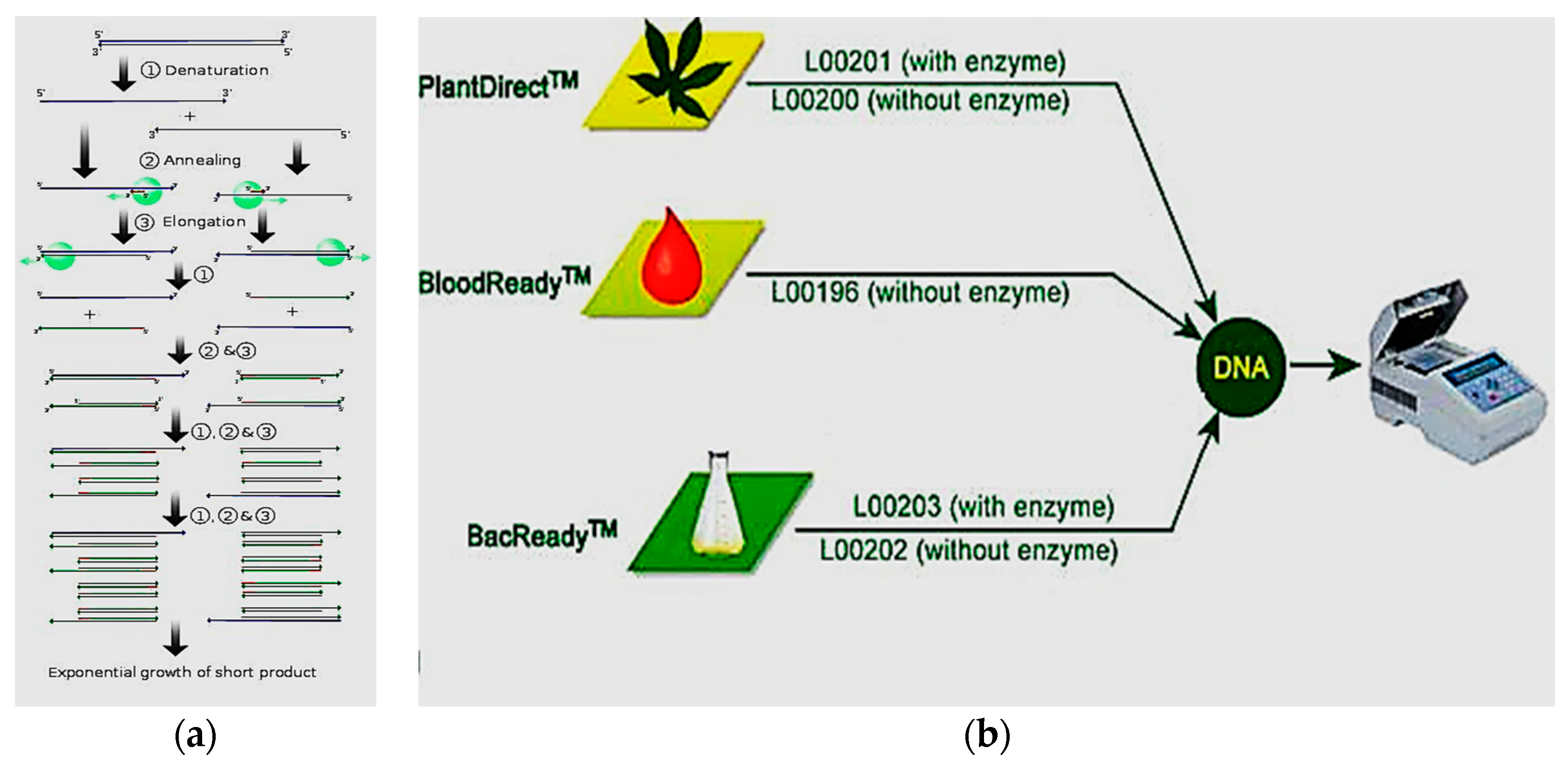
The polymerase chain reaction (PCR) is very popular detection method for detection of pathogens [25,26]. Specific bacteria based on their nucleic acid sequence [27,28,29], protozoa [30,31], and viruses [32,33] are targeted when PCR is used for pathogen detection. Different PCR methods are available for pathogen detection, called, reverse transcript PCR (RT-PCR) [34,35], real-time PCR [36], and multiplex PCR [37]. Figure 1 shows a schematic diagram of the PCR cycle to extract the DNA from bacteria for detection of the pathogen. However, the method requires costly instruments, and amplification, isolation, and quantification of DNA technology make it a complex method to perform. It also requires trained personnel to operate the whole procedure.
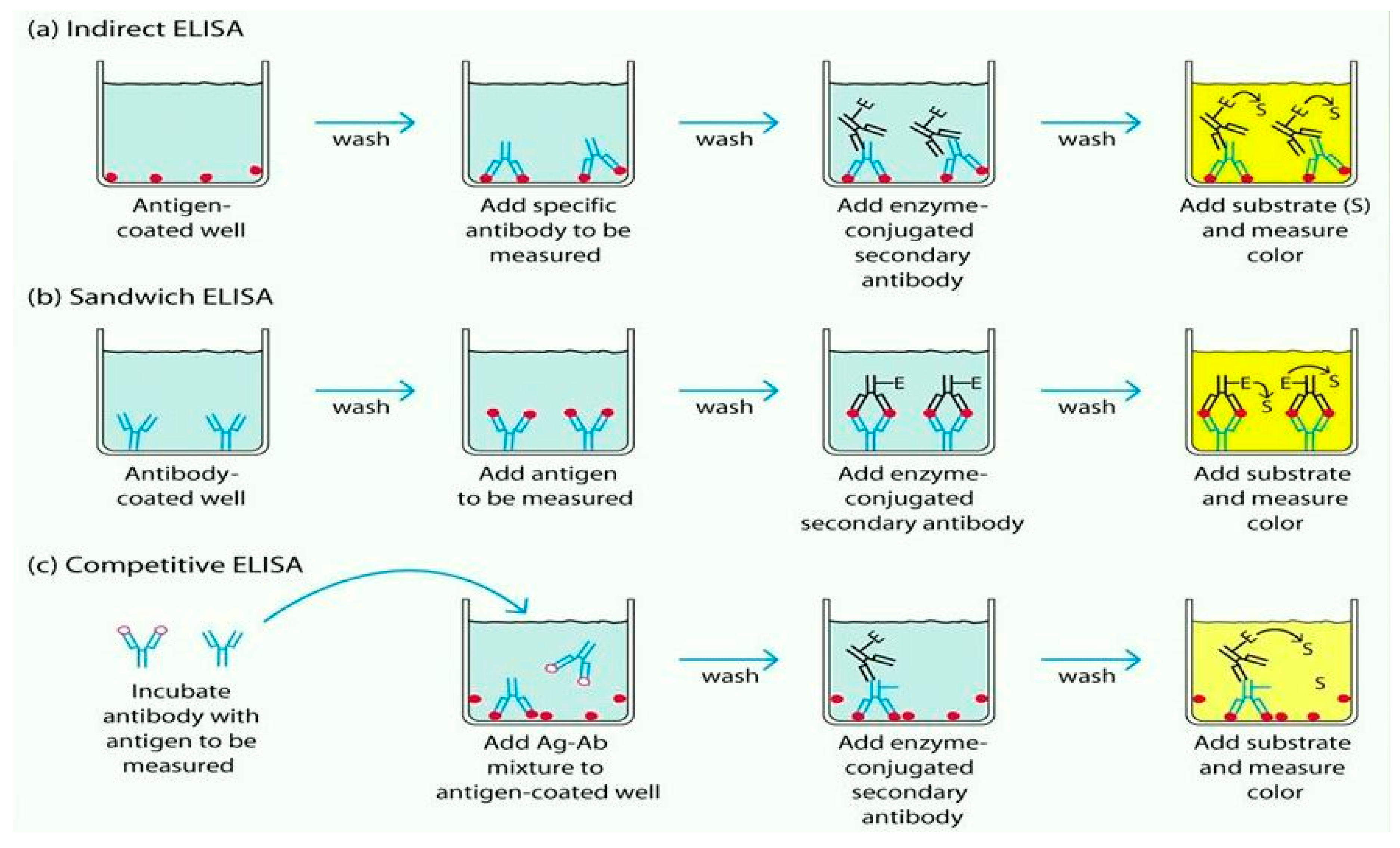
The immunological detection technique is used for the detection of pathogens [38,39]. The antigen-antibody bindings are utilized widely in immunological detection for pathogens from Gram-negative bacteria. This method has been successfully used to detect Salmonella and E. coli [40,41]. Enzyme immunoassay (EIA) [42,43], enzyme-linked fluorescent assay (ELFA) [44,45], enzyme-linked immunosorbent assay (ELISA) [46,47], flow injection immunoassay [48], and other immunological methods [49,50] are mostly used for immunological detection. They require less time to prepare the assay than a culturing technique. However, real-time pathogen detection is not possible with this method [51]. Figure 2 shows a schematic diagram of the immunology-based technique in different ELISA.
2.2. Detection Methods of Endotoxin
The rabbit pyrogen test and limulus amoebocyte lysate (LAL) [52,53] are used to detect endotoxins, and are considered conventional methods. The United States Food and Drug Administration (FDA) has approved these methods as the standard method. In 1920, the rabbit pyrogen test was first developed. A test solution is injected into a rabbit’s body and one waits for some time to see any change of the body temperature to detect an endotoxin. However, an animal rights group has opposed using this method, to stop killing rabbits. Additionally, the method is time-consuming and expensive. Now the method is losing favour, and only the LAL test is used, first discovered 67 years ago in 1950 by Dr Frederik Bang [54]. He first observed that horseshoe-crab blood forms clots when it is exposed to endotoxins.
Endotoxin-contaminated food or water is mixed with horseshoe-crab blood to derive the amoebocyte extraction, than one waits to watch for any response from endotoxins. The FDA has approved four tests: chromogenic assay, colorimetric [26,55] (lower protein), gel-clot, and turbidity metric (spectrophotometric). The particular reaction of amoebocyte/endotoxin has characterized these methods. The method of gel clots is based on the existence or nonexistence of gel formation in the sample: when endotoxins are present in the sample gelation occurs due to the coagulation of protein. Turbidity occurs due to the sharp division of an endotoxin, sensitive substrate, and the turbidimetric methods use this turbidity to detect endotoxins. There is another technique which is called the chromogenic technique, depending on the change of colour during the division of a complex into a peptide and a chromogen [56].
The concentration of Lipopolysaccharide (LPS) is expressed as EU/mL or EU/mg, where EU stands for endotoxin unit for biological activity in LPS. Suppose that in one EU, 10−15 g of LPS is contributed by Gram-negative bacteria. Therefore, at most 105 bacteria can be generated. The response of LAL test is quick and takes approximately 30 min to get the result. The detection limit is quite low, and the technique is highly sensitive compared to other detection methods. The major disadvantage is that it requires expert personnel to complete all the complex steps to avoid any external interference. Another disadvantage is that the testing kits are expensive for some sampling tests.
3. Biosensor-Based Method
The sensor measures physical and chemical quantities and converts them to an electrical signal. Sensors are a kind of transducer where they change one form of energy into another kind of energy. The sensor can measure much physical activity, such as temperature, humidity, acceleration, distance, and many more things. A biosensor is a device which measures living organisms or biological molecules, to detect chemicals present in living organisms.
The recognition is achieved by finding a molecular species to create a binding with the target pathogen for sensing. Different bioreceptors have been introduced in biosensors to increase the efficiency of the measurement. Bioreceptors play an important role in biosensor development, and different types of bioreceptor are discussed in the following section.
3.1. Different Types of Bioreceptor
It is important to design the specificity of a biosensor, and bioreceptors play a major role. They are responsible for binding the analyte to the sensor for measurements. Bioreceptors are categorized as: (1) antigen/antibody; (2) enzymes; (3) nucleic acids/DNA; (4) cellular structures/cells; (5) biomimetic; and (6) bacteriophage. Some of them are discussed below.
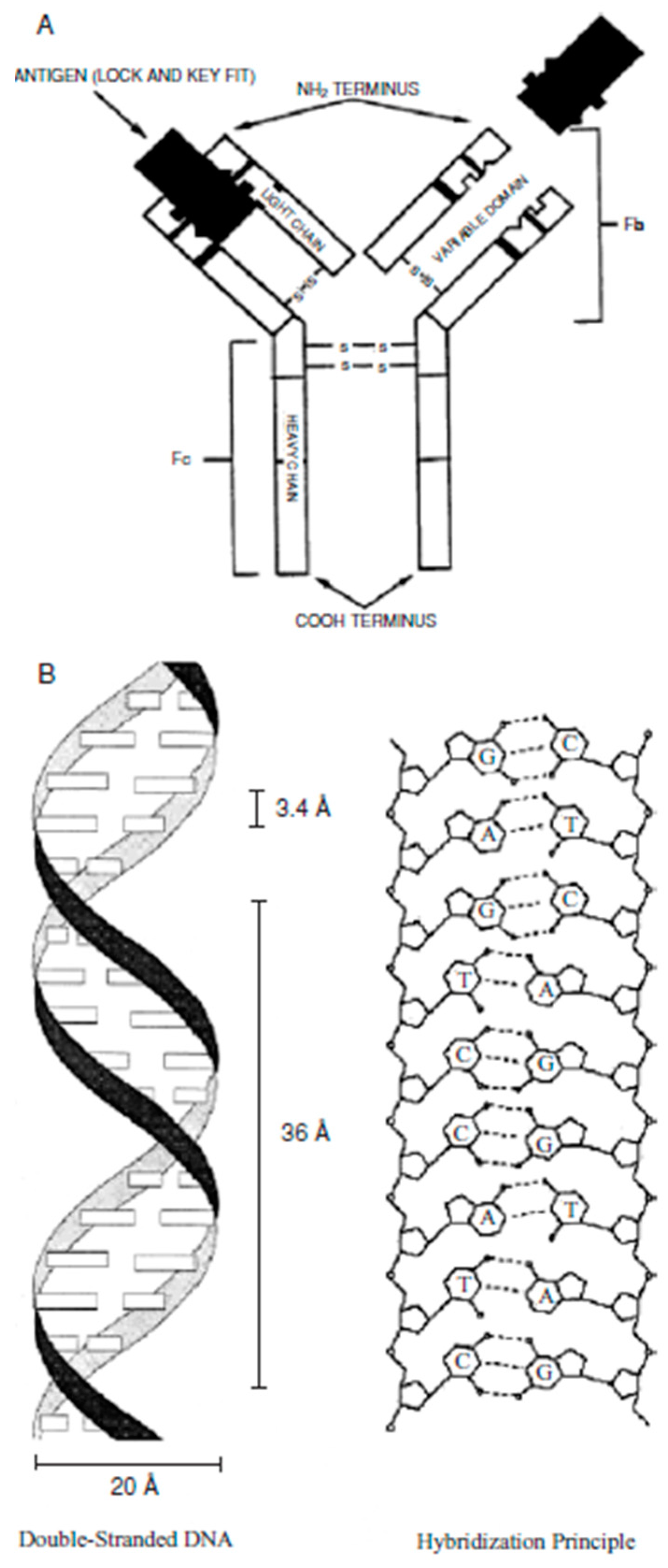
Enzyme-based bioreceptors involve enzymes which have specific bindings. All the enzymes are from the protein group except a small group of the catalytic ribonucleic molecule [56,57]. An antibody/antigen is a complex molecule, which is made up of hundreds of individual amino acids arranged in a highly ordered sequence. They have a particular binding ability for a specific structure, which is used as a bioreceptor [58]. A biomimetic-based bioreceptor is an artificial receptor that is designed and fabricated to mimic a biological receptor. Molecular imprinting, plastic membrane fabrication, and genetic engineering are used to design biomimetic-based bioreceptors [59]. An entire cell/microorganism of biorecognition, or a specific cellular component, is capable of creating a specific binding, which is used in cellular based bioreceptors [60], and nucleic acid/DNA based bioreceptor forms of the complementarity of adenine:thymine (A:T) and cytosine:guanosine (C:G) pairings inside the DNA, which ensures the specificity of the biorecognition [61,62]. Figure 3 shows schematic of antigen and nucleic-acid based bioreceptor.
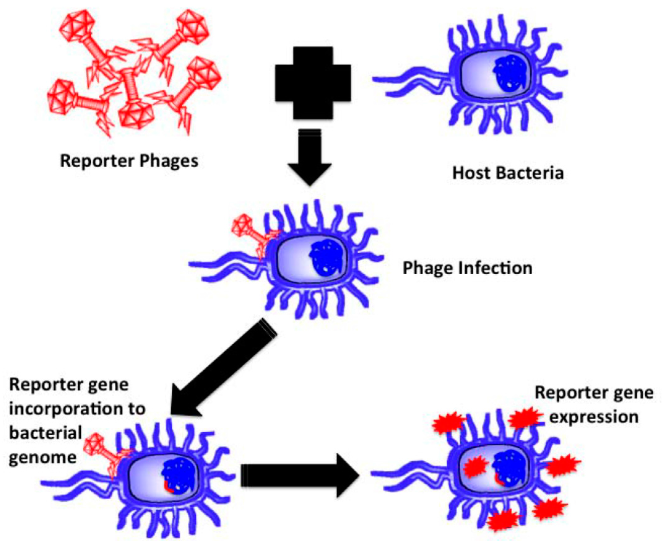
There are some obligate parasites, named phages, that do have no organism for metabolic purposes. They depend on their host bacteria for growth and propagation to become mature. Most of them know their specific host bacteria, whereas, a few of them create binding and killing within an entire bacterial genus [63]. Similar to other parasites, phages’ DNA has to be injected into the host bacteria to bind and take over the bacterial organisms (host) to the number of virions. The host DNA is occupied by the genome of the virions, and the genomes remain inactive temporarily until stimulated for propagation. Much research has reported on phage based biosensors for pathogen and endotoxin detection. Various analytical methods combine with bacteriophage-based probes to detect specific recognition sites. Some pathogens are detected in fresh milk [64,65], fresh tomato [66], and water [67], by using phage-based biosensors directly and efficiently. Figure 4 shows the basic steps of pathogen detection using a phage biosensor.
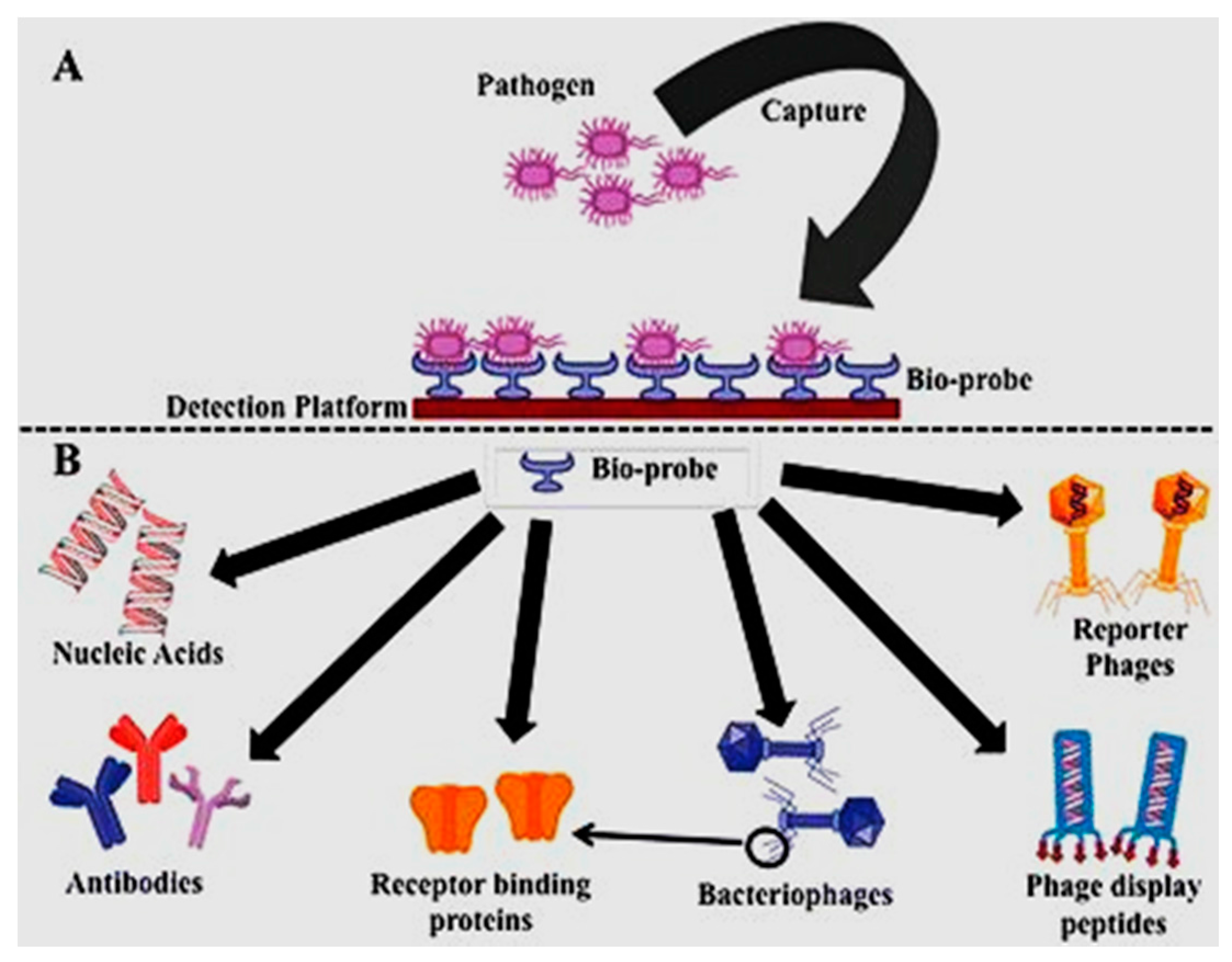
Figure 5 shows pathogen binding activity through different bioreceptors, used to design the specificity of the target pathogens on the transduction surface of the biosensors.
3.2. Types of Biosensing Methods
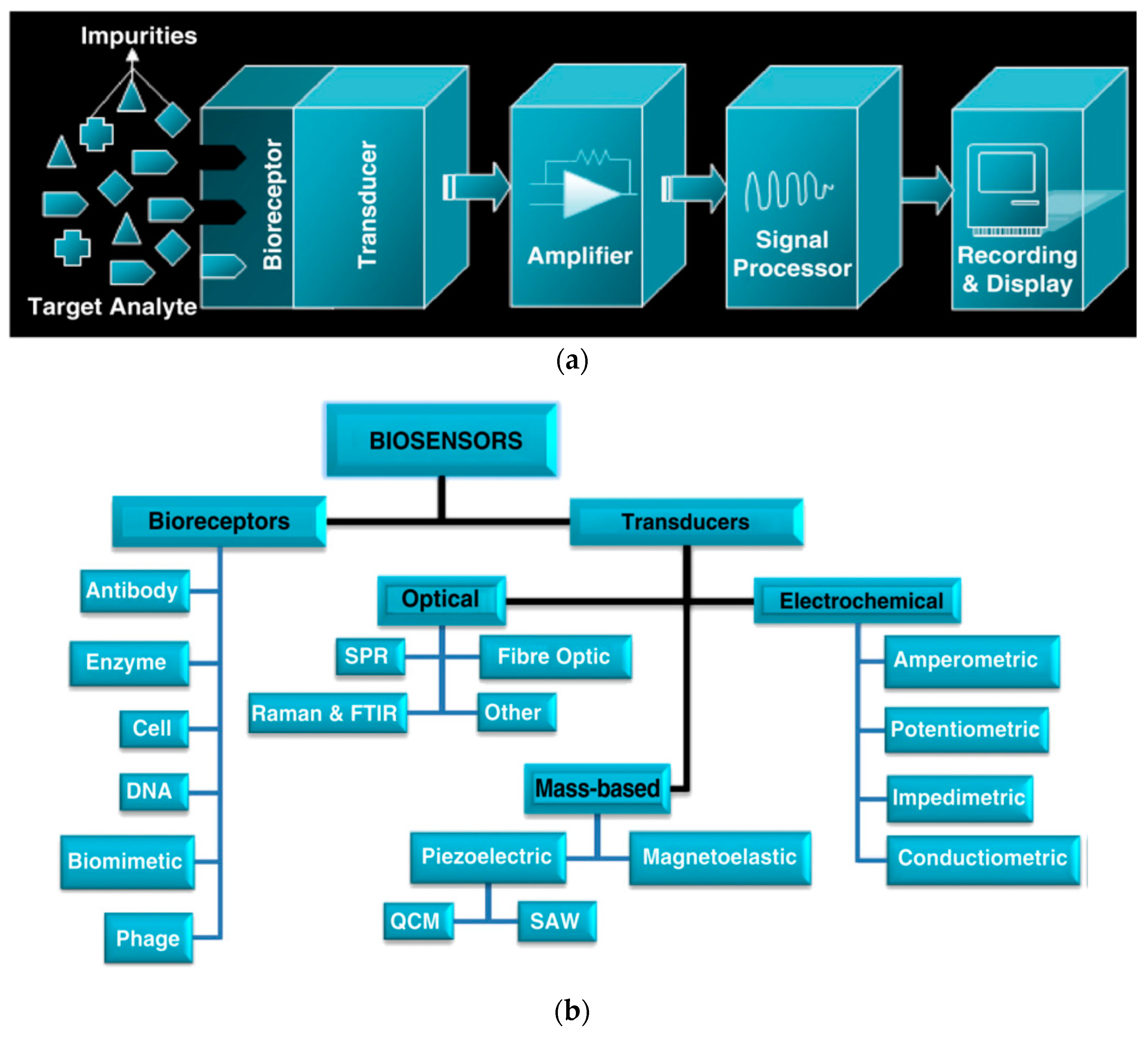
In the food industry, different biosensing methods are developed to detect pathogens, and have been widely used for some time [14,17,20]. Among all the methods, optical-, electrochemical-, and mass-based transduction are the sensitive and accurate methods. In biosensor application, a bioreceptor surface is required for recognition purposes to specify different pathogens [20]. Figure 6 shows the different stages of biosensing methods and their classification in terms of sensing methods.
3.2.1. Biosensors Based on the Optical Method
This method is very successful and reliable to detect pathogens and endotoxins [71]. The basic optics characteristics such as reflection, refraction, absorption, dispersion, etc., are used to develop optical biosensors. Optical fibre, Raman infrared spectroscopy, surface plasmon resonance, and others are used as transducers to develop optical biosensors [71].
Laser light propagates through the tapered optical fibre on the detection surface and then that emitted light is detected. The propagated light goes through the fibre or waveguide and detects foodborne pathogens and different endotoxins. These methods were used to develop optical-based biosensors, to detect pathogens such as E. coli [72,73], Salmonella [74,75], Listeria [76,77], and others [78,79]. Su et al. [80] and Marazuela et al. [81] have reported the detection of endotoxins where an optical-based biosensor was used as a detection method.
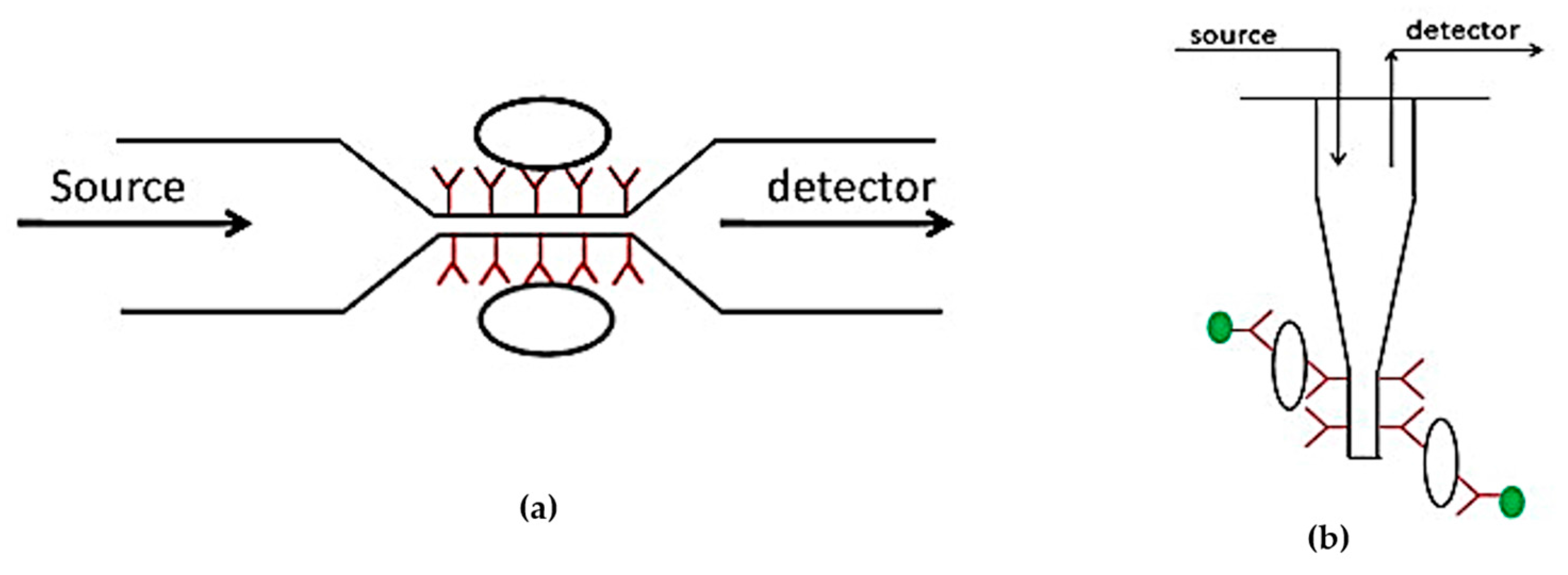
Reflectance spectroscopy is used to detect pathogens and endotoxins for surface plasmon resonance (SPR). SPR measures the changes of the reflected angle of the light when the cells create binding on the specific receptor [82]. Wang et al. [83], Torun et al. [84], and Zhang et al. [85] have reported many studies on pathogen and endotoxin detection based on SPR methods. There are other pathogens, and endotoxin detection methods are available, such as optical micro-ring resonators [86], Raman and FTIR spectroscopy [87], and fluorescence detection [88]. Figure 7 shows optical methods to develop a biosensor for pathogen detection.
3.2.2. Biosensors Based on the Electrochemical Method
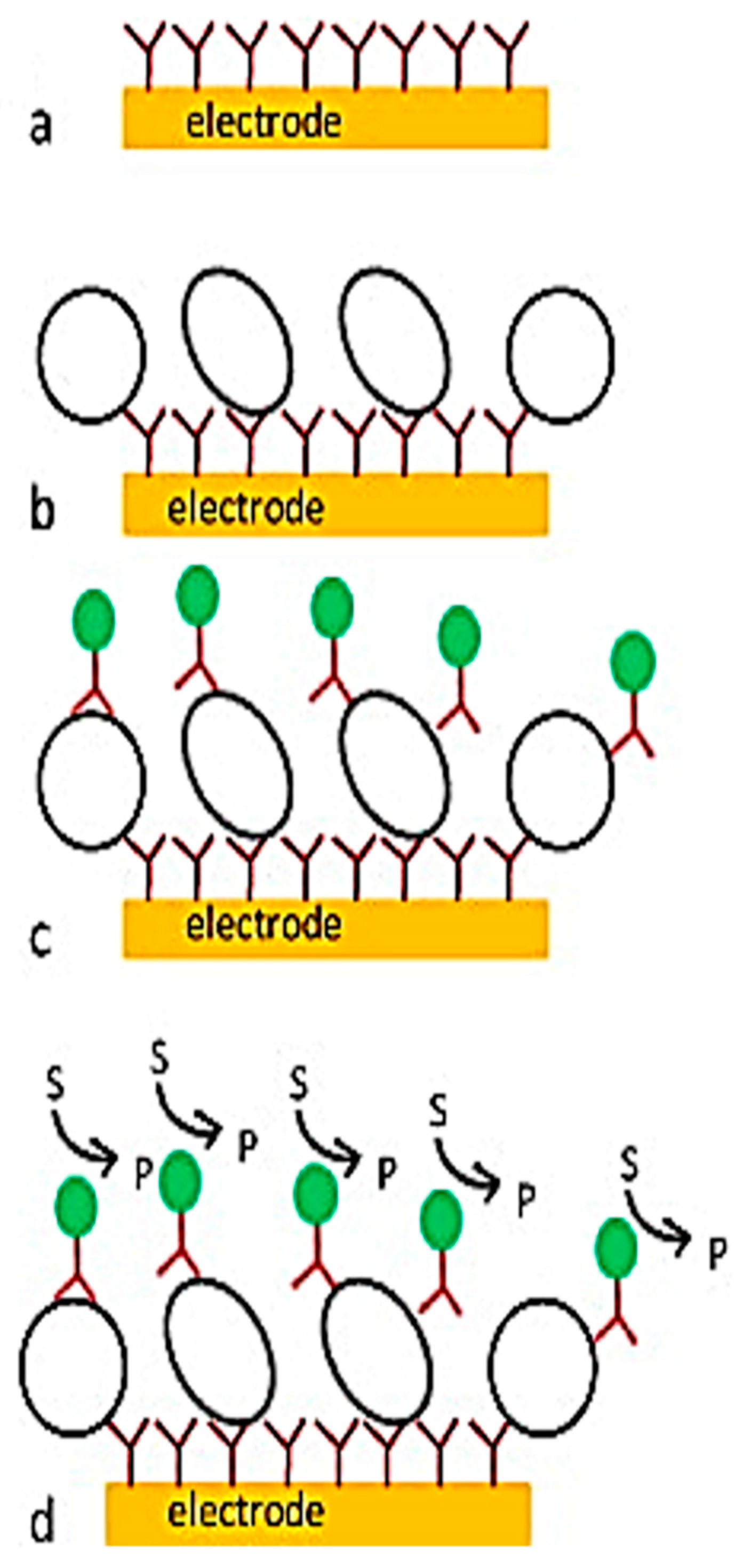
When the sensing electrode interacts with the sample, the changed potential and current are observed; this biosensor is called electrochemical biosensors [90]. Different types of electrochemical biosensors are available, such as the potential (potentiometric), current (amperometric), conductance (conductometric), and impedance (impedimetric) types [91,92,93]. The advantages of this biosensor are that it is low cost, miniature and robust to liquid samples. Compared to optical biosensors, the selectivity and sensitivity are a little restricted, but their use coupled with other bio-sensing methods can increase the performance of detection [20]. Figure 8 shows the schematic diagram of an electrochemical sensor and the steps enabling the pathogens to be detected in real-time.
Biosensors Based on the Amperometric Principle
One common type of an electrochemical biosensor for detection of pathogens is based on the amperometric principle. In this method, a current flow is measured which relates to the concentration of a measured analyte, such as a pathogen. The sensor based on amperometric principle applies a constant value of potential difference between the electrodes, and the resulting flow of current is measured [94]. The applied potential drives the movement of electrons and the rate of flow is measured. The amperometric-based biosensor has been used to detect E. coli [95,96], Salmonella [97], and others [98]. Usually, it offers improved sensitivity compared to the method based on the potentiometer. The developed method has been used to detect specifically E. coli, Mycrobacterium smegmatis, and Bacillus cereous has been reported in [99].
Biosensors Based on Impedimetric Measurement
In this type of biosensor, the impedance of the sensor is measured, which is affected by biological reaction. In recent years, the impedance-based biosensor has attracted widespread attention [100]. Electrochemical impedance spectroscopy (EIS) has a significant role in biosensor-based impedance measurement. EIS is usually used to determine the electrical properties of any material along with their interfaces using surface-modified electrodes [101]. A small alternating voltage is applied across the electrode terminals over a wide frequency range. The current between the electrodes passes through the material under observation. The impedance is measured and is then analysed using the EIS technique. The method has been used to detect pathogens [100,102,103,104,105]. In Wang et al. [106] have reported the new trend of biosensors based on impedance measurement. Detection of endotoxin has been reported in [80]. Pathogens and phthalates in water have been detected using the method and are reported in [107,108,109,110,111,112,113].
Biosensors Based on the Potentiometric Principle
In the potentiometric biosensor, the outcome of the bio-recognition activity is converted into a potential signal. The method uses an ion-sensitive field-effect transistor (ISFET) or ion-selective electrodes (ISE), where the ions accumulate at the interface of the ion-selective membrane and produce the primary output signal. In this method, the amount of current flowing through the electrodes is approximately zero. Since enzyme reaction ions are produced, which results in a potential across the electrodes. Research on biosensors for pathogen detection using this principle are reported in [114,115,116].
3.2.3. Biosensors Based on Mass-Sensitivity
Biosensors including the quartz crystal microbalance (QCM) and surface acoustic wave (SAW) belong to this category.
QCM-Based Biosensors
A QCM [117] based biosensor operates on the piezoelectric principle. An electric signal is applied between two gold plates on a quartz crystal to produce a vibration at a specific resonance frequency [118], which is measured. When the QCM is used as a biosensor, the resonance frequency of the crystal changes in the presence of the measurand, as it changes the mass of the crystal. The shift in frequency of the crystal is measured. The QCM-based biosensor has been used to detect pathogens [119] and endotoxins [120].
SAW-Based Biosensor
The surface acoustic wave (SAW) based interdigital transducer [IDT] [121] produces acoustic waves on the surface from the piezoelectric substrate and changes due to the measurand are detected. Acoustic waves are generated and detected on the piezoelectric surface using IDTs. SAW-based biosensors have been used for various applications including for detection of pathogens and endotoxins. Rocha-Gaso et al. [122] reported pathogen detection and Lange et al. [82] and Hammer et al. [123] have reported endotoxin detection by this method.
4. Nanomaterial-Based Sensors
In recent years, great advances have been made in nanomaterial-based biosensors, where the sensing electrode is modified by a nanomaterial to achieve a quick electron transfer due to the stimulation of different biomarkers. Due to this advantage, much research has been reported where the nanomaterials are coupled with biomolecules to develop nanomaterial-based biosensors to detect dangerous pathogens. Salmonella, E. coli [137], and L. monocytogenes are the most studied pathogens, developing their detection methods in food. However, the respiratory syncytial [138,139] virus and parasites (Giardia and Cryptosporidium) [140] are other pathogens which are responsible for contaminating food. Yang et al. [141] reported a sensor to detect Salmonella spp., where gold (Au) nanoparticles were used on a glassy carbon electrode. The sensing surface was selective for Salmonella spp. With the increased sensitivity and selectivity. Nanowire-based sensors have attracted great interest in recent years. Wang et al. [141] developed TiO2 nanowire microelectrodes for rapid and sensitive detection of Listeria monocytogenes. This method has an excellent stability, a low detection limit, and can avoid interference from other foodborne pathogens. Reduced graphene sheets (RGSs) contain a unique nanostructure, which is similar to a solid honeycomb crystal structure with excellent electrical conductivity, chemical stability, and mechanical strength. Wan et al. [142] developed an RGS-based sensor which can detect sulphate-reducing pathogens without the obvious response to Vibrio angillarum with excellent sensitivity, selectivity, stability, and good analytical performance. Additionally, nanopore-material-based sensors have shown a unique advantage for pathogen detection. Wang et al. [143] immobilized ssDNA which is functionalized for E. coli O157: H7 on an aluminium anodized oxide nanopore membrane.
Therefore, nano-materials have been used to develop a cost-effective biosensor for in situ measurement. Some more nanomaterial-based biosensor have been reported where nanoribbons [144], nanowires [145], carbon nanotubes [146,147,148], etc., are used to manufacture the biosensor for pathogen detection. The nanomaterial’s surface bonds with the recognition element to develop a biosensing platform, and their combined mechanism generates signal transduction for the monitoring of pathogens in food or water. Figure 9 shows the steps of pathogen detection.
Table 3 shows different biosensors with their advantages, limitations, and cost.
5. Conclusions
Detection of pathogens and toxins at an early stage is crucial to avoid food poisoning and other problems. There are different methods available for their detection. Conventional methods are mainly laboratory-based and usually take a long time to detect pathogens and toxins. Many new approaches, such as electrochemical-, optical-, and nanomaterial-based biosensors have been developed. Each developed method has its advantages and disadvantages. The adopted method should be reliable, accurate, and selective to a particular pathogen/toxin, as well as fast enough to obtain reliable results. The paper has reviewed different methods along with the sensors for detection available to scientific communities.
Conflicts of Interest
The authors declare no conflict of interest.
References
- Dwivedi, H.P.; Jaykus, L.-A. Detection of pathogens in foods: The current state-of-the-art and future directions. Crit. Rev. Microbiol. 2011, 37, 40–63. [Google Scholar] [CrossRef] [PubMed]
- Sayad, A.A.; Ibrahim, F.; Uddin, S.M.; Pei, K.X.; Mohktar, M.S.; Madou, M.; Thong, K.L. A microfluidic lab-on-a-disc integrated loop mediated isothermal amplification for foodborne pathogen detection. Sens. Actuators B Chem. 2016, 227, 600–609. [Google Scholar] [CrossRef]
- Oliver, S.P.; Jayarao, B.M.; Almeida, R.A. Foodborne pathogens in milk and the dairy farm environment: Food safety and public health implications. Foodbourne Pathog. Dis. 2005, 2, 115–129. [Google Scholar] [CrossRef] [PubMed]
- Services, U.S.D.O.H.H. Cdc: Estimates of Foodborne Illness in the United States. Available online: https://www.cdc.gov/foodborneburden/2011-foodborne-estimates.html (accessed on 14 August 2017).
- On, S.; Lim, E.; Lopez, L.; Cressey, P.; Pirie, R. Annual Report Concerning Foodborne Disease in New Zealand; Enviromental Science and Research Limited (ESR): Christchurch, New Zealand, 2011; p. 130. [Google Scholar]
- Organization, W.H. Foodborne Diseases. Available online: http://www.who.int/foodsafety/areas_work/foodborne-diseases/en/ (accessed on 14 August 2017).
- Kirk, M.D.; Pires, S.M.; Black, R.E.; Caipo, M.; Crump, J.A.; Devleesschauwer, B.; Döpfer, D.; Fazil, A.; Fischer-Walker, C.L.; Hald, T. World health organization estimates of the global and regional disease burden of 22 foodborne bacterial, protozoal, and viral diseases, 2010: A data synthesis. PLoS Med. 2015, 12, e1001921. [Google Scholar] [CrossRef] [PubMed] [Green Version]
- Scallan, E.; Hoekstra, R.M.; Angulo, F.J.; Tauxe, R.V.; Widdowson, M.A.; Roy, S.L.; Jones, J.L.; Griffin, P.M. Foodborne illness acquired in the united states-major pathogens. Emerg. Infect. Dis. 2011, 17, 7–15. [Google Scholar] [CrossRef] [PubMed]
- Butler, A.J.; Thomas, M.K.; Pintar, K.D. Expert elicitation as a means to attribute 28 enteric pathogens to foodborne, waterborne, animal contact, and person-to-person transmission routes in canada. Foodborne Pathog. Dis. 2015, 12, 335–344. [Google Scholar] [CrossRef] [PubMed]
- Kirk, M.; Glass, K.; Ford, L.; Brown, K.; Hall, G. Foodborne Illness in Australia: Annual Incidence Circa 2000; Australian Government Department of Health and Aging: Canberra, Australia, 2005; p. 57.
- Vally, H.; Glass, K.; Ford, L.; Hall, G.; Kirk, M.D.; Shadbolt, C.; Veitch, M.; Fullerton, K.E.; Musto, J.; Becker, N. Proportion of illness acquired by foodborne transmission for nine enteric pathogens in australia: An expert elicitation. Foodborne Pathog. Dis. 2014, 11, 727–733. [Google Scholar] [CrossRef] [PubMed]
- Adak, G.K.; Long, S.M.; O’Brien, S.J. Trends in indigenous foodborne disease and deaths, england and wales: 1992 to 2000. Gut 2002, 51, 832–841. [Google Scholar] [CrossRef] [PubMed]
- Havelaar, A.H.; Galindo, A.V.; Kurowicka, D.; Cooke, R.M. Attribution of foodborne pathogens using structured expert elicitation. Foodborne Pathog. Dis. 2008, 5, 649–659. [Google Scholar] [CrossRef] [PubMed]
- Alocilja, E.; Radke, S. Market analysis of biosensor for food safety. Biosens. Bioelectron. 2003, 18, 841–846. [Google Scholar] [CrossRef]
- Wagner, A.B. Bacterial Food Poisoning. Available online: http://aggie-horticulture.tamu.edu/food-technology/bacterial-food-poisoning/ (accessed on 14 August 2017).
- Seydel, U.; Schromm, A.B.; Blunck, R.; Brandenburg, K. Chemical structure, molecular conformation, and bioactivity of endotoxins. Chem. Immunol. 2000, 74, 5–24. [Google Scholar] [PubMed]
- Lazcka, O.; Del Campo, F.J.; Munoz, F.X. Pathogen detection: A perspective of traditional methods and biosensors. Biosens. Bioelectron. 2007, 22, 1205–1217. [Google Scholar] [CrossRef] [PubMed]
- Wolff, B.J.; Bramley, A.M.; Thurman, K.A.; Whitney, C.G.; Whitaker, B.; Self, W.H.; Arnold, S.R.; Trabue, C.; Wunderink, R.G.; McCullers, J. Improved detection of respiratory pathogens by use of high-quality sputum with taqman array card technology. J. Clin. Microbiol. 2017, 55, 110–121. [Google Scholar] [CrossRef] [PubMed]
- Chen, C.; Liu, P.; Zhao, X.; Du, W.; Feng, X.; Liu, B.-F. A self-contained microfluidic in-gel loop-mediated isothermal amplification for multiplexed pathogen detection. Sens. Actuators B Chem. 2017, 239, 1–8. [Google Scholar] [CrossRef]
- Velusamy, V.; Arshak, K.; Korostynska, O.; Oliwa, K.; Adley, C. An overview of foodborne pathogen detection: In the perspective of biosensors. Biotechnol. Adv. 2010, 28, 232–254. [Google Scholar] [CrossRef] [PubMed]
- Leonard, P.; Hearty, S.; Brennan, J.; Dunne, L.; Quinn, J.; Chakraborty, T.; O’Kennedy, R. Advances in biosensors for detection of pathogens in food and water. Enzyme Microb. Technol. 2003, 32, 3–13. [Google Scholar] [CrossRef]
- Lee, K.-M.; Runyon, M.; Herrman, T.J.; Phillips, R.; Hsieh, J. Review of salmonella detection and identification methods: Aspects of rapid emergency response and food safety. Food Control 2015, 47, 264–276. [Google Scholar] [CrossRef]
- Leoni, E.; Legnani, P.P. Comparison of selective procedures for isolation and enumeration of legionella species from hot water systems. J. Appl. Microbiol. 2001, 90, 27–33. [Google Scholar] [CrossRef] [PubMed]
- Ratnam, S.; March, S.B.; Ahmed, R.; Bezanson, G.S.; Kasatiya, S. Characterization of Escherichia coli serotype 0157:H7. J. Clin. Microbiol. 1988, 26, 2006–2012. [Google Scholar] [PubMed]
- Fratamico, P.M. Comparison of culture, polymerase chain reaction (PCR), taqman salmonella, and transia card salmonella assays for detection of salmonella spp. in naturally-contaminated ground chicken, ground turkey, and ground beef. Mol. Cell. Probes 2003, 17, 215–221. [Google Scholar] [CrossRef]
- Oh, S.J.; Park, B.H.; Jung, J.H.; Choi, G.; Lee, D.C.; Seo, T.S. Centrifugal loop-mediated isothermal amplification microdevice for rapid, multiplex and colorimetric foodborne pathogen detection. Biosens. Bioelectron. 2016, 75, 293–300. [Google Scholar] [CrossRef] [PubMed]
- Jensen, M.A.; Webster, J.A.; Straus, N. Rapid identification of bacteria on the basis of polymerase chain reaction-amplified ribosomal DNA spacer polymorphisms. Appl. Environ. Microbiol. 1993, 59, 945–952. [Google Scholar] [PubMed]
- Belgrader, P.; Benett, W.; Hadley, D.; Richards, J.; Stratton, P.; Mariella, R.; Milanovich, F. Infectious disease—PCR detection of bacteria in seven minutes. Science 1999, 284, 449–450. [Google Scholar] [CrossRef] [PubMed]
- Naravaneni, R.; Jamil, K. Rapid detection of food-borne pathogens by using molecular techniques. J. Med. Microbiol. 2005, 54, 51–54. [Google Scholar] [CrossRef] [PubMed]
- Russell, S.; Frasca, S.; Sunila, I.; French, R.A. Application of a multiplex PCR for the detection of protozoan pathogens of the eastern oyster crassostrea virginica in field samples. Dis. Aquat. Org. 2004, 59, 85–91. [Google Scholar] [CrossRef] [PubMed]
- Lee, S.H.; Joung, M.; Yoon, S.; Choi, K.; Park, W.Y.; Yu, J.R. Multiplex PCR detection of waterborne intestinal protozoa: Microsporidia, cyclospora, and cryptosporidium. Korean J. Parasitol. 2010, 48, 297–301. [Google Scholar] [CrossRef] [PubMed]
- Traore, O.; Arnal, C.; Mignotte, B.; Maul, A.; Laveran, H.; Billaudel, S.; Schwartzbrod, L. Reverse transcriptase PCR detection of astrovirus, hepatitis a virus, and poliovirus in experimentally contaminated mussels: Comparison of several extraction and concentration methods. Appl. Environ. Microbiol. 1998, 64, 3118–3122. [Google Scholar] [PubMed]
- Morales-Rayas, R.; Wolffs, P.F.G.; Griffiths, M.W. Simultaneous separation and detection of hepatitis a virus and norovirus in produce. Int. J. Food Microbiol. 2010, 139, 48–55. [Google Scholar] [CrossRef] [PubMed]
- Yaron, S.; Matthews, K.R. A reverse transcriptase-polymerase chain reaction assay for detection of viable Escherichia coli O157: H7: Investigation of specific target genes. J. Appl. Microbiol. 2002, 92, 633–640. [Google Scholar] [CrossRef] [PubMed]
- Choi, S.H.; Lee, S.B. Development of reverse transcriptase-polymerase chain reaction of fima gene to detect viable salmonella in milk. J. Anim. Sci. Technol. 2004, 46, 841–848. [Google Scholar]
- Lambertz, S.T.; Nilsson, C.; Hallanvuo, S.; Lindblad, M. Real-time PCR method for detection of pathogenic yersinia enterocolitica in food. Appl. Environ. Microbiol. 2008, 74, 6060–6067. [Google Scholar] [CrossRef] [PubMed]
- Mukhopadhyay, A.; Mukhopadhyay, U.K. Novel multiplex PCR approaches for the simultaneous detection of human pathogens: Escherichia coli O157: H7 and listeria monocytogenes. J. Microbiol. Methods 2007, 68, 193–200. [Google Scholar] [CrossRef] [PubMed]
- Iqbal, S.S.; Mayo, M.W.; Bruno, J.G.; Bronk, B.V.; Batt, C.A.; Chambers, J.P. A review of molecular recognition technologies for detection of biological threat agents. Biosens. Bioelectron. 2000, 15, 549–578. [Google Scholar] [CrossRef]
- Gracias, K.S.; McKillip, J.L. A review of conventional detection and enumeration methods for pathogenic bacteria in food. Can. J. Microbiol. 2004, 50, 883–890. [Google Scholar] [CrossRef] [PubMed]
- Chen, C.S.; Durst, R.A. Simultaneous detection of Escherichia coli O157: H7, salmonella spp. And listeria monocytogenes with an array-based immunosorbent assay using universal protein g-liposomal nanovesicles. Talanta 2006, 69, 232–238. [Google Scholar] [CrossRef] [PubMed]
- Magliulo, M.; Simoni, P.; Guardigli, M.; Michelini, E.; Luciani, M.; Lelli, R.; Roda, A. A rapid multiplexed chemiluminescent immunoassay for the detection of Escherichia coli O157: H7, yersinia enterocolitica, salmonella typhimurium, and listeria monocytogenes pathogen bacteria. J. Agric. Food Chem. 2007, 55, 4933–4939. [Google Scholar] [CrossRef] [PubMed]
- Qadri, A.; Ghosh, S.; Prakash, K.; Kumar, R.; Moudgil, K.D.; Talwar, G.P. Sandwich enzyme immunoassays for detection of salmonella-typhi. J. Immunoass. 1990, 11, 251–270. [Google Scholar] [CrossRef] [PubMed]
- Chapman, P.A.; Malo, A.T.C.; Siddons, C.A.; Harkin, M. Use of commercial enzyme immunoassays and immunomagnetic separation systems for detecting Escherichia coli O157 in bovine fecal samples. Appl. Environ. Microbiol. 1997, 63, 2549–2553. [Google Scholar] [PubMed]
- Rozand, C.; Feng, P.C.H. Specificity analysis of a novel phage-derived ligand in an enzyme-linked fluorescent assay for the detection of Escherichia coli O157: H7. J. Food Protect. 2009, 72, 1078–1081. [Google Scholar] [CrossRef]
- De Giusti, M.; Tufi, D.; Aurigemma, C.; Del Cimmuto, A.; Trinti, F.; Mannocci, A.; Boccia, A. Detection of Escherichia coli O157 in raw and cooked meat: Comparison of conventional direct culture method and enzyme linked fluorescent assay (ELFA). Ital. J. Public Health 2011, 8, 28. [Google Scholar]
- Vazquez, F.; Gonzalez, E.A.; Garabal, J.I.; Valderrama, S.; Blanco, J.; Baloda, S.B. Development and evaluation of an ELISA to detect Escherichia coli K88 (F4) fimbrial antibody levels. J. Med. Microbiol. 1996, 44, 453–463. [Google Scholar] [CrossRef] [PubMed]
- Song, C.; Liu, C.; Wu, S.; Li, H.; Guo, H.; Yang, B.; Qiu, S.; Li, J.; Liu, L.; Zeng, H. Development of a lateral flow colloidal gold immunoassay strip for the simultaneous detection of shigella boydii and Escherichia coli O157: H7 in bread, milk and jelly samples. Food Control 2016, 59, 345–351. [Google Scholar] [CrossRef]
- Abdel-Hamid, I.; Ivnitski, D.; Atanasov, P.; Wilkins, E. Highly sensitive flow-injection immunoassay system for rapid detection of bacteria. Anal. Chim. Acta 1999, 399, 99–108. [Google Scholar] [CrossRef]
- Valdivieso-Garcia, A.; Desruisseau, A.; Riche, E.; Fukuda, S.; Tatsumi, H. Evaluation of a 24-h bioluminescent enzyme immunoassay for the rapid detection of salmonella in chicken carcass rinses. J. Food Protect. 2003, 66, 1996–2004. [Google Scholar] [CrossRef]
- Rasooly, A.; Rasooly, R.S. Detection and analysis of staphylococcal enterotoxin a in food by western immunoblotting. Int. J. Food Microbiol. 1998, 41, 205–212. [Google Scholar] [CrossRef]
- Meng, J.H.; Doyle, M.P. Introduction. Microbiological food safety. Microbes Infect. 2002, 4, 395–397. [Google Scholar] [CrossRef]
- Ding, J.L.; Ho, B. Endotoxin Detection—From Limulus Amebocyte Lysate to Recombinant Factor C; Springer: Berlin, Germany, 2010; Volume 53. [Google Scholar]
- Thorne, P.S.; Perry, S.S.; Saito, R.; O’Shaughnessy, P.T.; Mehaffy, J.; Metwali, N.; Keefe, T.; Donham, K.J.; Reynolds, S.J. Evaluation of the limulus amebocyte lysate and recombinant factor c assays for assessment of airborne endotoxin. Appl. Environ. Microbiol. 2010, 76, 4988–4995. [Google Scholar] [CrossRef] [PubMed]
- Bang, F.B.; Frost, J.L. The toxic effect of a marine bacterium on limulus and the formation of blood clots. Mar. Biol. Lab. 1953, 105, 361–362. [Google Scholar]
- Alhogail, S.; Suaifan, G.A.; Zourob, M. Rapid colorimetric sensing platform for the detection of listeria monocytogenes foodborne pathogen. Biosens. Bioelectron. 2016, 86, 1061–1066. [Google Scholar] [CrossRef] [PubMed]
- Kotanen, C.N.; Moussy, F.G.; Carrara, S.; Guiseppi-Elie, A. Implantable enzyme amperometric biosensors. Biosens. Bioelectron. 2012, 35, 14–26. [Google Scholar] [CrossRef] [PubMed]
- Chemburu, S.; Wilkins, E.; Abdel-Hamid, I. Detection of pathogenic bacteria in food samples using highly-dispersed carbon particles. Biosens. Bioelectron. 2005, 21, 491–499. [Google Scholar] [CrossRef] [PubMed]
- Holford, T.R.J.; Davis, F.; Higson, S.P.J. Recent trends in antibody based sensors. Biosens. Bioelectron. 2012, 34, 12–24. [Google Scholar] [CrossRef] [PubMed]
- Silbert, L.; Ben Shlush, I.; Israel, E.; Porgador, A.; Kolusheva, S.; Jelinek, R. Rapid chromatic detection of bacteria by use of a new biomimetic polymer sensor. Appl. Environ. Microbiol. 2006, 72, 7339–7344. [Google Scholar] [CrossRef] [PubMed]
- Banerjee, P.; Bhunia, A.K. Cell-based biosensor for rapid screening of pathogens and toxins. Biosens. Bioelectron. 2010, 26, 99–106. [Google Scholar] [CrossRef] [PubMed]
- Chen, S.H.; Wu, V.C.H.; Chuang, Y.C.; Lin, C.S. Using oligonucleotide-functionalized au nanoparticles to rapidly detect foodborne pathogens on a piezoelectric biosensor. J. Microbiol. Methods 2008, 73, 7–17. [Google Scholar] [CrossRef] [PubMed]
- Zhang, D.; Alocilja, E.C. Characterization of nanoporous silicon-based DNA biosensor for the detection of salmonella enteritidis. IEEE Sens. J. 2008, 8, 775–780. [Google Scholar] [CrossRef]
- Zink, R.; Loessner, M.J. Classification of virulent and temperate bacteriophages of listeria spp. On the basis of morphology and protein analysis. Appl. Environ. Microbiol. 1992, 58, 296–302. [Google Scholar] [PubMed]
- Balasubramanian, S.; Sorokulova, I.B.; Vodyanoy, V.J.; Simonian, A.L. Lytic phage as a specific and selective probe for detection of staphylococcus aureus—A surface plasmon resonance spectroscopic study. Biosens. Bioelectron. 2007, 22, 948–955. [Google Scholar] [CrossRef] [PubMed]
- Goodridge, L.; Chen, J.; Griffiths, M. The use of a fluorescent bacteriophage assay for detection of Escherichia coli O157: H7 in inoculated ground beef and raw milk. Int. J. Food Microbiol. 1999, 47, 43–50. [Google Scholar] [CrossRef]
- Li, S.; Li, Y.; Chen, H.; Horikawa, S.; Shen, W.; Simonian, A.; Chin, B.A. Direct detection of salmonella typhimurium on fresh produce using phage-based magnetoelastic biosensors. Biosens. Bioelectron. 2010, 26, 1313–1319. [Google Scholar] [CrossRef] [PubMed]
- Edgar, R.; McKinstry, M.; Hwang, J.; Oppenheim, A.B.; Fekete, R.A.; Giulian, G.; Merril, C.; Nagashima, K.; Adhya, S. High-sensitivity bacterial detection using biotin-tagged phage and quantum-dot nanocomplexes. Proc. Natl. Acad. Sci. USA 2006, 103, 4841–4845. [Google Scholar] [CrossRef] [PubMed]
- Vo-Dinh, T. Biosensors and biochips. In Biomems and Biomedical Nanotechnology; Springer: Berlin, Germany, 2006; pp. 1–20. [Google Scholar]
- Singh, A.; Poshtiban, S.; Evoy, S. Recent advances in bacteriophage based biosensors for food-borne pathogen detection. Sensors 2013, 13, 1763–1786. [Google Scholar] [CrossRef] [PubMed]
- Singh, A.; Arutyunov, D.; Szymanski, C.M.; Evoy, S. Bacteriophage based probes for pathogen detection. Analyst 2012, 137, 3405–3421. [Google Scholar] [CrossRef] [PubMed]
- Narsaiah, K.; Jha, S.N.; Bhardwaj, R.; Sharma, R.; Kumar, R. Optical biosensors for food quality and safety assurance—A review. J. Food Sci. Technol. 2012, 49, 383–406. [Google Scholar] [CrossRef] [PubMed]
- Tripathi, S.M.; Bock, W.J.; Mikulic, P.; Chinnappan, R.; Ng, A.; Tolba, M.; Zourob, M. Long period grating based biosensor for the detection of Escherichia coli bacteria. Biosens. Bioelectron. 2012, 35, 308–312. [Google Scholar] [CrossRef] [PubMed]
- Bharadwaj, R.; Sai, V.V.R.; Thakare, K.; Dhawangale, A.; Kundu, T.; Titus, S.; Verma, P.K.; Mukherji, S. Evanescent wave absorbance based fiber optic biosensor for label-free detection of E. coli at 280 nm wavelength. Biosens. Bioelectron. 2011, 26, 3367–3370. [Google Scholar] [CrossRef] [PubMed]
- Valadez, A.M.; Lana, C.A.; Tu, S.I.; Morgan, M.T.; Bhunia, A.K. Evanescent wave fiber optic biosensor for salmonella detection in food. Sensors 2009, 9, 5810–5824. [Google Scholar] [CrossRef] [PubMed]
- Ko, S.H.; Grant, S.A. A novel fret-based optical fiber biosensor for rapid detection of salmonella typhimurium. Biosens. Bioelectron. 2006, 21, 1283–1290. [Google Scholar] [CrossRef] [PubMed]
- Geng, T.; Morgan, M.T.; Bhunia, A.K. Detection of low levels of listeria monocytogenes cells by using a fiber-optic immunosensor. Appl. Environ. Microbiol. 2004, 70, 6138–6146. [Google Scholar] [CrossRef] [PubMed]
- Ohk, S.H.; Koo, O.K.; Sen, T.; Yamamoto, C.M.; Bhunia, A.K. Antibody-aptamer functionalized fibre-optic biosensor for specific detection of listeria monocytogenes from food. J. Appl. Microbiol. 2010, 109, 808–817. [Google Scholar] [CrossRef] [PubMed]
- Villalobos, P.; Chavez, M.I.; Olguin, Y.; Sanchez, E.; Valdes, E.; Galindo, R.; Young, M.E. The application of polymerized lipid vesicles as colorimetric biosensors for real-time detection of pathogens in drinking water. Electron. J. Biotechnol. 2012, 15, 4. [Google Scholar] [CrossRef]
- Gnanaprakasa, T.J.; Oyarzabal, O.A.; Olsen, E.V.; Pedrosa, V.A.; Simonian, A.L. Tethered DNA scaffolds on optical sensor platforms for detection of hipO gene from Campylobacter jejuni. Sens. Actuators B Chem. 2011, 156, 304–311. [Google Scholar] [CrossRef]
- Su, W.; Lin, M.; Lee, H.; Cho, M.; Choe, W.S.; Lee, Y. Determination of endotoxin through an aptamer-based impedance biosensor. Biosens. Bioelectron. 2012, 32, 32–36. [Google Scholar] [CrossRef] [PubMed]
- Marazuela, M.D.; Moreno-Bondi, M.C. Fiber-optic biosensors—An overview. Anal. Bioanal. Chem. 2002, 372, 664–682. [Google Scholar] [CrossRef] [PubMed]
- Länge, K.; Rapp, B.E.; Rapp, M. Surface acoustic wave biosensors: A review. Anal. Bioanal. Chem. 2008, 391, 1509–1519. [Google Scholar] [CrossRef] [PubMed]
- Wang, Y.X.; Ye, Z.Z.; Si, C.Y.; Ying, Y.B. Subtractive inhibition assay for the detection of E. Coli O157: H7 using surface plasmon resonance. Sensors 2011, 11, 2728–2739. [Google Scholar] [CrossRef] [PubMed]
- Torun, O.; Boyaci, I.H.; Temur, E.; Tamer, U. Comparison of sensing strategies in spr biosensor for rapid and sensitive enumeration of bacteria. Biosens. Bioelectron. 2012, 37, 53–60. [Google Scholar] [CrossRef] [PubMed]
- Zhang, D.C.; Yan, Y.R.; Li, Q.; Yu, T.X.; Cheng, W.; Wang, L.; Ju, H.X.; Ding, S.J. Label-free and high-sensitive detection of salmonella using a surface plasmon resonance DNA-based biosensor. J. Biotechnol. 2012, 160, 123–128. [Google Scholar] [CrossRef] [PubMed]
- Ramachandran, A.; Wang, S.; Clarke, J.; Ja, S.J.; Goad, D.; Wald, L.; Flood, E.M.; Knobbe, E.; Hryniewicz, J.V.; Chu, S.T.; et al. A universal biosensing platform based on optical micro-ring resonators. Biosens. Bioelectron. 2008, 23, 939–944. [Google Scholar] [CrossRef] [PubMed]
- Goeller, L.J.; Riley, M.R. Discrimination of bacteria and bacteriophages by raman spectroscopy and surface-enhanced raman spectroscopy. Appl. Spectrosc. 2007, 61, 679–685. [Google Scholar] [CrossRef] [PubMed]
- Li, Y.; Dick, W.A.; Tuovinen, O.H. Fluorescence microscopy for visualization of soil microorganisms—A review. Biol. Fertil. Soils 2004, 39, 301–311. [Google Scholar]
- Sharma, H.; Mutharasan, R. Review of biosensors for foodborne pathogens and toxins. Sens. Actuators B Chem. 2013, 183, 535–549. [Google Scholar] [CrossRef]
- Wang, D.; Wang, Z.; Chen, J.; Kinchla, A.J.; Nugen, S.R. Rapid detection of salmonella using a redox cycling-based electrochemical method. Food Control 2016, 62, 81–88. [Google Scholar] [CrossRef]
- Arora, P.; Sindhu, A.; Dilbaghi, N.; Chaudhury, A. Biosensors as innovative tools for the detection of food borne pathogens. Biosens. Bioelectron. 2011, 28, 1–12. [Google Scholar] [CrossRef] [PubMed]
- Nag, A.; Zia, A.I.; Li, X.; Mukhopadhyay, S.C.; Kosel, J. Novel sensing approach for LPG leakage detection: Part II: Effects of particle size, composition, and coating layer thickness. IEEE Sens. J. 2016, 16, 1088–1094. [Google Scholar] [CrossRef]
- Nag, A.; Zia, A.I.; Li, X.; Mukhopadhyay, S.C.; Kosel, J. Novel sensing approach for LPG leakage detection: Part I: Operating mechanism and preliminary results. IEEE Sens. J. 2016, 16, 996–1003. [Google Scholar] [CrossRef]
- Aizawa, M. Principles and applications of electrochemical and optical biosensors. Anal. Chim. Acta 1991, 250, 249–256. [Google Scholar] [CrossRef]
- Gau, J.J.; Lan, E.H.; Dunn, B.; Ho, C.M.; Woo, J.C.S. A mems based amperometric detector for E. coli bacteria using self-assembled monolayers. Biosens. Bioelectron. 2001, 16, 745–755. [Google Scholar] [CrossRef]
- Li, Y.; Cheng, P.; Gong, J.H.; Fang, L.C.; Deng, J.; Liang, W.B.; Zheng, J.S. Amperometric immunosensor for the detection of Escherichia coli O157: H7 in food specimens. Anal. Biochem. 2012, 421, 227–233. [Google Scholar] [CrossRef] [PubMed]
- Jizhou, S.; Chao, B.; Lan, Q.; Shanhong, X. A micro amperometric immunosensor immobilized with electropolymerized staphylococcal protein a for the detection of salmonella typhimurium. In Proceedings of the 2009 4th Ieee International Conference on Nano/Micro Engineered and Molecular Systems, Shenzhen, China, 5–8 January 2009; pp. 295–298. [Google Scholar]
- Pohanka, M.; Skladai, P. Electrochemical biosensors—Principles and applications. J. Appl. Biomed. 2008, 6, 57–64. [Google Scholar]
- Neufeld, T.; Schwartz-Mittelmann, A.; Biran, D.; Ron, E.; Rishpon, J. Combined phage typing and amperometric detection of released enzymatic activity for the specific identification and quantification of bacteria. Anal. Chem. 2003, 75, 580–585. [Google Scholar] [CrossRef] [PubMed]
- Yang, L.; Bashir, R. Electrical/electrochemical impedance for rapid detection of foodborne pathogenic bacteria. Biotechnol. Adv. 2008, 26, 135–150. [Google Scholar] [CrossRef] [PubMed]
- Barsoukov, E.; Macdonald, J.R. Impedance Spectroscopy Theory, Experiment and Applications, 2nd ed.; John Wiley & Sons Ltd.: Hoboken, NJ, USA, 2005; p. 595. [Google Scholar]
- Radke, S.; Alocilja, E. Design and fabrication of a microimpedance biosensor for bacterial detection. IEEE Sens. J. 2004, 4, 434–440. [Google Scholar] [CrossRef]
- Yang, L.J.; Li, Y.B.; Griffis, C.L.; Johnson, M.G. Interdigitated microelectrode (IME) impedance sensor for the detection of viable salmonella typhimurium. Biosens. Bioelectron. 2004, 19, 1139–1147. [Google Scholar] [CrossRef] [PubMed]
- Mannoor, M.S.; Zhang, S.Y.; Link, A.J.; McAlpine, M.C. Electrical detection of pathogenic bacteria via immobilized antimicrobial peptides. Proc. Natl. Acad. Sci. USA 2010, 107, 19207–19212. [Google Scholar] [CrossRef] [PubMed]
- Primiceri, E.; Chiriacò, M.S.; de Feo, F.; Santovito, E.; Fusco, V.; Maruccio, G. A multipurpose biochip for food pathogen detection. Anal. Methods 2016, 8, 3055–3060. [Google Scholar] [CrossRef]
- Wang, Y.; Ye, Z.; Ying, Y. New trends in impedimetric biosensors for the detection of foodborne pathogenic bacteria. Sensors 2012, 12, 3449–3471. [Google Scholar] [CrossRef] [PubMed]
- Alahi, M.E.E.; Xie, L.; Mukhopadhyay, S.; Burkitt, L. A temperature compensated smart nitrate-sensor for agricultural industry. IEEE Trans. Ind. Electron. 2017, 64, 7333–7341. [Google Scholar] [CrossRef]
- Zia, A.I.; Mukhopadhyay, S.C.; Yu, P.-L.; Al-Bahadly, I.H.; Gooneratne, C.P.; Kosel, J. Rapid and molecular selective electrochemical sensing of phthalates in aqueous solution. Biosens. Bioelectron. 2015, 67, 342–349. [Google Scholar] [CrossRef] [PubMed]
- Wang, X.; Wang, Y.; Leung, H.; Mukhopadhyay, S.C.; Tian, M.; Zhou, J. Mechanism and experiment of planar electrode sensors in water pollutant measurement. IEEE Trans. Instrum. Meas. 2015, 64, 516–523. [Google Scholar] [CrossRef]
- Zia, A.I.; Rahman, M.S.A.; Mukhopadhyay, S.C.; Yu, P.-L.; Al-Bahadly, I.H.; Gooneratne, C.P.; Kosel, J.; Liao, T.-S. Technique for rapid detection of phthalates in water and beverages. J. Food Eng. 2013, 116, 515–523. [Google Scholar] [CrossRef]
- Rahman, M.S.A.; Mukhopadhyay, S.C.; Yu, P.-L.; Goicoechea, J.; Matias, I.R.; Gooneratne, C.P.; Kosel, J. Detection of bacterial endotoxin in food: New planar interdigital sensors based approach. J. Food Eng. 2013, 114, 346–360. [Google Scholar] [CrossRef]
- Syaifudin, A.M.; Mukhopadhyay, S.C.; Yu, P.-L.; Haji-Sheikh, M.J.; Chuang, C.-H.; Vanderford, J.D.; Huang, Y.-W. Measurements and performance evaluation of novel interdigital sensors for different chemicals related to food poisoning. IEEE Sens. J. 2011, 11, 2957–2965. [Google Scholar] [CrossRef]
- Syaifudin, A.M.; Jayasundera, K.; Mukhopadhyay, S. A low cost novel sensing system for detection of dangerous marine biotoxins in seafood. Sens. Actuators B Chem. 2009, 137, 67–75. [Google Scholar] [CrossRef]
- Ercole, C.; Del Gallo, M.; Mosiello, L.; Baccella, S.; Lepidi, A. Escherichia coli detection in vegetable food by a potentiometric biosensor. Sens. Actuators B Chem. 2003, 91, 163–168. [Google Scholar] [CrossRef]
- Sandifer, J.R.; Voycheck, J.J. A review of biosensor and industrial applications of ph-isfets and an evaluation of honeywell’s “durafet”. Mikrochim. Acta 1999, 131, 91–98. [Google Scholar] [CrossRef]
- Gehring, A.G.; Patterson, D.L.; Tu, S.I. Use of a light-addressable potentiometric sensor for the detection of Escherichia coli O157: H7. Anal. Biochem. 1998, 258, 293–298. [Google Scholar] [CrossRef] [PubMed]
- Masdor, N.A.; Altintas, Z.; Tothill, I.E. Sensitive detection of campylobacter jejuni using nanoparticles enhanced QCM sensor. Biosens. Bioelectron. 2016, 78, 328–336. [Google Scholar] [CrossRef] [PubMed]
- Mutlu, S. Mass Sensitive Biosensors: Principles and Applications in Food; CRC Press: Boca Raton, FL, USA, 2010. [Google Scholar]
- Carmon, K.S.; Baltus, R.E.; Luck, L.A. A piezoelectric quartz crystal biosensor: The use of two single cysteine mutants of the periplasmic Escherichia coli glucose/galactose receptor as target proteins for the detection of glucose. Biochemistry 2004, 43, 14249–14256. [Google Scholar] [CrossRef] [PubMed]
- Shen, Z.H.; Huang, M.C.; Xiao, C.D.; Zhang, Y.; Zeng, X.Q.; Wang, P.G. Nonlabeled quartz crystal microbalance biosensor for bacterial detection using carbohydrate and lectin recognitions. Anal. Chem. 2007, 79, 2312–2319. [Google Scholar] [CrossRef] [PubMed]
- White, R.M.; Voltmer, F.W. Direct piezoelectric coupling to surface elastic waves. Appl. Phys. Lett. 1965, 7, 314–316. [Google Scholar] [CrossRef]
- Rocha-Gaso, M.I.; March-Iborra, C.; Montoya-Baides, A.; Arnau-Vives, A. Surface generated acoustic wave biosensors for the detection of pathogens: A review. Sensors 2009, 9, 5740–5769. [Google Scholar] [CrossRef] [PubMed]
- Hammer, M.U.; Brauser, A.; Olak, C.; Brezesinski, G.; Goldmann, T.; Gutsmann, T.; Andra, J. Lipopolysaccharide interaction is decisive for the activity of the antimicrobial peptide NK-2 against escherichia coil and proteus mirabilis. Biochem. J. 2010, 427, 477–488. [Google Scholar] [CrossRef] [PubMed]
- Silk, T.M.; Donnelly, C.W. Increased detection of acid-injured Escherichia coli O157: H7 in autoclaved apple cider by using nonselective repair on trypticase soy agar. J. Food Protect. 1997, 60, 1483–1486. [Google Scholar] [CrossRef]
- Goodridge, L.; Chen, J.R.; Griffiths, M. Development and characterization of a fluorescent-bacteriophage assay for detection of Escherichia coli O157: H7. Appl. Environ. Microbiol. 1999, 65, 1397–1404. [Google Scholar] [PubMed]
- Czajka, J.; Batt, C.A. A solid phase fluorescent capillary immunoassay for the detection of Escherichia coli O157: H7 in ground beef and apple cider. J. Appl. Microbiol. 1996, 81, 601–607. [Google Scholar] [CrossRef]
- Yu, L.S.L.; Reed, S.A.; Golden, M.H. Time-resolved fluorescence immunoassay (TRFIA) for the detection of Escherichia coli O157: H7 in apple cider. J. Microbiol. Methods 2002, 49, 63–68. [Google Scholar] [CrossRef]
- Blais, B.W.; Leggate, J.; Bosley, J.; Martinez-Perez, A. Comparison of fluorogenic and chromogenic assay systems in the detection of Escherichia coli O157 by a novel polymyxin-based elisa. Lett. Appl. Microbiol. 2004, 39, 516–522. [Google Scholar] [CrossRef] [PubMed]
- Daly, P.; Collier, T.; Doyle, S. PCR-elisa detection of Escherichia coli in milk. Lett. Appl. Microbiol. 2002, 34, 222–226. [Google Scholar] [CrossRef] [PubMed]
- Fu, Z.; Rogelj, S.; Kieft, T.L. Rapid detection of Escherichia coli O157: H7 by immunomagnetic separation and real-time PCR. Int. J. Food Microbiol. 2005, 99, 47–57. [Google Scholar] [CrossRef] [PubMed]
- Tims, T.B.; Lim, D.V. Confirmation of viable E. coli O157: H7 by enrichment and PCR after rapid biosensor detection. J. Microbiol. Methods 2003, 55, 141–147. [Google Scholar] [CrossRef]
- Homola, J.; Hegnerová, K.; Vala, M. Surface plasmon resonance biosensors for detection of foodborne pathogens and toxins. Proc. SPIE 2009, 7167, 716705. [Google Scholar]
- Brooks, B.W.; Devenish, J.; Lutze-Wallace, C.L.; Milnes, D.; Robertson, R.H.; Berlie-Surujballi, G. Evaluation of a monoclonal antibody-based enzyme-linked immunosorbent assay for detection of Campylobacter fetus in bovine preputial washing and vaginal mucus samples. Vet. Microbiol. 2004, 103, 77–84. [Google Scholar] [CrossRef] [PubMed]
- Ivnitski, D.; Abdel-Hamid, I.; Atanasov, P.; Wilkins, E. Biosensors for detection of pathogenic bacteria. Biosens. Bioelectron. 1999, 14, 599–624. [Google Scholar] [CrossRef]
- Mubammad-Tahir, Z.; Alocilja, E.C. A conductometric biosensor for biosecurity. Biosens. Bioelectron. 2003, 18, 813–819. [Google Scholar] [CrossRef]
- Radke, S.A.; Alocilja, E.C. A high density microelectrode array biosensor for detection of E. coli O157: H7. Biosens. Bioelectron. 2005, 20, 1662–1667. [Google Scholar] [CrossRef] [PubMed]
- Jiang, T.; Song, Y.; Wei, T.; Li, H.; Du, D.; Zhu, M.-J.; Lin, Y. Sensitive detection of Escherichia coli O157: H7 using pt–au bimetal nanoparticles with peroxidase-like amplification. Biosens. Bioelectron. 2016, 77, 687–694. [Google Scholar] [CrossRef] [PubMed]
- Agrawal, A.; Tripp, R.A.; Anderson, L.J.; Nie, S. Real-time detection of virus particles and viral protein expression with two-color nanoparticle probes. J. Virol. 2005, 79, 8625–8628. [Google Scholar] [CrossRef] [PubMed]
- Bentzen, E.L.; House, F.; Utley, T.J.; Crowe, J.E.; Wright, D.W. Progression of respiratory syncytial virus infection monitored by fluorescent quantum dot probes. Nano Lett. 2005, 5, 591–595. [Google Scholar] [CrossRef] [PubMed]
- Zhu, L.; Ang, S.; Liu, W.-T. Quantum dots as a novel immunofluorescent detection system for cryptosporidium parvum and giardia lamblia. Appl. Environ. Microbiol. 2004, 70, 597–598. [Google Scholar] [CrossRef] [PubMed]
- Yang, G.-J.; Huang, J.-L.; Meng, W.-J.; Shen, M.; Jiao, X.-A. A reusable capacitive immunosensor for detection of salmonella spp. Based on grafted ethylene diamine and self-assembled gold nanoparticle monolayers. Anal. Chim. Acta 2009, 647, 159–166. [Google Scholar] [CrossRef] [PubMed]
- Wan, Y.; Lin, Z.; Zhang, D.; Wang, Y.; Hou, B. Impedimetric immunosensor doped with reduced graphene sheets fabricated by controllable electrodeposition for the non-labelled detection of bacteria. Biosens. Bioelectron. 2011, 26, 1959–1964. [Google Scholar] [CrossRef] [PubMed]
- Wang, L.; Liu, Q.; Hu, Z.; Zhang, Y.; Wu, C.; Yang, M.; Wang, P. A novel electrochemical biosensor based on dynamic polymerase-extending hybridization for E. coli O157: H7 DNA detection. Talanta 2009, 78, 647–652. [Google Scholar] [CrossRef] [PubMed]
- Norman, R.S.; Stone, J.W.; Gole, A.; Murphy, C.J.; Sabo-Attwood, T.L. Targeted photothermal lysis of the pathogenic bacteria, pseudomonas aeruginosa, with gold nanorods. Nano Lett. 2008, 8, 302–306. [Google Scholar] [CrossRef] [PubMed]
- Tok, J.B.H.; Chuang, F.; Kao, M.C.; Rose, K.A.; Pannu, S.S.; Sha, M.Y.; Chakarova, G.; Penn, S.G.; Dougherty, G.M. Metallic striped nanowires as multiplexed immunoassay platforms for pathogen detection. Angew. Chem. Int. Ed. 2006, 45, 6900–6904. [Google Scholar] [CrossRef] [PubMed]
- So, H.M.; Park, D.W.; Jeon, E.K.; Kim, Y.H.; Kim, B.S.; Lee, C.K.; Choi, S.Y.; Kim, S.C.; Chang, H.; Lee, J.O. Detection and titer estimation of Escherichia coli using aptamer-functionalized single-walled carbon-nanotube field-effect transistors. Small 2008, 4, 197–201. [Google Scholar] [CrossRef] [PubMed]
- Yamada, K.; Choi, W.; Lee, I.; Cho, B.-K.; Jun, S. Rapid detection of multiple foodborne pathogens using a nanoparticle-functionalized multi-junction biosensor. Biosens. Bioelectron. 2016, 77, 137–143. [Google Scholar] [CrossRef] [PubMed]
- Zhao, Y.; Wang, H.; Zhang, P.; Sun, C.; Wang, X.; Wang, X.; Yang, R.; Wang, C.; Zhou, L. Rapid multiplex detection of 10 foodborne pathogens with an up-converting phosphor technology-based 10-channel lateral flow assay. Sci. Rep. 2016, 6, 21342. [Google Scholar] [CrossRef] [PubMed]
- Mandal, P.; Biswas, A.; Choi, K.; Pal, U. Methods for rapid detection of foodborne pathogens: An overview. Am. J. Food Technol. 2011, 6, 87–102. [Google Scholar] [CrossRef]
- Zhang, G. Foodborne pathogenic bacteria detection: An evaluation of current and developing methods. Meducator 2013, 1, 24. [Google Scholar]
- Pérez-López, B.; Merkoçi, A. Nanomaterials based biosensors for food analysis applications. Trends Food Sci. Technol. 2011, 22, 625–639. [Google Scholar] [CrossRef]
Figure 1.
Steps of (a) PCR cycle and (b) DNA extraction.

Figure 2.
Immunology-based technique using indirect ELISA, sandwich ELISA, and competitive ELISA through schematic diagrams.
Figure 2.
Immunology-based technique using indirect ELISA, sandwich ELISA, and competitive ELISA through schematic diagrams.

Figure 3.
Schematic of two different bioreceptors: (A) antigen and (B) nuclei acid [68].
Figure 3.
Schematic of two different bioreceptors: (A) antigen and (B) nuclei acid [68].

Figure 4.
Detection of a pathogen by the phage-based method [69].
Figure 4.
Detection of a pathogen by the phage-based method [69].

Figure 5.
(A) Capturing the pathogen on the sensing platform; and (B) varieties of bio-probe surface for pathogen detection [70].
Figure 5.
(A) Capturing the pathogen on the sensing platform; and (B) varieties of bio-probe surface for pathogen detection [70].

Figure 6.
(a) Stages of a biosensor; and (b) classification of biosensors for different transducers [20].
Figure 6.
(a) Stages of a biosensor; and (b) classification of biosensors for different transducers [20].

Figure 7.
(a) Schematic diagram of a tapered optical biosensor; (b) Schematic diagram of a tapered tip optical biosensor [89].
Figure 7.
(a) Schematic diagram of a tapered optical biosensor; (b) Schematic diagram of a tapered tip optical biosensor [89].

Figure 8.
Schematic diagram of an electrochemical biosensor. (a) Recognition molecules are immobilized on the electrode surface; (b) antigen bonds with the recognition element; and (c,d) electrical signal is generated due to the redox reaction in between the binding antigen and the recognition element [89].

Figure 9.
Steps of nanomaterial-based biosensing of pathogen.


Table 1.
The estimated foodborne illness due to different pathogens from a few countries [5].
Table 1.
The estimated foodborne illness due to different pathogens from a few countries [5].
| Hazard | Percentage Foodborne (%) | |||||
|---|---|---|---|---|---|---|
| WHO (2015) a | USA (2011) | Canada (2015) | Australia (2005, 2014) | England and Wales (2002) | Netherlands (2008) | |
| Bacteria | ||||||
| Bacillus cereus | 100 | 100 | 99 | 100 | 100 | 90 |
| Campylobacter spp. | 51–76 | 80 | 62 | 77 b | 80 | 42 |
| Clostridium perfringens | 100 | 100 | 93 | 98 b | 94 | 91 |
| Shiga-toxin-producing Escherichia coli(STEC) O157:H7 | 40–60 c | 68 | 61 | 56 b,c | 63 | 40 |
| STEC non-O157 | 40–60 c | 82 | 60 | 56 b,c | 63 | 42 |
| Listeria monocytogenes | 100 | 99 | 77 | 98 b | 99 | 69 |
| Salmonella non-typhoidal | 46–76 | 94 | 63 | 72 b | 92 | 55 |
| Shigella spp. | 7–36 | 31 | 26 | 12 b | 8 | NE |
| Staphylococcus aureus | 100 | 100 | 78 | 100 | 96 | 87 |
| Yersinia enterocolitica | NE | 90 | 83 | 75 | 90 | NE |
| Parasitic | ||||||
| Cryptosporidium parvum | 8–16 | 8 | 11 | 10 | 6 | 12 |
| Giardia lamblia | 1–14 | 7 | 7 | 5 | 10 | 13 |
| Viruses | ||||||
| Hepatitis A virus | 2–42 | 7 | 30 | 12 b | 11 | 11 |
| Norovirus | 1–26 | 26 | 18 | 18 b | NE | 17 |
a = The WHO study estimated for 14 international regions; b = Marked estimation is from 2014. Rest are from their earlier publication; c = Estimate was derived for total STEC; NE = not estimated.
Table 2.
Existing E. coli method of detection, sampling, and their response summary.
| Name of the Method | Sample | Limit of Detection (CFU mL−1) | Response Time (Approx.) | Reference |
|---|---|---|---|---|
| Conventional Methods | ||||
| Culturing | Apple cider | Low CFUs | 1 day to 7 days | [124] |
| Fluorescence-based bacteriophage assay | Broth | 10.00–100.00 | 10 h | [125] |
| Capillary-based immunoassay | Ground beef and apple cider | 0.5–1.00 | 7 h | [126] |
| Fluorescence-based immunoassay | Apple cider | 10.00–100.00 | 6 h | [127] |
| ELISA | Ground beef, pork, turkey, Fermented sausage, salad, oriental salad and sausage | 1.20 × 10³ | 1 day | [128] |
| ELISA based PCR | Milk | 100.00 | 5 h | [129] |
| Real-Time based PCR | Ground beef | 5 cell | 5 h 20 min | [130] |
| Biosensing Methods | ||||
| Fibre Optic-based Immunosensor | Broth | 2.90 × 10³ | 10 h | [131] |
| SPR | Milk | 102 | 60 min | [132] |
| QCM-based Immunosensor | Cow’s preputial washing and vaginal mucus | 103 | 170 min | [133] |
| Amperometric | Solid Food | 100.00–600.00 | 30 min | [134] |
| Conductimetric | Water | 79.00 | 10 min | [135] |
| Impedimetric | Romaine lettuce wash water | 104 in culture and 107 in water | 10 min | [136] |
Table 3.
Summary of various biosensors with their advantages and limitations.
| Method of Detection | Advantages | Limitations | Cost | References |
|---|---|---|---|---|
| Optical methods | Sensitivity is high, nearly can detect in real time and detection system is label-free | Cost is very high | High | [149,150] |
| Electrochemical methods | Requires large quantity of sample numbers, might be automatic and detection system is label-free | Specificity is low and not suitable for low sensitivity and needs a lot of washing steps | Low | [149,150] |
| Mass-based methods | Cheaper than other methods, easy operation, can be able to detect in real-time; moreover, detection is label-free | Specificity and sensitivity are low, requires long incubation time and problematic to regenerate the crystal surface | Low | [149,150] |
| Nanomaterial-based Sensors | User-friendly measurement and measurement can be done in real time | Concerns are there regarding the toxicity of the nanomaterial and may not be possible to regenerate the sensor | Medium | [151] |
© 2017 by the authors. Licensee MDPI, Basel, Switzerland. This article is an open access article distributed under the terms and conditions of the Creative Commons Attribution (CC BY) license (http://creativecommons.org/licenses/by/4.0/).
Share and Cite
MDPI and ACS Style
Alahi, M.E.E.; Mukhopadhyay, S.C. Detection Methodologies for Pathogen and Toxins: A Review. Sensors 2017, 17, 1885. https://doi.org/10.3390/s17081885
AMA Style
Alahi MEE, Mukhopadhyay SC. Detection Methodologies for Pathogen and Toxins: A Review. Sensors. 2017; 17(8):1885. https://doi.org/10.3390/s17081885
Chicago/Turabian StyleAlahi, Md Eshrat E., and Subhas Chandra Mukhopadhyay. 2017. "Detection Methodologies for Pathogen and Toxins: A Review" Sensors 17, no. 8: 1885. https://doi.org/10.3390/s17081885
Note that from the first issue of 2016, this journal uses article numbers instead of page numbers. See further details here.