
An Evaluation of Sensor Performance for Harmful Compounds by Using Photo-Induced Electron Transfer from Photosynthetic Membranes to Electrodes

 and
and 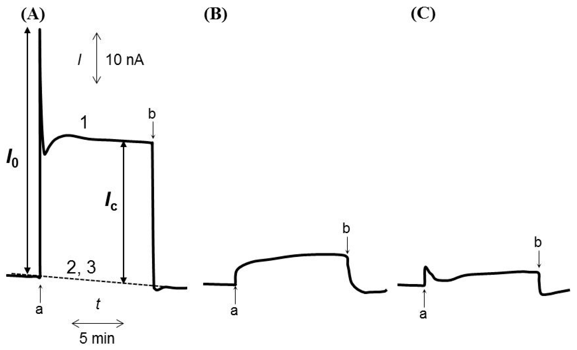
Abstract
:1. Introduction
2. Materials and Methods
2.1. Preparation of Chromatophore Vesicles and Reagents
2.2. Fabrication of R. sphaeroides Chromatophore-Entrapped and Mediator-Embedded CPEs
2.3. Electrochemical Measurements
3. Results and Discussion
3.1. Characteristics of Photo-Induced Electron Transfer from R. sphaeroides Chromatophores to CPEs through Exogenous Electron Acceptors
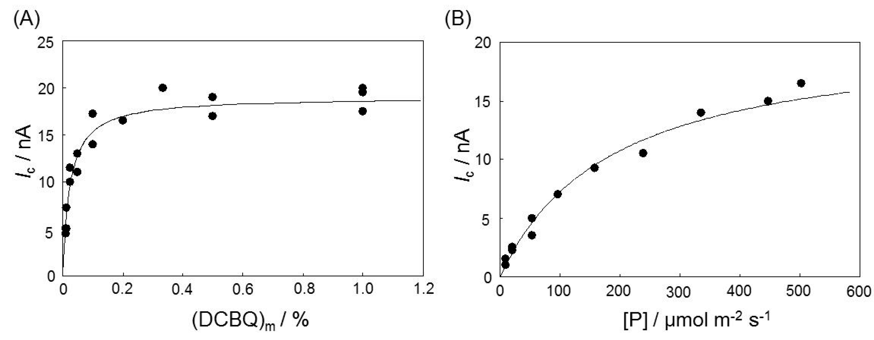
3.2. Effects on the Photocurrent of Varying R. sphaeroides Chromatophore Amount, DCBQ Fraction, and Light Intensity
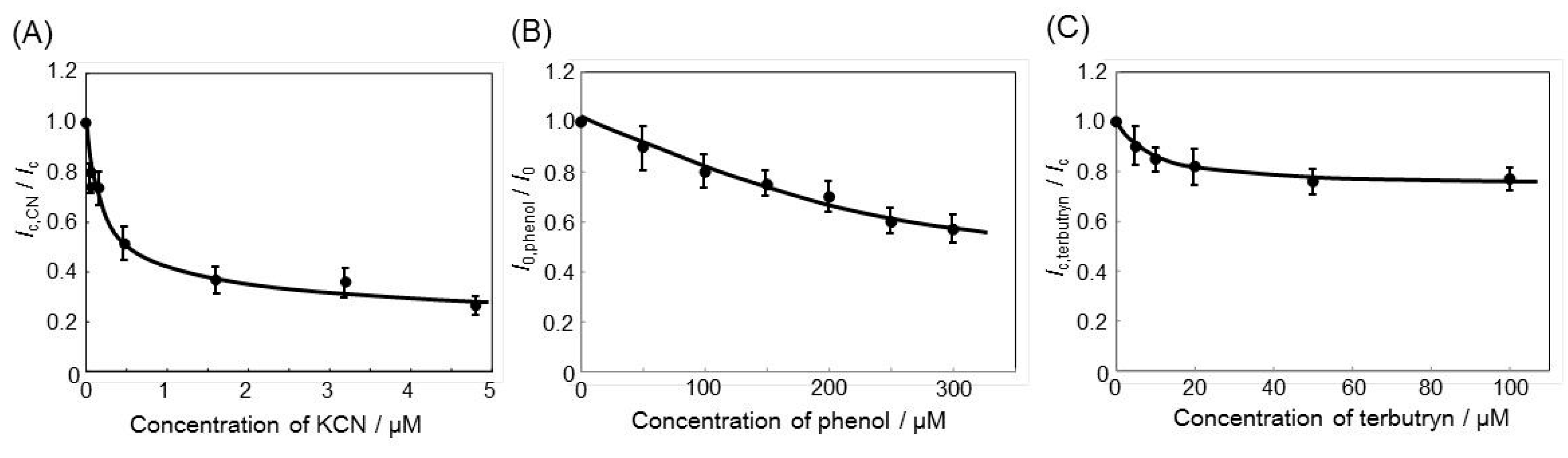
3.3. Effect of CN−, Phenol or Terbutryn on Electron Transfer from R. sphaeroides Chromatophores to CPE via DCBQ
4. Conclusions
Acknowledgments
Author Contributions
Conflicts of Interest
References
- US EPA. Method Guidance and Recommendations for Whole Effluent Toxicity Testing; United State Environmental Protection Agency: Washington, DC, USA, 2000. [Google Scholar]
- Rand, G.M. (Ed.) Fundamentals of Aquatic Toxicology: Effects, Environmental Fate and Risk Assessment, 2nd ed.; Taylor and Francis: Washington, DC, USA, 1995.
- Grothe, D.R.; Dickson, K.L.; Reed-Judkins, D.K. (Eds.) Whole Effluent Toxicity Testing: An Evaluation of Methods and Prediction of Receiving System Impacts; SETAC Press: Pensacola, FL, USA, 1996.
- Bowyer, J.R.; Camilleri, P.; Vermaas, W.F.J. Herbicides, Topics in Photosynthesis; Baker, N.R., Percival, M.P., Eds.; Elsevier Science Publishers: New York, NY, USA, 1991; Volume 10, pp. 27–85. [Google Scholar]
- Peters, H.; Dannert, C.S.; Schmid, R.D. The photoreaction center of Rhodobacter sphaeroides: A “biosensor protein” for the determination of photosystem-II herbicides? Mater. Sci. Eng. C 1997, 4, 227–232. [Google Scholar] [CrossRef]
- Brown, A.E.; Gilbert, C.W.; Guy, R.; Arntzen, C.J. Triazine herbicide resistance in the photosynthetic bacterium Rhodopseudomonas sphaeroides. Proc. Natl. Acad. Sci. USA 1984, 81, 6310–6314. [Google Scholar] [CrossRef] [PubMed]
- Broser, M.; Glöckner, C.; Gabdulkhakov, A.; Guskov, A.; Buchta, J.; Kern, J.; Müh, F.; Dau, H.; Saenger, W.; Zouni, A. Structural Basis of Cyanobacterial Photosystem II Inhibition by the Herbicide Terbutryn. J. Biol. Chem. 2011, 286, 15964–15972. [Google Scholar] [CrossRef] [PubMed]
- Swainsbury, D.J.K.; Friebe, V.M.; Frese, R.N.; Jones, M.R. Evaluation of a biohybrid photoelectrochemical cell employing the purple bacterial reaction center as a biosensor for herbicides. Biosens. Bioelectron. 2014, 58, 172–178. [Google Scholar] [CrossRef] [PubMed]
- Giardi, M.T.; Koblízek, M.; Masojídek, J. Photosystem II-based biosensors for the detection of pollutants. Biosens. Bioelectron. 2001, 16, 1027–1033. [Google Scholar] [CrossRef]
- Nagy, L.; Magyar, M.; Szabó, T.; Hajdu, K.; Giotta, L.; Dorogi, M.; Milano, F. Photosynthetic Machineries in Nano-Systems. Curr. Protein Pept. Sci. 2014, 15, 363–373. [Google Scholar] [CrossRef] [PubMed]
- Sardesai, V.M. Toxitants Occurring Naturally in Foods and Additives. In Introduction to Clinical Nutrition, 3rd ed.; CRC Press: Boca Raton, FL, USA, 2011; pp. 515–536. [Google Scholar]
- Barr, R.; Crane, F.L. Inhibition or Inactivation of Higher-Plant Chloroplast Electron Transport. In Handbook of Photosynthesis, 2nd ed.; Pessarakli, M., Ed.; CRC Press: Boca Raton, FL, USA, 2005; pp. 149–168. [Google Scholar]
- Mishra, S.; Dubey, R.S. Heavy Metal Toxicity Induced Alterations in Photosynthetic Metabolism. In Handbook of Photosynthesis, 2nd ed.; Pessarakli, M., Ed.; CRC Press: Boca Raton, FL, USA, 2005; pp. 845–864. [Google Scholar]
- Clijsters, H.; Assche, F.V. Inhibition of photosynthesis by heavy metals. Photosynth. Res. 1985, 7, 31–40. [Google Scholar] [CrossRef] [PubMed]
- Kumar, K.S.; Dahms, H.U.; Lee, J.S.; Kim, H.C.; Lee, W.C.; Shin, K.H. Algal photosynthetic responses to toxic metals and herbicides assessed by chlorophyll a fluorescence. Ecotoxicol. Environ. Saf. 2014, 104, 51–71. [Google Scholar] [CrossRef] [PubMed]
- Saphon, S.; Jackson, J.B.; Lerbs, V.; Witt, H.T. The functional unit of electrical events and phosphorylation in chromatophores from Rhodopseudomonas sphaeroides. Biochim. Biophys. Acta 1975, 408, 58–66. [Google Scholar] [CrossRef]
- Drews, G.; Golecki, J.R. Structure, Molecular Organization, and Biosynthesis of Membranes of Purple Bacteria. In Anoxygenic Photosynthetic Bacteria; Blankenship, R.E., Madigan, M.T., Bauer, C.E., Eds.; Kluwer Academic Publishing: Dordrecht, The Netherlands, 1995; pp. 231–257. [Google Scholar]
- Kasuno, M.; Torimura, M.; Tsukatani, Y.; Murakami, D.; Hanada, S.; Matsushita, T.; Tao, H. Characterization of the photoinduced electron transfer reaction from the photosynthetic system in Rhodobacter sphaeroides to an exogenous electron acceptor. J. Electroanal. Chem. 2009, 636, 101–106. [Google Scholar] [CrossRef]
- Ikeda, T.; Hamada, H.; Miki, K.; Senda, M. Glucose Oxidase-Immobilized Benzoquinone-Carbon Paste Electrode as a Glucose Sensor. Agric. Biol. Chem. 1985, 49, 541–543. [Google Scholar] [CrossRef]
- Ikeda, T.; Hamada, H.; Senda, M. Electrocatalytic Oxidation of Glucose at a Glucose Oxidase-Immobilized Benzoquinone-Mixed Carbon Paste Electrode. Agric. Biol. Chem. 1986, 50, 883–890. [Google Scholar] [CrossRef]
- Amako, K.; Yanai, H.; Ikeda, T. Dimethylbenzoquinone-mediated photoelectrochemical oxidation of water at a carbon paste electrode coated with photosystem II membranes. J. Electroanal. Chem. 1993, 362, 71–77. [Google Scholar] [CrossRef]
- Clayton, R.K. Absorption spectra of photosynthetic bacteria and their chlorophylls. In Bacterial Photosynthesis; Gest, H., Pietro, A.S., Vernon, L.P., Eds.; Antioch Press: Yellow Springs, OH, USA, 1963; pp. 495–500. [Google Scholar]
- Kano, K. Redox potentials of proteins and other compounds of bioelectrochemical interest in aqueous solutions. Rev. Porarogr. 2002, 48, 29–46. [Google Scholar] [CrossRef]
- Izawa, S. Acceptors and donors for chloroplast electron transport. In Methods in Enzymology; Pietro, A.S., Ed.; Academic Press: New York, NY, USA, 1980; Volume 69, pp. 413–433. [Google Scholar]
- Yasukawa, T.; Uchida, I.; Matsue, T. Permeation of Redox Species Through a Cell Membrane of a Single, Living Algal Protoplast Studied by Microamperometry. Biochim. Biophys. Acta 1998, 1369, 152–158. [Google Scholar] [CrossRef]
- Longatte, G.; Fu, H.Y.; Buriez, O.; Labbé, E.; Wollman, F.A.; Amatore, C.; Rappaport, F.; Guille-Collignon, M.; Lemaître, F. Evaluation of photosynthetic electrons derivation by exogenous redox mediators. Biophys. Chem. 2015, 205, 1–8. [Google Scholar] [CrossRef] [PubMed]
- Osyczka, A.; Moser, C.C.; Dutton, P.L. Novel cyanide inhibition at cytochrome c1 of Rhodobacter capsulatus cytochrome bc1. Biochim. Biophys. Acta 2004, 1655, 71–76. [Google Scholar] [CrossRef] [PubMed]
- Sekar, N.; Umasankar, Y.; Ramasamy, R.P. Photocurrent generation by immobilized cyanobacteria via direct electron transport in photo-bioelectrochemical cells. Phys. Chem. Chem. Phys. 2014, 16, 7862–7871. [Google Scholar] [CrossRef] [PubMed]
- Escher, B.I.; Snozzi, M.; Häberli, K.; Schwarzenbach, R.P. A new method for simultaneous quantification of uncoupling and inhibitory activity of organic pollutants in energy-transducing membranes. Environ. Toxicol. Chem. 1997, 16, 405–414. [Google Scholar] [CrossRef]
- Matorin, D.N.; Plekhanov, S.E.; Bratkovskaya, L.B.; Yakovleva, O.V.; Alekseev, A.A. The Effect of Phenols on the Parameters of Chlorophyll Fluorescence and Reactions of P700 in Green Algae Scenedesmus Quadricauda. Biophysics 2014, 59, 374–379. [Google Scholar] [CrossRef]
- Gabellini, N.; Hauska, G. Characterization of cytochrome b in the isolated ubiquinol-cytochrome c2 oxidoreductase from Rhodopseudomonas Sphaeroides GA. FEBS Lett. 1983, 153, 146–150. [Google Scholar] [CrossRef]
- World Health Organization. Guidelines for Drinking-Water Quality, 4th ed.; World Health Organization: Geneva, Switzerland, 2011. [Google Scholar]
- Mak, K.K.W.; Yanase, H.; Renneberg, R. Cyanide fishing and cyanide detection in coral reef fish using chemical tests and biosensors. Biosens. Bioelectron. 2005, 20, 2581–2593. [Google Scholar] [CrossRef] [PubMed]
- Karim, F.; Fakhruddin, A.N.M. Recent advances in the development of biosensor for phenol: A review. Rev. Environ. Sci. Biotechnol. 2012, 11, 261–274. [Google Scholar] [CrossRef]
- Stein, R.R.; Camilleri, A.L.; Bogacz, J.P.; Wraight, C.A. Herbicide-quinone competition in the acceptor complex of photosynthetic reaction centers from rhodopseudomonas sphaeroides: A bacterial model for PS-II-herbicide activity in plants. J. Cell. Biochem. 1984, 24, 243–259. [Google Scholar] [CrossRef] [PubMed]
- Husu, I.; Magyar, M.; Szabó, T.; Fiser, B.; Gómez-Bengoa, E.; Nagy, L. Structure and binding efficiency relations of QB site inhibitors of photosynthetic reaction centres. Gen. Physiol. Biophys. 2015, 34, 119–133. [Google Scholar] [CrossRef] [PubMed]


© 2016 by the authors; licensee MDPI, Basel, Switzerland. This article is an open access article distributed under the terms and conditions of the Creative Commons by Attribution (CC-BY) license (http://creativecommons.org/licenses/by/4.0/).
Share and Cite
Kasuno, M.; Kimura, H.; Yasutomo, H.; Torimura, M.; Murakami, D.; Tsukatani, Y.; Hanada, S.; Matsushita, T.; Tao, H. An Evaluation of Sensor Performance for Harmful Compounds by Using Photo-Induced Electron Transfer from Photosynthetic Membranes to Electrodes. Sensors 2016, 16, 438. https://doi.org/10.3390/s16040438
Kasuno M, Kimura H, Yasutomo H, Torimura M, Murakami D, Tsukatani Y, Hanada S, Matsushita T, Tao H. An Evaluation of Sensor Performance for Harmful Compounds by Using Photo-Induced Electron Transfer from Photosynthetic Membranes to Electrodes. Sensors. 2016; 16(4):438. https://doi.org/10.3390/s16040438
Chicago/Turabian StyleKasuno, Megumi, Hiroki Kimura, Hisataka Yasutomo, Masaki Torimura, Daisuke Murakami, Yusuke Tsukatani, Satoshi Hanada, Takayuki Matsushita, and Hiroaki Tao. 2016. "An Evaluation of Sensor Performance for Harmful Compounds by Using Photo-Induced Electron Transfer from Photosynthetic Membranes to Electrodes" Sensors 16, no. 4: 438. https://doi.org/10.3390/s16040438