1. Introduction
Rutoside (RUT) (also known as rutin, quercetin-3-
O-glucoside, and sophorin) is a natural flavonoid glycoside that has quercetin as the aglycone part, and a disaccharide, rutinose [α-L-rhamnopyranosyl-(1→6)-β-
d-glucopyranose], as the glucidic part. Rutoside is found in fruits and vegetables, such as
Sophora japonica,
Fagopyrum esculentum,
Rutagraveolens,
Hypericum perforatum,
Ginkgo biloba,
Sambucus nigra,
Tilia spp., and
Crataegus spp. [
1,
2,
3,
4,
5,
6,
7,
8]. RUT is used in therapeutical practice for its capillary protective (increasing the resistance and decreasing the capillary permeability) and antioxidative properties (direct chelation mechanism) in capillary fragility, venous insufficiency, hemorrhoids, or epistaxis [
9,
10].
Beyond its scientifically proven effectiveness, rutoside processing in different pharmaceutical forms is still hard to be made, due to its low oral absorption and bioavailability, as a result of poor solubility and chemical instability [
11,
12]. During the last years, a lot of studies were aimed at enhancing the stability, solubility, absorbability, and bioavailability of rutoside, by using rutoside derivates, particle size reduction, the use of surfactants, or sugar coating [
13,
14,
15]. A facile and effective method for increasing the solubility, stability, and antioxidant activity of rutoside is its inclusion in the cyclodextrins cavities by forming inclusion complexes [
16,
17,
18]. Cyclodextrins are cyclic oligosaccharides able to include various molecules in their internal hydrophobic cavity, frequently used to improve the water solubility of encapsulated ingredients due to their hydrophilic external surface [
19]. Different studies reported an enhancement of the aqueous solubility, permeability, and absorption of rutoside, and many others proved to improve the chemical stability of rutoside, all by complexing it with β-cyclodextrin [
17,
20,
21,
22]. It appears that the dimension of the rutoside molecule is suitable to fit perfectly in the cavity of β-cyclodextrin, the α-cyclodextrin cavity being too small, and the γ-cyclodextrin too large [
21,
23,
24]. Although, until now, the studies of the preparation and structural characterization of inclusion complexes between rutoside and cyclodextrins by different ways have been advanced, the further development of the inclusion compounds by new methods is required, especially to study the solubility, stability, and antioxidant capacity of rutoside.
In this context, the use of rutoside to form inclusion complexes with CDs has been paid much attention. In the literature, a lot of studies have reported the formulation of rutoside inclusion compounds using various CDs with significant enhancement of its bioavailability. The inclusion complexes between rutoside and β-CD and HP-βCD prepared in a molar ratio of 1:1 by the co-precipitation method show an improved solubility, antioxidative activity, and photostability of rutoside [
17]. The RUT- β-CD inclusion complex obtained via a co-grinding technique in a 1:1 molar ratio was found to have a greater stability and solubility compared with free compound but a lower antibacterial activity [
25]. The phase-solubility studies of 1:1 stoichiometric inclusion complexes of rutoside with β-CD, HP-β-CD, HP-α-CD, and HP-γ-CD have showed greater stability constants [
23]. The authors claimed that the complexes RUT- HP-β-CD and RUT- HP-γ-CD were found to have the greatest stability constants, followed by RUT-β-CD and RUT-α-CD, and in consequence an enhanced antioxidant activity. The inclusion complexes of rutin with β-cyclodextrin and hydroxypropyl-β-cyclodextrin by the kneading method, followed by the evaluation of the antioxidant, antiproliferative, and pro-apoptotic activity against the B164A5 murine melanoma cell line shows an increase of the antioxidant activity compared with free rutoside [
26]. Most of these studies have not fully investigated the stability and the antioxidant activity of the inclusion complexes and furthermore the pharmaco-technical of the tablets, including RUT-β-CD and RUT-HP-β-CD inclusion complexes, as active ingredients.
Considering the so-far available studies, the aim of the present study was to prove the enhancement of the stability and the antioxidant properties of RUT by complexation with two cyclodextrins, namely β-cyclodextrin (β-CD) and hydroxypropyl-β-cyclodextrin (HP-β-CD). Therefore, the prepared inclusion complexes of RUT in β-CD and HP-β-CD, using the lyophilization method of complexation in solution, were compared with the simple physical mixtures, obtained in the same molar ratio of 1:1. The complexes were identified and characterized by scanning electron microscopy (SEM), differential scanning calorimetry (DSC), and Fourier-transform infrared spectroscopy (FT-IR) analyses. Finally, the inclusion complexes were processed under the form of tablets, showing the preformulation, formulation, and manufacture studies, ending with the quality control performed on the final products.
Moreover, the antioxidant activity of rutoside inclusion complexes with β-CD and HP-β-CD was evaluated by three in vitro methods: (i) one using the radical 2,2-diphenyl-1-picryl-hydrazyl (DPPH); (ii) the second test was made with 2, 2′-azino-bis(3-ethylbenzothiazolin-6-sulfonic) acid (ABTS), determining the radical scavenging activity; and (iii) the third one through ferric reducing antioxidant power (FRAP) assay.
2. Materials and Methods
2.1. Materials
RUT, β-CD and HP-β-CD, DPPH, ABTS, and TPTZ were purchased from Sigma-Aldrich Chemie GmbH, (Steinheim, Germany). The methanol and distilled water were of analytical grade.
For sample weighing, a Mettler Toledo AT261 balance (with 0.01 mg sensitivity) was used.
2.2. Preparation of the Inclusion Complexes
The lyophilization method of complexation in solution was used to prepare inclusion complexes between rutoside and β-CD, and RUT and HP-β-CD, respectively, in a molar ratio of 1:1. The synthesized inclusion complexes were noted as: RUT-β-CD and RUT-HP-β-CD. RUT-β-CD inclusion complex was prepared as follows: 0.25 mmol of β-CD was dissolved in 100 mL distilled water, and, separately, 0.25 mmol of RUT was dissolved in 20 mL of methanol. Using the same procedure as in the case of RUT-β-CD complex, the inclusion complex between RUT and HP-β-CD was synthesized using 0.25 mmol of HP-β-CD dissolved in 100 mL of distilled water and 0.25 mmol RUT dissolved in 20 mL of methanol. The two methanol solution samples were added stepwise to each aqueous solution of β-CD and HP-β-CD, and the two obtained suspensions were stirred at room temperature, for 8 h, at 1000 rpm, in a Heidolph MR 3001K magnetic stirrer. The final two obtained suspensions were lyophilized at −60 °C, for 12 h, in a Christ ALPHA 1–2, Braun Biotech International (Osterode, Germany) lyophilizer.
2.3. Preparation of RUT-β-CD and RUT-HP-β-CD Physical Mixtures
The physical mixtures, in the same molar ratio (1:1) as the inclusion complexes, were prepared by mixing the raw materials accurately weighted, for 5 min, at room temperature, in a mortar. Samples notation: RUT-β-CD physical mixture and RUT-HP-β-CD physical mixture.
2.4. Measurements and Characterization
The obtained inclusion complexes (RUT-β-CD and RUT-HP-β-CD), physical mixtures (RUT-β-CD and RUT-HP-β-CD), and pure compounds (RUT, β-CD, and HT- β-CD) were identified and characterized by scanning electron microscopy (SEM, TECSAN Brno, Czech Republic), differential scanning calorimetry (DSC, Setaram Instrumentation, Caluire, France), and Fourier-transform infrared spectroscopy (FT-IR) analyses. Additionally, the antioxidant properties of rutoside were evaluated.
2.4.1. SEM Analysis
To determine the morphology of the samples, the scanning electron microscope, VEGA II model, produced by TESCAN, (Brno, Czech Republic) was used, working in the following conditions: accelerating voltage of 30 kV, at a working distance of 9.9 mm, with a beam current of 60 μA, a probe current from 1:8 to 1:12, current absorbed up to ≈100 pA, and secondary electrons detector. The SEM images were registered at magnifications between 100× and 4000×. The samples were processed by adhesion on a support covered with a graphite double adhesive conducting tape, eliminating the excessing materials by blowing with a compressed nitrogen gun of chromatographic purity.
2.4.2. Differential Scanning Calorimetry
DSC measurements of pure compounds and their physical mixtures (RUT-β-CD and RUT-HP-β-CD) and inclusion complexes (RUT-β-CD and RUT-HP-β-CD) were performed on SETARAM DSC 131EVO (Setaram Instrumentation, Caluire, France), equipped with a cooler system based on liquid nitrogen (95 °C/min), a data detection system Platinum™ Software, and a DSC Standard Cell RC module. Samples of approximately 5 mg sealed in aluminum pans were heated with a heating rate of 10 °C/min, in the temperature range of 30–210 °C. An inert nitrogen atmosphere with a flow rate of 50.0 mL/min was used. As a reference, an empty sealed aluminum pan was used. The instrument calibration regarding the temperature and the enthalpy was made with the standard material indium (99.98%) with the known melting point Tm = 156.65 °C. All samples (RUT, β-CD, and HT-β-CD, physical mixtures and inclusion complexes) were analyzed in duplicate.
2.4.3. FT-IR Analysis
The Fourier Transform Infrared spectra were registered using a JASCO FT/IR-4200 spectrometer (Tokyo, Japan) with an ATR PRO450-S accessory, on a spectral range of 4000–400 cm−1 using a resolution of 4 cm−1. The pure compounds (RUT, β-CD and HT-β-CD), the physical mixtures (RUT-β-CD and RUT-HP-β-CD), and the inclusion complexes (RUT-β-CD and RUT-HP-β-CD) were analyzed in duplicate.
2.4.4. Antioxidant Activity of the RUT-β-CD and RUT-HP-β-CD Inclusion Complexes
DPPH Radical Scavenging Activity
The DPPH method may be used either in aqueous or nonpolar organic solvents, and is useful to examine both hydrophilic and lipophilic antioxidants. DPPH is a stable free radical, due to the spare electron delocalization over the whole molecule; thus, it does not dimerize, as is the case with most free radicals. Delocalization on the DPPH molecule causes the appearance of a deep purple color, with an absorption band at a maximum of about 520 nm. When DPPH reacts with a hydrogen donor, the reduced molecular form (DPPH-H) occurs, accompanied by the loss of the purple color. Therefore, the decrease in absorption linearly depends on the antioxidant capacity [
27,
28,
29].
The antioxidant activity of the rutoside-cyclodextrins inclusion complexes was determined using the DPPH free radical scavenging assay described by Margina et al. [
30] with some modifications. Briefly, the samples contained 10 μL of 1 mM ethanolic solution of each tested complex or compound treated with 100 µL 0.5 mM DPPH solution. The absorbance of the obtained mixture was measured after 5 min and then from 5 to 5 min, for 40 min, at 505 nm. Ascorbic acid was used as the positive control, in this case the reduction of DPPH was evaluated for different concentrations: 2.8 mM–87.5 µM. The results are presented as the decrease percentage in optical density at 40 min from the initial one.
The DPPH scavenging effect (radical scavenging activity
RSA%) was calculated using Equation (1):
where
Abs0 is the absorbance of DPPH solution (reference) at
t0 (initial optical density);
Absp is the absorbance of the samples measured at different period of times
t (sample optical density).
ABTS Radical Scavenging Activity
The cationic ABTS radical (ABTS•+), which shows absorption at 743 nm (giving a blue-green color), is formed by the loss of an electron through the nitrogen atom of 2, 2′-azino-bis (3-ethylbenzothiazolin-6-sulfonic) acid. In the presence of a hydrogen donor antioxidant compound, the nitrogen atom binds the hydrogen atom, leading to the discoloration of the solution.
The studied compounds were dissolved in ethanol solution 96%, the concentration of the obtained solution being 1 mM. The stock solution containing 7 mM ABTS and 2.45 mM K
2S
2O
8 was maintained in the dark, at 4 °C, for 16 h, to generate the ABTS•
+ free radical. The working reagent was obtained by diluting the stock solution with distilled water to OD
714 nm = 0.8. The samples (50 μL) were treated with 100 µL of working reagent and optical density was measured initially and after an incubation of 5 and 10 min, at 714 nm. The results were expressed as %
DOD (decrease percentage in optical density), this being directly proportional to the antioxidant capacity of the samples, using Equation (2) [
31]:
FRAP Assay
The mechanism of ferric reducing antioxidant power depends on electron transfer and is based on the ability of antioxidants to reduce Fe
3+ to Fe
2+ in the presence of 2,4,6-trys(2-pyridyl)-s-triazine (TPTZ), when an intense blue ferrous ion–TPTZ complex (absorption at 593 nm) is formed [
32]. The reaction takes place at acidic medium pH = 3.6, which ensures the iron solubility, increasing the redox potential, which is the main reaction mechanism.
For the FRAP assay, the method described by Nair et al. was used [
33]. The reagent was obtained by mixing 10 mL of 300 mM acetate buffer (pH 3.6 adjusted with acetic acid), 1 mL of 20 mM ferric chloride hexahydrate, and 1 mL of 10 mM TPTZ in 40 mM HCl. Then, 100 µL methanolic solutions of each sample weremixed with 3 mL of the prepared FRAP solution, then incubated for 30 min at 37 °C, protected from light. The absorbance at 593 nm was recorded at 5 and 30 min. The calibration curves were obtained on FeSO
4 methanol sample solutions. The antioxidant activity was reported as µM Fe
2+ produced by 300-µL methanol sample solutions, from a constant Fe
3+ concentration.
2.4.5. Preformulation and Precompression Studies for Tablets Containing RUT-β-CD and RUT-HP-β-CD Inclusion Complexes
The preformulation studies give useful information for the design of an optimum drug delivery system. For the present study, it was chosen to manufacture the tablets containing RUT-β-CD and RUT-HP-β-CD inclusion complexes by the direct compression method. In order to obtain high-quality tablets, the direct compression material must have good physical properties, including the fundamental ones, flowability and compressibility. As excipients, the following were selected: (i) Avicel PH 102, as filler, which improves the flow of powders, and also shows good binder attributes; and (ii) Polyplasdone XL-10, a non-ionic crosslinked PVP homopolymer, for its superdisintegrant properties due to the porous particle morphology, and magnesium stearate and talc, as lubricants.
Assessment of Bulk Powder Physical Properties
Particle size directly influences the flowability of the powder, and also, the content uniformity and the dissolution rate of the tablets. It was determined by the sieving and sorting method, using a CISA Sieve Shaker Mod. RP 10, produced by Cisa Cedaceria Industrial, Spain. The method consists in the passing of the material by mechanical shaking through a set of sieves with well-known mesh sizes, placed under each other, in ascending order of the finesse degree.
Flowability, a critical characteristic, was evaluated by determining the angle of repose, flowing time, and rate, needed for 60 g of powder to flow through an orifice with a standardized diameter. The study was performed with an Automated Powder and Granulate Testing System PTG-S3, fabricated by Pharma Test Apparatebau GmbH, Germany.
Compressibility, also a fundamental property, is established by volumetric characteristics like bulk and tapped density, calculating the Hausner ratio (HR) and Carr Index (CI), which are also used as flowability indicators. It was performed with Vankel Tap Density Tester, produced by Vankel Industries Inc., USA. Bulk density was determined by measuring the volume occupied by 50 g of powder poured into a graduated cylinder, which is, then, subjected to a given number of mechanical shocks, measuring the tapped volume of the material. Hausner ratio (HR) is the ratio between tapped density and bulk density, with a value under 1.25 indicating that the powder is free flowing. Carr index is given by the equation: CI% = (tapped density − bulk density) × 100/tapped density, and the smaller its value is, the better the flowability and compressibility are.
Moisture content is assessed as the loss on drying, by the Karl Fisher method with a HR 73 Mettler Toledo halogen humidity analyzer.
2.4.6. Formulation of the Tablets Containing RUT-β-CD and RUT-HP-β-CD Inclusion Complexes
After it was concluded that the powder has suitable characteristics to be processed by direct compression, the final formulations of the tablets were established, and they are presented in the
Table 1.
2.4.7. Tablets Manufacturing
The immediate release tablets were obtained from the composed powder by direct compression technology, using a medium compression force of 14 kN, in a single-post eccentric machine Korsch EK-O type, equipped with 7-mm flat punches. The machine was adjusted to obtain tablets with an average mass of 200 mg, corresponding to a content of 40 mg rutoside/tablet. After manufacturing, the obtained tablets were subjected to quality control tests.
2.4.8. Quality Parameters of the Tablets
Organoleptic Evaluation
The appearance, color, taste, and smell of the tablets were evaluated according to the European Pharmacopoeia specifications [
34].
Weight Variation
Mass uniformity is also influenced by the compression parameters. The testing was done by individually weighing 30 tablets of each formulation, then calculating the average mass and finally a comparison among them. The tablets comply with the test if not more than one individual mass is outside the limits of 85–115% of the average mass [
35].
Hardness
The tablet crushing strength, even it is not a compendial standard, is very important to be measured, as the disintegration time, the dissolution rate, and the resistance of tablets to breakage under conditions of storage, transportation, or handling before usage all depend on it. Tablet hardness was measured with a VK 200 Tablet Hardness Tester. A tablet was placed in the hardness tester and the load required to crush the tablet was registered.
Friability
This test was performed to evaluate the ability of tablets to withstand abrasion in packing, handling, and transporting. An initial weight of 10 tablets was taken and these were placed in the Wankel Friabilator, rotating at 25 rpm for 4 min, and then weighed again. The difference in the weight was noted and expressed as a percentage. It should be preferably below 1.0%.
2.4.9. In Vitro Disintegration Time
The
in vitro disintegration time [
35] of a tablet was determined using an Erweka DT 3 apparatus, produced by Erweka
® GmbH, Germany. The test was carried out on 6 tablets of each formulations using distilled water at 37 ± 0.5 °C as a disintegration media and the time taken for complete disintegration of the tablet, with no palpable mass remaining on the screen, was measured in seconds.
2.4.10. In Vitro Dissolution Rate
The dissolution rate was determined using a USP paddle Apparatus II (dissolution tester ERWEKA DT 800), in 900 mL of pH 6.8 phosphate buffer solution [
35]. The apparatus was settled at 37 ± 0.5 °C, with a paddle rotating speed of 50 rpm. The amount of dissolved rutoside was determined after 30 min, by the UV-VIS spectrophotometric method (UV-VIS Nicolet Evolution 100 spectrometer) at the absorption maximum of 257 nm.
3. Results
3.1. Organoleptic Evaluation of the RUT-β-CD and RUT-HP-β-CD Inclusion Complexes
The two physical mixtures obtained by mixing RUT with β-CD and RUT with HT-β-CD were yellow crystalline powders, odorless, with a bitter taste. By the lyophilization method, very fine and smooth yellow powders, odorless and bitter tasting, were obtained.
3.2. Identification and Characterization of the RUT-β-CD and RUT-HP-β-CD Inclusion Complexes
3.2.1. Scanning Electron Microscopy (SEM)
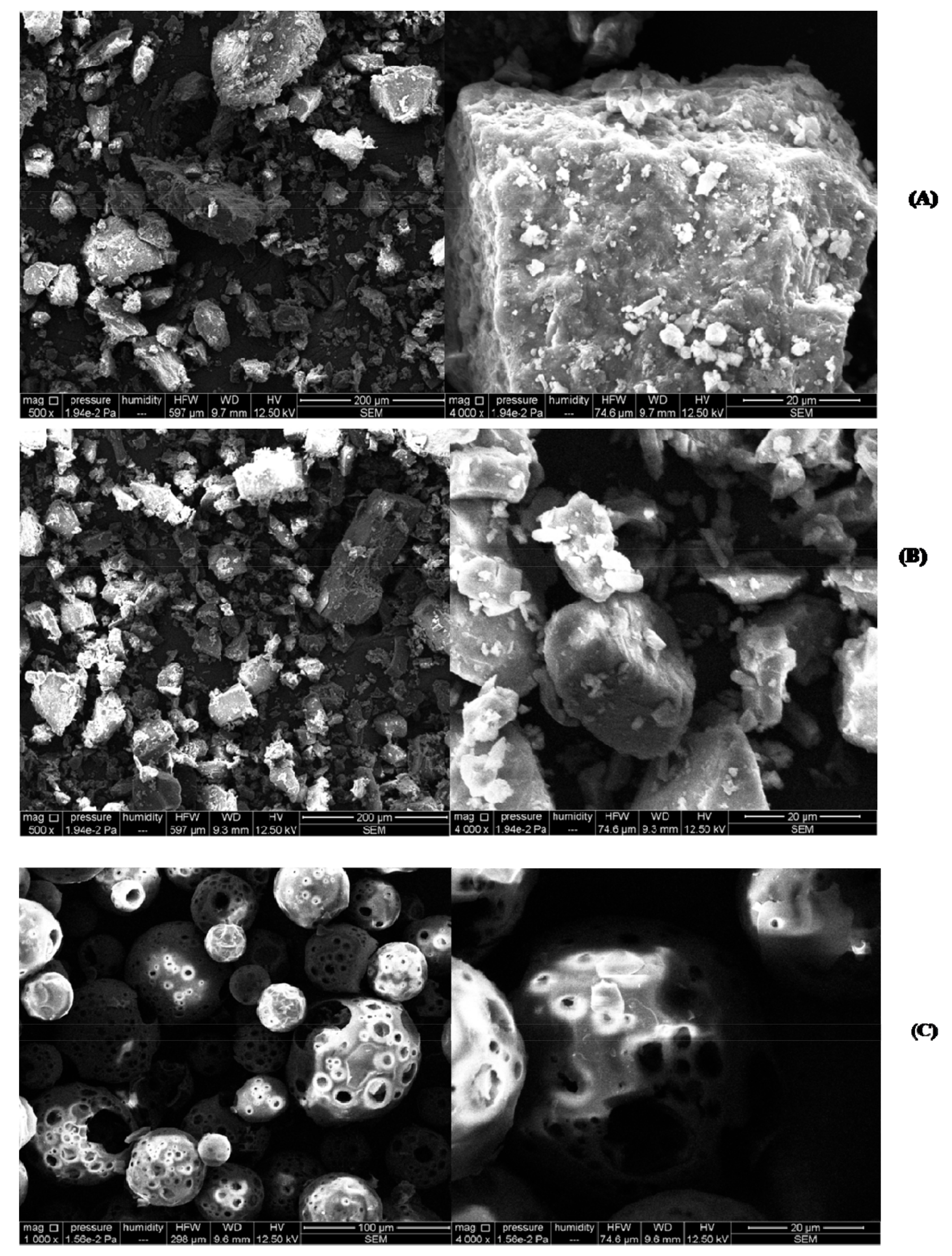
The SEM images of RUT, β-CD, HP-β-CD, RUT-β-CD, and RUT-HP-β-CD physical mixtures, and RUT-β-CD and RUT-HP-β-CD complexes are shown in
Figure 1 and
Figure 2 (between 500× and 4000× magnifications).
The SEM images of RUT, at 500× and 4000×, from
Figure 1A show the presence of non-homogeneous prismatic crystalline particles, with different sizes between 10 and 200 μm. SEM images at different magnifications for β-CD (
Figure 1B) showed also crystalline particles, with various shapes and sizes between 5 and 20 μm [
36], and HP-β-CD (
Figure 1C) showed spherical particles, with an amorphous character [
37]. The SEM images for both physical mixtures (
Figure 1D,E) showed the crystalline structures of rutoside adhering to the surface of the β-CD and HP-β-CD, confirming the affinity between them, but it is clear that no new substrate occurred. In both mixtures, the individual characteristics of raw material particles remained unchanged, leading to the idea that no inclusion took place yet. The SEM images show only both crystalline structures of the individual components. The same morphologies were found in the literature for similar compounds [
36,
38,
39].
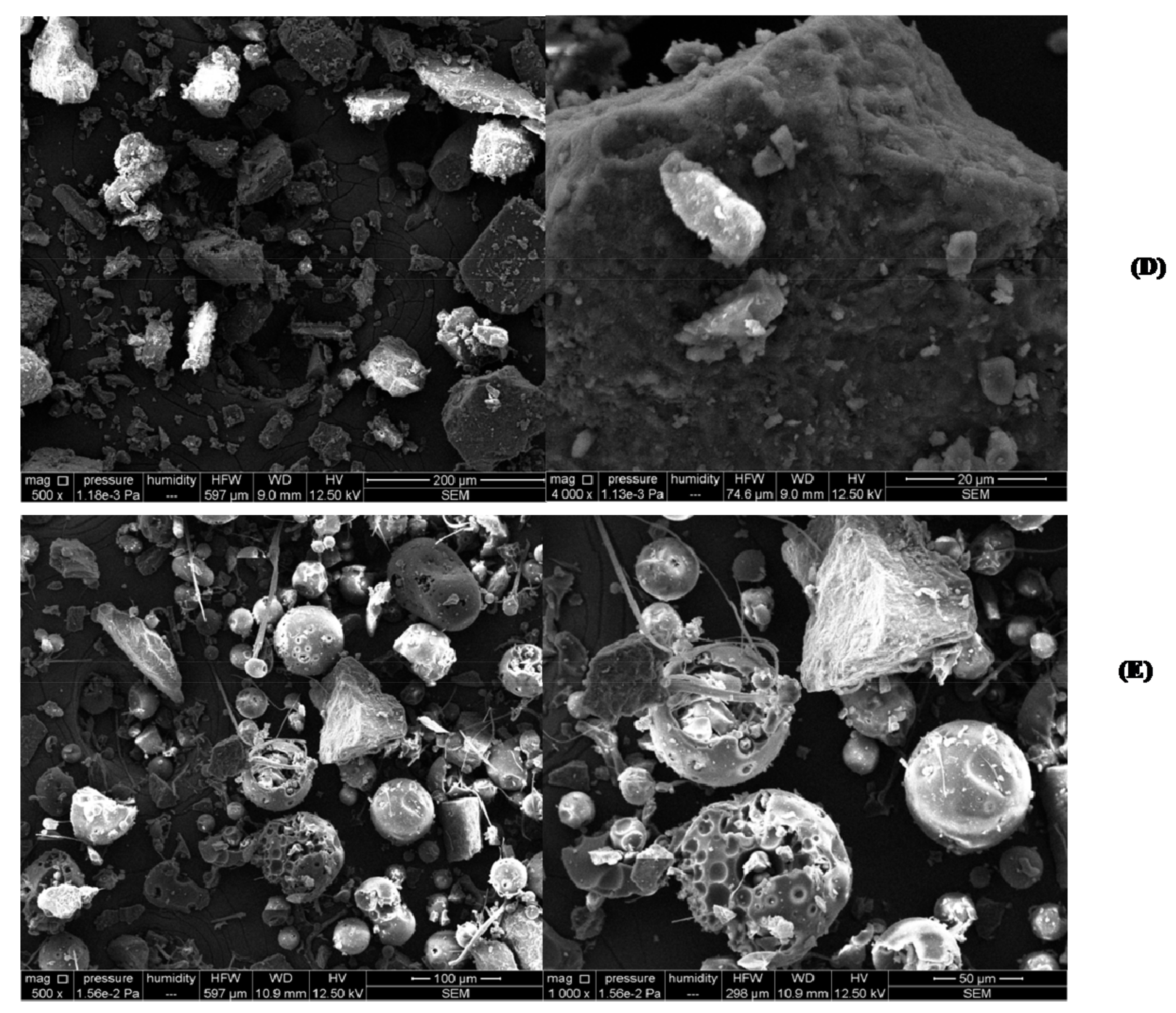
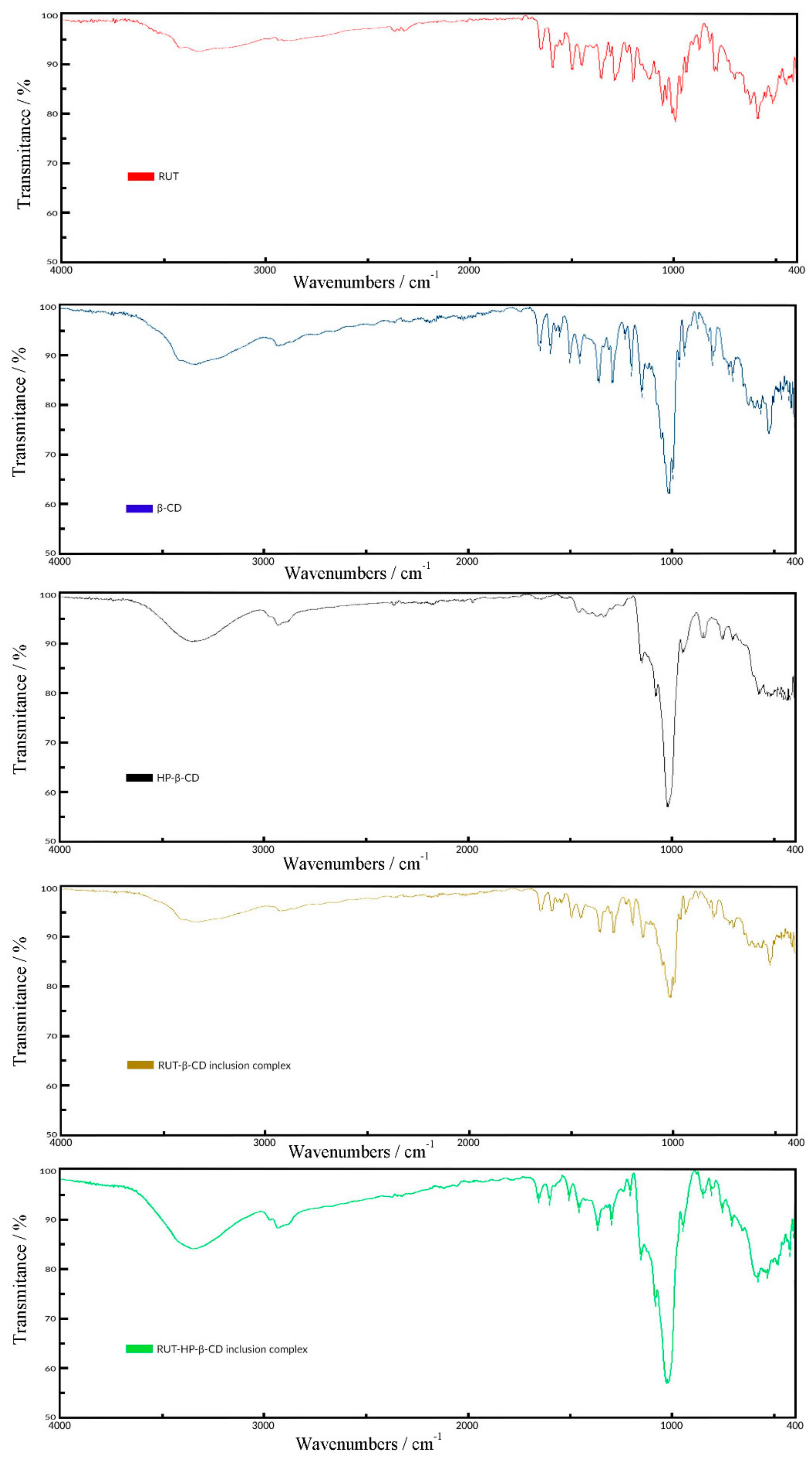
Analyzing the SEM images for RUT-β-CD (
Figure 2A) and RUT-HP-β-CD (
Figure 2B) inclusion complexes, it can be noticed the presence of some homogeneous slightly amorphous powders with fine and regular crystals. It is clear that a new system, with a drastic decrease of particle size and a partial loss of crystallinity towards the initial ingredients, was forming. The important changes in the morphology and shape of the particles reveal the presence of a new solid phase, being a specific characteristic for the formation of the inclusion complexes.
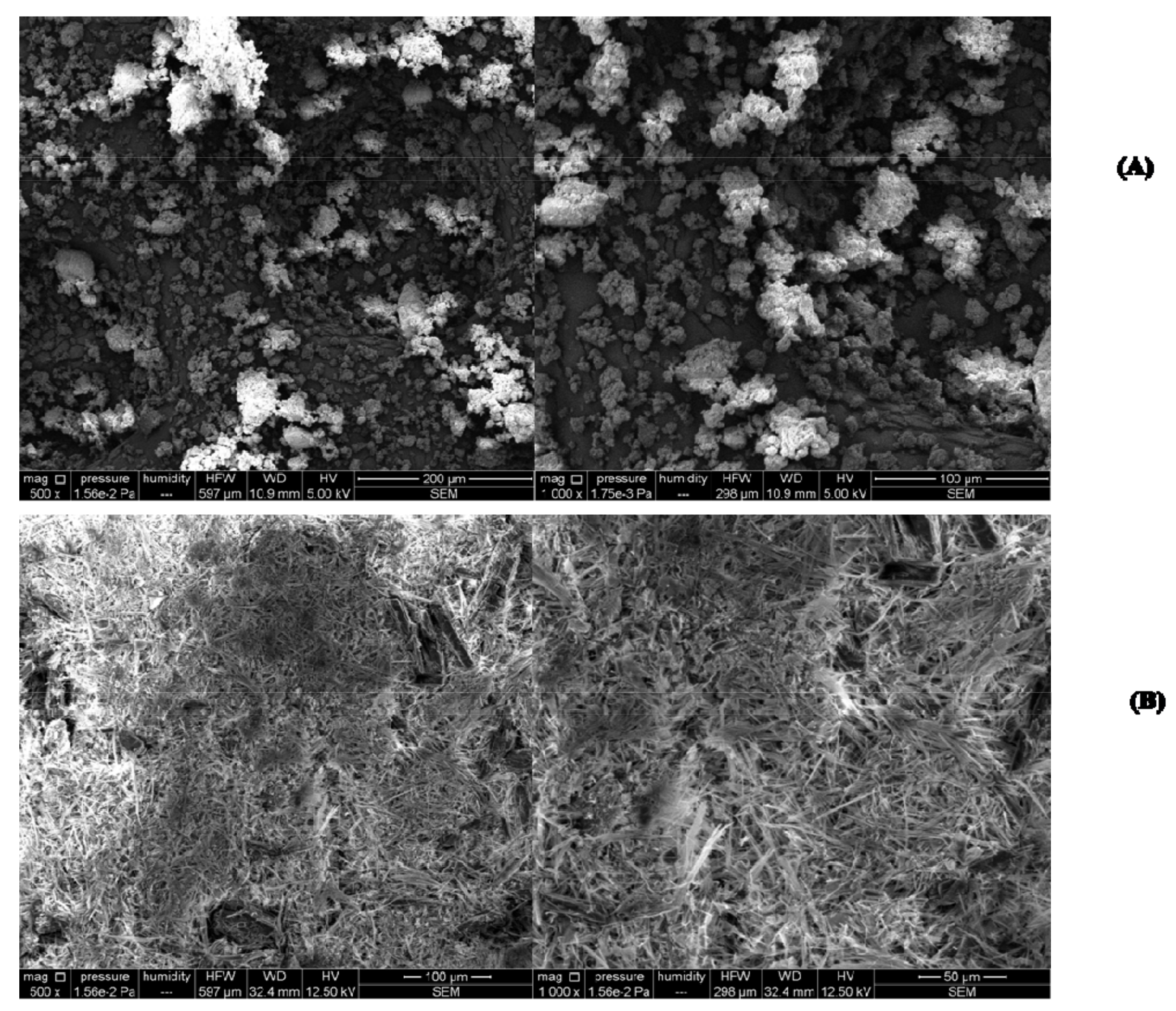
3.2.2. Fourier-Transform Infrared Spectroscopy (FT-IR)
The characteristic FT-IR bands of pure compounds (rutoside, β-cyclodextrin, hydroxypopyl-β-cyclodextrin), and of the two inclusion complexes are shown in
Figure 3. The FT-IR spectrum of rutoside (
Figure 3 (red line)) shows two broad bands at 3413 and 3334 cm
−1, respectively, due to O-H bounds vibrations. The weak peak at 2938 cm
−1 can be assigned to alkene C-H stretch. The band at around 1649 cm
−1 is characteristic of the double bound C=O from quercetol molecule, and the band at around 1450 cm
−1 can be assigned to C=C bound. In the range of 500–1200 cm
−1, a series of strong intensity bands are associated with the stretching vibration of the C-C and C-O bounds from the dihydroxiphenyl portion and from the disaccharide structure, and wagging vibration of the C-H bonds.
In the FT-IR spectrum of β-CD (
Figure 3 (blue line)), the strong bands at 1021, 1077, and 1151 cm
−1 are attributed to the stretching vibration of the C-C, C-O bonds and wagging vibration of the C-H bonds [
25]. The large band at 3327 cm
−1 is due to the O-H stretching vibrations [
36,
40]. The band at 2925 cm
−1 was assigned to the C-H stretching vibration. The absorption band at 1647 cm
−1 was due to the H-O-H bending vibration.
The FT-IR spectrum of HP-β-CD (
Figure 3 (black line)) is very similar to the one of β-CD. Several features can be pointed: the large band at 3336 cm
−1 is assigned to the O-H stretching vibrations. The strong band at 1022 cm
−1 is due to the C-O bound vibration. The band at around 2927 cm
−1 is attributed to the alkylic part.
In FT-IR spectra of the two physical mixtures of the compounds (RUT-β-CD and RUT-HP-β-CD), all the above peaks of the compounds appear at the same wavenumbers, indicating no interaction between drug and β-CD or HP-β-CD. The spectra of both physical mixtures showed an approximate superposition of individual spectra of pure compounds.
The FT-IR spectra of RUT-β-CD (
Figure 3 (olive line)) inclusion complex is similar with the one of the β-CD, but with a decrease in the intensity and a disappearance of some characteristic bands of RUT, which may be due to the encapsulation of the drug in the β-CD matrix. The differences in the positions and intensity of the ranges 500–1150 cm
−1 were recorded for RUT-β-CD inclusion complex in comparison with the pure compounds. In this domain, the bands from the FT-IR spectrum of the inclusion complex were shifted and were broadened.
The FT-IR spectrum of RUT-HP-β-CD (
Figure 3 (green line)) inclusion complex shows a high resemblance with the one of the RUT-β-CD inclusion complex due to the structural similarity of the two cyclodextrines. The peak of alcohol is strong; the peaks of C-OH and amine are weak. The lower intensity of these peaks from FT-IR spectrum of the RUT-HP-β-CD inclusion complex is due to the interaction to the hydroxyl group with the secondary amine [
41]. These results showed that the inclusion complexes of RUT with β-CD and HT-β-CD were successfully formed.
3.2.3. Differential Scanning Calorimetry (DSC)
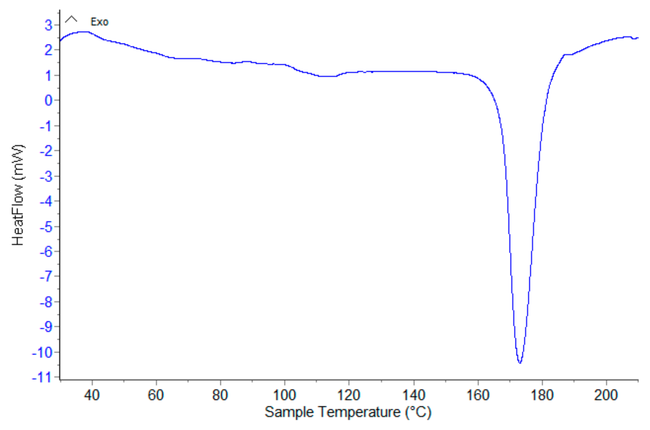
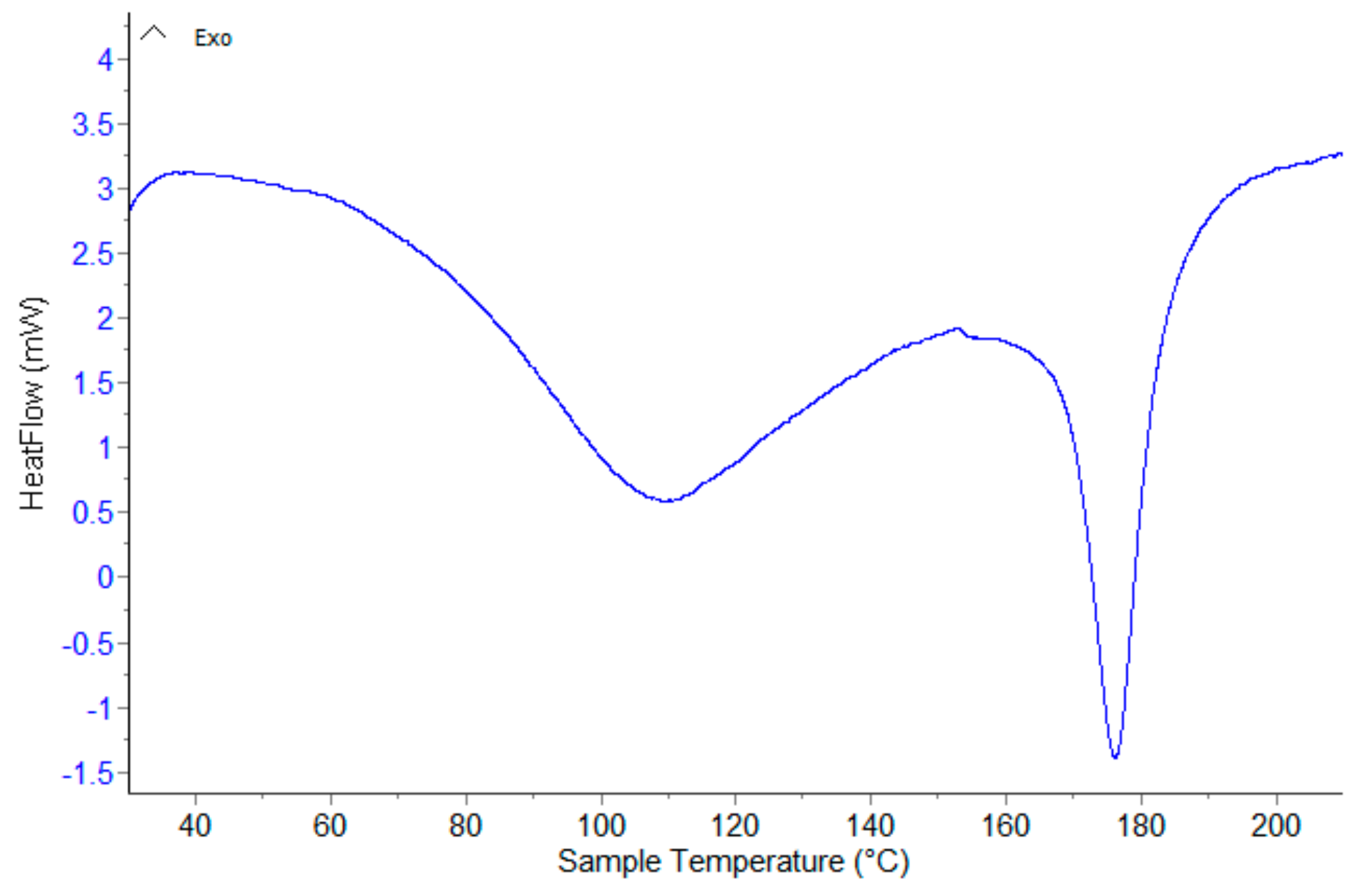
The DSC curve of rutoside (
Figure A1) exhibits a sharp endothermic peat at 173 °C, (peak temperature of the DSC curve recorded at a heating rate of 10 K min
−1) corresponding to its melting point, with the melting heat of Δ
H = 137.2 Jg
−1 evaluated from the peak area (
Figure A1 from
Appendix A).
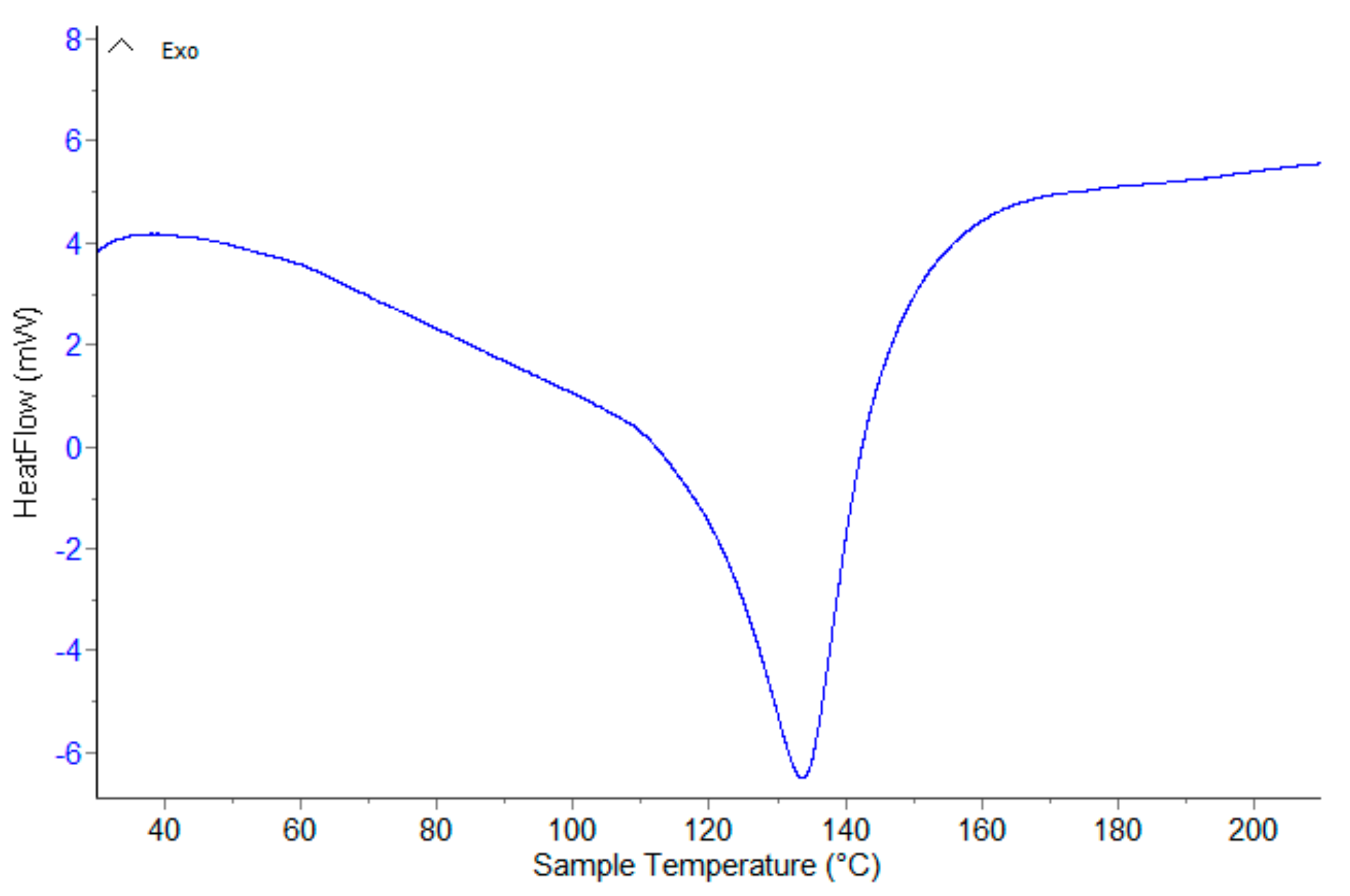
The DSC curve of β-CD (
Figure A2) shows an endothermic peak at 133.9 °C with an associated heat of the endothermal process of 222.5 °C, which is due to the release of water from the β-CD cavity [
42,
43]. No other endothermal effects appear.
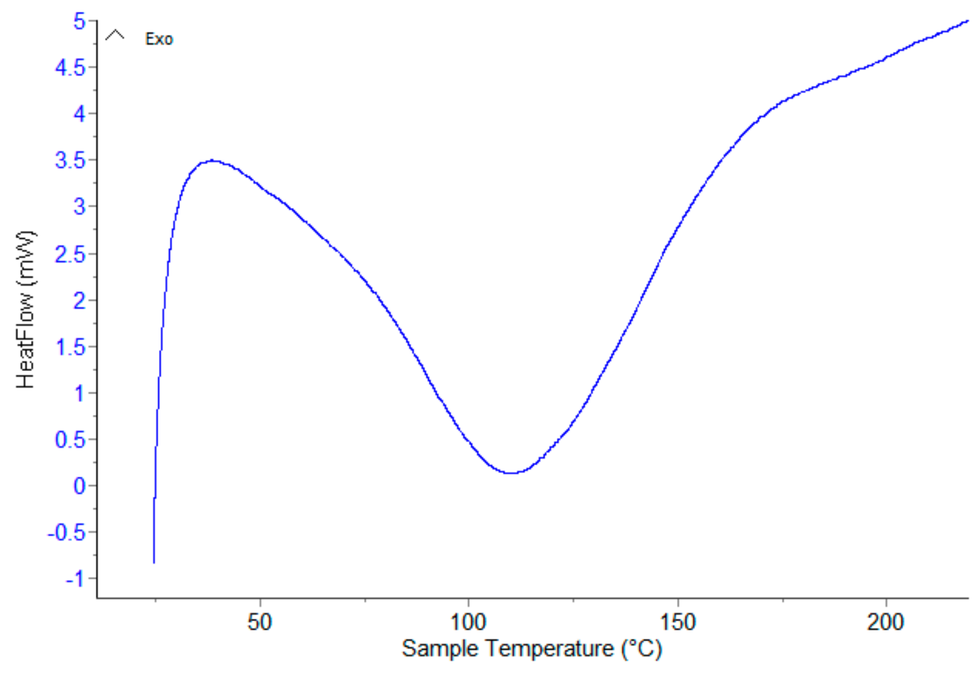
The DSC curve of HP-β-CD (
Figure A3) showed a large endothermal peak associated with water loss at 109.3 °C. In the 30–210 °C temperature range, no other peaks appear in the DSC curve of HP-β-CD.
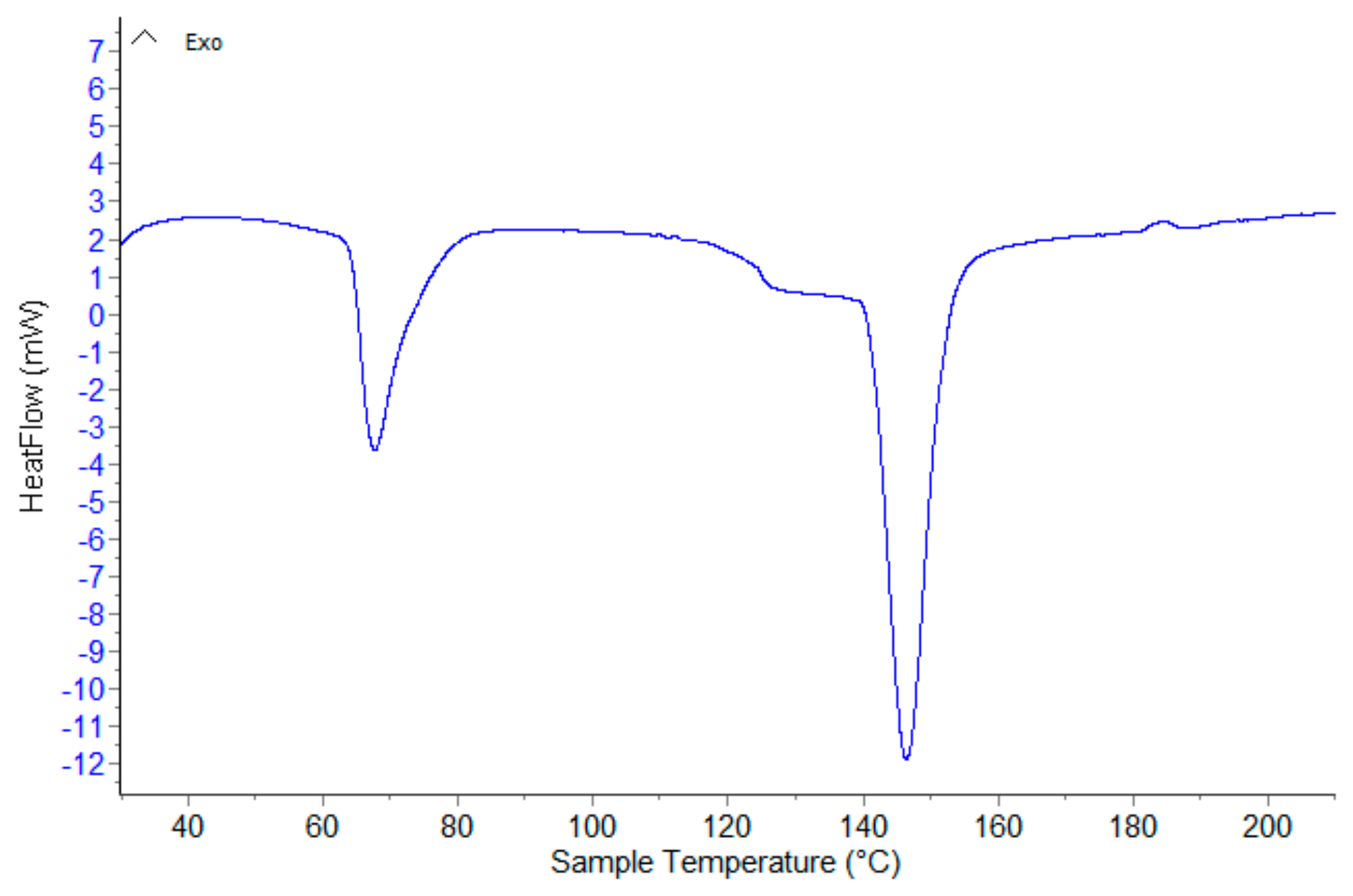
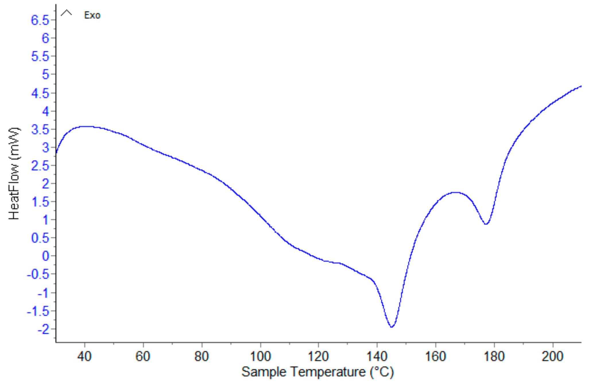
The DSC curves of the two physical mixtures were similar to the individual DSC curves of pure compounds. For RUT-β-CD physical mixture (
Figure A4), the melting peak of RUT appears at a lower temperature (146.4 °C). The melting endotherm of RUT is attenuated in this case. This is proof that a consistent amount of RUT is included in the β-CD, even in a mixing procedure. From
Table 2, it was observed that the peak attributed to the water molecules was at 67.7 °C lower than the parent β-CD, suggesting that the water molecules from the β-CD cavity are replaced by RUT molecules. The small differences in the peak temperatures of RUT-HP-β-CD observed from the DSC curves (
Figure A5) is proof that the complexation process did not occur in the physical mixtures.
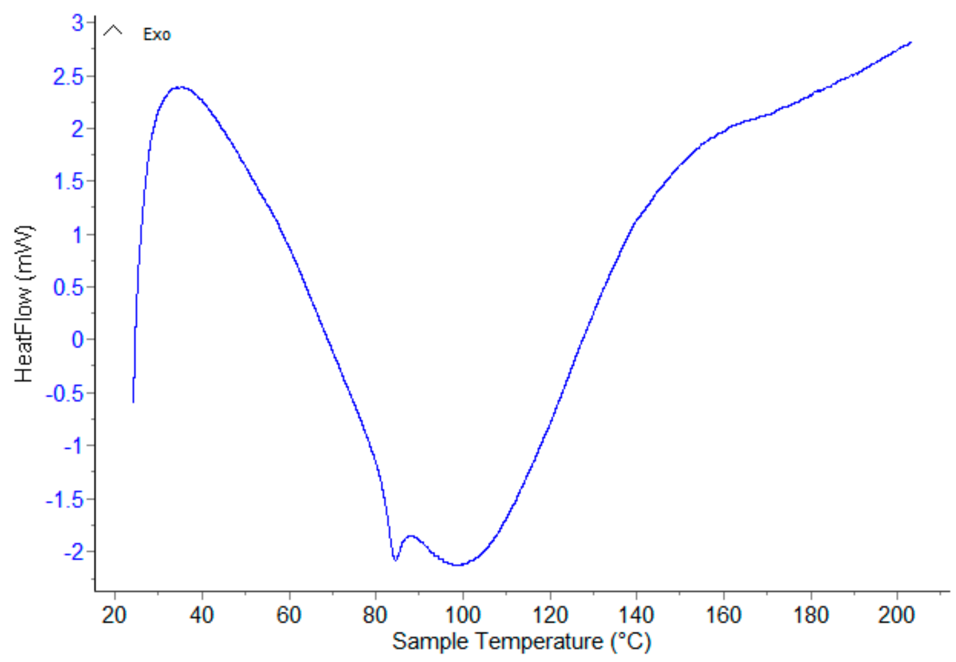
A difference pattern was observed in the DSC curves of the inclusion complexes. In the RUT-β-CD inclusion complex (
Figure A6), the melting point of RUT is still present but is shifted to a higher temperature (178.3 °C). The decrease of the sharpness and the intensity of the characteristic endothermic peak of RUT indicate a conversion of the crystalline RUT into an amorphous form, due to the lyophilization process. The DSC curve of the RUT-HP-β-CD inclusion compound (
Figure A7) shows the disappearance of the RUT melting point, suggesting in this case the formation of an inclusion complex. The dehydration process of the inclusion complex appears at a lower temperature than physical mixtures and the parent β-CD. The DSC thermal feature of the two compounds (RUT and HP- β-CD) practically disappears in the DSC curve of the inclusion compound, a clear suggestion of a new compound. The shift at a higher temperature in the case of RUT-β-CD complex or the disappearance for RUT-HP-β-CD complex of the RUT melting point is proof that the RUT molecule is included in β-CD or HP-β-CD cavities, the inclusion process being complete in the complexes.
3.3. Antioxidant Activity of the Complexes
3.3.1. DPPH Radical Scavenging Activity
Table 3 and
Figure 4 show the percentage decrease of the optical density (% radical scavenging activities) of samples and vitamin C used as a positive control.
All the studied samples exhibit an antioxidant activity, with linear increases in time. It is considered that the ascorbic acid presents strong antioxidative activities, but according to the registered results, rutoside and its inclusion compounds showed a two times better effectiveness than the highest concentration of positive control. After 40 min, RUT exhibited a relatively high DPPH scavenging activity (45.78%) [
44]. Both physical mixtures (47.37% for RUT-β-CD physical mixture and 48.24% for RUT-HP-β-CD physical mixture) showed relatively similar reducing power activity as RUT. However, the inclusion complexes presented the highest inhibition of oxidation, having an excellent electron donating ability, revealing a strong antioxidant activity (50.20% for RUT-β-CD inclusion complex and 54.79% for RUT-HP-β-CD inclusion complex), probably due to the increase of rutoside solubility by complexation with cyclodextrins. Still, it can be concluded that the RUT-HP-β-CD inclusion complex had the best behavior, proving the highest DPPH radical scavenging activity.
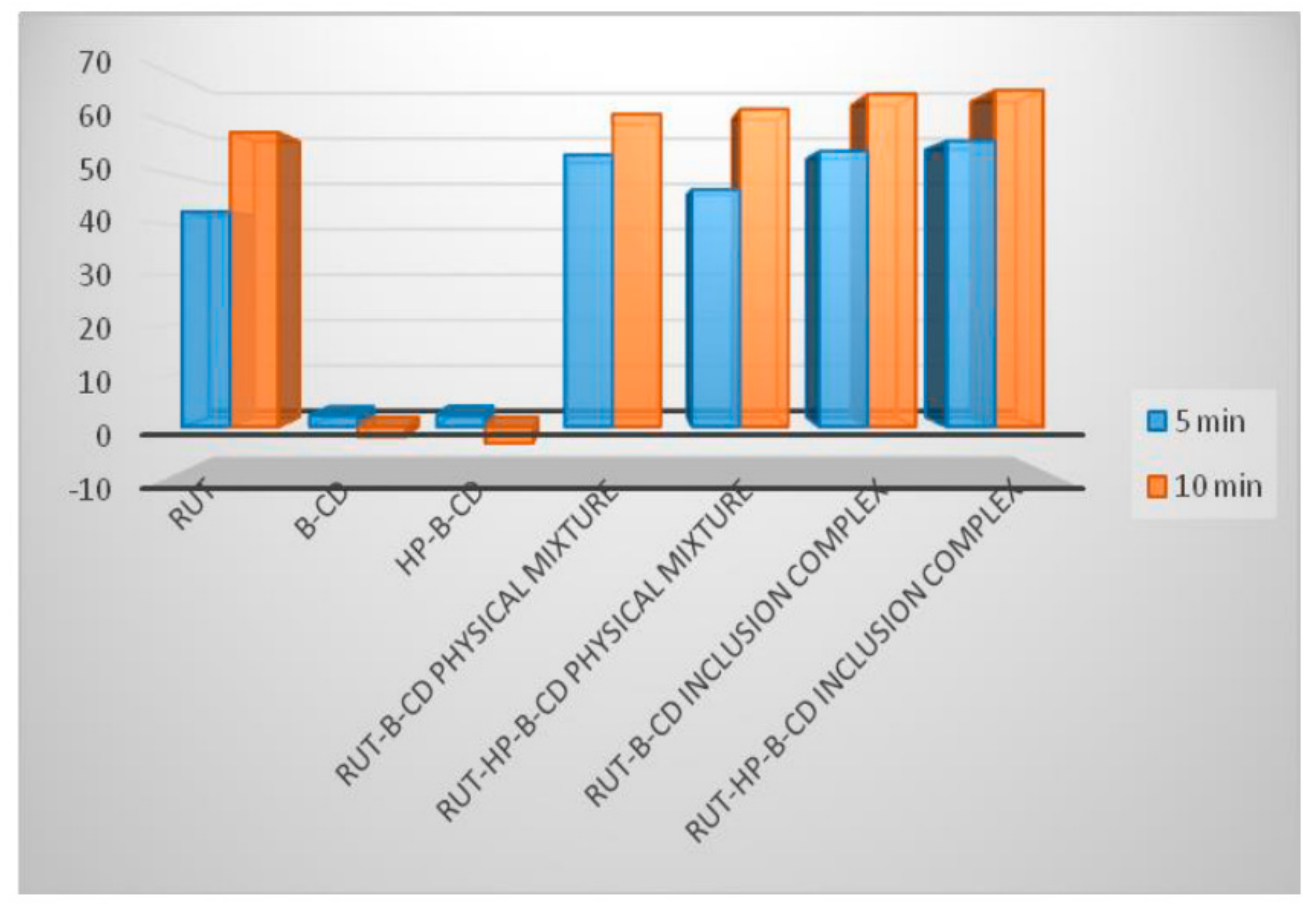
3.3.2. ABTS Radical Scavenging Activity
Table 4 and
Figure 5 present the percentage decrease of the optical density (% radical scavenging activities) displayed by the samples, at time of 5 and 10 min.
It has been demonstrated that the ABTS assay is more sensitive in identifying antioxidant activity because of the faster reaction kinetics, and its response to antioxidants is higher. The results proved that both cyclodextrins, β-CD and HP-β-CD, do not possess antioxidant capacities, but they have a significant influence on the ABTS radical scavenging activity of rutoside. The
in vitro antioxidant assay of RUT revealed its antioxidant potential (42.41% RSA at 5 min and 58.08 at 10 min), which is considerably increased when it is associated with any of the two CDs [
45]. Another remark evidenced in
Figure 4 is that also the physical mixtures performed better than RUT alone in scavenging the ABTS
• + radical cation. As can be noticed, the most effective inhibition is provided by RUT inclusion complexes, with β-CD and HP-β-CD being the most active samples after 10 min (65.60% for RUT-β-CD inclusion complex and 66.37% for RUT-HP-β-CD inclusion complex). From the ABTS assay, it can be considered that the inclusion complexes are free radical scavengers of fast action.
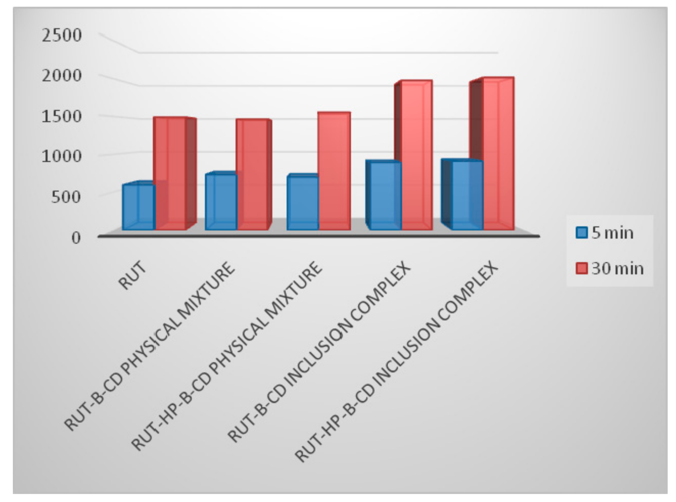
3.3.3. FRAP Assay
Table 5 and
Figure 6 show the FRAP values determined at a time of 5 and 30 min.
Table 5 presents the FRAP values (µmol equivalent of Fe
2+ (FeSO
4) /L), calculated using the Fe
2+ calibration curves in methanol.
The results provided by the FRAP assay strengthen the conclusions obtained by the other two tests that the complexation process produces a significant increase of rutoside antioxidant activity, slightly higher for RUT-HP-β-CD than RUT-β-CD inclusion complex [
46]. The complexes also presented a higher reducing ability (around 20% higher) after 5, and even after 30 min, revealing an improved antioxidant efficacy.
3.4. Precompression Studies for Tablets Containing RUT-β-CD and RUT-HP-β-CD Inclusion Complexes
Different studies were performed on the bulk powder for direct compression, in order to establish the behavior of the material during the compression process and to choose the adequate compression parameters (
Table 6).
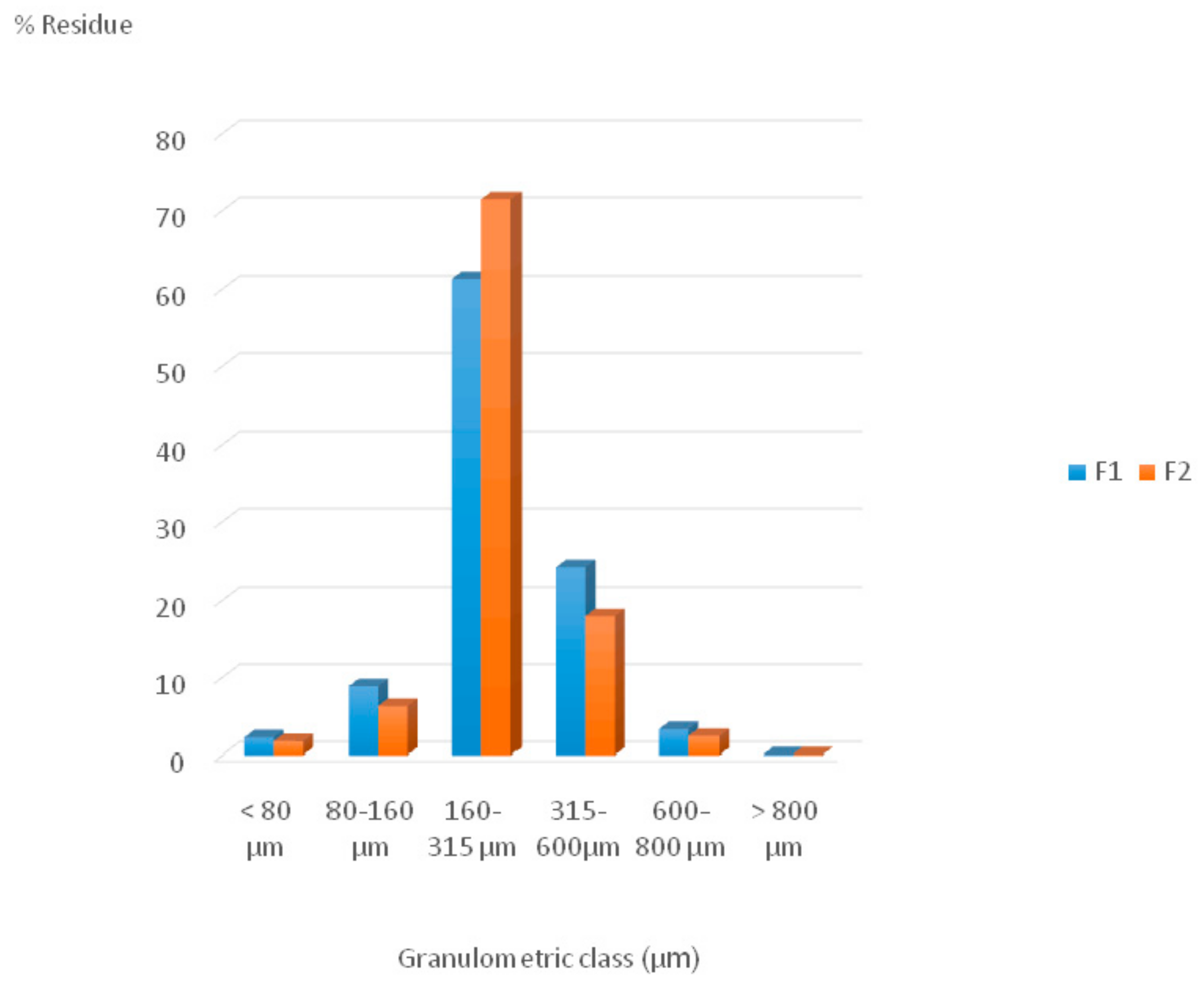
Concerning the particles dimensions, the histogram exhibited in
Figure 7 was obtained by representing the distribution of particle size on granulometric classes for both studied formulations.
Figure 7 shows that both compression powders contain a considerable part of the particles with a median particle size between 160 and 600 μm, with most of them fitting in the smaller fraction on 160–315 μm.
The values of the flow time, angle of repose, and flow rate, obtained after 5 determinations on 60 g of each powder, with a 15 rpm stirring rate, on a 10-mm nozzle, are very similar for both formulations. The flowability could not be measured using the 10-mm nozzle without stirring or with a 5-rpm and 10-rpm speed rate, because the powders were not flowing steadily and consistently under these conditions.
Flowing time was 10.4 s for F1 and 11.1 s for F2, the angle of repose around 29° for both formulations, and the flowing rate was 5.741 g/s for F1 and 5.382 g/s for F2, values that indicate an excellent flow, but considering the fact that stirring was needed, the powders have a rather poor to moderate free flow. The results were practically the same for both studied materials, leading to the idea that the type of cyclodextrin does not have such a great influence on the flowability of the compression blend.
F1 had lower bulk and tap densities as compared to
F2.
F2 presented a lower value for CI (13.6) and HR (1.15) than
F1 (18.2 and 1.21), therefore it is considered to have a very good compressibility and a good flow. Meanwhile,
F1 is rated as having a ‘fair’ flow and a good compressibility [
47]. In terms of HR and CI, both formulations have a required free flow and good compressive properties.
Both formulations displayed similar moister contents of 3.90% (F1) and 3.66 (F2), low values which are considered ideal for preprocessed powders for tablets.
Making the correlations between all the physical parameters determined for the tableting mixtures, it can affirm that smaller values of CI and HR for
F2 are due to the broader particle-size distribution for a smaller particle-size fraction (160–315 µm), to an increased bulk and tapped densities, and, also, to a lower moisture content [
48]. Considering these remarks, it can be predicted that
F2 will have a better compression behavior compared to
F1, but the differences between the two formulations are not too significant. It is obvious that in both bulk and tapped, densities increased with decreasing particle size independent of the used CD type. A linear relationship between the mean particle size, moisture content, volumetric properties, flowability, and compressibility of the powders can be observed, with all being characteristic for smooth fluid powders. The results registered for the pharmaco-technical properties of the direct compression mixtures suggest that the two lubricants (magnesium stearate and talc) were well selected, having a good influence on the powder’s flowability.
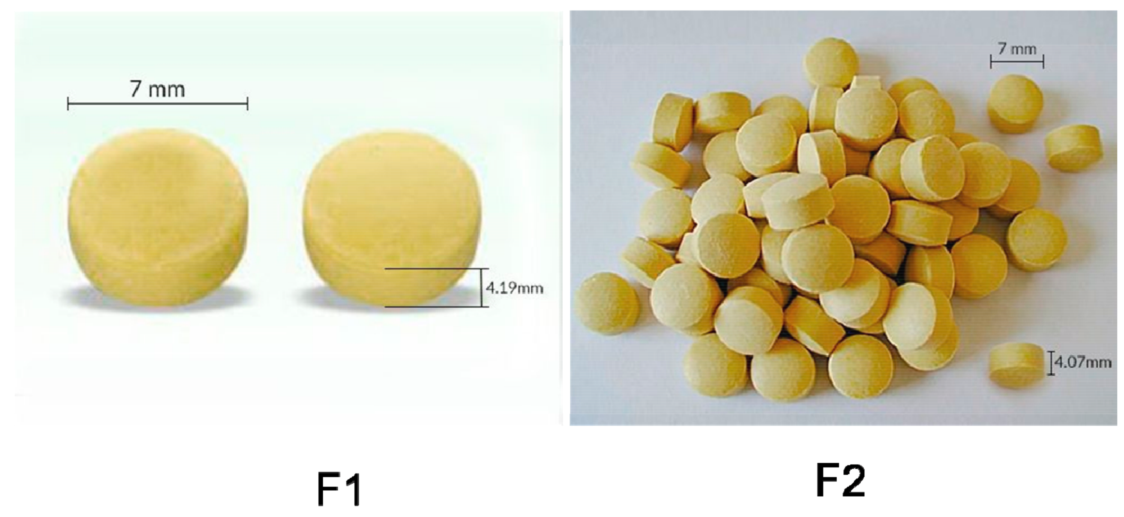
3.5. Quality Parameters of the Tablets
The obtained tablets quality performances were characterized by different physical and mechanical properties and the results of the tests are presented in
Table 7 and the digital images of the tablets (
F1 and
F2) are shown in
Figure 8.
Based on the above results, the average thickness of tablets is 4.19 SD ± 0.22 for F1 and 4.07 SD ± 0.09 for F2, indicating a uniformity between the two formulations and, also, between the tablets from the same lot. In the case of diameter, the differences are even smaller, showing the tablets of both formulations comply with the pharmacopoeial standard. The uniformity in dimensional properties of the tablets proves that the compression parameters were correctly chosen and no undesirable phenomenon occurred during the process.
Regarding the mass uniformity, the average weight of both formulations (197 SD ± 3.20 for F1 and 199 SD ± 2.88 for F2) is in accordance with the pharmacopoeial criteria, proving that the materials had a good flowability, which generated a uniform filling of the mold, ensuring the consistency of dosage units during compression.
Both formulations present a good mechanical resistance (69.07 SD ± 2.63 for
F1 and 71.20 SD ± 2.18 for
F2) and an excellent friability (0.34 SD ± 0.17 for
F1 and 0.41 SD ± 0.06), leading to the conclusion that the tablets withstand mechanical shocks during the manufacturing and handling. With friability being closely related to tablet hardness, it is obvious that the studied tablets have the ability to avoid fracture and breaking. This demonstrates that the ingredients and the compression force were properly chosen [
48,
49].
The disintegration time was below 4 min for both formulations (228 SD ± 2 for
F1 and 216 SD ± 3 for
F2), indicating a short period of time is needed for the disintegration process of the tablets. The fast disintegration was ensured by the use of Polyplasdone XL-10, the chosen superdisintegrant in both formulations and also by the two cyclodextrins, which increased rutoside’s solubility in water. Both formulations meet the imposed pharmacopoeia limits for immediate-release dosage forms [
50,
51].
The results obtained for the tested characteristics prove that high-quality tablets including the RUT-β-CD and RUT-HP-β-CD inclusion complexes as active ingredients were developed. Both formulations’ pharmaco-technical attributes were satisfactory and within the limits imposed by rules inforce. They show a good mechanical resistance with a low friability, qualities that ensure the tablets’ integrity, and excellent disintegration times, being able to consider them rapidly disintegrating tablets. As the results are very similar for both formulations, it can be admitted that the physical and mechanical properties of the tablets are not influenced by the type of used cyclodextrin.
The dissolved quantity of rutoside rapidly increased within 30 min. The registered dissolution rates after 30 min are characteristic for highly soluble ingredients, and based on the fact that rutoside is a poorly water-soluble drug, the conclusion is that by encapsulating it in the cavity of both CDs, its solubility is significantly enhanced [
51]. The RUT-HP-β-CD inclusion complex tablets showed a higher dissolution rate than RUT-β-CD tablets. Beside the influence of CDs on the dissolution profiles of the tablets, probably, a very important role isalso the used method of direct compression, the direct compression excipient Avicel PH 102, and the superdisintegrant Polyplasdone XL-10.
4. Conclusions
In summary, two inclusion complexes in a 1:1 molar ratio, (i) one of rutoside with β-cyclodextrin (RUT-β-CD) and (ii) another one of rutoside with hydroxypropyl-β-cyclodextrin (RUT-HT-β-CD), using the lyophilization method, were successfully prepared. FT-IR spectra showed obvious differences in the positions and intensities in the ranges of 500–1150 cm−1 in both inclusion complexes, but their spectra were similar with the ones recorded for pure cyclodextrines, strengthening the idea that rutoside was successfully included in their cavity. The morphologies, evaluated by SEM analysis, revealed important changes in the structure and shape of the binary lyophilized systems, proving that new compounds were formed. The recorded DSC curves showed that the complexation was complete in case of RUT-HP-β-CD, and only partial in RUT-β-CD complex. From the antioxidant activities, it was noticed that the two inclusion complexes had a remarkably improved antioxidant activity than rutoside alone or its simple physical mixtures with each CD. Thus, the encapsulation of rutoside in the CDs cavity leads to important increases of its antioxidant properties, probably due to its improved solubility. The physico-chemical and mechanical properties of the solid dosage pharmaceutical forms (F1 and F2) were evaluated in the preformulation studies, the results proving that the new two obtained materials are suitable for the direct compression process. Both drug delivery systems presented a good mechanical resistance with a low friability, and a weight variation within the compendial limits. A rapid disintegration was registered for both series of tables and it can be considered that the main purpose of this study was completely achieved, as the disintegration is the critical quality attribute for tablets. The dissolution rates were satisfying and within the imposed pharmacopeial limits for both formulations. As the performances were very similar for both formulations, the conclusion is that the pharmaco-technical properties of the tablets are not influenced by the type of the used cyclodextrin, but the RUT-HP-β-CD tablets present a higher dissolution rate.


 ,
, 


