
Preparation and Characteristics of Alginate Microparticles for Food, Pharmaceutical and Cosmetic Applications


Faculty of Chemical Engineering and Technology, Institute of Organic Chemistry and Technology, Cracow University of Technology, 31-155 Krakow, Poland
*
Author to whom correspondence should be addressed.
Polymers 2022, 14(18), 3834; https://doi.org/10.3390/polym14183834
Submission received: 23 August 2022
/
Revised: 8 September 2022
/
Accepted: 11 September 2022
/
Published: 14 September 2022
(This article belongs to the Special Issue Research Progress in Polymer Hydrogels)
Abstract
:Alginates are the most widely used natural polymers in the pharmaceutical, food and cosmetic industries. Usually, they are applied as a thickening, gel-forming and stabilizing agent. Moreover, the alginate-based formulations such as matrices, membranes, nanospheres or microcapsules are often used as delivery systems. Alginate microparticles (AMP) are biocompatible, biodegradable and nontoxic carriers, applied to encapsulate hydrophilic active substances, including probiotics. Here, we report the methods most frequently used for AMP production and encapsulation of different actives. The technological parameters important in the process of AMP preparation, such as alginate concentration, the type and concentration of other reagents (cross-linking agents, oils, emulsifiers and pH regulators), agitation speed or cross-linking time, are reviewed. Furthermore, the advantages and disadvantages of alginate microparticles as delivery systems are discussed, and an overview of the active ingredients enclosed in the alginate carriers are presented.
1. Introduction
“Microparticle” is the term used for spherical particles with diameters in the micrometer range, typically from 1 to 1000 µm. Polymeric microparticles are usually formed by a polymer matrix in which a smaller amount of an active compound could be immobilized [1]. Generally, taking into account the method of microparticle preparation, their morphology, and the distribution of the encapsulated actives, microparticles can be divided into two categories: “microspheres” and “microcapsules” [1,2,3,4].
Microspheres usually are characterized as matrix systems in which the active substance is homogeneously dispersed. In contrast, microcapsules are heterogenous particles where a membrane shell surrounds the core (solid or liquid) and forms a reservoir with an encapsulated active compound [1,5,6]. In some cases, to overcome problems with the mechanical stability of such, the carriers and low-actives-loading, polymer-coated microspheres are obtained [7,8].
Compared with other effective carriers of the active compounds, such as nanoparticles, the advantage of the microcarriers is that they do not penetrate into the interstitium, and thus act locally [5,9]. The choice of microcapsules over nanocapsules in the case of cosmetic products can be an ideal solution for epidermal action, where the encapsulated ingredient is designed to act on the top of the epidermis. Moreover, nanocapsules are not suitable, due to their small size, to encapsulate active ingredients with larger than nano sizes, including bacteria.
The microparticles can be prepared from a large variety of starting materials, both natural and synthetic origin, and with different preparation techniques. Most drug delivery systems are prepared using natural polymers, such as polysaccharides (e.g., starch, dextran, alginate, hyaluronic acid and chitosan) or proteins (e.g., collagen, gelatin and albumin) [10,11]. Among synthetic polymers applied to obtain the carriers, there are poly(lactide-co-glycolide) (PLGA), (3-hydroxybutyrate-co-3-hydroxyvalerate) (PHBV), poly(sebacic anhydride) and poly(ε-caprolactone) [2]. Moreover, thermosensitive polymers, e.g., poly(N-isopropyacrylamide) (PNIPAAm) [12], and pH-sensitive polymers (Eudragit L100 and Eudragit S100) are used [13,14].
Among the natural raw materials used to obtain microparticles, alginates are the most popular and deserve special attention. In the food industry, alginate-based formulations are used as texture modifiers [15,16,17] or to improve the stability and long-term efficacy of active compounds [18,19,20]. In the case of the cosmetic and pharmaceutic industries, alginate systems are used to improve stability and to protect the encapsulated compounds against external conditions, e.g., UV light, temperature [21,22,23], or gastric environments in the case of oral application [24,25,26,27]. The alginate microparticles (AMP) are biocompatible, biodegradable and nontoxic delivery systems allowed to encapsulate different active substances, including probiotics [28,29,30].
The objective of this article was to provide a review of the techniques used for AMP production. The most popular alginate encapsulation methods, such as the spray-drying technique, extrusion and emulsification are described. The technological parameters important in the microencapsulation process and their impact on the quality of the alginate carriers are discussed. Particular attention was paid to the microencapsulation of alginates based on the emulsification technique, as the process allows to obtain small microparticles, containing both hydrophilic and hydrophobic ingredients with high efficiency. Moreover, the examples of the actives enclosed in the AMP, and their application in the food, pharmaceutical and cosmetic industries, are presented. Most scientific articles focus on applications of AMP in the food and pharmaceutical industries [28,31,32,33]. Recently, the environmental application of alginate carriers has also appeared more often [34]. However, the cosmetics industry has not been the subject of wide scientific interest so far.
2. Alginate Characteristics
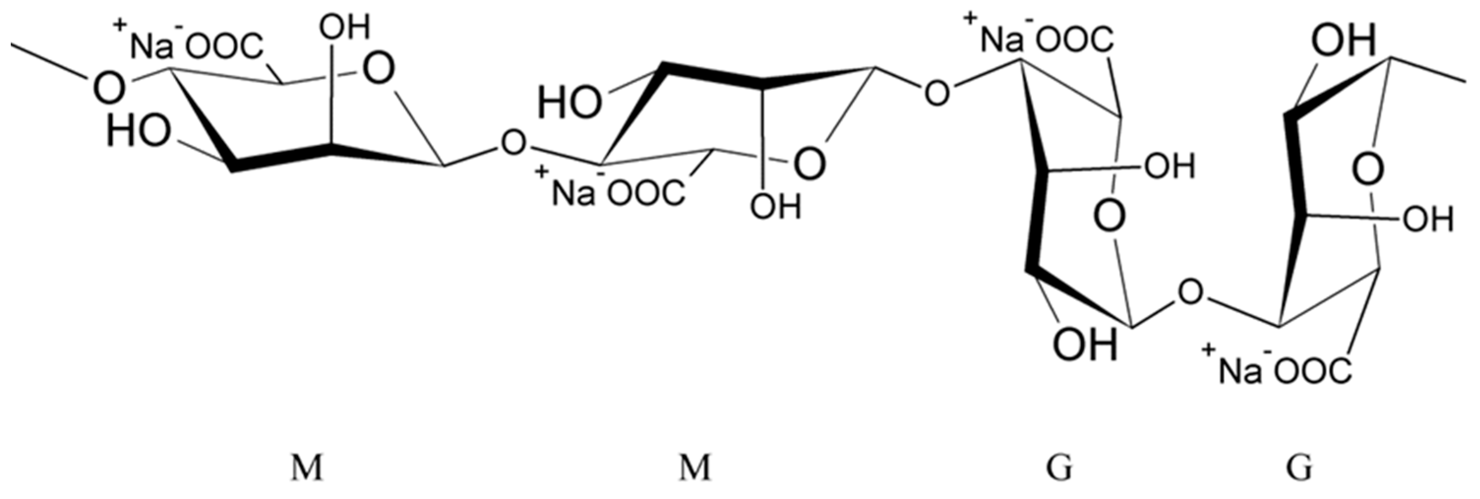
Alginates are natural polysaccharides, polyanionic polymers, obtained from marine algae [35,36], usually from three species of marine brown algae, Phaeophyceae: Macrocystis pyrifera, Laminaria digitata and Laminaria saccharina. The structure of alginates depends on the source of the sea algae, i.e., its species, geographic origin, or seasonal varieties [37]. Alginates are unbranched copolymers of D-Mannuronic acid (M block) and L-Guluronic acid (G block) linked by β (1–4) glycosidic bonds [37,38,39,40,41,42,43]. These blocks are arranged in an irregular block pattern with different GG, MM and MG block proportions (Figure 1) [30,35,37,38,42]. Blocks M and G are placed in the different locations along the chain (e.g., MMMM, GGGG, MMGG, GMGM) and in various amounts [38].
Cross-Linking of Alginates
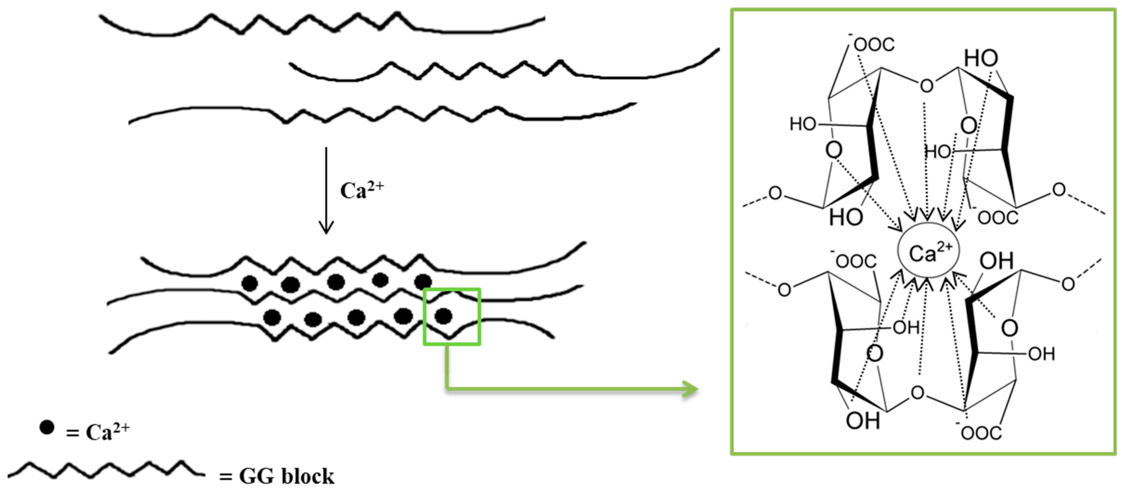
Monovalent metal ions with alginates form soluble salts, while divalent and multivalent cations (except Mg2+) form gels or precipitate. The affinity of alginates to various cations and selective ion binding are the basis of the alginates’ ability to form hydrogels. Alginates containing a large number of guluronic acid blocks form gels with much greater strength compared with mannuronate-rich alginates because G blocks have a stronger affinity with divalent ions than M blocks [35]. The interaction of alginates with divalent cations, which are in particular calcium cations, leads to the formation of biodegradable gels [38]. Polymerization is based on the cross-linking of copolymers through ionic bonds between Ca2+ cations and alginate anions [38,44]. Each Na+ cation ionically binds to only one carboxyl group of the alginate chain, while the Ca2+ cation interacts with two carboxyl groups that come from different polymer chains. The exchange of Na+ ions into Ca2+ ions is relatively easy and takes place when an aqueous solution of sodium alginate is mixed with a solution containing calcium ions [41]. The mechanical properties of the formed gels are determined by both the concentration of divalent ions and the alginic acid salts in the reaction mixture [38,44]. The distinctive molecular structure resulting from these interactions is referred to as the “egg–box” model (Figure 2) [38,44,45,46]. It should be underlined that only the G blocks take part in the cross-linking process. The homopolymers of G blocks form ordered, three-dimensional regions wherein Ca2+ ions are embedded like eggs in a cardboard box [47].
The immediate cross-linking of alginates, due to the action of calcium ions, causes the formation of particles of different diameters and different porosity [48]. The gel strength increases with the increase in G blocks content. Moreover, the parameters of the cross-linking process are important. Temperatures ranging from 60 to 80 °C are needed to dissolve alginates in the water. Moreover, it is known that alginate gels are insoluble in acidic environments [49]. Additionally, the technique used to obtain alginate particles influences the capsule size, which can range in size from nanoparticles to macroparticles. Macroparticles are particles larger than 1000 microns in size and are easily visible [50,51]. They are applied especially in the field of dietary supplements [52] or drugs [53,54,55,56]. The size of microparticles ranges from 100 nanometres to 1000 μm [57,58,59], and particles with a diameter below 100 nanometres are classified as nanoparticles [60].
3. Alginate Encapsulation Techniques
- (a)
- Physical methods such as spray drying, extrusion, lyophilization, supercritical fluid precipitation and solvent evaporation;
- (b)
- Physico-chemical methods including coacervation, liposomes and ionic gelation;
- (c)
- Chemical methods such as interfacial polymerization and molecular inclusion complexation.
In the case of alginate capsules preparation, the most frequently used methods are the spray-drying technique and either extrusion or emulsification/gelation [61].
3.1. Spray-Drying Technique
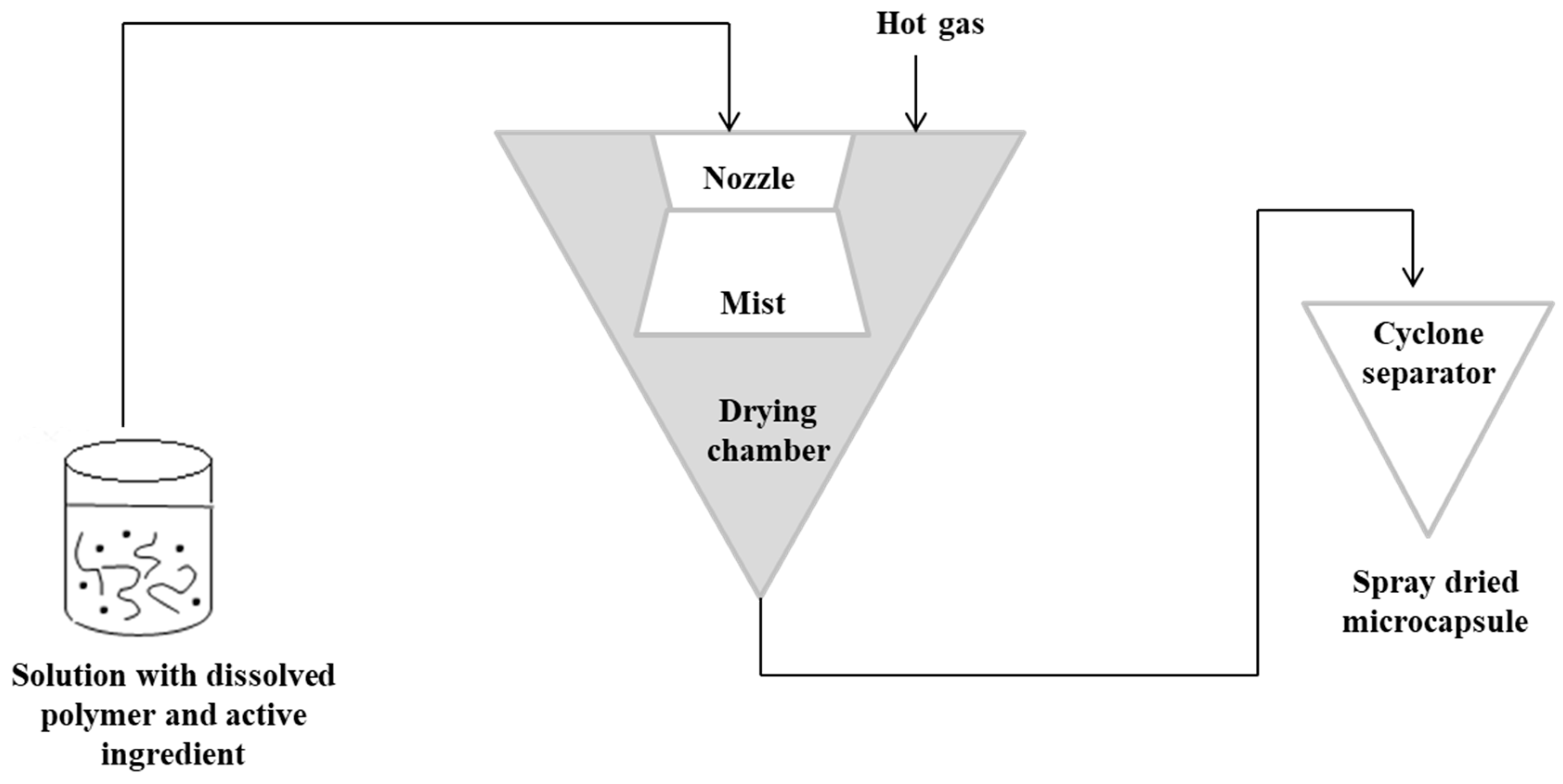
The spray-drying technique is the oldest technique used in alginate capsules. The concept of this method was first described in Samuel Percy’s patent in 1872 [64]. The process was introduced commercially in the 1920s, but spray drying was fully applied on a large scale in the early 1980s [65,66]. In this method, initially, a solution containing the active ingredient should be prepared with the dissolved polymer matrix. The solution is then pressurized and sprayed to form a “mist” in the drying chamber. Hot gas (air or nitrogen) is also blown into the drying chamber. The hot gas makes it possible to evaporate the solvent. The capsules are then transported to a cyclone separator for recovery [3]. In this technique, the control of the product’s feed, gas flow and temperature is important [67,68]. Figure 3 shows a schematic representation of the spray-drying procedure [3,63].
The spray-drying technique is applicable to encapsulate different kind of actives. Among substances encapsulated in alginate capsules, there were lipids [69] and hydrocarbons such as cellulase [70], carvacrol [71], or insulin [72]. Unfortunately, the main disadvantage of this method is the impossibility to encapsulate substances sensitive to elevated temperature and pressure.
3.2. Extrusion Technique
Another technique that has been used for a long time for producing microparticles is the extrusion technique [73]. It consists of the preparation of a hydrocolloid solution (an aqueous dispersion of a natural or synthetic polymer), e.g., sodium alginate. Then, the material for encapsulation is added to the solution, and the suspension is pressed into droplets into the gelling or hardener solution, e.g., calcium chloride [3,30,39,49,67,74,75,76,77]. Depending on the scale (laboratory or pilot scale), a syringe needle or an extruder is used for the process, respectively [39]. The size and shape of the obtained beads depend on factors such as the diameter of the needle and the distance of the needle from the hardener solution. This technique is the most popular in laboratory scale because of simplicity and low cost of production [78]. However, the main limitation in the case of its application in the large scale is the fact that microparticles are formed slowly [30].
3.3. Emulsification Technique
An alternative method commonly used to obtain alginate capsules is emulsification. The emulsification technique is more expensive than the extrusion method because a large amount of oil is needed to prepare the emulsion [79]. In this technique, the discontinuous phase (mixture of hydrocolloid and encapsulated material) is added to a large volume of continuous oil phase oil [3,30,39,46,49,67]. In the case of food applications, vegetable oils are used as a continuous phase, most often canola, sunflower or corn oils [78,80]. Additionally, to stabilize the droplets of the internal phase, an emulsifier should be added to the mixture formed. The obtained water-in-oil emulsion is continuously homogenized by stirring [3,30,39,49,67]. The speed of the emulsion mixing is a critical step because it affects both the shape and size of the capsules formed [81]. Very large capsules (around 1000 μm in diameter or larger) can result in poorly coated structures and a coarse texture [82], which in turn affects the dispersion quality of the capsules in the final product [79]. The final emulsion containing alginate droplets is broken by adding calcium chloride solution and centrifuged, thereby separating the oil and water phases to obtain microspheres [83,84]. Figure 4 shows a diagram of the encapsulation process using extrusion and emulsification techniques [46,67].
The main difference between the two techniques is the sequence of combining the alginate and calcium chloride solutions. The polymer solution containing the active ingredient is introduced into the calcium chloride solution in the case of the extrusion technique as opposed to the emulsification technique, where the CaCl2 solution is dripped into the polymer solution. Depending on the method used to obtain them, the calcium alginate beads are called balls (gelled droplets obtained by extrusion technique) or capsules, in the case of the products of the emulsification technique [85]. The main differences between these alginate beads are listed below [49,78,85,86]:
- The balls have a porous network, while the capsules have a liquid core (water or oil);
- The dimensions of the balls are much larger than in the case of the capsules;
- The balls are uniform in size and shape in contrast to differing in size in capsules.
The differences in the morphology (structure) of both the microparticle types are perfectly illustrated in images obtained by electron microscopy TEM and SEM [87].
To sum up, some advantages and disadvantages of the above-presented methods of AMP preparation are summarized in Table 1.
The data presented in Table 1 indicate that the currently used alginate encapsulation techniques are not universal. All methods have some pros and cons, but among the others, the emulsification is more universal. The method allows to obtain small microparticles, containing both hydrophilic and hydrophobic ingredients with high process efficiency. Moreover, the microencapsulation of alginates based on the emulsification technique could be modified by differing the polymer gelling process.
4. Microencapsulation of Alginates Based on Emulsification Technique
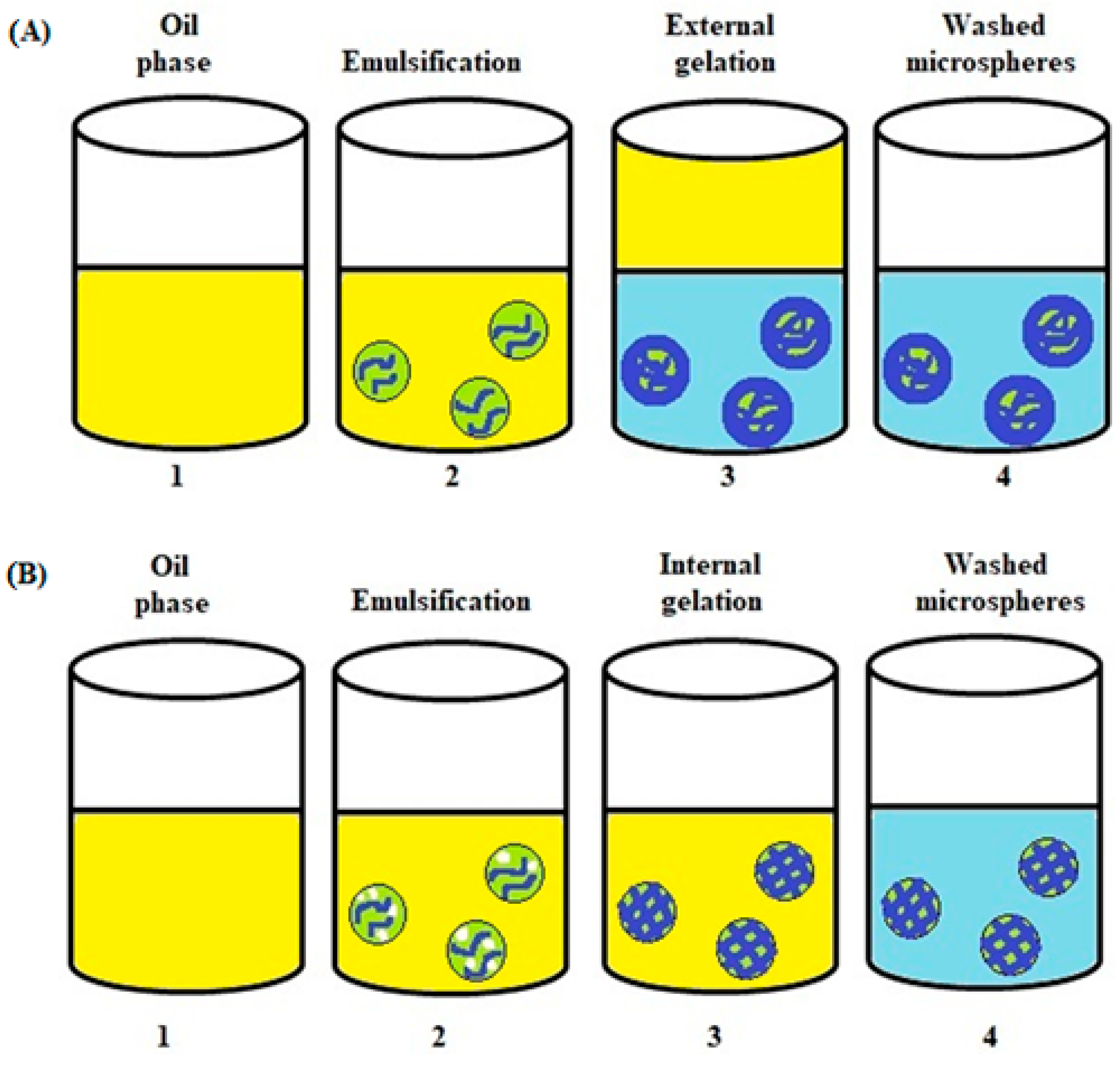
The emulsification process is based on the mechanism of external or internal gelling. Both water-in-oil (W/O) and oil-in-water (O/W) emulsions could be used to prepare microparticles. Depending on the gelling system used, the obtained microparticles can have different properties and thus different applications. The properties differentiating the external and internal gelling are: pore size, stiffness, permeability and strength of the matrix. Alginate microspheres prepared with external gelling have smaller pores and a denser surface structure than inside the capsule. In turn, microspheres obtained by internal gelling have a more homogeneous structure, which is associated with a more uniform distribution of cations in the particle [91,92,93,94].
4.1. Alginate Microspheres Formed in a W/O Emulsion—External Gelation
In the external gelation emulsification method, an alginate solution containing the encapsulated active substance is emulsified in the oil phase to form a W/O emulsion. Next, a cross-linking compound, e.g., calcium chloride solution, is gradually added to the emulsion [42,95,96,97]. The addition of CaCl2 solution causes the droplets of the alginate emulsion to gel and form microspheres [4,98]. The calcium chloride particles in the solution migrate according to the principle of diffusion to the phase interface of the droplets of the alginate emulsion (the dispersed phase), where they dissolve in the aqueous phase and cause gelation to form the microspheres [98]. This step is followed with filtration and rinsing to remove residual oil. In this method, the obtained microspheres consist of a soft core and a rigid outer matrix [29,99].
4.2. Alginate Microspheres Formed in a W/O Emulsion—Internal Gelation
In an internal gelation process, insoluble calcium crystals (e.g., calcium carbonate) are dispersed in an aqueous polysaccharide solution and serve as an internal calcium source. This mixture is emulsified in an oil phase containing a surfactant. Upon pH reduction, from 7.5 to 6.5, calcium (Ca2+) is released from the calcium complex causing gelation to form calcium alginate. To lower the pH, usually an oil-soluble organic acid such as acetic acid is gently added to the oil, causing immediate diffusion into the water phase. In this way, the immediate lowering of the pH of the dispersed droplet, solubilizing the crystalline calcium, causes rapid gelling [4,29,98,100].
Considering the gelation process, two successive reactions follow after diffusion of the oil-soluble acid through the oil–water interface. Protons that diffuse into the aqueous gel phase encounter evenly spaced calcium microcrystals, usually CaCO3, which causes the immediate release of calcium ions (1) and in situ gelling of the alginate, creating a homogenous gel network (2). Below is the course of the reactions taking place [29,100]:
2H+ + CaCO3 → Ca2+ + H2O + CO2
Ca2+ + Na+G → Ca2+G + Na+
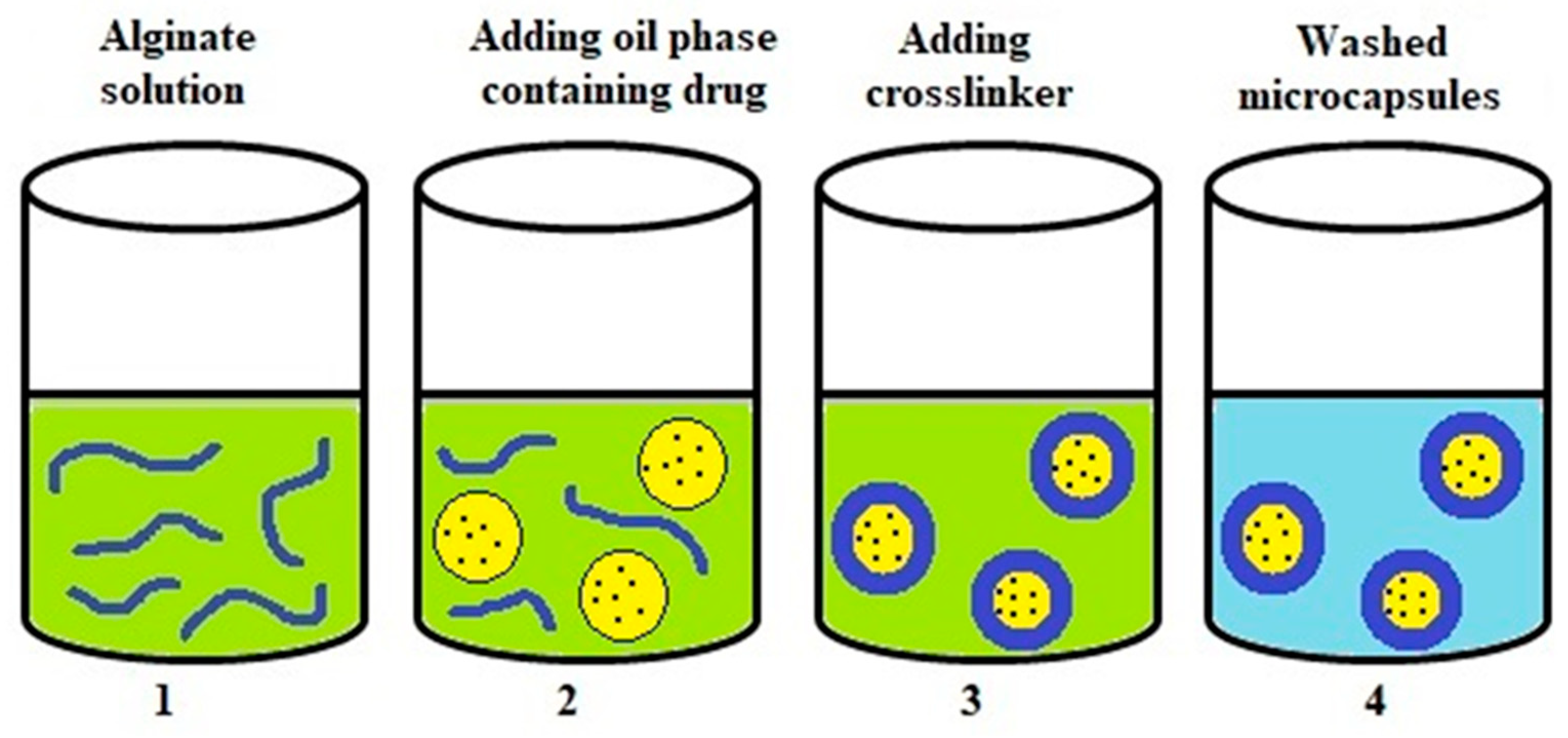
4.3. Gelation at the Interfacial Area of Emulsion (Complexation)
One method of forming an emulsion is to deposit the polymer at the interface of the emulsion droplets and then remove the solvent used [102]. The polymer coating that deposits the polymer at the interface is stabilized with covalent or physical intermolecular forces due to the addition of a cross-linking agent. The scheme of creating microcapsules in this way is shown in Figure 6 [4,29].
In this method, microcapsules are synthesized in an aqueous solution and at the interface of oil drops. The preparation of alginate microcapsules is accomplished by mixing the encapsulated component (usually hydrophobic drugs) with an organic solvent such as ethanol or acetone to form the inner phase (oil phase) of the microcapsules [103]. The previously prepared mixture is added to the alginate solution containing, among others, Tween 80 as a surfactant. It is then subjected to homogenization or sonication to produce an O/W emulsion. The calcium chloride solution is slowly added to the formed emulsion, which causes the alginate to cross-link and form microcapsules. The core of the so-prepared microcapsules consists of an oily liquid surrounded by a single polymer layer. The microcapsule suspension is equilibrated for several hours. Finally, the solvent is removed under reduced pressure and the microcapsules formed are washed and isolated [42,104,105]. Capsules of different row sizes can be obtained with this method. It has been used for encapsulating, among others, testosterone [106] or essential oils [107].
In the case of the emulsification technique, core/shell microparticles with a liquid core are formed. However, the latest few reports also characterized the AMP with a solid core. For example, in a recent paper by Dubashynskaya et al., microparticles consisting of a core made of partially deacetylated chitin nanowhiskers and an alginate shell were used for the topical drug delivery of metronidazole [108].
5. Factors Influencing the Alginate Microparticle Formation Process
During the manufacture of AMP, regardless of the technique used, various factors are key parameters. The alginate concentration, type and concentration of cross-linking vector, type of oil, emulsifiers, pH regulator, agitation speed and cross-linking time influence the quality of alginate microparticles (size, shape, stability and encapsulation efficiency).
5.1. Concentration of Sodium Alginate
Chan et al. [109] highlighted the effect of alginate concentration on the size of beads prepared by use of the extrusion method. They observed that the concentration of alginate below 1% resulted in the formation of nonspherical capsules, probably due to the lack of a sufficient number of carboxyl groups for the gelling process. On the other hand, when the concentration of sodium alginate was higher (>5%), the increase in the viscosity of the aqueous phase resulted in larger droplets with broad distribution. This is consistent with the studies by Liu et al. [110], who investigated the effect of alginate concentration on the size of capsules produced with the emulsification technique/internal gelation.
In the case of the microencapsulation of bacteria, Mandal et al. [111], investigated the effect of alginate concentration on the survival of microencapsulated bacteria in the emulsification technique/external gelation. They used alginate concentration in the range of 2–4%. The obtained results show that an increase in alginate concentration results in better resistance to unfavorable conditions in the gastrointestinal tract, which may be due to the more rigid structure of the microspheres with the use of a higher concentration of alginate. Similar results were obtained by Lotfipour et al. [112], who conducted research on the microencapsulation of bacteria with the extrusion technique. They additionally found that the mean bead diameters increased significantly with increasing alginate concentration, which can be attributed to the greater viscosity of the resulting gel. According to research in this area, an increase in the viscosity of the starter gel leads to the production of larger beads with extrusion. Therefore, for a given application, the alginate concentration must be controlled in terms of particle size, shape and particle distribution.
5.2. Type and Concentration of Cross-Linking Agent
Alginate gels cross-link in the presence of divalent cations. Chan et al. [109] investigated the effect of various cations on the properties of alginate gels. In the study, they used cations of calcium, barium, strontium, potassium, aluminum, lithium, ammonium and copper. It was found that only a few cations (Ca2+, Sr2+, Ba2+ and Cu2+) can form spherical alginate shells. Aluminum ions did not form spherical capsules, while potassium, lithium and ammonium ions did not form gels with alginate under experimental conditions. Earlier studies have shown that several divalent cations can bind to alginates of different affinity in the following order: Mg2+ < Mn2+ < Zn2+, Ni2+, Co2+ < Fe2+ < Ca2 + < Sr2+ < Ba2+ < Cd2+ < Cu2+ < Pb2+ [113,114]. In addition, Chan et al. [109] investigated the influence of cation type on the strength of formed alginate gel. The gel strength was expressed in Young’s modulus. Cations of copper (802 kPa), in comparison with barium (592 kPa), strontium (464 kPa) and calcium (339 kPa), formed the strongest gels. This is in accordance with results obtained by Harper et al. [113,115]. They concluded that the size of used cation affects the strength of the alginate gel. The larger cations (such as Ba2+ and Sr2+) provide stronger binding forces in the junction zones, thus forming stronger gels. Although cations of barium and copper formed stronger alginate gels, calcium is the most popular cation used to cross-link alginates. Ca2+ is considered clinically safe, readily available and economical [100]. Additionally, the alginate microspheres and microcapsules could be chemically cross-linked by covalent bonds (using, for example, epichlorohydrin) [116,117], offering the potential for microgel preparation with highly controllable porosities and cell interactions [118], or by ionic bonds with cationic polymers such as N,N′-methylene bisacrylamide [119].
Walczak et al. [41] conducted research on the effect of CaCl2 concentration on calcium alginate cross-linking using the extrusion method. It has been observed that with an increase in calcium chloride concentration (0.075–0.5 M), the number of microparticles obtained decreased, while their hardness increased. The gel’s hardness increase is a consequence of alginate cross-linking increasing along CaCl2 concentration. On the other hand, calcium chloride concentration (1–4%) had no significant effect on the size and an encapsulation efficiency of the alginate beads. Won et al. [120] studied the effect of CaCl2 concentration on the efficiency of lipase encapsulation with the extrusion method. They did not observe a significant effect of CaCl2 concentration on the immobilization efficiency. These results are similar to those obtained by Lotfipour et al. [112], who studied the influence of calcium chloride concentration on the encapsulation of bacteria in alginate beads with the extrusion technique. It seems that the excess of Ca2+ ion concentration above a certain level (3 M) does not affect the formation of gel networks, and as a consequence, the efficiency of encapsulation [120].
5.3. Type and Concentration of the Emulsifier
Surfactants pose two functions in emulsification processes. The first one is to lower the interfacial tension between the water and oil phases and to facilitate the dispersion of a viscous solution of alginate in the oil. The second one is to stabilize the emulsion droplets against coalescence [100,110]. Various emulsifiers have been found useful in the emulsifying/gelling processes of alginate emulsions. Table 2 shows examples of emulsifiers used in the production of AMP.
The data in the table show that the most commonly used surfactants in the microemulsion method are nonionic sorbitan esters. This is most likely due to the fact that they are the most popular surfactants used to stabilize microemulsions. Another important parameter in the case of the microparticles obtaining process is the concentration of these emulsifiers.
Liu et al. [110] conducted a study in which they found that the size of calcium alginate capsules in the emulsification technique/internal gelation significantly decreased from 218 µm to 76.7 µm with an increase in the surfactant concentration (Span 80) from 0.5–2.0%. Since the surfactant is adsorbed on the surface of the droplets of the dispersed phase and forms a film to prevent their coalescence, lower surfactant concentration results in not completely covering the surface of the droplets, which declines droplets stability. Coalescence causes larger droplets. On the other hand, emulsifier concentration above 2.0% is of little benefit to the formulation as the oil-soluble acid (glacial acetic acid) should diffuse across the interface of water and oil to initiate the gelling reaction. Therefore, a high concentration of the surfactant may result in resistance to mass transfer to protons, prolonging gelation and resulting in low particle production [110]. A similar finding was presented by Alnaief et al. [122]. An increase in the emulsifier concentration leads to a reduction in the interfacial tensions between the alginate and the oil phase of the emulsion, and thus to the reduction in the average size of the microspheres.
5.4. Effect of an Oil Type on Alginate Capsule Properties
Food-grade and mineral oils are commonly applied in the process of alginate microspheres preparation. However, mineral oils have a potentially greater risk to the environment [132], and therefore food-grade oils are used more frequently. Among food-grade oils, the following are used: corn oil [129], canola oil [133], sunflower oil [20], soy bean oil [84] and olive oil [134]. The effect of the oil phase on the properties of microparticles has not been extensively studied. It is reported that the mean particle diameter obtained depends on the viscosity of the oil used; therefore, the choice of oil depends on the required particle size. For example, Wang et al. found that the particles were larger and additionally had a wider particle size distribution when olive oil was used compared with liquid paraffin oil, because the viscosity of olive oil compared with liquid paraffin oil was higher [134].
5.5. pH Values
The pH range for encapsulation depends on several parameters such as the type of polysaccharide, the ion vector and the kind of encapsulating agent [100]. In general, the complex of cationic and anionic ions is formed as a result of electrostatic interactions between charged particles [29,135]. The strength of the polyelectrolyte complex is significantly influenced by the pH value of the solution [29,136]. For a given hydrocolloid, the pH range that will be optimal and will provide the greatest number of ionized or protonated groups required for interaction should be checked. In the case of alginate, the pKa ranges from 3.4 to 4.4. When emulsion pH is higher than pKa, alginate carboxylate groups are ionized and electrostatically linked to cationic polymers and calcium ions [29,137]. Additionally, negatively charged carboxylate groups dominate in a weakly acidic environment with a pH value of about 5 [29,138]. Neutral pH values are most appropriate in the case of the encapsulation of living cells. Acid-tolerant cultures, such as lactic acid bacteria, can be immobilized in the lower pH range, down to 5 [139]. In the case of proteins, the isoelectric point must be taken into account [100].
In the case of the emulsification method/internal gelation, an important factor is the selection of the appropriate oil-soluble organic acid. There are many different oil-soluble organic acids that can be used, but acetic, citric and lactic acids are the most common ones [100]. The acid concentration must be carefully calculated to promote the release of calcium from the calcium complex without overdosing, otherwise it may potentially damage the enclosure [139].
5.6. Agitation Speed
In addition to the factors related to the composition of the reaction mixture, the technological parameters of the emulsification process also affect the quality of the microcapsules obtained. The literature sources indicate that increasing the mixing speed has a positive effect on reducing the size of microparticles. The high speed of mixing produces finer microparticles, which is related to greater energy that ensures the dispersion between the oil phase and the water phase in an emulsion [140]. The results of Ahmed et al. [141,142] indicate that the rotor speed was the most important parameter for controlling the size of the microparticles. It has been shown that increasing the agitation speed from 200 to 600 rpm generally reduces the size of the microspheres as they produce smaller emulsion droplets due to higher shear forces and increased turbulence in the emulsion. Similar conclusions have been presented by Rodrigues et al. [143], who prepared microspheres containing Aeromonas hydrophila bacteria using the emulsification technique/external gelation. As a result of increasing the rotation speed from 500 rpm to 2000 rpm, the average particle size decreased from 230 to 30.1 µm. Moreover, Alnaief et al. [122] made similar conclusions regarding the size of the microparticles. They explained that increasing the agitation speed from 200 to 1400 rpm gave a large energy input in order to create a larger interface area; thus, the dispersed droplets became finer and the microspheres were smaller in size. As reported by Shukla et al., who prepared alginate microspheres containing diloxanide furoate, using the emulsification technique/external gelation increased agitation speed (750–1500 rpm) and had a positive effect on the reduction in microspheres (from 455.92 to 348.24 µm). However, when the mixing speed (1500 rpm) was too high, the formation of irregularly shaped microspheres occurred as an effect of particle aggregation [127,144,145]. Therefore, in order to obtain satisfactory results, it is necessary to select the value of the mixing rate individually, proper to each application.
5.7. Cross-Linking Time
Mali et al. drew attention to the effect of different cross-linking times on alginate microspheres loaded with domperidone prepared with the emulsification technique/external gelation. Their results show that increasing the gelation time from 5–15 min slightly increased the size (57.63–58.86 µm) of the alginate microspheres. However, it was found that with increasing cross-linking time, the encapsulation efficiency decreased (from 50.73% to 18.24%). This can be attributed to incomplete emulsification as a result of the higher viscosity of the internal aqueous phase [124]. Gedam et al. reached similar conclusions by preparing alginate microspheres filled with risedronate sodium using the emulsification technique/external gelation. As a result of increasing the cross-linking time from 5 to 10 min, the encapsulation efficiency decreased, and the size of the microparticles increased. Extended cross-linking time may be responsible for relatively greater cross-linking of sodium alginate guluronic acid units, which in turn could increase the viscosity of the preparation and cause the formation of larger microspheres [123,146]. On the other hand, too short cross-linking time may cause incomplete cross-linking of the alginate and, consequently, reduce the efficiency of encapsulation [29].
In turn, Lin et al. prepared alginate beads filled with astaxanthin using the extrusion technique. It was shown that with the increasing cross-linking time (from 15 to 60 min), no significant difference was observed, both in the mean bead size and the microencapsulation efficiency [127]. Similar conclusions were presented by Lotfipour et al. regarding the influence of mixing time on microencapsulation of bacteria with extrusion. Therefore, a short mixing time may not be sufficient to fully cross-link and generate intense electrolyte interactions. Consequently, larger particles with exposed pores are obtained, resulting in compound losses in the microparticle and reduced encapsulation efficiency [29,147,148]. On the other hand, if the mixing time is too long, it may increase the viscosity of the alginate phase, reduce the voids in the alginate matrix and increase the porosity and thus the leakage of active substance molecules from the alginate droplets to the medium [29,149,150].
6. Application of Alginate Microparticles
The AMP are very popular vehicles used for the encapsulation of active substances in food and pharmaceutic products (Table 3 and Table 4). Primarily, the carriers were applied in the food industry and later on in pharmaceutical applications. In the case of cosmetics, alginate microparticles are not very popular yet, but as naturally originating, nontoxic, biodegradable and biocompatible systems, additionally showing a moisturizing and protective effect on skin, they have potential to grow their application [151].
6.1. The Application of AMP as Active Substance Carriers in the Food Industry
Alginates are very popular polymers in food industry. They are applied as thickening, gel-forming and stabilizing agents. Moreover, the alginate-based formulations, being biocompatible, biodegradable and nontoxic, are used as active compounds carriers.
With regard to the actives encapsulated in AMP, the probiotic micro-organisms are the most often entrapped [152,153,154,155,156,157,158]. The data collected in Table 3 show that the most common bacteria enclosed in alginate carriers are probiotics of the genus Lactobacillus, among others such as L. plantarum, L. acidophilus and L. reuteri. In the alginate systems, the probiotics have been incorporated into various formulations such as freeze-dried powders, tablets, pellets and microcapsules [159,160]. The microencapsulation process not only protects probiotics against unfavorable conditions in the digestive tract but also improves their viability and increases survival rate [30,152,161]. It should be underlined that the size of probiotic bacteria usually ranges from 1 to 4 μm [162]. For such reason, it would not be possible to encapsulate them in nano-sized carriers.

Table 3.
The application of alginate carriers in the food industry.
| Capsules Components | Concentration | Active Ingredients | Encapsulation Technique | References | |
|---|---|---|---|---|---|
| [%] | [M] | ||||
| sodium alginate chitosan calcium chloride | 0.05 0.05 - | - - 0.0002 | Nisin | Extrusion | [20] |
| sodium alginate guar gum calcium chloride | 1.5–2.5 0.2–0.6 - | - - 0.1 | Nisin | Extrusion | [163] |
| sodium alginate polyvinyl alcohol whey protein concentrate calcium chloride | 31.2 31.2 18.8 5 | - - - - | Sea buckthorn berries bio-oil and amaranth seeds bio-oil | Extrusion | [164] |
| sodium alginate calcium chloride fructooligosaccharides, isomaltooligosaccharides or peptide coated | 1–3 - - | - 0.1 - | L. acidophilus, L. casei, B. bifidum and B. longum | Extrusion | [165] |
| sodium alginate inulin or Jerusalem artichoke calcium chloride chitosan coated | 3 3 - - | - - 0.1 - | L. acidophilus | Extrusion | [152] |
| sodium alginate starch lecithin calcium chloride | 2 2 0–4 - | - - - 0.1 | L. casei, L. plantarum, L. acidophilus, L. gasperi, L. bulgaricus, B. adolescenti and L. lactis | Extrusion | [166] |
| sodium alginate xanthan gum fructose maltose glycerol calcium chloride | 2 0.15 3 3 5.5 | - - - - - | L. plantarum | Extrusion | [167] |
| sodium alginate sugarbeet calcium chloride | 2 2 - | - - 0.1 | Staphylococcus succinus and Enterococcus fecium | Extrusion | [168] |
| sodium alginate calcium chloride | 2 0.5 | - - | Saccharomyces cerevisiae | Extrusion | [169] |
| sodium alginate guar gum calcium chloride whey protein and chitosan coated | 3 5 - 2 | - - 0.1 - | Yarrowialipolytica, Kluyveromyces lactis, Lipomycesstarkeyi, Saccharomycopsisfibuligera and Brettanomycescustersianus | Extrusion | [170] |
| sodium alginate calcium chloride | 1.875 - | - 1.5 | L. bulgaricus and Streptococcus thermophilus | Extrusion | [171] |
| sodium alginate calcium chloride | 1.875 - | - 1.5 | Streptococcus lactis, Streptococcus lactis subsp. diacetylactis and Streptococcus cremoris | Extrusion | [172] |
| sodium alginate calcium chloride | 3 - | - 0.5 | L. reuteri and B. longum | Extrusion | [173] |
| sodium alginate calcium chloride | 1 - | - 0.1 | Streptococcus lactis and Streptococcus cremoris | Extrusion | [174] |
| sodium alginate calcium chloride | 0.75–2 - | - 0.1; 0.2; 1 | L. acidophilus | Extrusion | [175] |
| sodium alginate locust bean gum xanthan gum calcium chloride chitosan coated | 2 - - - - | - - - 0.1 - | L. rhamnosus | Extrusion | [176] |
| sodium alginate calcium chloride | 3 - | - 0.15 | L. plantarum | Extrusion | [177] |
| sodium alginate pancreatic digested casein fructooligosacharides isomaltooligosaccharides calcium chloride | 1–3 0–1 0–3 0–3 - | - - - - 1 | L. casei, L. acidophilus, B. longum and B. bifidum | Extrusion | [178] |
| sodium alginate calcium chloride | 3 - | - 0.5 | L. reuteri | Extrusion | [179] |
| sodium alginate calcium chloride | 2 0.5; 0.8 | - - | L. plantarum | Extrusion | [180] |
| sodium alginate Hi-maize resistant starch calcium chloride | 2 2 - | - - 0.1 | L. acidophilus | Extrusion | [181] |
| sodium alginate fruktooligosacharides pancreatic digested casein calcium chloride | 1–3 0–3 0–1 - | - - - 0.1 | B. bifidum | Extrusion | [182] |
| sodium alginate calcium chloride | 3 - | - 0.5 | L. reuteri | Extrusion | [183] |
| sodium alginate starch calcium chloride | 2 2 - | - - 0.5 | L. reuteri | Extrusion | [183] |
| sodium alginate glycerol xanthan gum Tween 20 calcium chloride chitosan coated | 2 5 0.26 0.1 - 0.8 | - - - - 0.5 - | L. bulgaricus | Extrusion | [128] |
| sodium alginate calcium chloride | 2 - | - 1 | L. acidophilus and B. bifidum | Extrusion | [184] |
| sodium alginate or palmitoylated alginate calcium chloride | 2 30 | - - | B. longum | Extrusion | [185] |
| sodium alginate calcium chloride | 1 - | - 0.1 | L. lactis subsp. cremoris | Extrusion | [186] |
| sodium alginate glycerol xanthan gum calcium chloride chitosan or gelatin coated | 1–3 5 0.9 - - | - - - 1 - | L. plantarum | Extrusion | [187] |
| sodium alginate calcium chloride | - 2 | 0.1 - | L. acidophilus | Extrusion | [188] |
| whey proteins concentrate sodium alginate calcium chloride | 2.5–4 0.125 - | - - 0.1 | L. acidophilus | Extrusion | [188] |
| sodium alginate whey protein isolate | - - | - - | L. plantarum | Spray-drying | [189] |
| sodium alginate calcium chloride | 2 - | - 0.1 | L. rhamnosus | Spray-drying | [190] |
| sodium alginate chitosan Tween 40 calcium chloride | 1–2 - 0.5–1.5 - | - - - 0.5 | Coriander essential oil | Emulsification (external gelation) | [97] |
| sodium alginate and alginate-resistant starch sunflower oil Span 80 Tween 80 calcium chloride | 1 - 1 1 25 | - - - - - | Nisin | Emulsification (external gelation) | [19] |
| sodium alginate CaCO3 Span 80 polyglycerol polyricinoleate (PGPR) sunflower oil glacial acetic acid Tween 20 calcium chloride | 2 - - 4–15 - - - 0.03–01 | - - - - - - - - | Cocoa extract | Emulsification (internal gelation) | [131] |
| sodium alginate vegetable oil Tween 80 calcium chloride | - - - - | - - - 0.0625 | L. acidophilus and L. rhamnosus | Emulsification (external gelation) | [191] |
| sodium alginate Hi-maize resistant starch Tween 80 calcium chloride | 2 2 0.02 - | - - - 0.1 | L. acidophilus and Bifidobacterium spp. | Emulsification (external gelation) | [83] |
| sodium alginate guar gum xanthan gum locust bean gum carrageenan gum vegetable oil Tween 80 calcium chloride | 3 - - - - - - - | - - - - - - - 0.01 | L. rhamnosus, B. longum, L. salivarius, L. plantarum, B. lactis, L. paracasei, L. acidophilus and B. bifidum | Emulsification (external gelation) | [192] |
| sodium alginate Hi-maize resistant starch corn oil Tween 80 calcium chloride chitosan coated | 3 2 - - - - | - - - - 0.1 - | L. acidophilus and L. casei | Emulsification (external gelation) | [129] |
| sodium alginate faxseed mucilage canola oil Tween 80 calcium chloride | 1–3 0.9 - 0.05 - | - - - - 0.15 | L. casei | Emulsification (external gelation) | [133] |
| sodium alginate vegetable oil Tween 80 calcium chloride | 3.6 - 2 - | - - - 0.05 | L. bulgaricus | Emulsification (external gelation) | [193] |
| sodium alginate Hi-maize resistant starch canola oil lecithin calcium chloride | 2 2 - 0.02 - | - - - - 0.1 | L. casei and B. lactis | Emulsification (external gelation) | [194] |
| sodium alginate corn oil Tween 80 calcium chloride | 3 - 0.02 - | - - - 0.1 | L. reuteri | Emulsification (external gelation) | [174] |
| sodium alginate Hi-maize starch calcium chloride | 2 2 - | - - 0.1 | L. acidophilus and B. lactis | Emulsification (external gelation) | [195] |
| sodium alginate soybean oil Tween 80 calcium chloride | 2–4 - 0.2 - | - - - 0.05 | L. casei | Emulsification (external gelation) | [111] |
| sodium alginate sodium citrate calcium citrate canola oil glacial acetic acid | 2 0.1 1 - 0.667 | - - - - - | Lactococcus lactis subsp. cremoris | Emulsification (internal gelation) | [196] |
| 3% sodium alginate corn oil Tween 80 calcium chloride | 3 - 0.02 - | - - - 0.1 | L. reuteri | Emulsification (external gelation) | [183] |
| sodium alginate starch corn oil Tween 80 calcium chloride | 2 2 - 0.02 - | - - - - 0.1 | L. reuteri | Emulsification (external gelation) | [183] |
| sodium alginate corn starch soybean oil Tween 80 calcium chloride | 4 2 - - - | - - - - 0.1 | L. acidophilus | Emulsification (external gelation) | [197] |
| sodium alginate CaCO3 canola oil Tween 80 glacial acetic acid | 1.5 - - 1.5 - | - - - - - | B. animalis subsp. lactis | Emulsification (internal gelation) | [198] |
6.2. The Application of AMP as Active Substance Carriers in the Pharmaceutical Industry
Alginate-based formulations as delivery systems have been originally developed for pharmaceutical applications to provide controlled release and improve the stability of drugs. Moreover, as in the case of food applications, the probiotic micro-organisms are mostly entrapped in such carriers (Table 4).
Table 4.
The application of alginate carriers in the pharmaceutical industry.
| Capsules Components | Concentration | Active Ingredients | Encapsulation Technique | References | |
|---|---|---|---|---|---|
| [%] | [M] | ||||
| sodium alginate citric acid dicalcium phosphate succinic acid latex bovine serum albumin | 2–4 0.6 0.2 4 0.05–0.5 0.15 | - - - - - - | Cellulase | Spray-drying | [70] |
| sodium alginate pectin Tween 80 | 3 3 - | - - - | Carvacrol | Spray-drying | [71] |
| sodium alginate | 2 | - | Insulin | Spray-drying | [72] |
| sodium alginate Tween 80 succinic acid calcium phosphate dibasic dihydrate | 4 - 2 0.2 | - - - - | Corn oil | Spray-drying | [69] |
| sodium alginate calcium chloride chitosan coated | 2 2.5 - | - - - | Bovine serum albumin | Spray-drying | [25] |
| sodium alginate sun flower oil oleic acid ester calcium chloride | 3 - 1 4 | - - - - | Catechin | Extrusion | [18] |
| sodium alginate pectin glycerol Tween 80 calcium chloride | 0.5–2 2 1 0.1 5 | - - - - - | α-tocopherol | Extrusion | [199] |
| sodium alginate calcium chloride | 1 2.5 | - - | Invertase | Extrusion | [200] |
| sodium alginate calcium chloride whey proteins coated | 20 - 20 | - 0.1 - | L. plantarum | Extrusion | [201] |
| sodium alginate alginate/psyllium blend alginate/fenugreek blend calcium chloride | 2 1.5/0.5 1.5/0.5 4 | - - - - | L. plantarum | Extrusion | [202] |
| xanthan gum sodium alginate Tween 80 calcium chloride chitosan coated | 0.15 1.8 1 - - | - - - 0.1 - | L. plantarum | Extrusion | [203] |
| sodium alginate calcium chloride chitosan coated | 2 - - | - 0.1 - | L. acidophilus, B. bifidum and L. casei | Extrusion | [204] |
| sodium alginate calcium chloride | 2–4 - | - 0.1 | B. longum | Extrusion | [205] |
| sodium alginate calcium chloride | 1.8 - | - 0.1 | Saccharomyces boulardii | Extrusion | [206] |
| sodium alginate calcium chloride poly-L-lysine and alginate coated | 1.5 - 0.1 and 0.1 | - 0.1 - | L. reuteri | Extrusion | [207] |
| sodium alginate oligosaccharides (GOS, FOS, IMO, XLO) calcium chloride | 2 1.8 2 | - - - | L. fermentum | Extrusion | [208] |
| sodium alginate zein calcium chloride | 1.4 1–9 - | - - 0.1 | B. bifidum | Extrusion | [209] |
| sodium alginate Tween 80 glycerol calcium chloride | 2 - - - | - - - 2 | Testosterone | Emulsification (complexation) | [106] |
| sodium alginate chitosan Tween 80 ethanol calcium chloride | 0.3–0.6 0.3–0.6 1 - 0.67 | - - - - - | Lemongrass oil and turmeric oil | Emulsification (complexation) | [107] |
| sodium alginate CaCO3 soybean oil glacial acetic acid Tween 80 chitosan coated | 1.5 - - - 1 0.4 | - 0.000625 - - - - | B. longum | Emulsification (internal gelation) | [84] |
| sodium alginate corn starch vegetable oil Tween 80 calcium chloride | 1 2 - 0.2 - | - - - - 0.1 | L. casei and B. bifidum | Emulsification (external gelation) | [210] |
| gelatin genipin tempered oil Span 85 calcium chloride sodium alginate coated | 13 - - 0.5 - 1 | - 0.00125 - - 0.05 - | B. adolescentis | Emulsification (external gelation) | [211] |
6.3. The Application of AMP as Active Substance Carriers in the Cosmetic Industry
In the case of cosmetics, alginate microparticles are not very popular yet, but as naturally originating systems additionally showing a moisturizing and protective effect on skin, their application is still growing. Currently, in the case of skin care products, AMP are used as carriers, both to protect unstable active compounds and to increase products’ efficacy [212,213,214,215]. Additionally, AMP are used to enclosed unpleasant-smelling substances and, as a result, to obtain products with little odor or completely without it [214]. The examples of AMP cosmetic applications are presented in Table 5.
The data presented in Table 3, Table 4 and Table 5 show that extrusion, among others, is the most commonly used method for encapsulating substances in alginate systems, while the least used technique is spray drying. It is probably, due to the fact, that extrusion is a cheap method in which there is no need to use additional components, such organic solvents, emulsifiers, or large amounts of oil.
Comparing the methods of probiotics microencapsulation (Table 3, Table 4 and Table 5), it is clear that extrusion is the most popular technique, probably because it does not damage bacteria cells and gives high probiotic viability. In turn, external gelation is the most frequently chosen method of emulsification process. This may be due to the fact that during external gelation, it is not necessary to use any additional acid, which could also cause interactions with microorganisms and, consequently, their reduced survival in microparticles.
6.4. Other AMP Applications
In recent years, the use of alginate microcapsules has been intensively developed in agriculture [219,220,221] for encapsulating plant-growth-promoting bacteria. Plants can grow better in the presence of plant probiotic bacteria because these bacteria carry out a number of various important functions, such as nitrogen fixation and mineral dissolution, among others. Other designs include the application of AMP as phase-change materials for thermal energy storage [222]. The urgent need to meet increasing energy demand and mitigate the environmental impact of fossil fuel combustion is driving large-scale research into efficient and sustainable methods of storing thermal energy. Among various technologies, systems using phase-change materials present one of the most promising strategies. Another potential application of alginate microparticles is metallic protection. The common application includes self-healing coatings for corrosion prevention in metal substrates. Hia et al. developed surface-modified alginate multicore microcapsules for metallic protection [223]. AMP are also used in the separation of metal ions, such as Mo(VI) [224], Sb(III) [225], Pu(IV) [226], Platinum Group Metals [227] and Cu2+ [228], among others, and for photocatalytic wastewater treatment [229].
6.5. Advantages and Disadvantages of Alginate Microparticles
The alginates used as raw material to obtain the microparticles show a lot of advantages but also a few disadvantages. Among the advantages of using AMP as a carrier of active compounds, it should be mentioned that:
- There are mild microencapsulation conditions and no high temperatures (emulsification and extrusion), which is especially important in the case of the encapsulation of bacteria [200], because they are very sensitive to high temperatures;
- Alginates improve the stability and increase the efficiency of delivery to the body of microencapsulated active compounds [72], including probiotic microorganisms;
- Alginates protect bacterial cells against the external environment [152], reduce susceptibility to contamination and protect against damage [205], such as the presence of gastric juices, acidity and low pH [239,240], and in the case of cosmetics, protect probiotics against the effects of preservatives;
On the other hands, among the disadvantages of AMP, attention should be paid to:
- The low efficiency of capsules loaded with bacteria, difficulties in application on an industrial scale due to the high cost of bacteria and the microencapsulation techniques [72];
7. Summary
The manuscript presents a review of the current state of alginate polymer applications in the form of alginate microparticles (AMP) in the food, pharmaceutic and cosmetic industries. The most important properties of alginates, including safety and biocompatibility, were described. The AMP preparation methods, their characteristics and industrial applications were discussed. This overview provides an understanding of encapsulation techniques (such as extrusion, emulsification and spray-drying processes) and the process parameters that are critical to microcapsules production and affect particle size, morphology and encapsulation efficiency. The influence of the microencapsulation process parameters on microparticle quality was discussed. Finally, a summary of the active compounds encapsulated in the alginate carriers was presented, and the advantages and some disadvantages of the alginate particles as delivery systems were summarized.
It was shown that alginate as natural biopolymer forms a highly versatile, biocompatible, biodegradable and nontoxic matrix, effectively protecting the active components against external factors, and thereby enhancing their stability and bioavailability. However, there are only few studies devoted to the use of alginate capsules as carriers of active substances in cosmetic products, despite the fact that alginate carriers can be an ideal solution, especially as probiotic carriers, in the case of skin-microbiome-friendly cosmetics.
In addition, given the various applications in which AMP are already used for due to their attractive physicochemical properties and the continuously evolving methods of producing them, alginates will make a significant contribution to revolutionizing research in the future.
Another future interest is the development of the alginate microencapsulation techniques. The upcoming process for the production of microcapsules and microspheres, with great potential also in alginate microcapsule production, is the microfluidic technique. The process allows the preparation of microgels with a wide range of morphologies and allows the continuous fine-tuning of the shape of the microparticles by simply changing the gelation conditions, e.g., the viscosity of the gelation bath, collecting height or interfacial tension [246,247].
Author Contributions
A.Ł., M.M. and E.S.—conceptualization; A.Ł., M.M. and E.S. —writing—original draft preparation; A.Ł.—writing—review and editing; A.Ł.—visualization; M.M. and E.S.—supervision. All authors have read and agreed to the published version of the manuscript.
Funding
This research received no external funding.
Institutional Review Board Statement
Not applicable.
Informed Consent Statement
Not applicable.
Data Availability Statement
Not applicable.
Conflicts of Interest
The authors declare no conflict of interest.
References
- Campos, E.; Branquinho, J.; Carreira, A.S.; Carvalho, A.; Coimbra, P.; Ferreira, P.; Gil, M.H. Designing Polymeric Microparticles for Biomedical and Industrial Applications. Eur. Polym. J. 2013, 49, 2005–2021. [Google Scholar] [CrossRef]
- Vilos, C.; Velasquez, L.A. Therapeutic Strategies Based on Polymeric Microparticles. J. Biomed. Biotechnol. 2012, 2012, 672760. [Google Scholar] [CrossRef] [PubMed]
- Solanki, H.K.; Pawar, D.D.; Shah, D.A.; Prajapati, V.D.; Jani, G.K.; Mulla, A.M.; Thakar, P.M. Development of Microencapsulation Delivery System for Long-Term Preservation of Probiotics as Biotherapeutics Agent. BioMed Res. Int. 2013, 2013, 620719. [Google Scholar] [CrossRef] [PubMed]
- Paques, J.P.; Van Der Linden, E.; Van Rijn, C.J.M.; Sagis, L.M.C. Preparation Methods of Alginate Nanoparticles. Adv. Colloid Interface Sci. 2014, 209, 163–171. [Google Scholar] [CrossRef] [PubMed]
- Lengyel, M.; Kállai-Szabó, N.; Antal, V.; Laki, A.J.; Antal, I. Microparticles, Microspheres, and Microcapsules for Advanced Drug Delivery. Sci. Pharm. 2019, 87, 20. [Google Scholar] [CrossRef]
- Coelho, J.F.; Ferreira, P.C.; Alves, P.; Cordeiro, R.; Fonseca, A.C.; Góis, J.R.; Gil, M.H. Drug Delivery Systems: Advanced Technologies Potentially Applicable in Personalized Treatments. EPMA J. 2010, 1, 164–209. [Google Scholar] [CrossRef]
- Matricardi, P.; Di Meo, C.; Coviello, T.; Alhaique, F. Recent Advances and Perspectives on Coated Alginate Microspheres for Modified Drug Delivery. Expert Opin. Drug Deliv. 2008, 5, 417–425. [Google Scholar] [CrossRef]
- Teoh, P.L.; Mirhosseini, H.; Mustafa, S.; Hussin, A.S.M.; Manap, M.Y.A. Recent Approaches in the Development of Encapsulated Delivery Systems for Probiotics. Food Biotechnol. 2011, 25, 77–101. [Google Scholar] [CrossRef]
- Wang, B.; Hu, L.; Siahaan, T.J. Drug Delivery: Principles and Applications, 2nd ed.; John Wiley & Sons: Hoboken, NJ, USA, 2016. [Google Scholar] [CrossRef]
- Park, J.H.; Ye, M.; Park, K. Biodegradable Polymers for Microencapsulation of Drugs. Molecules 2005, 10, 146–161. [Google Scholar] [CrossRef] [Green Version]
- Gasperini, L.; Mano, J.F.; Reis, R.L. Natural Polymers for the Microencapsulation of Cells. J. R. Soc. Interface 2014, 11, 20140817. [Google Scholar] [CrossRef]
- Curcio, M.; Spizzirri, U.G.; Iemma, F.; Puoci, F.; Cirillo, G.; Parisi, O.I.; Picci, N. Grafted Thermo-Responsive Gelatin Microspheres as Delivery Systems in Triggered Drug Release. Eur. J. Pharm. Biopharm. 2010, 76, 48–55. [Google Scholar] [CrossRef] [PubMed]
- Ammar, H.O.; Ghorab, M.; Kamel, R.; Salama, A.H. A Trial for the Design and Optimization of PH-Sensitive Microparticles for Intestinal Delivery of Cinnarizine. Drug Deliv. Transl. Res. 2016, 6, 195–209. [Google Scholar] [CrossRef] [PubMed]
- Kumar, A.; Montemagno, C.; Choi, H.J. Smart Microparticles with a PH-Responsive Macropore for Targeted Oral Drug Delivery. Sci. Rep. 2017, 7, 3059. [Google Scholar] [CrossRef] [PubMed]
- Tyle, P. Effect of Size, Shape and Hardness of Particles in Suspension on Oral Texture and Palatability. Acta Psychol. 1993, 84, 111–118. [Google Scholar] [CrossRef]
- Leon, A.M.; Medina, W.T.; Park, D.J.; Aguilera, J.M. Mechanical Properties of Whey Protein/Na Alginate Gel Microparticles. J. Food Eng. 2016, 188, 1–7. [Google Scholar] [CrossRef]
- Leon, A.M.; Medina, W.T.; Park, D.J.; Aguilera, J.M. Properties of Microparticles from a Whey Protein Isolate/Alginate Emulsion Gel. Food Sci. Technol. Int. 2018, 24, 414–423. [Google Scholar] [CrossRef]
- Kim, E.S.; Lee, J.S.; Lee, H.G. Calcium-Alginate Microparticles for Sustained Release of Catechin Prepared via an Emulsion Gelation Technique. Food Sci. Biotechnol. 2016, 25, 1337–1343. [Google Scholar] [CrossRef]
- Hosseini, S.M.; Hosseini, H.; Mohammadifar, M.A.; German, J.B.; Mortazavian, A.M.; Mohammadi, A.; Khosravi-Darani, K.; Shojaee-Aliabadi, S.; Khaksar, R. Preparation and Characterization of Alginate and Alginate-Resistant Starch Microparticles Containing Nisin. Carbohydr. Polym. 2014, 103, 573–580. [Google Scholar] [CrossRef]
- Chandrasekar, V.; Coupland, J.N.; Anantheswaran, R.C. Characterization of Nisin Containing Chitosan-Alginate Microparticles. Food Hydrocoll. 2017, 69, 301–307. [Google Scholar] [CrossRef]
- Cheng, S.Y.; Yuen, M.C.W.; Kan, C.W.; Cheuk, K.K.L.; Chui, C.H.; Lam, K.H. Cosmetic Textiles with Biological Benefits: Gelatin Microcapsules Containing Vitamin C. Int. J. Mol. Med. 2009, 24, 411–419. [Google Scholar] [CrossRef]
- Chelaru, C.; Ignat, M.; Albu, M.; Meghea, A. Polymeric Microcapsules for Cosmetic Applications, Based on Lemon Essential Oil (Citrus limon). UPB Sci. Bull. Ser. B Chem. Mater. Sci. 2015, 77, 101–112. [Google Scholar]
- Yingngam, B.; Kacha, W.; Rungseevijitprapa, W.; Sudta, P.; Prasitpuriprecha, C.; Brantner, A. Response Surface Optimization of Spray-Dried Citronella Oil Microcapsules with Reduced Volatility and Irritation for Cosmetic Textile Uses. Powder Technol. 2019, 355, 372–385. [Google Scholar] [CrossRef]
- Tran, V.T.; Benoît, J.P.; Venier-Julienne, M.C. Why and How to Prepare Biodegradable, Monodispersed, Polymeric Microparticles in the Field of Pharmacy? Int. J. Pharm. 2011, 407, 1–11. [Google Scholar] [CrossRef]
- Coppi, G.; Iannuccelli, V.; Leo, E.; Bernabei, M.T.; Cameroni, R. Chitosan-Alginate Microparticles as a Protein Carrier. Drug Dev. Ind. Pharm. 2001, 27, 393–400. [Google Scholar] [CrossRef]
- Coppi, G.; Iannuccelli, V.; Leo, E.; Bernabei, M.T.; Cameroni, R. Protein Immobilization in Crosslinked Alginate Microparticles. J. Microencapsul. 2002, 19, 37–44. [Google Scholar] [CrossRef] [PubMed]
- Agüero, L.; Zaldivar-Silva, D.; Peña, L.; Dias, M. Alginate Microparticles as Oral Colon Drug Delivery Device: A Review. Carbohydr. Polym. 2017, 168, 32–43. [Google Scholar] [CrossRef]
- Puscaselu, R.G.; Lobiuc, A.; Dimian, M.; Covasa, M. Alginate: From Food Industry to Biomedical Applications and Management of Metabolic Disorders. Polymers 2020, 12, 2417. [Google Scholar] [CrossRef]
- Choukaife, H.; Doolaanea, A.A.; Alfatama, M. Alginate Nanoformulation: Influence of Process and Selected Variables. Pharmaceuticals 2020, 13, 335. [Google Scholar] [CrossRef]
- Iravani, S.; Korbekandi, H.; Mirmohammadi, S.V. Technology and Potential Applications of Probiotic Encapsulation in Fermented Milk Products. J. Food Sci. Technol. 2015, 52, 4679–4696. [Google Scholar] [CrossRef]
- Yang, M.; Liang, Z.; Wang, L.; Qi, M.; Luo, Z.; Li, L. Microencapsulation Delivery System in Food Industry-Challenge and the Way Forward. Adv. Polym. Technol. 2020, 2020, 7531810. [Google Scholar] [CrossRef]
- Dhamecha, D.; Movsas, R.; Sano, U.; Menon, J.U. Applications of Alginate Microspheres in Therapeutics Delivery and Cell Culture: Past, Present and Future. Int. J. Pharm. 2019, 569, 118627. [Google Scholar] [CrossRef] [PubMed]
- Martau, G.A.; Mihai, M.; Vodnar, D.C. The Use of Chitosan, Alginate, and Pectin in the Biomedical and Food Sector-Biocompatibility, Bioadhesiveness, and Biodegradability. Polymers 2019, 11, 1837. [Google Scholar] [CrossRef] [PubMed]
- Wang, B.; Wan, Y.; Zheng, Y.; Lee, X.; Liu, T.; Yu, Z.; Huang, J.; Ok, Y.S.; Chen, J.; Gao, B. Alginate-Based Composites for Environmental Applications: A Critical Review. Crit. Rev. Environ. Sci. Technol. 2019, 49, 318–356. [Google Scholar] [CrossRef] [PubMed]
- Tønnesen, H.H.; Karlsen, J. Alginate in Drug Delivery Systems. Drug Dev. Ind. Pharm. 2002, 28, 621–630. [Google Scholar] [CrossRef]
- Gomathi, T.; Susi, S.; Abirami, D.; Sudha, P.N. Size Optimization And Thermal Studies on Calcium Alginate Nanoparticles. IOSR J. Pharm. 2017, 48, 1–7. [Google Scholar]
- Van Vlierberghe, S.; Graulus, G.J.; Samal, S.K.; Van Nieuwenhove, I.; Dubruel, P. Porous Hydrogel Biomedical Foam Scaffolds for Tissue Repair. In Biomedical Foams for Tissue Engineering Applications; Woodhead Publishing Limited: Thorston, UK, 2014; pp. 335–390. [Google Scholar] [CrossRef]
- Wyrębska, Ł.; Szuster, L.; Stawska, H. Synteza i Aplikacja Nowych Pochodnych Wybranych Polisacharydów Część I: Przegląd Literatury. Technol. i Jakość Wyr. 2014, 59, 3–16. [Google Scholar]
- Asgari, S.; Pourjavadi, A.; Licht, T.R.; Boisen, A.; Ajalloueian, F. Polymeric Carriers for Enhanced Delivery of Probiotics. Adv. Drug Deliv. Rev. 2020, 161–162, 1–21. [Google Scholar] [CrossRef]
- El Din, K.S. Deposition and Structural Formation of 3D Alginate Tissue Scaffolds. Ph.D. Thesis, Drexel University, Philadelphia, PA, USA, 2005. [Google Scholar]
- Walczak, J.; Marchewka, J.; Laska, J. Hydrożele Alginianowe Sieciowane Jonowo i Kowalencyjnie. Eng. Biomater. 2015, 18, 17–23. [Google Scholar]
- Paques, J.P. Formation of Alginate Nanospheres. Ph.D. Thesis, Wageningen University, Wageningen, NL, USA, 2014. [Google Scholar]
- Kim, S.; Jeong, C.; Cho, S.; Kim, S.B. Effects of Thermal Treatment on the Physical Properties of Edible Calcium Alginate Gel Beads: Response Surface Methodological Approach. Foods 2019, 8, 578. [Google Scholar] [CrossRef]
- Jarmoluk, A.; Wrzeszcz, K.; Zimoch, A.; Marycz, K. Sieciowanie Alginianu Sodu Do Hydrożeli Dla Inżynierii Tkankowej. Przem. Chem. 2013, 92, 1018–1022. [Google Scholar]
- Gombotz, W.R.; Wee, S.F. Protein Release from Alginate Matrices. Adv. Drug Deliv. Rev. 2012, 64, 194–205. [Google Scholar] [CrossRef]
- Cook, M.T.; Tzortzis, G.; Charalampopoulos, D.; Khutoryanskiy, V.V. Microencapsulation of Probiotics for Gastrointestinal Delivery. J. Control. Release 2012, 162, 56–67. [Google Scholar] [CrossRef] [PubMed]
- Emmerichs, N.; Wingender, J.; Flemming, H.C.; Mayer, C. Interaction between Alginates and Manganese Cations: Identification of Preferred Cation Binding Sites. Int. J. Biol. Macromol. 2004, 34, 73–79. [Google Scholar] [CrossRef] [PubMed]
- Tu, J.; Bolla, S.; Barr, J.; Miedema, J.; Li, X.; Jasti, B. Alginate Microparticles Prepared by Spray-Coagulation Method: Preparation, Drug Loading and Release Characterization. Int. J. Pharm. 2005, 303, 171–181. [Google Scholar] [CrossRef]
- Gbassi, G.K.; Vandamme, T. Probiotic Encapsulation Technology: From Microencapsulation to Release into the Gut. Pharmaceutics 2012, 4, 149–163. [Google Scholar] [CrossRef]
- Keyan, K.; Ramachandran, T.; Shumugasundaram, O.L.; Balasubramaniam, M.; Ragavendra, T. Microencapsulation of PCMs in Textiles: A Review. J. Text. Apparel Technol. Manag. 2012, 7, 1–10. [Google Scholar]
- Devi, N.; Sarmah, M.; Khatun, B.; Maji, T.K. Encapsulation of Active Ingredients in Polysaccharide–Protein Complex Coacervates. Adv. Colloid Interface Sci. 2017, 239, 136–145. [Google Scholar] [CrossRef]
- Avula, B.; Parveen, I.; Zhao, J.; Wang, M.; Techen, N.; Wang, Y.H.; Riaz, M.; Bae, J.Y.; Shami, A.A.; Chittiboyina, A.G.; et al. A Comprehensive Workflow for the Analysis of Bio-Macromolecular Supplements: Case Study of 20 Whey Protein Products. J. Diet. Suppl. 2022, 19, 515–533. [Google Scholar] [CrossRef]
- Drobník, J. Biodegradable Soluble Macromolecules as Drug Carriers. Adv. Drug Deliv. Rev. 1989, 3, 229–245. [Google Scholar] [CrossRef]
- Kiyohara, H.; Nonaka, K.; Sekiya, M.; Matsumoto, T.; Nagai, T.; Tabuchi, Y.; Yamada, H. Polysaccharide-Containing Macromolecules in a Kampo (Traditional Japanese Herbal) Medicine, Hochuekkito: Dual Active Ingredients for Modulation of Immune Functions on Intestinal Peyer’s Patches and Epithelial Cells. Evid.-Based Complement. Altern. Med. 2011, 2011, 492691. [Google Scholar] [CrossRef]
- Sharma, N.; Sharma, A.; Bhatnagar, A.; Nishad, D.; Karwasra, R.; Khanna, K.; Sharma, D.; Kumar, N.; Jain, G.K. Novel Gum Acacia Based Macroparticles for Colon Delivery of Mesalazine: Development and Gammascintigraphy Study. J. Drug Deliv. Sci. Technol. 2019, 54, 101224. [Google Scholar] [CrossRef]
- MacEwan, S.R.; Callahan, D.J.; Chilkoti, A. Stimulus-Responsive Macromolecules and Nanoparticles for Cancer Drug Delivery. Nanomedicine 2010, 5, 793–806. [Google Scholar] [CrossRef] [PubMed]
- Kaurav, H.; Hari Kumar, S.L.; Kaur, A. Mucoadhesive Microspheres as Carriers in Drug Delivery: A Review. Int. J. Drug Dev. Res. 2012, 4, 21–34. [Google Scholar]
- Gurung, B.D.; Kakar, S. An Overview on Microspheres. Int. J. Health Clin. Res. 2020, 3, 11–24. [Google Scholar]
- Popović, D.A.; Milinčić, D.D.; Pešić, M.B.; Kalušević, A.M.; Tešić, Ž.L.; Nedović, V.A. Encapsulation Technologies for Polyphenol-Loaded Microparticles in Food Industry. In Green Food Processing Techniques; Academic Press: Cambridge, MA, USA, 2019; pp. 335–367. [Google Scholar] [CrossRef]
- Tiloke, C.; Phulukdaree, A.; Chuturgoon, A.A. The Chemotherapeutic Potential of Gold Nanoparticles Against Human Carcinomas: A Review. In Nanoarchitectonics for Smart Delivery and Drug Targeting; Elsevier Inc.: Amsterdam, The Netherlands, 2016; pp. 3–31. [Google Scholar] [CrossRef]
- Tyagi, V.V.; Kaushik, S.C.; Tyagi, S.K.; Akiyama, T. Development of Phase Change Materials Based Microencapsulated Technology for Buildings: A Review. Renew. Sustain. Energy Rev. 2011, 15, 1373–1391. [Google Scholar] [CrossRef]
- Baranauskaite, J.; Ockun, M.A.; Uner, B.; Tas, C.; Ivanauskas, L. Effect of the Amount of Polysorbate 80 and Oregano Essential Oil on the Emulsion Stability and Characterization Properties of Sodium Alginate Microcapsules. Molecules 2021, 26, 6304. [Google Scholar] [CrossRef]
- Ozkan, G.; Franco, P.; De Marco, I.; Xiao, J.; Capanoglu, E. A Review of Microencapsulation Methods for Food Antioxidants: Principles, Advantages, Drawbacks and Applications. Food Chem. 2019, 272, 494–506. [Google Scholar] [CrossRef]
- Percy, S.R. Improvement in Drying and Concentrating Liquid Substances by Atomizing. US Patent No. 125406, 1872. [Google Scholar]
- Piñón-Balderrama, C.I.; Leyva-Porras, C.; Terán-Figueroa, Y.; Espinosa-Solís, V.; Álvarez-Salas, C.; Saavedra-Leos, M.Z. Encapsulation of Active Ingredients in Food Industry by Spray-Drying and Nano Spray-Drying Technologies. Processes 2020, 8, 889. [Google Scholar] [CrossRef]
- Keshani, S.; Daud, W.R.W.; Nourouzi, M.M.; Namvar, F.; Ghasemi, M. Spray Drying: An Overview on Wall Deposition, Process and Modeling. J. Food Eng. 2015, 146, 152–162. [Google Scholar] [CrossRef]
- Martín, M.J.; Lara-Villoslada, F.; Ruiz, M.A.; Morales, M.E. Microencapsulation of Bacteria: A Review of Different Technologies and Their Impact on the Probiotic Effects. Innov. Food Sci. Emerg. Technol. 2015, 27, 15–25. [Google Scholar] [CrossRef]
- Kailasapathy, K. Microencapsulation of Probiotic Bacteria: Technology and Potential Applications. Curr. Issues Intest. Microbiol. 2002, 3, 39–48. [Google Scholar] [PubMed]
- Strobel, S.A.; Scher, H.B.; Nitin, N.; Jeoh, T. In Situ Cross-Linking of Alginate during Spray-Drying to Microencapsulate Lipids in Powder. Food Hydrocoll. 2016, 58, 141–149. [Google Scholar] [CrossRef]
- Santa-Maria, M.; Scher, H.; Jeoh, T. Microencapsulation of Bioactives in Cross-Linked Alginate Matrices by Spray Drying. J. Microencapsul. 2012, 29, 286–295. [Google Scholar] [CrossRef] [PubMed]
- Sun, X.; Cameron, R.G.; Bai, J. Effect of Spray-Drying Temperature on Physicochemical, Antioxidant and Antimicrobial Properties of Pectin/Sodium Alginate Microencapsulated Carvacrol. Food Hydrocoll. 2020, 100, 105420. [Google Scholar] [CrossRef]
- Bowey, K.; Swift, B.E.; Flynn, L.E.; Neufeld, R.J. Characterization of Biologically Active Insulin-Loaded Alginate Microparticles Prepared by Spray Drying. Drug Dev. Ind. Pharm. 2013, 39, 457–465. [Google Scholar] [CrossRef] [PubMed]
- King, A.H. Encapsulation of Food Ingredients. A Review of Available Technology, Focusing on Hydrocolloids. ACS Symp. Ser. 1995, 590, 26–39. [Google Scholar]
- Heidebach, T.; Först, P.; Kulozik, U. Microencapsulation of Probiotic Cells for Food Applications. Crit. Rev. Food Sci. Nutr. 2012, 52, 291–311. [Google Scholar] [CrossRef]
- Gouin, S. Microencapsulation: Industrial Appraisal of Existing Technologies and Trends. Trends Food Sci. Technol. 2004, 15, 330–347. [Google Scholar] [CrossRef]
- Kailasapathy, K. Encapsulation Technologies for Functional Foods and Nutraceutical Product Development. CAB Rev. Perspect. Agric. Vet. Sci. Nutr. Nat. Resour. 2009, 4, 1–19. [Google Scholar] [CrossRef]
- de Vos, P.; Faas, M.M.; Spasojevic, M.; Sikkema, J. Encapsulation for Preservation of Functionality and Targeted Delivery of Bioactive Food Components. Int. Dairy J. 2010, 20, 292–302. [Google Scholar] [CrossRef]
- Krasaekoopt, W.; Bhandari, B.; Deeth, H. Evaluation of Encapsulation Techniques of Probiotics for Yoghurt. Int. Dairy J. 2003, 13, 3–13. [Google Scholar] [CrossRef]
- Mortazavian, A.; Razavi, S.H.; Ehsani, M.R.; Sohrabvandi, S. Principles and Methods of Microencapsulation of Probiotic Microorganisms. Iran. J. Biotechnol. 2007, 5, 1–18. [Google Scholar]
- Groboillot, A.F.; Champagne, C.P.; Darling, G.D.; Poncelet, D.; Neufeld, R.J. Membrane Formation by Interfacial Cross-Linking of Chitosan for Microencapsulation of Lactococcus Iactis. Biotechnol. Bioeng. 1993, 42, 1157–1163. [Google Scholar] [CrossRef] [PubMed]
- Shima, M.; Morita, Y.; Yamashita, M.; Adachi, S. Protection of Lactobacillus Acidophilus from the Low PH of a Model Gastric Juice by Incorporation in a W/O/W Emulsion. Food Hydrocoll. 2006, 20, 1164–1169. [Google Scholar] [CrossRef]
- Mortazavian, A.M.; Ehsani, M.R.; Azizi, A.; Razavi, S.H.; Mousavi, S.M.; Sohrabvandi, S.; Reinheimer, J.A. Viability of Calcium-Alginate-Microencapsulated Probiotic Bacteria in Iranian Yogurt Drink (Doogh) during Refrigerated Storage and under Simulated Gastrointestinal Conditions. Aust. J. Dairy Technol. 2008, 63, 25–30. [Google Scholar]
- Sultana, K.; Godward, G.; Reynolds, N.; Arumugaswamy, R.; Peiris, P.; Kailasapathy, K. Encapsulation of Probiotic Bacteria with Alginate-Starch and Evaluation of Survival in Simulated Gastrointestinal Conditions and in Yoghurt. Int. J. Food Microbiol. 2000, 62, 47–55. [Google Scholar] [CrossRef]
- Ji, R.; Wu, J.; Zhang, J.; Wang, T.; Zhang, X.; Shao, L.; Chen, D.; Wang, J. Extending Viability of Bifidobacterium Longumin Chitosan-Coated Alginate Microcapsules Using Emulsification and Internal Gelation Encapsulation Technology. Front. Microbiol. 2019, 10, 1389. [Google Scholar] [CrossRef]
- Kearney, L.; Upton, M.; McLoughlin, A. Enhancing the Viability of Lactobacillus Plantarum Inoculum by Immobilizing the Cells in Calcium-Alginate Beads Incorporating Cryoprotectants. Appl. Environ. Microbiol. 1990, 56, 3112–3116. [Google Scholar] [CrossRef]
- Gentile, F.T.; Doherty, E.J.; Rein, D.H.; Shoichet, M.S.; Winn, S.R. Reactive Polymers Polymer Science for Macroencapsulation of Cells for Central Nervous System Transplantation. React. Polym. 1995, 25, 207–227. [Google Scholar] [CrossRef]
- Pupa, P.; Apiwatsiri, P.; Sirichokchatchawan, W.; Pirarat, N.; Muangsin, N.; Shah, A.A.; Prapasarakul, N. The Efficacy of Three Double-Microencapsulation Methods for Preservation of Probiotic Bacteria. Sci. Rep. 2021, 11, 13753. [Google Scholar] [CrossRef]
- Przybysławska, M.; Winnicka, K. Technologie Otrzymywania Mikrokapsułek. Farm. Pol. 2012, 68, 283–289. Available online: https://www.ptfarm.pl/en/wydawnictwa/czasopisma/farmacja-polska/103/-/14209 (accessed on 22 August 2022).
- Madene, A.; Jacquot, M.; Scher, J.; Desobry, S. Flavour Encapsulation and Controlled Release-A Review. Int. J. Food Sci. Technol. 2006, 41, 1–21. [Google Scholar] [CrossRef]
- Dong, Q.Y.; Chen, M.Y.; Xin, Y.; Qin, X.Y.; Cheng, Z.; Shi, L.E.; Tang, Z.X. Alginate-Based and Protein-Based Materials for Probiotics Encapsulation: A Review. Int. J. Food Sci. Technol. 2013, 48, 1339–1351. [Google Scholar] [CrossRef]
- Chan, L.W.; Lee, H.Y.; Heng, P.W.S. Mechanisms of External and Internal Gelation and Their Impact on the Functions of Alginate as a Coat and Delivery System. Carbohydr. Polym. 2006, 63, 176–187. [Google Scholar] [CrossRef]
- Liu, X.; Xue, W.; Liu, Q.; Yu, W.; Fu, Y.; Xiong, X.; Ma, X.; Yuan, Q. Swelling Behaviour of Alginate-Chitosan Microcapsules Prepared by External Gelation or Internal Gelation Technology. Carbohydr. Polym. 2004, 56, 459–464. [Google Scholar] [CrossRef]
- Quong, D.; Neufeld, R.J.; Skjåk-Bræk, G.; Poncelet, D. External versus Internal Source of Calcium during the Gelation of Alginate Beads for DNA Encapsulation. Biotechnol. Bioeng. 1998, 57, 438–446. [Google Scholar] [CrossRef]
- Liu, X.D.; Yu, W.Y.; Zhang, Y.; Xue, W.M.; Yu, W.T.; Xiong, Y.; Ma, X.J.; Chen, Y.; Yuan, Q. Characterization of Structure and Diffusion Behaviour of Ca-Alginate Beads Prepared with External or Internal Calcium Sources. J. Microencapsul. 2002, 19, 775–782. [Google Scholar] [CrossRef] [PubMed]
- Kaklamani, G.; Cheneler, D.; Grover, L.M.; Adams, M.J.; Bowen, J. Mechanical Properties of Alginate Hydrogels Manufactured Using External Gelation. J. Mech. Behav. Biomed. Mater. 2014, 36, 135–142. [Google Scholar] [CrossRef]
- Mahdi, M.H.; Diryak, R.; Kontogiorgos, V.; Morris, G.A.; Smith, A.M. In Situ Rheological Measurements of the External Gelation of Alginate. Food Hydrocoll. 2016, 55, 77–80. [Google Scholar] [CrossRef]
- Dima, C.; Gitin, L.; Alexe, P.; Dima, S. Encapsulation of Coriander Essential Oil in Alginate and Alginate/Chitosan Microspheres by Emulsification External Gelation Method. Insid. Food Symp. 2013, 9, 1–6. [Google Scholar]
- Paques, J.P. Alginate Nanospheres Prepared by Internal or External Gelation with Nanoparticles. In Microencapsulation and Microspheres for Food Applications; Elsevier Inc.: Amsterdam, The Netherlands, 2015; pp. 39–55. [Google Scholar] [CrossRef]
- Leong, J.Y.; Lam, W.H.; Ho, K.W.; Voo, W.P.; Lee, M.F.X.; Lim, H.P.; Lim, S.L.; Tey, B.T.; Poncelet, D.; Chan, E.S. Advances in Fabricating Spherical Alginate Hydrogels with Controlled Particle Designs by Ionotropic Gelation as Encapsulation Systems. Particuology 2016, 24, 44–60. [Google Scholar] [CrossRef]
- Reis, C.P.; Neufeld, R.J.; Vilela, S.; Ribeiro, A.J.; Veiga, F. Review and Current Status of Emulsion/Dispersion Technology Using an Internal Gelation Process for the Design of Alginate Particles. J. Microencapsul. 2006, 23, 245–257. [Google Scholar] [CrossRef] [PubMed]
- Baek, S.; Joo, S.H.; Toborek, M. Treatment of Antibiotic-Resistant Bacteria by Encapsulation of ZnO Nanoparticles in an Alginate Biopolymer: Insights into Treatment Mechanisms. J. Hazard. Mater. 2019, 373, 122–130. [Google Scholar] [CrossRef]
- Fessi, H.; Puisieux, F.; Devissaguet, J.P.; Ammoury, N.; Benita, S. Nanocapsule Formation by Interfacial Polymer Deposition Following Solvent Displacement. Int. J. Pharm. 1989, 55, R1–R4. [Google Scholar] [CrossRef]
- Ahmadi, M.; Madrakian, T.; Ghavami, S. Preparation and Characterization of Simvastatin Nanocapsules: Encapsulation of Hydrophobic Drugs in Calcium Alginate. Methods in Molecular Biology; Humana: New York, NY, USA, 2018; pp. 47–56. [Google Scholar] [CrossRef]
- Šafaříkova, M.; Roy, I.; Gupta, M.N.; Šafařík, I. Magnetic Alginate Microparticles for Purification of α-Amylases. J. Biotechnol. 2003, 105, 255–260. [Google Scholar] [CrossRef] [PubMed]
- Lertsutthiwong, P.; Noomun, K.; Jongaroonngamsang, N.; Rojsitthisak, P.; Nimmannit, U. Preparation of Alginate Nanocapsules Containing Turmeric Oil. Carbohydr. Polym. 2008, 74, 209–214. [Google Scholar] [CrossRef]
- Bhowmik, B.B.; Sa, B.; Mukherjee, A. Preparation and in Vitro Characterization of Slow Release Testosterone Nanocapsules in Alginates. Acta Pharm. 2006, 56, 417–429. [Google Scholar]
- Natrajan, D.; Srinivasan, S.; Sundar, K.; Ravindran, A. Formulation of Essential Oil-Loaded Chitosan-Alginate Nanocapsules. J. Food Drug Anal. 2015, 23, 560–568. [Google Scholar] [CrossRef] [Green Version]
- Dubashynskaya, N.V.; Petrova, V.A.; Romanov, D.P.; Skorik, Y.A. PH-Sensitive Drug Delivery System Based on Chitin Nanowhiskers–Sodium Alginate Polyelectrolyte Complex. Materials 2022, 15, 5860. [Google Scholar] [CrossRef]
- Chan, E.S.; Lim, T.K.; Voo, W.P.; Pogaku, R.; Tey, B.T.; Zhang, Z. Effect of Formulation of Alginate Beads on Their Mechanical Behavior and Stiffness. Particuology 2011, 9, 228–234. [Google Scholar] [CrossRef]
- Liu, X.D.; Bao, D.C.; Xue, W.M.; Xiong, Y.; Yu, W.T.; Yu, X.J.; Ma, X.J.; Yuan, Q. Preparation of Uniform Calcium Alginate Gel Beads by Membrane Emulsification Coupled with Internal Gelation. J. Appl. Polym. Sci. 2002, 87, 848–852. [Google Scholar] [CrossRef]
- Mandal, S.; Puniya, A.K.; Singh, K. Effect of Alginate Concentrations on Survival of Microencapsulated Lactobacillus Casei NCDC-298. Int. Dairy J. 2006, 16, 1190–1195. [Google Scholar] [CrossRef]
- Lotfipour, F.; Mirzaeei, S.; Maghsoodi, M. Evaluation of the Effect of CaCl2 and Alginate Concentrations and Hardening Time on the Characteristics of Lactobacillus Acidophilus Loaded Alginate Beads Using Response Surface Analysis. Adv. Pharm. Bull. 2012, 2, 71–78. [Google Scholar] [CrossRef]
- Zhang, H.; Cheng, J.; Ao, Q. Preparation of Alginate-Based Biomaterials and Their Applications in Biomedicine. Mar. Drugs 2021, 19, 264. [Google Scholar] [CrossRef]
- Li, L.; Fang, Y.; Vreeker, R.; Appelqvist, I.; Mendes, E. Reexamining the Egg-Box Model in Calcium-Alginate Gels with X-ray Diffraction. Biomacromolecules 2007, 8, 464–468. [Google Scholar] [CrossRef]
- Harper, B.A.; Barbut, S.; Lim, L.T.; Marcone, M.F. Effect of Various Gelling Cations on the Physical Properties of “Wet” Alginate Films. J. Food Sci. 2014, 79, 562–567. [Google Scholar] [CrossRef] [PubMed]
- Mu, B.; Lu, C.; Liu, P. Disintegration-Controllable Stimuli-Responsive Polyelectrolyte Multilayer Microcapsules via Covalent Layer-by-Layer Assembly. Colloids Surf. B Biointerfaces 2011, 82, 385–390. [Google Scholar] [CrossRef]
- Merakchi, A.; Bettayeb, S.; Drouiche, N.; Adour, L.; Lounici, H. Cross-Linking and Modification of Sodium Alginate Biopolymer for Dye Removal in Aqueous Solution. Polym. Bull. 2019, 76, 3535–3554. [Google Scholar] [CrossRef]
- Kesselman, L.R.B.; Shinwary, S.; Selvaganapathy, P.R.; Hoare, T. Synthesis of Monodisperse, Covalently Cross-Linked, Degradable “Smart” Microgels Using Microfluidics. Small 2012, 8, 1092–1098. [Google Scholar] [CrossRef]
- Yang, L.; Ma, X.; Guo, N. Sodium Alginate/Na+-Rectorite Composite Microspheres: Preparation, Characterization, and Dye Adsorption. Carbohydr. Polym. 2012, 90, 853–858. [Google Scholar] [CrossRef]
- Won, K.; Kim, S.; Kim, K.J.; Park, H.W.; Moon, S.J. Optimization of Lipase Entrapment in Ca-Alginate Gel Beads. Process. Biochem. 2005, 40, 2149–2154. [Google Scholar] [CrossRef]
- Esquisabel, A.; Hernandez, R.M.; Igartua, M.; Gascon, A.R.; Calvo, B.; Pedraz, J.L. Effect of Lecithins on BCG-Alginate-PLL Microcapsule Particle Size and Stability upon Storage. J. Microencapsul. 2000, 17, 363–372. [Google Scholar] [CrossRef]
- Alnaief, M.; Alzaitoun, M.A.; García-González, C.A.; Smirnova, I. Preparation of Biodegradable Nanoporous Microspherical Aerogel Based on Alginate. Carbohydr. Polym. 2011, 84, 1011–1018. [Google Scholar] [CrossRef]
- Gedam, S.; Jadhav, P.; Talele, S.; Jadhav, A. Effect of Crosslinking Agent on Development of Gastroretentive Mucoadhesive Microspheres of Risedronate Sodium. Int. J. Appl. Pharm. 2018, 10, 133–140. [Google Scholar] [CrossRef]
- Mali, K.K.; Dias, R.J.; Ghorpade, V.S.; Havaldar, V.D. Sodium Alginate Microspheres Containing Multicomponent Inclusion Complex of Domperidone. Lat. Am. J. Pharm. 2010, 29, 1199–1207. [Google Scholar]
- Lemoine, D.; Wauters, F.; Bouchend’homme, S.; Préat, V. Preparation and Characterization of Alginate Microspheres Containing a Model Antigen. Int. J. Pharm. 1998, 176, 9–19. [Google Scholar] [CrossRef]
- Chan, L.W.; Lee, H.Y.; Heng, P.W.S. Production of Alginate Microspheres by Internal Gelation Using an Emulsification Method. Int. J. Pharm. 2002, 242, 259–262. [Google Scholar] [CrossRef]
- Lin, S.; Chen, Y.; Chen, R.; Chen, L.; Ho, H. Improving the Stability of Astaxanthin by Microencapsulation in Calcium Alginate Beads. PLoS ONE 2016, 11, e0153685. [Google Scholar] [CrossRef]
- Lee, J.S.; Cha, D.S.; Park, H.J. Survival of Freeze-Dried Lactobacillus Bulgaricus KFRI 673 in Chitosan-Coated Calcium Alginate Microparticles. J. Agric. Food Chem. 2004, 52, 7300–7305. [Google Scholar] [CrossRef]
- Seyedain-Ardabili, M.; Sharifan, A.; Tarzi, B.G. The Production of Synbiotic Bread by Microencapsulation. Food Technol. Biotechnol. 2016, 54, 52–59. [Google Scholar] [CrossRef]
- Ribeiro, A.J.; Neufeld, R.J.; Arnaud, P.; Chaumeil, J.C. Microencapsulation of Lipophilic Drugs in Chitosan-Coated Alginate Microspheres. Int. J. Pharm. 1999, 187, 115–123. [Google Scholar] [CrossRef]
- Lupo, B.; Maestro, A.; Porras, M.; Gutiérrez, J.M.; González, C. Preparation of Alginate Microspheres by Emulsification/Internal Gelation to Encapsulate Cocoa Polyphenols. Food Hydrocoll. 2014, 38, 56–65. [Google Scholar] [CrossRef]
- Price, R.; Monshipouri, M. Internal Gelation Methodmfor Forming Multilayer Microspheres and Product Thereof. U.S. Patent No. 5,744,337, 28 April 1998. [Google Scholar]
- Shafizadeh, A.; Golestan, L.; Ahmadi, M.; Darjani, P.; Ghorbani, A. Encapsulation of Lactobacillus Casei in Alginate Microcapsules: Improvement of the Bacterial Viability under Simulated Gastrointestinal Conditions Using Flaxseed Mucilage. J. Food Meas. Charact. 2020, 14, 1901–1908. [Google Scholar] [CrossRef]
- Wang, L.Y.; Ma, G.H.; Su, Z.G. Preparation of Uniform Sized Chitosan Microspheres by Membrane Emulsification Technique and Application as a Carrier of Protein Drug. J. Control. Release 2005, 106, 62–75. [Google Scholar] [CrossRef]
- Seo, J.Y.; Lee, B.; Kang, T.W.; Noh, J.H.; Kim, M.J.; Ji, Y.B.; Ju, H.J.; Min, B.H.; Kim, M.S. Electrostatically Interactive Injectable Hydrogels for Drug Delivery. Tissue Eng. Regen. Med. 2018, 15, 513–520. [Google Scholar] [CrossRef] [PubMed]
- Gierszewska, M.; Ostrowska-Czubenko, J.; Chrzanowska, E. PH-Responsive Chitosan/Alginate Polyelectrolyte Complex Membranes Reinforced by Tripolyphosphate. Eur. Polym. J. 2018, 101, 282–290. [Google Scholar] [CrossRef]
- Jardim, K.V.; Palomec-Garfias, A.F.; Andrade, B.Y.G.; Chaker, J.A.; Báo, S.N.; Márquez-Beltrán, C.; Moya, S.E.; Parize, A.L.; Sousa, M.H. Novel Magneto-Responsive Nanoplatforms Based on MnFe2O4 Nanoparticles Layer-by-Layer Functionalized with Chitosan and Sodium Alginate for Magnetic Controlled Release of Curcumin. Mater. Sci. Eng. C 2018, 92, 184–195. [Google Scholar] [CrossRef]
- Katuwavila, N.P.; Perera, A.D.L.C.; Dahanayake, D.; Karunaratne, V.; Amaratunga, G.A.J.; Karunaratne, D.N. Alginate Nanoparticles Protect Ferrous from Oxidation: Potential Iron Delivery System. Int. J. Pharm. 2016, 513, 404–409. [Google Scholar] [CrossRef]
- Poncelet, D. Production of Alginate Beads by Emulsification/Internal Gelation. Ann. N. Y. Acad. Sci. 2001, 944, 74–82. [Google Scholar] [CrossRef]
- Zhai, P.; Chen, X.B.; Schreyer, D.J. Preparation and Characterization of Alginate Microspheres for Sustained Protein Delivery within Tissue Scaffolds. Biofabrication 2013, 5, 015009. [Google Scholar] [CrossRef]
- Ahmed, M.M.; El-Rasoul, S.A.; Auda, S.H.; Ibrahim, M.A. Emulsification/Internal Gelation as a Method for Preparation of Diclofenac Sodium-Sodium Alginate Microparticles. Saudi Pharm. J. 2013, 21, 61–69. [Google Scholar] [CrossRef] [PubMed]
- Perumal, D. Microencapsulation of Ibuprofen and Eudragit® RS 100 by the Emulsion Solvent Evaporation Technique. Int. J. Pharm. 2001, 218, 1–11. [Google Scholar] [CrossRef]
- Rodrigues, A.P.; Hirsch, D.; Figueiredo, H.C.P.; Logato, P.V.R.; Moraes, Â.M. Production and Characterisation of Alginate Microparticles Incorporating Aeromonas Hydrophila Designed for Fish Oral Vaccination. Process. Biochem. 2006, 41, 638–643. [Google Scholar] [CrossRef]
- Shukla, S.; Jain, D.; Verma, K.; Verma, S. Formulation and in Vitro Characterization of Alginate Microspheres Loaded with Diloxanide Furoate for Colon- Specific Drug Delivery. Asian J. Pharm. 2010, 4, 199–204. [Google Scholar] [CrossRef]
- Esposito, E.; Cortesi, R.; Luca, G.; Nastruzzi, C. Pectin-Based Microspheres: A Preformulatory Study. Ann. N. Y. Acad. Sci. 2001, 944, 160–179. [Google Scholar] [CrossRef]
- Rahman, Z.; Kohli, K.; Khar, R.K.; Ali, M.; Charoo, N.A.; Shamsher, A.A.A. Characterization of 5-Fluorouracil Microspheres for Colonic Delivery. AAPS PharmSciTech 2006, 7, 113–121. [Google Scholar] [CrossRef]
- Rastogi, R.; Sultana, Y.; Aqil, M.; Ali, A.; Kumar, S.; Chuttani, K.; Mishra, A.K. Alginate Microspheres of Isoniazid for Oral Sustained Drug Delivery. Int. J. Pharm. 2007, 334, 71–77. [Google Scholar] [CrossRef]
- Patil, S.B.; Sawant, K.K. Development, Optimization and in Vitro Evaluation of Alginate Mucoadhesive Microspheres of Carvedilol for Nasal Delivery. J. Microencapsul. 2009, 26, 432–443. [Google Scholar] [CrossRef]
- Lee, K.Y.; Mooney, D.J. Alginate: Properties and Biomedical Applications. Prog. Polym. Sci. 2012, 37, 106–126. [Google Scholar] [CrossRef] [Green Version]
- Venkatesan, J.; Bhatnagar, I.; Kim, S.K. Chitosan-Alginate Biocomposite Containing Fucoidan for Bone Tissue Engineering. Mar. Drugs 2014, 12, 300–316. [Google Scholar] [CrossRef]
- Schroeder, G. Nanotechnologia, Kosmetyki, Chemia Supramolekularna; Cursiva: Krakow, Poland, 2010. [Google Scholar]
- Jantarathin, S.; Borompichaichartkul, C.; Sanguandeekul, R. Microencapsulation of Probiotic and Prebiotic in Alginate-Chitosan Capsules and Its Effect on Viability under Heat Process in Shrimp Feeding. Mater. Today Proc. 2017, 4, 6166–6172. [Google Scholar] [CrossRef]
- Arslan, S.; Erbas, M.; Tontul, I.; Topuz, A. Microencapsulation of Probiotic Saccharomyces Cerevisiae Var: Boulardii with Different Wall Materials by Spray Drying. LWT-Food Sci. Technol. 2015, 63, 685–690. [Google Scholar] [CrossRef]
- Maleki, O.; Khaledabad, M.A.; Amiri, S.; Asl, A.K.; Makouie, S. Microencapsulation of Lactobacillus Rhamnosus ATCC 7469 in Whey Protein Isolate-Crystalline Nanocellulose-Inulin Composite Enhanced Gastrointestinal Survivability. LWT-Food Sci. Technol. 2020, 126, 109224. [Google Scholar] [CrossRef]
- Calinoiu, L.F.; Ştefanescu, B.E.; Pop, I.D.; Muntean, L.; Vodnar, D.C. Chitosan Coating Applications in Probiotic Microencapsulation. Coatings 2019, 9, 194. [Google Scholar] [CrossRef]
- Trząskowska, M. Probiotyki w Produktach Pochodzenia Roślinnego. Zywn. Nauk. Technol. Jakosc 2013, 4, 5–20. [Google Scholar]
- Fuller, R. Probiotics in Man and Animals. J. Appl. Bacteriol. 1989, 66, 365–378. [Google Scholar]
- Toczek, K.; Glibowski, P. Bakterie Probiotyczne w Żywności Nowe Kierunki Stosowania. Przem. Spożywczy 2015, 69, 42–45. [Google Scholar]
- Sánchez-Portilla, Z.; Melgoza-Contreras, L.M.; Reynoso-Camacho, R.; Pérez-Carreón, J.I.; Gutiérrez-Nava, A. Incorporation of Bifidobacterium Sp. into Powder Products through a Fluidized Bed Process for Enteric Targeted Release. J. Dairy Sci. 2020, 103, 11129–11137. [Google Scholar] [CrossRef]
- Muszyński, J.; Hudemowicz, P.; Gawlik, M. Mikroenkapsulacja–Innowacyjna Technologia Zwiększająca Biodostępność Probiotyków Dojelitowych. Gaz. Farm. 2019, 3, 1–3. [Google Scholar]
- Kołodziej, M.; Łukasik, J.; Szajewska, H. Mikroenkapsulacja Probiotyków. Pediatr. Pol. 2017, 92, 594–601. [Google Scholar] [CrossRef]
- Rokka, S.; Rantamäki, P. Protecting Probiotic Bacteria by Microencapsulation: Challenges for Industrial Applications. Eur. Food Res. Technol. 2010, 231, 1–12. [Google Scholar] [CrossRef]
- Narsaiah, K.; Jha, S.N.; Wilson, R.A.; Mandge, H.M.; Manikantan, M.R. Optimizing Microencapsulation of Nisin with Sodium Alginate and Guar Gum. J. Food Sci. Technol. 2014, 51, 4054–4059. [Google Scholar] [CrossRef] [PubMed]
- Dajnowiec, F.; Biegaj, M.; Banaszczyk, P.; Kubiak, A.; Zander, L. Otrzymywanie Mikrokapsułek Alginianowych Za Pomocą Ultradźwięków. Inżynieria Apar. Chem. 2015, 54, 29–30. [Google Scholar]
- Chen, K.N.; Chen, M.J.; Liu, J.E.R.; Lin, C.W.; Chiu, H.Y.I. Optimization of Incorporated Prebiotics as Coating Materials for Probiotic Microencapsulation. J. Food Sci. 2005, 70, M260–M266. [Google Scholar] [CrossRef]
- Donthidi, A.R.; Tester, R.F.; Aidoo, K.E. Effect of Lecithin and Starch on Alginate-Encapsulated Probiotic Bacteria. J. Microencapsul. 2010, 27, 67–77. [Google Scholar] [CrossRef]
- Trabelsi, I.; Bejar, W.; Ayadi, D.; Chouayekh, H.; Kammoun, R.; Bejar, S.; Ben Salah, R. Encapsulation in Alginate and Alginate Coated-Chitosan Improved the Survival of Newly Probiotic in Oxgall and Gastric Juice. Int. J. Biol. Macromol. 2013, 61, 36–42. [Google Scholar] [CrossRef]
- Sathyabama, S.; Kumar, M.R.; Devi, P.B.; Vijayabharathi, R.; Priyadharisini, V.B. Co-Encapsulation of Probiotics with Prebiotics on Alginate Matrix and Its Effect on Viability in Simulated Gastric Environment. LWT-Food Sci. Technol. 2014, 57, 419–425. [Google Scholar] [CrossRef]
- Bevilacqua, A.; Campaniello, D.; Speranza, B.; Racioppo, A.; Altieri, C.; Sinigaglia, M.; Corbo, M.R. Microencapsulation of Saccharomyces Cerevisiae into Alginate Beads: A Focus on Functional Properties of Released Cells. Foods 2020, 9, 1051. [Google Scholar] [CrossRef]
- Ragavan, M.L.; Das, N. Process Optimization for Microencapsulation of Probiotic Yeasts. Front. Biol. 2018, 13, 197–207. [Google Scholar] [CrossRef]
- Prevost, H.; Divies, C.; Rousseau, E. Continuous Yoghurt Production with Lactobacillus Bulgaricus and Streptococcus Thermophilus Entrapped in Ca-Alginate. Biotechnol. Lett. 1985, 7, 247–252. [Google Scholar] [CrossRef]
- Prevost, H.; Divies, C. Fresh Fermented Cheese Production with Continuous Pre-Fermented Milk by a Mixed Culture of Mesophilic Lactic Streptococci Entrapped in Ca-Alginate. Biotechnol. Lett. 1987, 9, 789–794. [Google Scholar] [CrossRef]
- Muthukumarasamy, P.; Holley, R.A. Survival of Escherichia Coli O157:H7 in Dry Fermented Sausages Containing Micro-Encapsulated Probiotic Lactic Acid Bacteria. Food Microbiol. 2007, 24, 82–88. [Google Scholar] [CrossRef] [PubMed]
- Steenson, L.R.; Klaenhammer*, T.R.; Swaisgood, H.E. Calcium Alginate-Immobilized Cultures of Lactic Streptococci Are Protected from Bacteriophages. J. Dairy Sci. 1987, 70, 1121–1127. [Google Scholar] [CrossRef]
- Chandramouli, V.; Kailasapathy, K.; Peiris, P.; Jones, M. An Improved Method of Microencapsulation and Its Evaluation to Protect Lactobacillus Spp. in Simulated Gastric Conditions. J. Microbiol. Methods 2004, 56, 27–35. [Google Scholar] [CrossRef] [PubMed]
- Cheow, W.S.; Kiew, T.Y.; Hadinoto, K. Controlled Release of Lactobacillus Rhamnosus Biofilm Probiotics from Alginate-Locust Bean Gum Microcapsules. Carbohydr. Polym. 2014, 103, 587–595. [Google Scholar] [CrossRef]
- Nualkaekul, S.; Lenton, D.; Cook, M.T.; Khutoryanskiy, V.V.; Charalampopoulos, D. Chitosan Coated Alginate Beads for the Survival of Microencapsulated Lactobacillus Plantarum in Pomegranate Juice. Carbohydr. Polym. 2012, 90, 1281–1287. [Google Scholar] [CrossRef]
- Chen, K.N.; Chen, M.J.; Lin, C.W. Optimal Combination of the Encapsulating Materials for Probiotic Microcapsules and Its Experimental Verification (R1). J. Food Eng. 2006, 76, 313–320. [Google Scholar] [CrossRef]
- Muthukumarasamy, P.; Holley, R.A. Microbiological and Sensory Quality of Dry Fermented Sausages Containing Alginate-Microencapsulated Lactobacillus Reuteri. Int. J. Food Microbiol. 2006, 111, 164–169. [Google Scholar] [CrossRef]
- Corbo, M.R.; Bevilacqua, A.; Speranza, B.; Di Maggio, B.; Gallo, M.; Sinigaglia, M. Use of Alginate Beads as Carriers for Lactic Acid Bacteria in a Structured System and Preliminary Validation in a Meat Product. Meat Sci. 2016, 111, 198–203. [Google Scholar] [CrossRef]
- Mirzaei, H.; Pourjafar, H.; Homayouni, A. Effect of Calcium Alginate and Resistant Starch Microencapsulation on the Survival Rate of Lactobacillus Acidophilus La5 and Sensory Properties in Iranian White Brined Cheese. Food Chem. 2012, 132, 1966–1970. [Google Scholar] [CrossRef]
- Chen, M.J.; Chen, K.N.; Kuo, Y.T. Optimal Thermotolerance of Bifidobacterium Bifidum in Gellan-Alginate Microparticles. Biotechnol. Bioeng. 2007, 98, 411–419. [Google Scholar] [CrossRef] [PubMed]
- Muthukumarasamy, P.; Wojtas, P.; A Holley, R. Stability of Lactobacillus Reuteri in Different Types of Microcapsules. J. Food Sci. 2006, 71, 20–24. [Google Scholar] [CrossRef]
- Özer, B.; Kirmaci, H.A.; Şenel, E.; Atamer, M.; Hayaloǧlu, A. Improving the Viability of Bifidobacterium Bifidum BB-12 and Lactobacillus Acidophilus LA-5 in White-Brined Cheese by Microencapsulation. Int. Dairy J. 2009, 19, 22–29. [Google Scholar] [CrossRef]
- Amine, K.M.; Champagne, C.P.; Raymond, Y.; St-Gelais, D.; Britten, M.; Fustier, P.; Salmieri, S.; Lacroix, M. Survival of Microencapsulated Bifidobacterium Longum in Cheddar Cheese during Production and Storage. Food Control 2014, 37, 193–199. [Google Scholar] [CrossRef] [Green Version]
- Yeung, T.W.; Arroyo-Maya, I.J.; McClements, D.J.; Sela, D.A. Microencapsulation of Probiotics in Hydrogel Particles: Enhancing Lactococcus Lactis Subsp. Cremoris LM0230 Viability Using Calcium Alginate Beads. Food Funct. 2016, 7, 1797–1804. [Google Scholar] [CrossRef] [PubMed]
- Trabelsi, I.; Ayadi, D.; Bejar, W.; Bejar, S.; Chouayekh, H.; Ben Salah, R. Effects of Lactobacillus Plantarum Immobilization in Alginate Coated with Chitosan and Gelatin on Antibacterial Activity. Int. J. Biol. Macromol. 2014, 64, 84–89. [Google Scholar] [CrossRef]
- De Araújo Etchepare, M.; Nunes, G.L.; Nicoloso, B.R.; Barin, J.S.; Moraes Flores, E.M.; de Oliveira Mello, R.; Ragagnin de Menezes, C. Improvement of the Viability of Encapsulated Probiotics Using Whey Proteins. LWT-Food Sci. Technol. 2020, 117, 108601. [Google Scholar] [CrossRef]
- Rajam, R.; Karthik, P.; Parthasarathi, S.; Joseph, G.S.; Anandharamakrishnan, C. Effect of Whey Protein-Alginate Wall Systems on Survival of Microencapsulated Lactobacillus Plantarum in Simulated Gastrointestinal Conditions. J. Funct. Foods 2012, 4, 891–898. [Google Scholar] [CrossRef]
- Ningtyas, D.W.; Bhandari, B.; Bansal, N.; Prakash, S. The Viability of Probiotic Lactobacillus Rhamnosus (Non-Encapsulated and Encapsulated) in Functional Reduced-Fat Cream Cheese and Its Textural Properties during Storage. Food Control 2019, 100, 8–16. [Google Scholar] [CrossRef]
- Mokarram, R.R.; Mortazavi, S.A.; Najafi, M.B.H.; Shahidi, F. The Influence of Multi Stage Alginate Coating on Survivability of Potential Probiotic Bacteria in Simulated Gastric and Intestinal Juice. Food Res. Int. 2009, 42, 1040–1045. [Google Scholar] [CrossRef]
- Ding, W.K.; Shah, N.P. Effect of Various Encapsulating Materials on the Stability of Probiotic Bacteria. J. Food Sci. 2009, 74, M100–M107. [Google Scholar] [CrossRef] [PubMed]
- Sheu, T.Y.; Marshall, R.T.; Heymann, H. Improving Survival of Culture Bacteria in Frozen Desserts by Microentrapment. J. Dairy Sci. 1993, 76, 1902–1907. [Google Scholar] [CrossRef]
- Homayouni, A.; Azizi, A.; Ehsani, M.R.; Yarmand, M.S.; Razavi, S.H. Effect of Microencapsulation and Resistant Starch on the Probiotic Survival and Sensory Properties of Synbiotic Ice Cream. Food Chem. 2008, 111, 50–55. [Google Scholar] [CrossRef]
- Kailasapathy, K.; Masondole, L. Survival of Free and Microencapsulated Lactobacillus Acidophilus and Bifidobacterium Lactis and Their Effect on Texture of Feta Cheese. Aust. J. Dairy Technol. 2005, 60, 252–258. [Google Scholar]
- Larisch, B.C.; Poncelet, D.; Champagne, C.P.; Neufeld, R.J. Microencapsulation of Lactococcus Lactis Subsp. Cremoris. J. Microencapsul. 1994, 11, 189–195. [Google Scholar] [CrossRef]
- Sabikhi, L.; Babu, R.; Thompkinson, D.K.; Kapila, S. Resistance of Microencapsulated Lactobacillus Acidophilus LA1 to Processing Treatments and Simulated Gut Conditions. Food Bioprocess. Technol. 2010, 3, 586–593. [Google Scholar] [CrossRef]
- Holkem, A.T.; Raddatz, G.C.; Barin, J.S.; Moraes Flores, É.M.; Muller, E.I.; Codevilla, C.F.; Jacob-Lopes, E.; Ferreira Grosso, C.R.; de Menezes, C.R. Production of Microcapsules Containing Bifidobacterium BB-12 by Emulsification/Internal Gelation. LWT-Food Sci. Technol. 2017, 76, 216–221. [Google Scholar] [CrossRef]
- Singh, J.; Kaur, K.; Kumar, P. Optimizing Microencapsulation of α-Tocopherol with Pectin and Sodium Alginate. J. Food Sci. Technol. 2018, 55, 3625–3631. [Google Scholar] [CrossRef]
- Santagapita, P.R.; Mazzobre, M.F.; Buera, M.P. Formulation and Drying of Alginate Beads for Controlled Release and Stabilization of Invertase. Biomacromolecules 2011, 12, 3147–3155. [Google Scholar] [CrossRef]
- Gbassi, G.K.; Vandamme, T.; Ennahar, S.; Marchioni, E. Microencapsulation of Lactobacillus plantarum spp in an Alginate Matrix Coated with Whey Proteins. Int. J. Food Microbiol. 2009, 129, 103–105. [Google Scholar] [CrossRef]
- Haghshenas, B.; Abdullah, N.; Nami, Y.; Radiah, D.; Rosli, R.; Khosroushahi, A.Y. Microencapsulation of Probiotic Bacteria Lactobacillus Plantarum 15HN Using Alginate-Psyllium-Fenugreek Polymeric Blends. J. Appl. Microbiol. 2015, 118, 1048–1057. [Google Scholar] [CrossRef] [PubMed]
- Fareez, I.M.; Lim, S.M.; Mishra, R.K.; Ramasamy, K. Chitosan Coated Alginate-Xanthan Gum Bead Enhanced PH and Thermotolerance of Lactobacillus Plantarum LAB12. Int. J. Biol. Macromol. 2015, 72, 1419–1428. [Google Scholar] [CrossRef]
- Krasaekoopt, W.; Bhandari, B.; Deeth, H. The Influence of Coating Materials on Some Properties of Alginate Beads and Survivability of Microencapsulated Probiotic Bacteria. Int. Dairy J. 2004, 14, 737–743. [Google Scholar] [CrossRef]
- Lee, K.; Heo, T.-R. Survival of Bifidobacterium Longum Immobilized in Calcium Alginate Beads in Simulated Gastric Juices and Bile Salt Solution. Appl. Environ. Microbiol. 2000, 66, 869–873. [Google Scholar] [CrossRef]
- Graff, S.; Chaumeil, J.C.; Boy, P.; Lai-Kuen, R.; Charrueau, C. Formulations for Protecting the Probiotic Saccharomyces Boulardii from Degradation in Acidic Condition. Biol. Pharm. Bull. 2008, 31, 266–272. [Google Scholar] [CrossRef] [PubMed] [Green Version]
- Martoni, C.; Bhathena, J.; Urbanska, A.M.; Prakash, S. Microencapsulated Bile Salt Hydrolase Producing Lactobacillus Reuteri for Oral Targeted Delivery in the Gastrointestinal Tract. Appl. Microbiol. Biotechnol. 2008, 81, 225–233. [Google Scholar] [CrossRef] [PubMed]
- Liao, N.; Luo, B.; Gao, J.; Li, X.; Zhao, Z.; Zhang, Y.; Ni, Y.; Tian, F. Oligosaccharides as Co-Encapsulating Agents: Effect on Oral Lactobacillus Fermentum Survival in a Simulated Gastrointestinal Tract. Biotechnol. Lett. 2019, 41, 263–272. [Google Scholar] [CrossRef]
- Riaz, T.; Iqbal, M.W.; Saeed, M.; Yasmin, I.; Hassanin, H.A.M.; Mahmood, S.; Rehman, A. In Vitro Survival of Bifidobacterium Bifidum Microencapsulated in Zein-Coated Alginate Hydrogel Microbeads. J. Microencapsul. 2019, 36, 192–203. [Google Scholar] [CrossRef]
- Zanjani, M.A.K.; Tarzi, B.G.; Sharifan, A.; Mohammadi, N. Microencapsulation of Probiotics by Calcium Alginate-Gelatinized Starch with Chitosan Coating and Evaluation of Survival in Simulated Human Gastro-Intestinal Condition. Iran. J. Pharm. Res. IJPR 2014, 13, 843–852. [Google Scholar]
- Annan, N.T.; Borza, A.D.; Hansen, L.T. Encapsulation in Alginate-Coated Gelatin Microspheres Improves Survival of the Probiotic Bifidobacterium Adolescentis 15703T during Exposure to Simulated Gastro-Intestinal Conditions. Food Res. Int. 2008, 41, 184–193. [Google Scholar] [CrossRef]
- Sikora, E.; Miastkowska, M.; Lasoń, E. Selected Skin Delivery Systems; Wydawnictwo PK: Kraków, Poland, 2020. [Google Scholar]
- Patravale, V.B.; Mandawgade, S.D. Novel Cosmetic Delivery Systems: An Application Update. Int. J. Cosmet. Sci. 2008, 30, 19–33. [Google Scholar] [CrossRef] [PubMed]
- Levi, S.; Rac, V.; Manojlovi, V.; Raki, V.; Bugarski, B.; Flock, T.; Krzyczmonik, K.E.; Nedovi, V. Limonene Encapsulation in Alginate/Poly (Vinyl Alcohol). Procedia Food Sci. 2011, 1, 1816–1820. [Google Scholar] [CrossRef]
- Martins, E.; Poncelet, D.; Rodrigues, R.C.; Martins, E.; Poncelet, D.; Rodrigues, R.C.; Renard, D. Oil Encapsulation Techniques Using Alginate as Encapsulating Agent: Applications and Drawbacks. J. Microencapsul. 2017, 38, 754–771. [Google Scholar] [CrossRef] [PubMed]
- Lebeer, S.; Oerlemans, E.; Claes, I.; Henkens, T. Topical Cream with Live Lactobacilli Modulates the Skin Microbiome and Reduce Acne Symptoms. bioRxiv 2018, 463307. [Google Scholar] [CrossRef]
- Lertsutthiwong, P.; Rojsitthisak, P. Chitosan-Alginate Nanocapsules for Encapsulation of Turmeric Oil. Die Pharm. Int. J. Pharm. Sci. 2011, 66, 911–915. [Google Scholar] [CrossRef]
- Shalaka, D.; Naik, S.R.; Amruta, A.; Parimal, K. Vitamin E Loaded Pectin Alginate Microspheres for Cosmetic Application. J. Pharm. Res. 2009, 2, 1098–1102. [Google Scholar]
- Riseh, R.S.; Skorik, Y.A.; Thakur, V.K.; Pour, M.M.; Tamanadar, E.; Noghabi, S.S. Encapsulation of Plant Biocontrol Bacteria with Alginate as a Main Polymer Material. Int. J. Mol. Sci. 2021, 22, 11165. [Google Scholar] [CrossRef]
- Riseh, R.S.; Ebrahimi-Zarandi, M.; Vazvani, M.G.; Skorik, Y.A. Reducing Drought Stress in Plants by Encapsulating Plant Growth-Promoting Bacteria with Polysaccharides. Int. J. Mol. Sci. 2021, 22, 12979. [Google Scholar] [CrossRef]
- Fathi, F.; Saberi-Riseh, R.; Khodaygan, P. Survivability and Controlled Release of Alginate-Microencapsulated Pseudomonas Fluorescens VUPF506 and Their Effects on Biocontrol of Rhizoctonia Solani on Potato. Int. J. Biol. Macromol. 2021, 183, 627–634. [Google Scholar] [CrossRef]
- Meng, L.; Ivanov, A.S.; Kim, S.; Zhao, X.; Kumar, N.; Young-gonzales, A.; Saito, T.; Bras, W.; Gluesenkamp, K.; Bocharova, V. Alginate—Sodium Sulfate Decahydrate Phase Change Composite with Extended Stability. ACS Appl. Polym. Mater. 2022, 4, 6563–6571. [Google Scholar] [CrossRef]
- Hia, I.L.; Lam, W.H.; Chai, S.P.; Chan, E.S.; Pasbakhsh, P. Surface Modified Alginate Multicore Microcapsules and Their Application in Self-Healing Epoxy Coatings for Metallic Protection. Mater. Chem. Phys. 2018, 215, 69–80. [Google Scholar] [CrossRef]
- Tohoku, T.; Section, A.; Agency, E. Selective Separation and Recovery of Mo(VI) by Hybrid Microcapsules Containing Organic Extractants-10179. In Proceedings of the WM2010 Conference, Phoenix, AZ, USA, 7–11 March 2010; pp. 3–13. [Google Scholar]
- Wang, L.; Li, H.; Yu, D.; Wang, Y.; Wang, W.; Wu, M. Hyperbranched Polyamide–Functionalized Sodium Alginate Microsphere as a Novel Adsorbent for the Removal of Antimony(III) in Wastewater. Environ. Sci. Pollut. Res. 2019, 26, 27372–27384. [Google Scholar] [CrossRef] [PubMed]
- Wu, Y.; Mimura, H.; Niibori, Y. Selective Uptake of Plutonium (IV) on Calcium Alginate Gel Polymer and TBP Microcapsule. J. Radioanal. Nucl. Chem. 2009, 281, 513–520. [Google Scholar] [CrossRef]
- Mimura, H.; Ohta, H.; Akiba, K.; Wakui, Y.; Onodera, Y. Uptake and Recovery of Platinum Group Metals Ions By Alginate Microcapsules Immobilizing Cyanex 302 Emulsions. J. Nucl. Sci. Technol. 2002, 39, 1008–1012. [Google Scholar] [CrossRef]
- Zhao, F.; Qin, X.; Feng, S. Preparation of Microgel/Sodium Alginate Composite Granular Hydrogels and Their Cu2+ Adsorption Properties. RSC Adv. 2016, 6, 100511–100518. [Google Scholar] [CrossRef]
- Hao, D.; Huang, Q.; Wei, W.; Bai, X.; Ni, B.J. A Reusable, Separation-Free and Biodegradable Calcium Alginate/g-C3N4 Microsphere for Sustainable Photocatalytic Wastewater Treatment. J. Clean. Prod. 2021, 314, 128033. [Google Scholar] [CrossRef]
- Goh, C.H.; Heng, P.W.S.; Chan, L.W. Alginates as a Useful Natural Polymer for Microencapsulation and Therapeutic Applications. Carbohydr. Polym. 2012, 88, 1–12. [Google Scholar] [CrossRef]
- Pourjafar, H.; Noori, N.; Gandomi, H.; Akhondzadeh, A. Viability of Microencapsulated and Non-Microencapsulated Lactobacilli in a Commercial Beverage. Biotechnol. Rep. 2020, 25, e00432. [Google Scholar] [CrossRef]
- Haghshenas, B.; Nami, Y.; Haghshenas, M. Effect of Addition of Inulin and Fenugreek on the Survival of Microencapsulated Enterococcus Durans 39C in Alginate-Psyllium Polymeric Blends in Simulated Digestive System and Yogurt. Asian J. Pharm. Sci. 2015, 10, 350–361. [Google Scholar] [CrossRef]
- Cavalheiro, C.P.; Ruiz-capillas, C.; Herrero, A.M.; Jiménez-colmenero, F.; Pintado, T.; Ragagnin, C.; Menezes, D.; Lucy, L.; Fries, M. Effect of Different Strategies of Lactobacillus Plantarum Incorporation in Chorizo Sausages. J. Sci. Food Agric. 2019, 99, 6706–6712. [Google Scholar] [CrossRef]
- Karabulut, I.; Candogan, K. Effects of Encapsulated Starter Cultures on Microbial and Physicochemical Properties of Traditionally Produced and Heat Treated Sausages (Sucuks). LWT-Food Sci. Technol. 2017, 75, 425–433. [Google Scholar] [CrossRef]
- Angiolillo, L.; Conte, A.; Nobile, M.A. Del. Microencapsulated Lactobacillus Reuteri Combined with Modified Atmosphere as a Way to Improve Tuna Burger Shelf Life. Int. J. Food Sci. Technol. 2017, 52, 1576–1584. [Google Scholar] [CrossRef]
- Kavas, N.; Kavas, G.; Kinik, Ö.; Ates, M.; Kaplan, M.; Şatir, G. Symbiotic Microencapsulation to Enhance Bifidobacterium Longum and Lactobacillus Paracasei Survival in Goat Cheese. Food Sci. Technol. 2022, 42, 1–7. [Google Scholar] [CrossRef]
- Gandomi, H.; Abbaszadeh, S.; Misaghi, A.; Bokaie, S.; Noori, N. Effect of Chitosan-Alginate Encapsulation with Inulin on Survival of Lactobacillus Rhamnosus GG during Apple Juice Storage and under Simulated Gastrointestinal Conditions. LWT-Food Sci. Technol. 2016, 69, 365–371. [Google Scholar] [CrossRef]
- Zanjani, K.A.M.; Reza, M.E.; Tarzi, B.G.; Sharifan, A. Promoting Lactobacillus Casei and Bifidobacterium Adolescentis Survival by Microencapsulation with Different Starches and Chitosan and Poly L-Lysine Coatings in Ice Cream. J. Process. Preserv. 2017, 42, e13318. [Google Scholar] [CrossRef]
- Barajas-álvarez, P.; González-ávila, M.; Espinosa-andrews, H.; Barajas-álvarez, P. Recent Advances in Probiotic Encapsulation to Improve Viability under Storage and Gastrointestinal Conditions and Their Impact on Functional Food Formulation Recent Advances in Probiotic Encapsulation to Improve Viability under Storage and Gastrointestina. Food Rev. Int. 2021, 1–22. [Google Scholar] [CrossRef]
- Polina, C.C.; Zamprogna, D.F.; Paliga, M.; Wisniewski, M.S.W.; Junges, A.; Steffens, J.; Cansian, R.L.; Backes, G.T. Encapsulation Methods for Probiotic Immobilization with Food Application Métodos de Encapsulação Para Imobilização de Probióticos Com Aplicação Em Alimentos. Brazilian J. Dev. 2021, 7, 22908–22929. [Google Scholar] [CrossRef]
- Salem, I.; Ramser, A.; Isham, N.; Ghannoum, M.A. The Gut Microbiome as a Major Regulator of the Gut-Skin Axis. Front. Biol. 2018, 9, 1459. [Google Scholar] [CrossRef]
- Prescott, S.L.; Larcombe, D.; Logan, A.C.; West, C.; Burks, W.; Caraballo, L.; Levin, M.; Van Etten, E.; Horwitz, P.; Kozyrskyj, A.; et al. The Skin Microbiome: Impact of Modern Environments on Skin Ecology, Barrier Integrity, and Systemic Immune Programming. World Allergy Organ. J. 2017, 10, 29. [Google Scholar] [CrossRef]
- Burgain, J.; Gaiani, C.; Linder, M.; Scher, J. Encapsulation of Probiotic Living Cells: From Laboratory Scale to Industrial Applications. J. Food Eng. 2011, 104, 467–483. [Google Scholar] [CrossRef]
- Aguirre Calvo, T.; Santagapita, P. Physicochemical Characterization of Alginate Beads Containing Sugars and Biopolymers. J. Qual. Reliab. Eng. 2016, 2016, 9184039. [Google Scholar] [CrossRef]
- Lopes, M.; Abrahim, B.; Veiga, F.; Seiça, R.; Cabral, L.M.; Arnaud, P.; Andrade, J.C.; Ribeiro, A.J. Preparation Methods and Applications behind Alginate-Based Particles. Expert Opin. Drug Deliv. 2017, 14, 769–782. [Google Scholar] [CrossRef] [PubMed]
- Hu, Y.; Azadi, G.; Ardekani, A.M. Microfluidic Fabrication of Shape-Tunable Alginate Microgels: Effect of Size and Impact Velocity. Carbohydr. Polym. 2015, 120, 38–45. [Google Scholar] [CrossRef] [PubMed]
- Hu, Y.; Wang, Q.; Wang, J.; Zhu, J.; Wang, H.; Yang, Y. Shape Controllable Microgel Particles Prepared by Microfluidic Combining External Ionic Crosslinking. Biomicrofluidics 2012, 6, 026502–265029. [Google Scholar] [CrossRef] [PubMed] [Green Version]
Figure 1.
Structure of sodium alginate.

Figure 2.
Scheme of an alginate gel formation by calcium cations (“egg–box” model).

Figure 3.
Scheme of the spray-drying encapsulation technique.

Figure 4.
Scheme of the encapsulation process by extrusion (on the (left)) and by emulsification technique (on the (right)).
Figure 4.
Scheme of the encapsulation process by extrusion (on the (left)) and by emulsification technique (on the (right)).

Figure 5.
Schematic representation of microsphere formation with emulsification coupled with external (A) and internal gelation (B). (A) 1. Continuous oil phase. 2. Emulsifying an alginate solution in the oil phase (creating a W/O emulsion). 3. Addition of CaCl2 solution to the oil phase (external gelation of the alginate microspheres). 4. Washed alginate microspheres. (B) 1. Continuous oil phase. 2. Emulsifying an alginate solution (containing an insoluble calcium source) in the oil phase (creating a W/O emulsion 3. Solubilization of the calcium source by adding acid and lowering the pH inside the beads (internal gelation of the alginate microspheres). 4. Washed alginate microspheres.

Figure 6.
Schematic representation of microcapsule formation. 1. Alginate solution. 2. The oil phase, containing the encapsulating ingredients, is emulsified in the alginate solution. 3. The addition of CaCl2 solution and formation of alginate microcapsules at the interface of the oil droplets. 4. The solvent is removed, and the microcapsules are isolated and washed.

| Method | Advantages | Disadvantages |
|---|---|---|
| Extrusion |
|
|
| Emulsification |
|
|
| Spray-drying |
|
|
Table 2.
Surfactants used in the production of alginate microparticles.
| Trade Name | Concentration of Emulsifier [%] | Encapsulation Techniques | References |
|---|---|---|---|
| Lecithin | 0.1; 0.5; 1.0; 2.0 | Emulsification/internal gelation | [121] |
| Span 80 | 0.5–2.0 | Emulsification/internal gelation | [110] |
| Span 80 | 0–3.0 | Emulsification/internal gelation | [122] |
| Span 80 | 2.0 | Emulsification/external gelation | [123] |
| Span 80 | 2.0 | Emulsification/external gelation | [124] |
| Span 85 | 2.0 | Emulsification/external gelation | [125] |
| Span 85 | - | Emulsification/internal gelation | [126] |
| Tween 20 | 0.5; 2.0 | Extrusion | [127] |
| Tween 20 | 0.1 | Extrusion | [128] |
| Tween 80 | 0.02 | Emulsification/external gelation | [83] |
| Tween 80 | - | Spray-drying | [69] |
| Tween 80 | - | Spray-drying | [71] |
| Tween 80 | - | Emulsification/complexation | [106] |
| Tween 80 | 0.2 | Emulsification/external gelation | [129] |
| Mixture: Span 80 and Tween 80 | 1.0 Span 80 and 1.0 Tween 80 | Emulsification/external gelation | [19] |
| Mixture: Span 80 and Tween 80 | 1.0 (mixture 60/40) | Emulsification/internal gelation | [130] |
| polyglycerol polyricinoleate (PGPR) | 4.0–15.0 | Emulsification/internal gelation | [131] |
Table 5.
The application of alginate carriers in the cosmetic industry.
| Capsules Components | Concentration | Actie Ingredients | Encapsulation Technique | References | |
|---|---|---|---|---|---|
| [%] | [M] | ||||
| sodium alginate Tween 20 calcium chloride | 1–3 0.5; 2 2; 10 | - - - | Astaxanthin | Extrusion | [127] |
| sodium alginate calcium chloride | - - | - - | Lactic acid bacteria (Lactobacillus) | Extrusion | [216] |
| sodium alginate solution ethanol Tween 80 calcium chloride | - - - - | - - - - | Tumeric oil | Emulsification | [217] |
| sodium alginate pectin Tween 80 calcium chloride | 0.5–2.5 5 0.1 5 | - - - - | Vitamin E | Emulsification | [218] |
Publisher’s Note: MDPI stays neutral with regard to jurisdictional claims in published maps and institutional affiliations. |
© 2022 by the authors. Licensee MDPI, Basel, Switzerland. This article is an open access article distributed under the terms and conditions of the Creative Commons Attribution (CC BY) license (https://creativecommons.org/licenses/by/4.0/).
Share and Cite
MDPI and ACS Style
Łętocha, A.; Miastkowska, M.; Sikora, E. Preparation and Characteristics of Alginate Microparticles for Food, Pharmaceutical and Cosmetic Applications. Polymers 2022, 14, 3834. https://doi.org/10.3390/polym14183834
AMA Style
Łętocha A, Miastkowska M, Sikora E. Preparation and Characteristics of Alginate Microparticles for Food, Pharmaceutical and Cosmetic Applications. Polymers. 2022; 14(18):3834. https://doi.org/10.3390/polym14183834
Chicago/Turabian StyleŁętocha, Anna, Małgorzata Miastkowska, and Elżbieta Sikora. 2022. "Preparation and Characteristics of Alginate Microparticles for Food, Pharmaceutical and Cosmetic Applications" Polymers 14, no. 18: 3834. https://doi.org/10.3390/polym14183834
Note that from the first issue of 2016, this journal uses article numbers instead of page numbers. See further details here.