
Effect of Chitosan and Aloe Vera Extract Concentrations on the Physicochemical Properties of Chitosan Biofilms

 ,
,  ,
,  and
and
Abstract
:1. Introduction
2. Materials and Methods
2.1. Materials
2.2. Chitosan Suspension
2.3. Preparation of Chitosan Film
2.4. Color Analysis
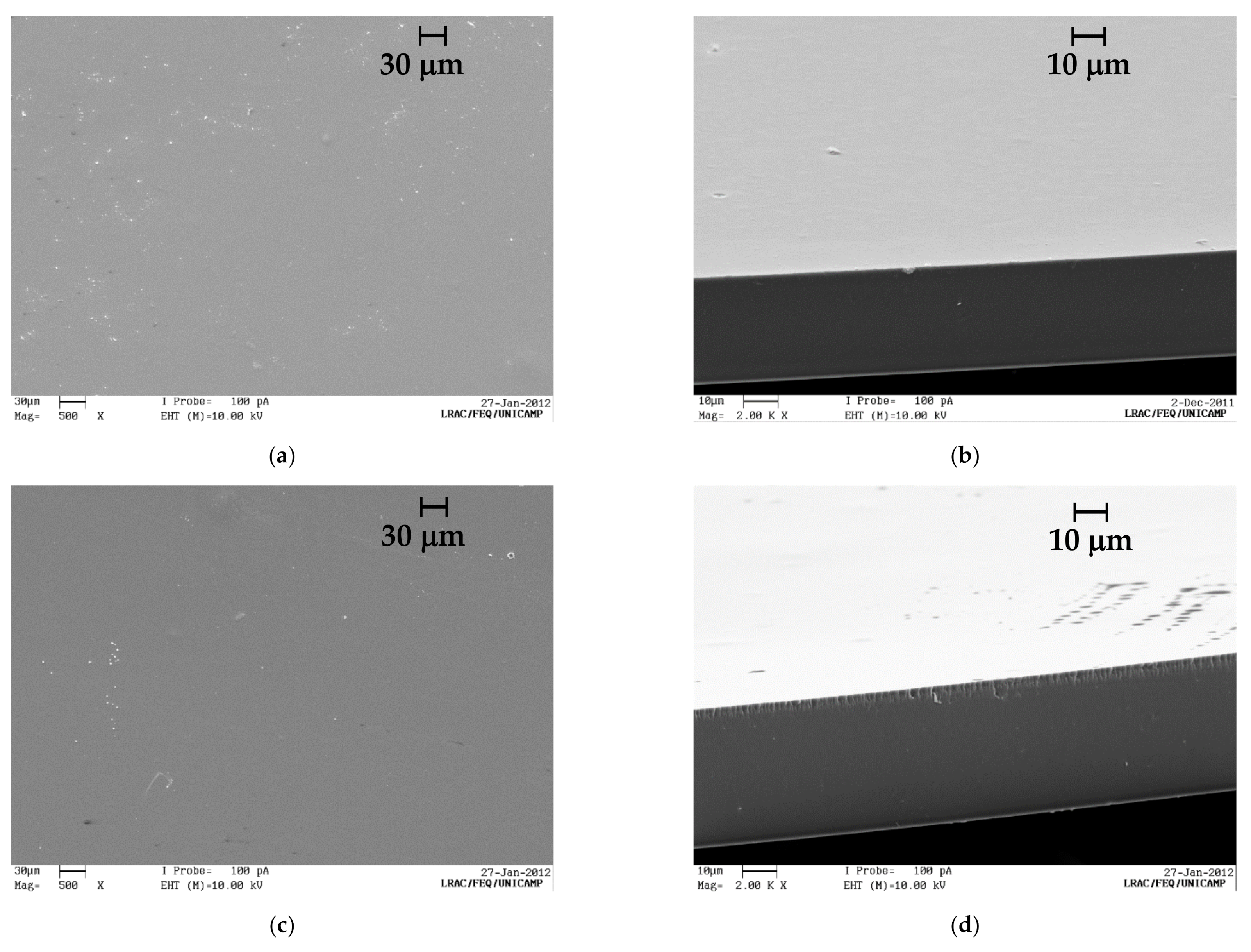
2.5. Scanning Electron Microscopy (SEM)
2.6. Barrier Properties
2.7. Mechanical Properties
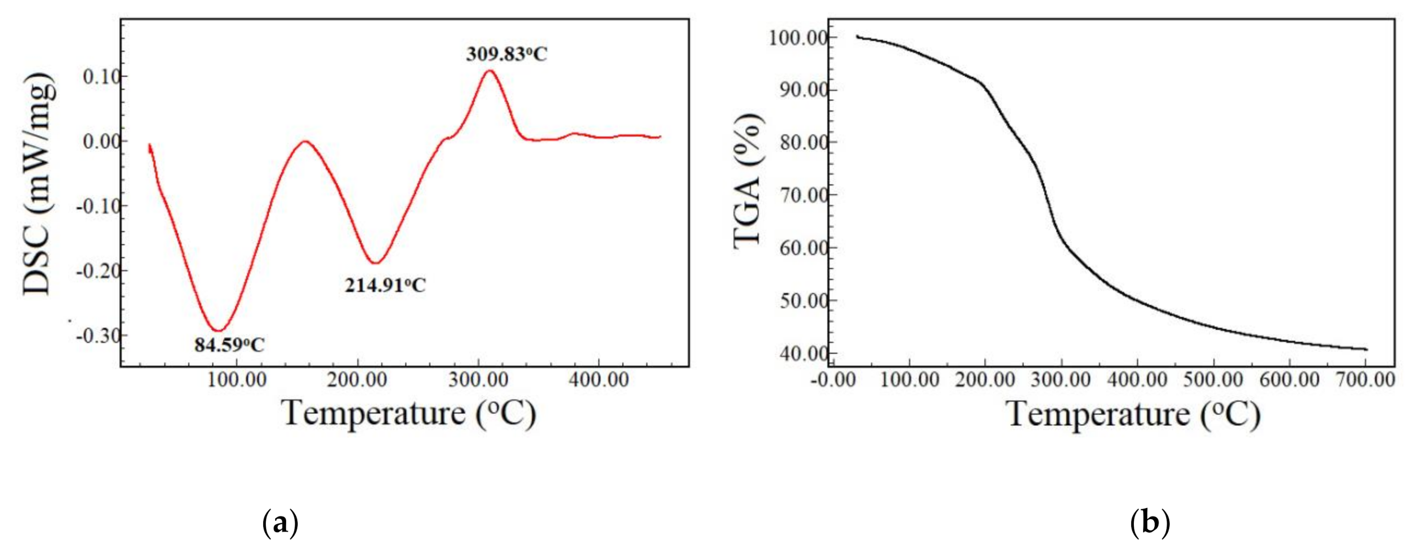
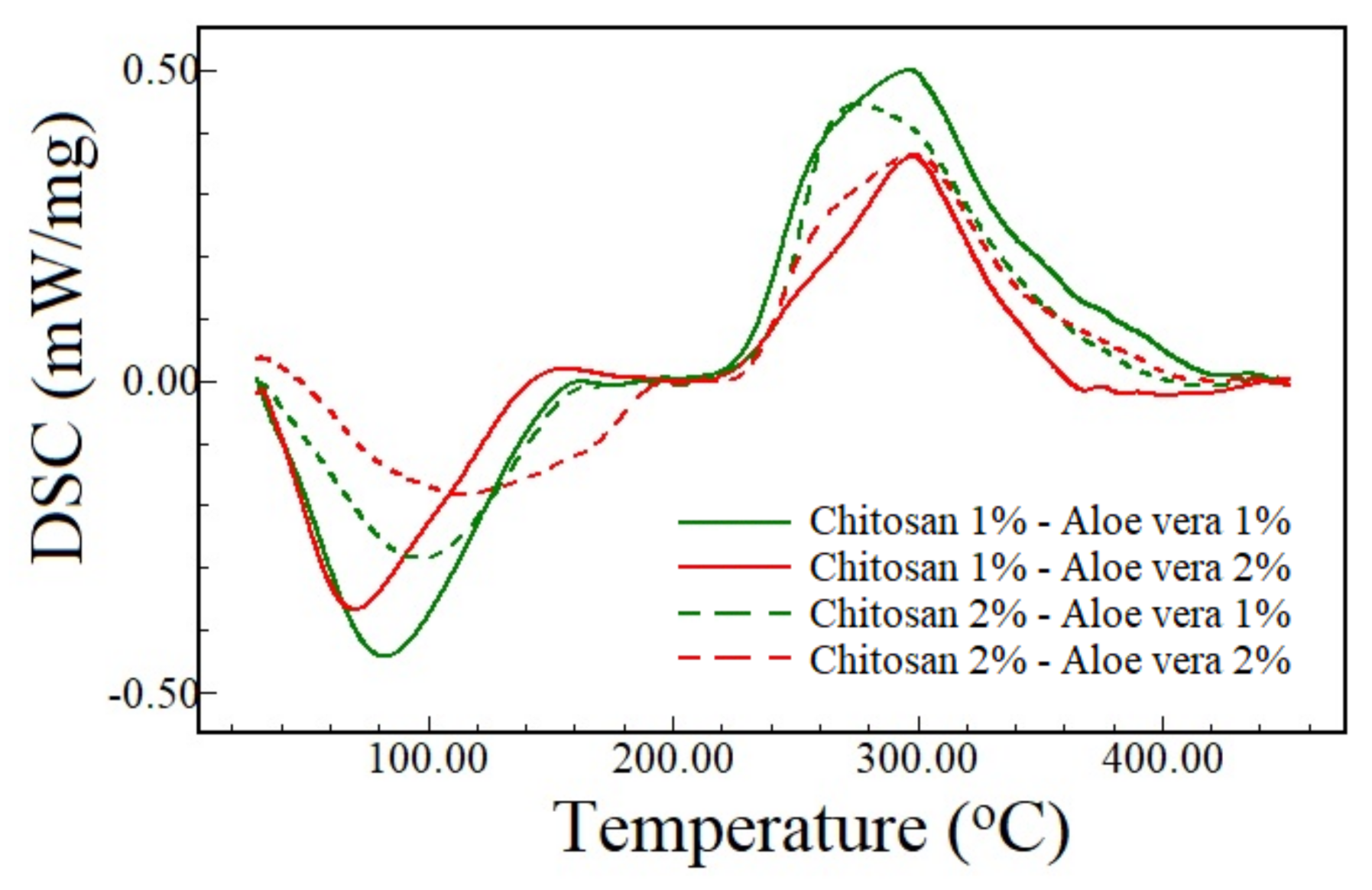
2.8. Thermal Analysis
2.9. Statistical Analysis
3. Results and Discussion
4. Conclusions
Author Contributions
Funding
Institutional Review Board Statement
Informed Consent Statement
Data Availability Statement
Conflicts of Interest
References
- Garcia-Orue, I.; Gainza, G.; Gutierrez, F.B.; Aguirre, J.J.; Evora, C.; Pedraz, J.L.; Hernandez, R.M.; Delgado, A.; Igartua, M. Novel nanofibrous dressings containing rhEGF and Aloe vera for wound healing applications. Int. J. Pharm. 2017, 523, 556–566. [Google Scholar] [CrossRef] [PubMed]
- Mi, F.-L.; Wu, Y.-B.; Shyu, S.-S.; Chao, A.-C.; Lai, J.-Y.; Su, C.-C. Asymmetric chitosan membranes prepared by dry/wet phase separation: A new type of wound dressing for controlled antibacterial release. J. Membr. Sci. 2003, 212, 237–254. [Google Scholar] [CrossRef]
- Barbosa, G.P.; Debone, H.S.; Severino, P.; Souto, E.B.; da Silva, C.F. Design and characterization of chitosan/zeolite composite films—Effect of zeolite type and zeolite dose on the film properties. Mater. Sci. Eng. C 2016, 60, 246–254. [Google Scholar] [CrossRef] [PubMed]
- Hissae Yassue-Cordeiro, P.; Zandonai, C.H.; Pereira Genesi, B.; Santos Lopes, P.; Sanchez-Lopez, E.; Garcia, M.L.; Camargo Fernandes-Machado, N.R.; Severino, P.; Souto, E.B.; Ferreira da Silva, C. Development of Chitosan/Silver Sulfadiazine/Zeolite Composite Films for Wound Dressing. Pharmaceutics 2019, 11, 535. [Google Scholar] [CrossRef] [Green Version]
- Muzzarelli, R.A.A.; Muzzarelli, C. Chitosan Chemistry: Relevance to the Biomedical Sciences. In Polysaccharides I: Structure, Characterization and Use; Heinze, T., Ed.; Springer: Berlin/Heidelberg, Germany, 2005; pp. 151–209. [Google Scholar] [CrossRef]
- Patrulea, V.; Ostafe, V.; Borchard, G.; Jordan, O. Chitosan as a starting material for wound healing applications. Eur. J. Pharm. Biopharm. 2015, 97, 417–426. [Google Scholar] [CrossRef] [Green Version]
- Howling, G.I.; Dettmar, P.W.; Goddard, P.A.; Hampson, F.C.; Dornish, M.; Wood, E.J. The effect of chitin and chitosan on the proliferation of human skin fibroblasts and keratinocytes in vitro. Biomaterials 2001, 22, 2959–2966. [Google Scholar] [CrossRef]
- Alemdaroğlu, C.; Değim, Z.; Celebi, N.; Zor, F.; Oztürk, S.; Erdoğan, D. An investigation on burn wound healing in rats with chitosan gel formulation containing epidermal growth factor. Burn. J. Int. Soc. Burn Inj. 2006, 32, 319–327. [Google Scholar] [CrossRef]
- Paul, W.; Sharma, C.; Tirunal, C. Chitosan and Alginate Wound Dressings: A Short Review. Trends Biomater. Artif. Organs 2004, 18, 18–23. [Google Scholar]
- Debone, H.S.; Lopes, P.S.; Severino, P.; Yoshida, C.M.P.; Souto, E.B.; da Silva, C.F. Chitosan/Copaiba oleoresin films for would dressing application. Int. J. Pharm. 2019, 555, 146–152. [Google Scholar] [CrossRef]
- Hamman, J.H. Composition and Applications of Aloe vera Leaf Gel. Molecules 2008, 13, 1599–1616. [Google Scholar] [CrossRef] [Green Version]
- Choi, S.; Chung, M.-H. A review on the relationship between aloe vera components and their biologic effects. Semin. Integr. Med. 2003, 1, 53–62. [Google Scholar] [CrossRef]
- Hashemi, S.A.; Madani, S.A.; Abediankenari, S. The Review on Properties of Aloe Vera in Healing of Cutaneous Wounds. Biomed. Res. Int. 2015, 2015, 714216. [Google Scholar] [CrossRef] [Green Version]
- Farzadinia, P.; Jofreh, N.; Khatamsaz, S.; Movahed, A.; Akbarzadeh, S.; Mohammadi, M.; Bargahi, A. Anti-inflammatory and Wound Healing Activities of Aloe vera, Honey and Milk Ointment on Second-Degree Burns in Rats. Int. J. Low. Extrem. Wounds 2016, 15, 241–247. [Google Scholar] [CrossRef] [Green Version]
- Langmead, L.; Makins, R.J.; Rampton, D.S. Anti-inflammatory effects of aloe vera gel in human colorectal mucosa in vitro. Aliment. Pharmacol. Ther. 2004, 19, 521–527. [Google Scholar] [CrossRef]
- Hosseinimehr, S.J.; Khorasani, G.; Azadbakht, M.; Zamani, P.; Ghasemi, M.; Ahmadi, A. Effect of aloe cream versus silver sulfadiazine for healing burn wounds in rats. Acta Dermatovenerol. Croat. ADC 2010, 18, 2–7. [Google Scholar]
- Singh, M.; Singh, S.; Nath, V.; Sahu, V.; Rawat, A.K. Antibacterial activity of some bryophytes used traditionally for the treatment of burn infections. Pharm. Biol. 2011, 49, 526–530. [Google Scholar] [CrossRef]
- Brandenburg, A.H.; Weller, C.L.; Testin, R.F. Edible Films and Coatings from Soy Protein. J. Food Sci. 1993, 58, 1086–1089. [Google Scholar] [CrossRef]
- Humbert, P.; Courderot-Masuyer, C.; Robin, S.; Oster, D.; Pegahi, R. Exudates absorption and proteases trapping in venous leg ulcers. J. Wound Care 2017, 26, 346–348. [Google Scholar] [CrossRef]
- Pereda, M.; Aranguren, M.I.; Marcovich, N.E. Caseinate films modified with tung oil. Food Hydrocoll. 2010, 24, 800–808. [Google Scholar] [CrossRef]
- Estevam, L.d.S.; Debone, H.S.; Yoshida, C.M.P.; Da Silva, C. Adsorption of bovine serum and bovine haemoglobin onto chitosan film. Adsorpt. Sci. Technol. 2012, 30, 785–792. [Google Scholar] [CrossRef]
- De Silva, M.F.; Lopes, P.S.; da Silva, C.F.; Yoshida, C.M. Active packaging material based on buriti oil–Mauritia flexuosa Lf (Arecaceae) incorporated into chitosan films. J. Appl. Polym. Sci. 2016, 133. [Google Scholar] [CrossRef]
- Yassue-Cordeiro, P.H.; Zandonai, C.H.; Silva, C.F.D.; Fernandes-Machado, N.R.C. Desenvolvimento e caracterização de filmes compósitos de quitosana e zeólitas com prata. Polímeros 2015, 25, 492–502. [Google Scholar] [CrossRef] [Green Version]
- Thomas, S.; Young, S. Exudate-handling mechanisms of two foam-film dressings. J. Wound Care 2008, 17, 309–315. [Google Scholar] [CrossRef] [PubMed]
- Kimura, V.T.; Miyasato, C.S.; Genesi, B.P.; Lopes, P.S.; Yoshida, C.M.P.; Silva, C.F.D. The effect of andiroba oil and chitosan concentration on the physical properties of chitosan emulsion film. Polímeros 2016, 26, 168–175. [Google Scholar] [CrossRef] [Green Version]
- Abdeen, Z. Swelling and Reswelling Characteristics of Cross-Linked Poly(vinyl alcohol)/Chitosan Hydrogel Film. J. Dispers. Sci. Technol. 2011, 32, 1337–1344. [Google Scholar] [CrossRef]
- Ghosh, A.; Azam Ali, M.; Walls, R. Modification of microstructural morphology and physical performance of chitosan films. Int. J. Biol. Macromol. 2010, 46, 179–186. [Google Scholar] [CrossRef]
- Campos, M.G.N.; Rawls, H.R.; Innocentini-Mei, L.H.; Satsangi, N. In vitro gentamicin sustained and controlled release from chitosan cross-linked films. J. Mater. Sci. Mater. Med. 2009, 20, 537–542. [Google Scholar] [CrossRef]
- Pereda, M.; Aranguren, M.I.; Marcovich, N.E. Water vapor absorption and permeability of films based on chitosan and sodium caseinate. J. Appl. Polym. Sci. 2009, 111, 2777–2784. [Google Scholar] [CrossRef]
- Batista, R.A.; Espitia, P.J.P.; Vergne, D.M.C.; Vicente, A.A.; Pereira, P.A.C.; Cerqueira, M.A.; Teixeira, J.A.; Jovanovic, J.; Severino, P.; Souto, E.B.; et al. Development and Evaluation of Superabsorbent Hydrogels Based on Natural Polymers. Polymers 2020, 12, 2173. [Google Scholar] [CrossRef]
- Khoshgozaran-Abras, S.; Azizi, M.H.; Hamidy, Z.; Bagheripoor-Fallah, N. Mechanical, physicochemical and color properties of chitosan based-films as a function of Aloe vera gel incorporation. Carbohydr. Polym. 2012, 87, 2058–2062. [Google Scholar] [CrossRef]
- Silva, S.S.; Popa, E.G.; Gomes, M.E.; Cerqueira, M.; Marques, A.P.; Caridade, S.G.; Teixeira, P.; Sousa, C.; Mano, J.F.; Reis, R.L. An investigation of the potential application of chitosan/aloe-based membranes for regenerative medicine. Acta Biomater. 2013, 9, 6790–6797. [Google Scholar] [CrossRef] [Green Version]
- Wang, L.; Khor, E.; Wee, A.; Lim, L.Y. Chitosan-alginate PEC membrane as a wound dressing: Assessment of incisional wound healing. J. Biomed. Mater. Res. 2002, 63, 610–618. [Google Scholar] [CrossRef]
- Pereda, M.; Ponce, A.G.; Marcovich, N.E.; Ruseckaite, R.A.; Martucci, J.F. Chitosan-gelatin composites and bi-layer films with potential antimicrobial activity. Food Hydrocoll. 2011, 25, 1372–1381. [Google Scholar] [CrossRef]
- Pires, A.L.R.; de Azevedo Motta, L.; Dias, A.M.A.; de Sousa, H.C.; Moraes, Â.M.; Braga, M.E.M. Towards wound dressings with improved properties: Effects of poly(dimethylsiloxane) on chitosan-alginate films loaded with thymol and beta-carotene. Mater. Sci. Eng. C Mater. Biol. Appl. 2018, 93, 595–605. [Google Scholar] [CrossRef]
- Martins, J.G.; de Oliveira, A.C.; Garcia, P.S.; Kipper, M.J.; Martins, A.F. Durable pectin/chitosan membranes with self-assembling, water resistance and enhanced mechanical properties. Carbohydr. Polym. 2018, 188, 136–142. [Google Scholar] [CrossRef]
- Pinzon, M.I.; Garcia, O.R.; Villa, C.C. The influence of Aloe vera gel incorporation on the physicochemical and mechanical properties of banana starch-chitosan edible films. J. Sci. Food Agric. 2018, 98, 4042–4049. [Google Scholar] [CrossRef]
- Gallardo-Rivera, R.; de los Ángeles Aguilar-Santamaría, M.; Silva-Bermúdez, P.; García-López, J.; Tecante, A.; Velasquillo, C.; Román-Guerrero, A.; Pérez-Alonso, C.; Vázquez-Torres, H.; Shirai, K. Polyelectrolyte complex of Aloe vera, chitosan, and alginate produced fibroblast and lymphocyte viabilities and migration. Carbohydr. Polym. 2018, 192, 84–94. [Google Scholar] [CrossRef]
- Jithendra, P.; Rajam, A.M.; Kalaivani, T.; Mandal, A.B.; Rose, C. Preparation and characterization of aloe vera blended collagen-chitosan composite scaffold for tissue engineering applications. ACS Appl. Mater. Interfaces 2013, 5, 7291–7298. [Google Scholar] [CrossRef]
- Georgieva, V.; Zvezdova, D.; Vlaev, L. Non-isothermal kinetics of thermal degradation of chitosan. Chem. Cent. J. 2012, 6, 81. [Google Scholar] [CrossRef] [Green Version]
- Ostrowska-Czubenko, J.; Gierszewska-Drużyńska, M. Effect of ionic crosslinking on the water state in hydrogel chitosan membranes. Carbohydr. Polym. 2009, 77, 590–598. [Google Scholar] [CrossRef]


| Sample | Chroma Parameter | Color Difference (∆E*) | ||
|---|---|---|---|---|
| L* | a* | b* | ||
| CH1-AV1 | 91.02 ± 2.73 a | −4.22 ± 0.32 a | 14.61 ± 1.58 ª | 7.19 |
| CH1-AV2 | 89.73 ± 3.02 a,b | −4.32 ± 0.35 a | 21.68 ± 1.97 b | 13.94 |
| CH2-AV1 | 87.50 ± 1.04 b,c | −3.34 ± 0.23 b | 26.39 ± 1.66 c | 12.48 |
| CH2-AV2 | 86.08 ± 1.16 c | −2.81 ± 0.26 c | 27.49 ± 2.31 c | 13.27 |
| Samples | MVTR (g/10 cm2/24 h) | ABS (g/10 cm2/24 h) | FHC (g/10 cm2/24 h) | WVP × 1011 (g/m.s.Pa) |
|---|---|---|---|---|
| CH1-AV1 | 12.19 ± 0.92 a | 1.61 ± 0.16 a | 13.80 ± 0.9 a | 4.50 ± 0.42 a |
| CH1-AV2 | 12.74 ± 1.07 a | 1.30 ± 0.09 b | 14.04 ± 1.1 a | 4.19 ± 0.07 a |
| CH2-AV1 | 31.90 ± 2.63 b | 2.52 ± 0.22 c | 34.42 ± 2.6 b | 6.56 ± 0.55 b |
| CH2-AV2 | 31.85 ± 0.92 b | 1.42 ± 0.14 a,b | 33.27 ± 0.9 b | 6.24 ± 0.27 b |
| Samples | Young Modulus (MPa) | Tensile Strength (n/m2) | Elongation at Break (%) |
|---|---|---|---|
| CH1-AV1 | 63.3 ± 6.8 a | 180.5 ± 26.0 ª | 2.9 ± 0.5 a |
| CH1-AV2 | 61.6 ± 5.6 a | 151.7 ± 33.6 ª | 2.7 ± 0.6 a |
| CH2-AV1 | 60.6 ± 7.3 a | 176.9 ± 23.2 ª | 6.7 ± 1.6 b |
| CH2-AV2 | 68.6 ± 10.0 a | 189.1± 33.1 ª | 5.6 ± 1.6 b |
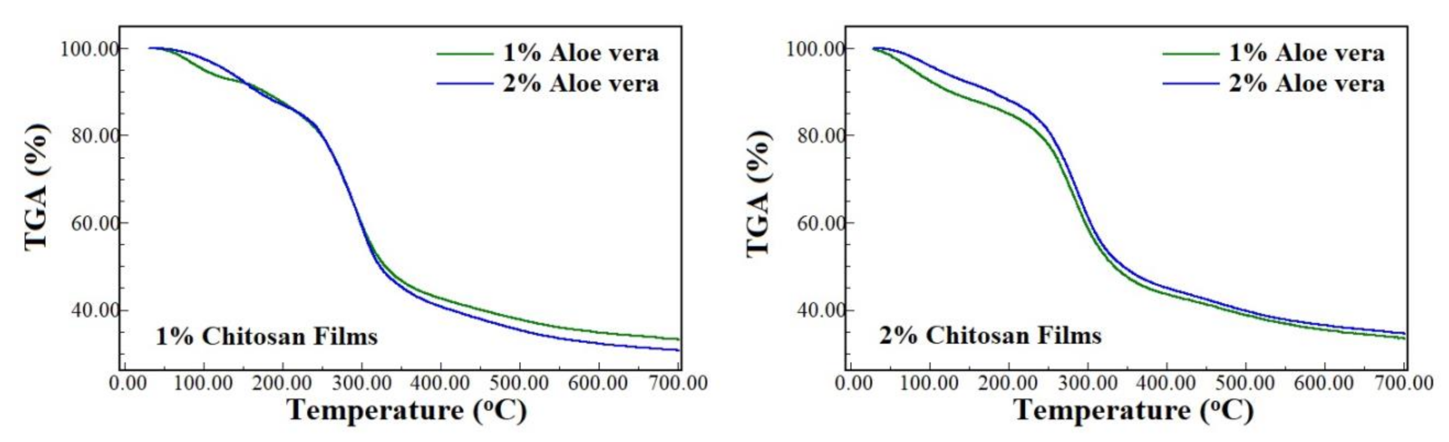
| Concentration (% w/w) | Percentage of Cumulative Weight Loss (%) | |||||||
|---|---|---|---|---|---|---|---|---|
| Chitosan | Aloe Vera | 100 °C | 200 °C | 300 °C | 400 °C | 500 °C | 600 °C | 700 °C |
| 1 | 1.0 | 5 | 12 | 40 | 57 | 62 | 65 | 67 |
| 2.0 | 2 | 13 | 41 | 59 | 65 | 68 | 69 | |
| 2 | 1.0 | 7 | 15 | 41 | 56 | 61 | 65 | 67 |
| 2.0 | 4 | 12 | 38 | 54 | 59 | 62 | 64 | |
| Aloe vera extract | 2 | 10 | 38 | 50 | 55 | 58 | 59 | |
Publisher’s Note: MDPI stays neutral with regard to jurisdictional claims in published maps and institutional affiliations. |
© 2021 by the authors. Licensee MDPI, Basel, Switzerland. This article is an open access article distributed under the terms and conditions of the Creative Commons Attribution (CC BY) license (https://creativecommons.org/licenses/by/4.0/).
Share and Cite
Yoshida, C.M.P.; Pacheco, M.S.; de Moraes, M.A.; Lopes, P.S.; Severino, P.; Souto, E.B.; da Silva, C.F. Effect of Chitosan and Aloe Vera Extract Concentrations on the Physicochemical Properties of Chitosan Biofilms. Polymers 2021, 13, 1187. https://doi.org/10.3390/polym13081187
Yoshida CMP, Pacheco MS, de Moraes MA, Lopes PS, Severino P, Souto EB, da Silva CF. Effect of Chitosan and Aloe Vera Extract Concentrations on the Physicochemical Properties of Chitosan Biofilms. Polymers. 2021; 13(8):1187. https://doi.org/10.3390/polym13081187
Chicago/Turabian StyleYoshida, Cristiana M. P., Murilo S. Pacheco, Mariana A. de Moraes, Patrícia S. Lopes, Patrícia Severino, Eliana B. Souto, and Classius F. da Silva. 2021. "Effect of Chitosan and Aloe Vera Extract Concentrations on the Physicochemical Properties of Chitosan Biofilms" Polymers 13, no. 8: 1187. https://doi.org/10.3390/polym13081187