Effect of Nardostachys jatamansi DC. on Apoptosis, Inflammation and Oxidative Stress Induced by Doxorubicin in Wistar Rats

 ,
,  , , , and
Abstract
:1. Introduction
2. Materials and Methods
2.1. Chemicals and Reagents
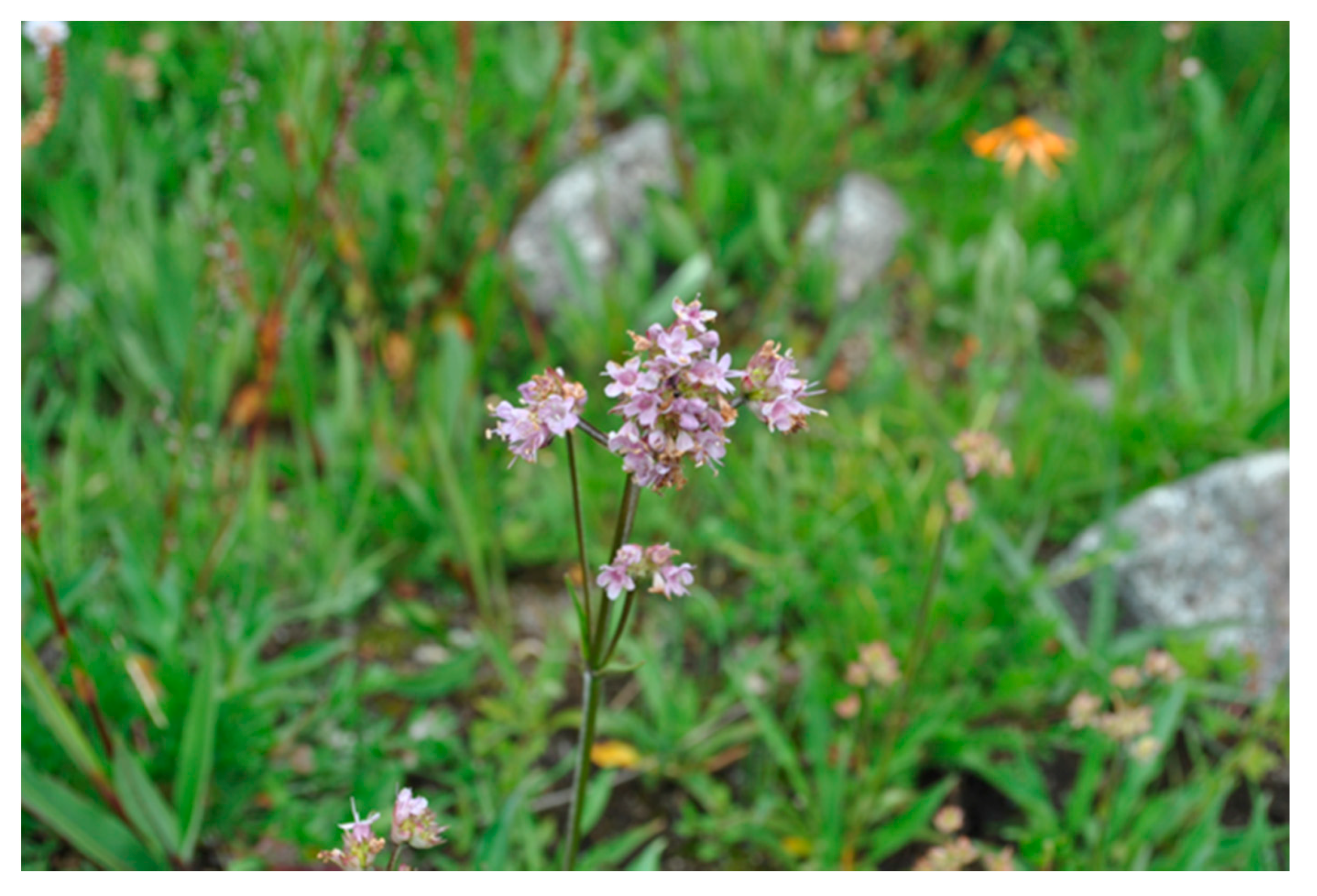
2.2. Plant Material
2.3. Extract Preparation
2.4. Standardization of MEJ
2.4.1. HPTLC Profiling
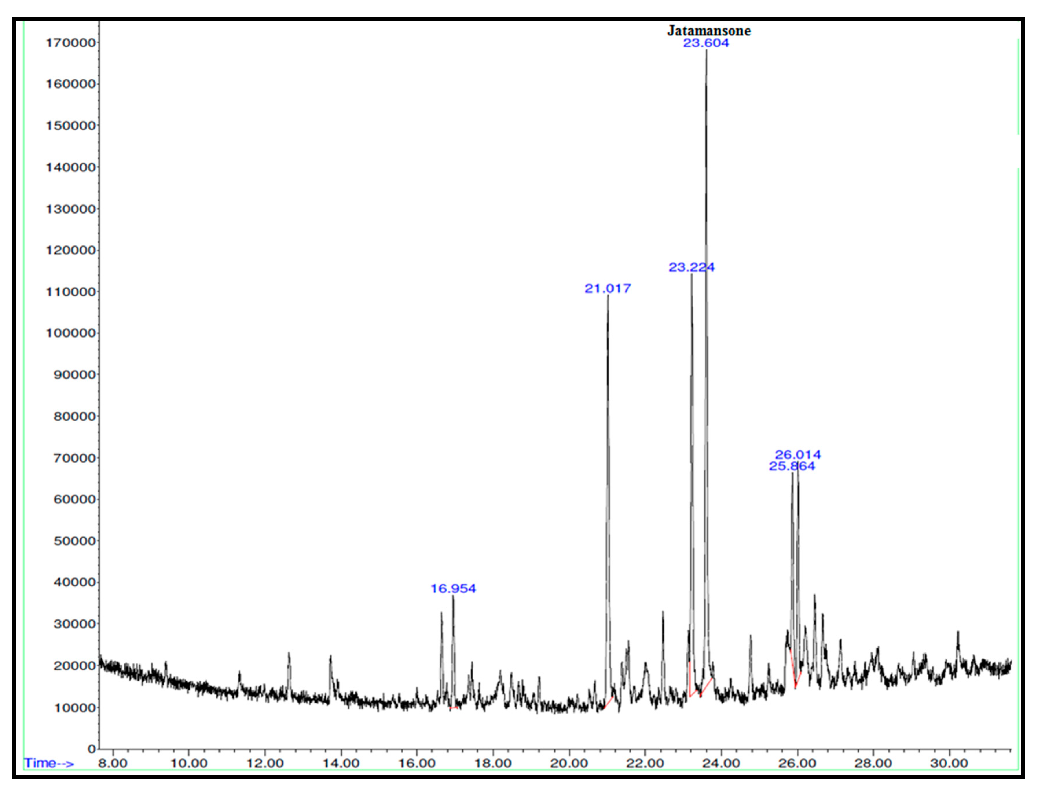
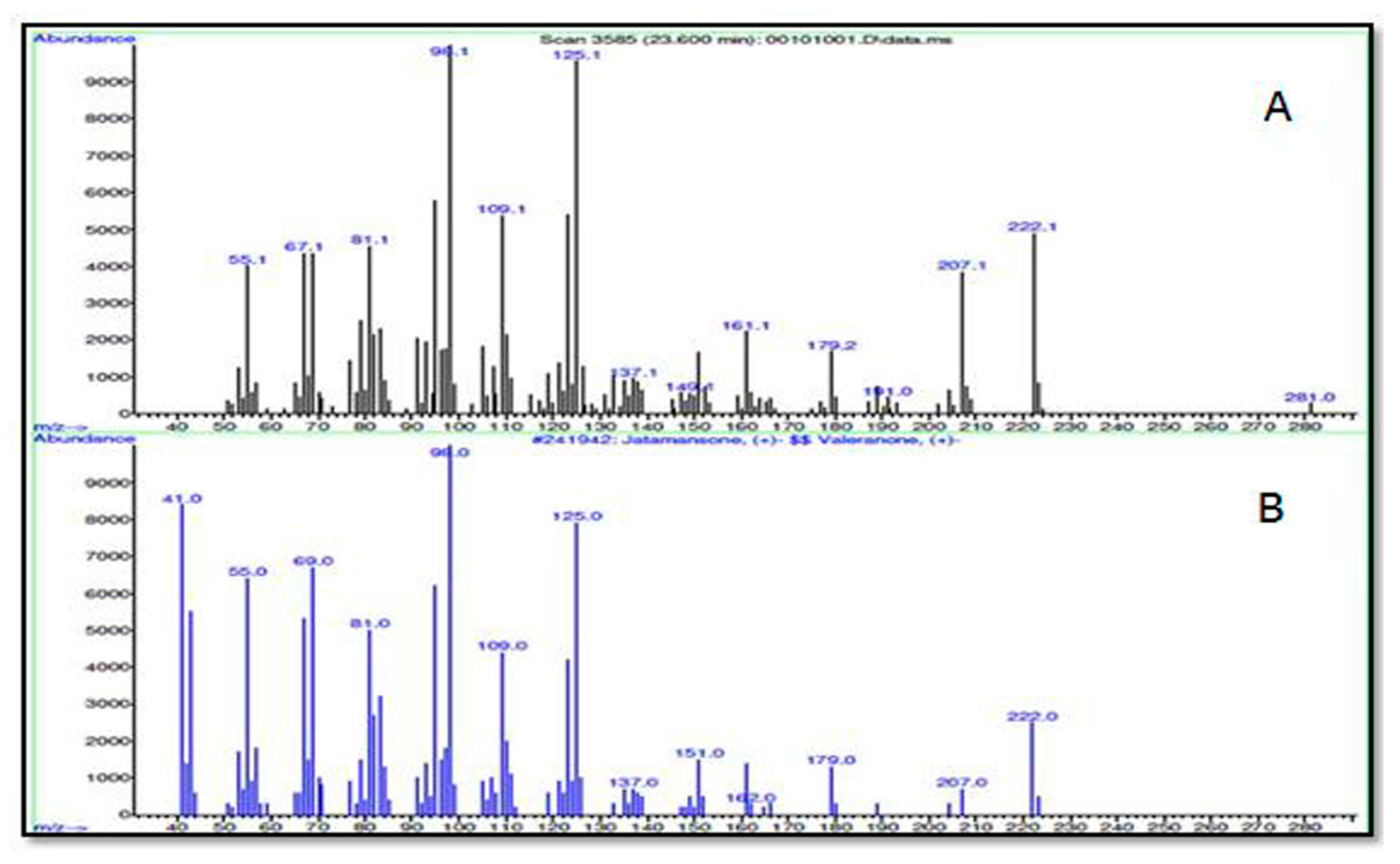
2.4.2. GC-MS Chemoprofiling
2.4.3. Determination of Total Phenolic Content and In Vitro Antixidant Potential
2.5. In Vivo Study
2.5.1. Animals
2.5.2. Treatment Schedule
2.5.3. Biochemical Estimation
2.6. Histopathology
2.7. Statistical Analysis
3. Results
3.1. Standardization of MEJ
3.2. In Vivo Study
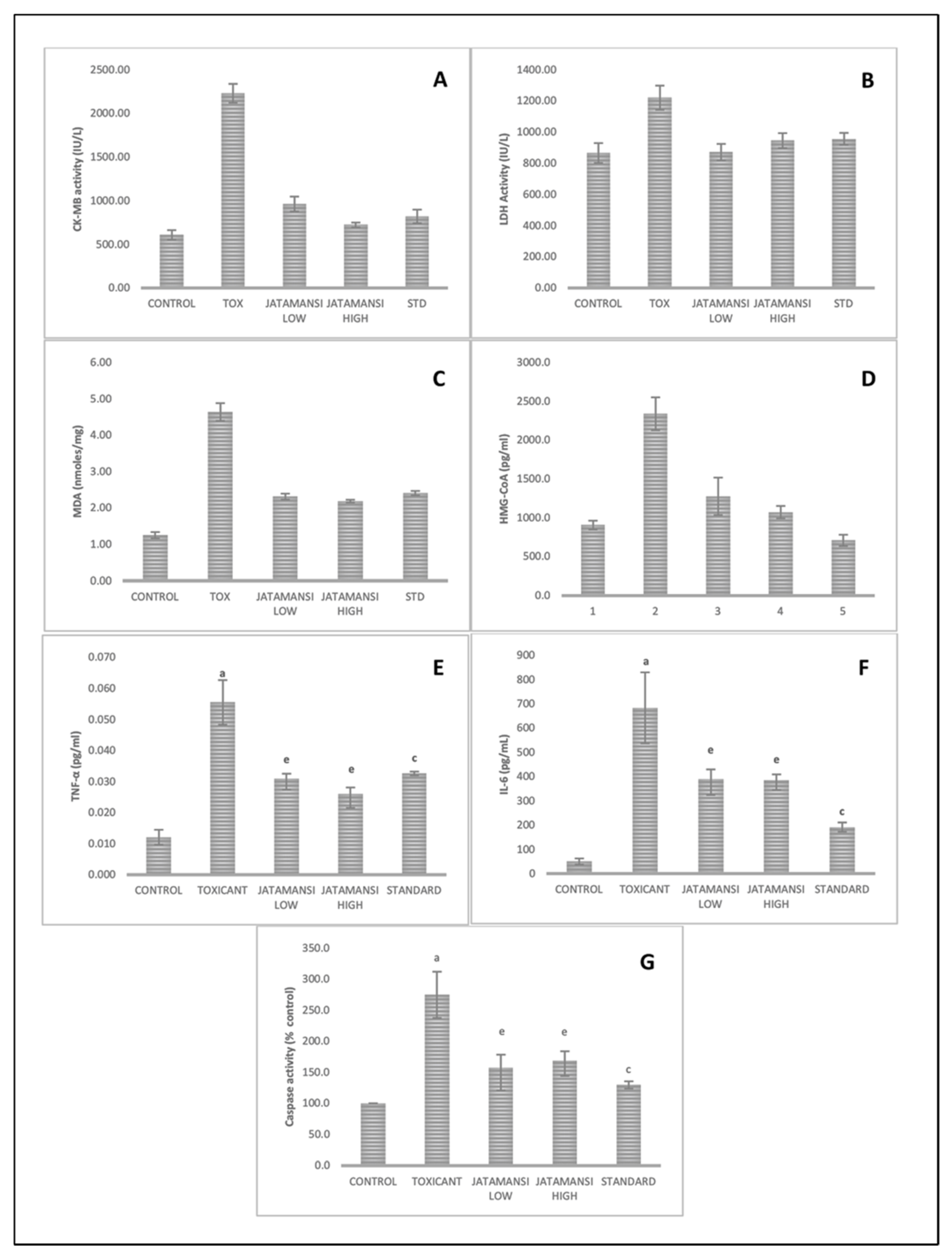
Biochemical Estimation
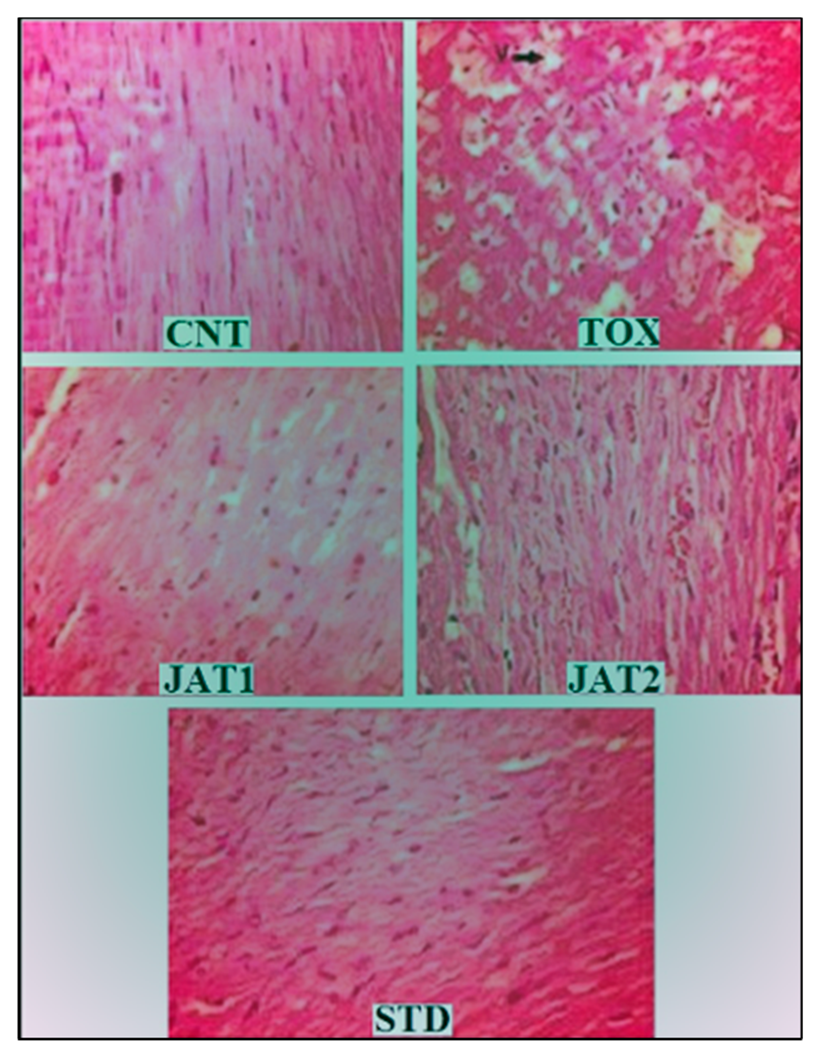
3.3. Histopathology
4. Dicussion
Supplementary Materials
Author Contributions
Funding
Acknowledgments
Conflicts of Interest
Abbreviations
| ANOVA | Analysis of Variance |
| CK-MB | Creatine kinase myocardial band |
| CPCSEA | Committee for the purpose of control and supervision of experiment on animals |
| DOX | Doxorubicin |
| DPPH | 2,2-diphenyl-1-picrylhydrazyl. |
| ELISA | Enzyme Linke Immunosorbent Assay |
| GC-MS | Gas chromatography–mass spectrometry |
| HMG-CoA | 3-hydroxy-3-methyl-glutaryl-coenzyme A |
| HPTLC | High-performance thin-layer chromatography |
| IL-6 | Interleukin-6 |
| MDA | Malondialdehyde |
| NO | Nitric Oxide |
| TBA | Thiobarbituric Acid |
| TNF-α | Tumor Necrosis Factor-Alpha |
Appendix A


References
- Keizer, H.G.; Pinedo, H.M.; Schuurhuis, G.J.; Joenje, H. DOX (adriamycin): A critical review of free radical-dependent mechanisms of cytotoxicity. Pharmacol. Ther. 1990, 47, 219–231. [Google Scholar] [CrossRef]
- Zucchi, R.; Danesi, R. Cardiac toxicity of antineoplastic anthracyclines. Curr. Med. Chem. Anticancer Agents 2003, 3, 151–171. [Google Scholar] [CrossRef] [PubMed]
- Lee, K.; Sung, R.Y.; Huang, W.Z.; Yang, M.; Pong, N.H.; Lee, S.M.; Chan, W.Y.; Zhao, H.; To, M.Y.; Fok, T.F.; et al. Thrombopoietin protects against in vitro and in vivo cardiotoxicity induced by DOX. Circulation 2006, 113, 2211–2220. [Google Scholar]
- Neilan, T.G.; Blake, S.L.; Ichinose, F.; Raher, M.J.; Buys, E.S.; Jassal, D.S.; Furutani, E.; Perez-Sanz, T.M.; Graveline, A.; Janssens, S.P.; et al. Disruption of nitric oxide synthase 3 protects against the cardiac injury, dysfunction, and mortality induced by DOX. Circulation 2007, 116, 506–514. [Google Scholar] [CrossRef] [PubMed] [Green Version]
- Arola, O.J.; Saraste, A.; Pulkki, K.; Kallajoki, M.; Parvinen, M.; Voipio-Pulkki, L.M. Acute DOX cardiotoxicity involves cardiomyocyte apoptosis. Cancer Res. 2000, 60, 1789–1792. [Google Scholar] [PubMed]
- Fisher, P.W.; Salloum, F.; Das, A.; Hyder, H.; Kukreja, R.C. Phosphodiesterase-5 inhibition with sildenafil attenuates cardiomyocyte apoptosis and left ventricular dysfunction in a chronic model of DOX cardiotoxicity. Circulation 2005, 111, 1601–1610. [Google Scholar] [CrossRef] [PubMed] [Green Version]
- Kawamura, T.; Hasegawa, K.; Morimoto, T.; Iwai-Kanai, E.; Miyamoto, S.; Kawase, Y.; Ono, K.; Wada, H.; Akao, M.; Kita, T. Expression of p300 protects cardiac myocytes from apoptosis in vivo. Biochem. Biophys. Res. Commun. 2004, 315, 733–738. [Google Scholar] [CrossRef] [Green Version]
- Wang, G.W.; Klein, J.B.; Kang, Y.J. Metallothionein inhibits DOX-induced mitochondrial cytochrome release and caspase-3 activation in cardiomyocytes. J. Pharmacol. Exp. Ther. 2001, 298, 461–468. [Google Scholar]
- Xu, X.; Persson, H.L.; Richardson, D.R. Molecular pharmacology of the interaction of anthracyclines with iron. Mol. Pharmacol. 2005, 68, 261–271. [Google Scholar] [CrossRef]
- Calderone, A.; De Champlain, J.; Rouleau, J.L. Adriamycin-induced changes to the myocardial beta-adrenergic system in the rabbit. J. Mol. Cell Cardiol. 1991, 23, 333–342. [Google Scholar] [CrossRef]
- Singal, P.K.; Iliskovic, N. DOX-induced cardiomyopathy. N. Engl. J. Med. 1998, 339, 900–905. [Google Scholar] [CrossRef] [PubMed]
- Takemura, G.; Fujiwara, H. DOX-induced cardiomyopathy from the cardiotoxic mechanisms to management. Prog. Cardiovasc. Dis. 2007, 49, 330–352. [Google Scholar] [CrossRef] [PubMed]
- Minotti, G.; Menna, P.; Salvatorelli, E.; Cairo, G.; Gianni, l. Anthracyclines: Molecular advances and pharmacologic developments in antitumor activity and cardiotoxicity. Pharmacol. Rev. 2004, 56, 185–229. [Google Scholar] [CrossRef] [PubMed] [Green Version]
- Subashini, R.; Yogeeta, S.; Gnanapragasam, A.; Devaki, T. Protective effect of Nardostachys jatamansi on oxidative injury and cellular abnormalities during doxorubicin-induced cardiac damage in rats. J. Pharm. Pharmacol. 2006, 58, 257–262. [Google Scholar] [CrossRef] [PubMed]
- Dixit, V.P.; Jain, P.; Joshi, S.C. Hypolipidaemic effects of Curcuma longa L and Nardostachys jatamansi, DC in triton-induced hyperlipidaemic rats. Indian J. Physiol. Pharmacol. 1988, 32, 299–304. [Google Scholar]
- Joshi, H.; Parle, M. Nardostachys jatamansi improves learning and memory in mice. J. Med. Food 2006, 9, 113–118. [Google Scholar] [CrossRef]
- Rao, V.S.; Rao, A.; Karanth, K.S. Anticonvulsant and neurotoxicity profile of Nardostachys jatamansi in rats. J. Ethnopharmacol. 2005, 102, 351–356. [Google Scholar] [CrossRef]
- Bhat, M.D.A.; Malik, S.A. Efficacy of Nardostachys jatamansi (D.Don) DC in essential hypertension: A randomized controlled study. Complement. Ther. Med. 2020, 53, 102532. [Google Scholar] [CrossRef]
- Bagchi, A.; Oshima, Y.; Hikino, H. Neolignans and lignans of Nardostachys jatamansi roots. Planta Med. 1991, 57, 96–97. [Google Scholar] [CrossRef]
- Lyle, N.; Bhattacharyya, D.; Sur, T.K.; Munshi, S.; Paul, S.; Chatterjee, S.; Gomes, A. Stress modulating antioxidant effect of Nardostachys jatamansi. Indian J. Biochem. Biophys. 2009, 46, 93–98. [Google Scholar]
- Bae, G.S.; Seo, S.W.; Kim, M.S.; Park, K.C.; Koo, B.S.; Jung, W.S.; Cho, G.H.; Oh, H.C.; Yun, S.W.; Kim, J.J.; et al. The roots of Nardostachys jatamansi inhibits lipopolysaccharide-induced endotoxin shock. J. Nat. Med. 2011, 65, 63–72. [Google Scholar] [CrossRef] [PubMed]
- Subashini, R.; Gnanapragasam, A.; Senthilkumar, S.; Yogeeta, S.K.; Devaki, T. Protective efficacy of Nardostachys jatamansi (rhizomes) on mitochondrial respiration and lysosomal hydrolases during doxorubicin induced myocardial injury in rats. J. Health Sci. 2007, 53, 67–76. [Google Scholar] [CrossRef] [Green Version]
- Subashini, R.; Ragavendran, B.; Gnanapragasam, A.; Yogeeta, S.K.; Devaki, T. Biochemical study on the protective potential of Nardostachys jatamansi extract on lipid profile and lipid metabolizing enzymes in doxorubicin intoxicated rats. Pharmazie 2007, 62, 382–387. [Google Scholar] [PubMed]
- Slikard, K.; Singleton, V.L. Total phenol analysis: Automation and comparison with manual methods. Am. J. Enol. Vitic. 1977, 28, 49–55. [Google Scholar]
- Kumaran, A.; Karunakaran, R.J. In vitro antioxidant activities of methanol extracts of five Phyllanthus species from India. LWT Food Sci. Technol. 2007, 40, 344–352. [Google Scholar] [CrossRef]
- Liu, W.; Fu, Y.J.; Zu, Y.G.; Tong, M.H.; Wu, N.; Liu, X.L.; Zhang, S. Supercritical carbon dioxide extraction of seed oil from Opuntia dillenii Haw and its antioxidant activity. Food Chem. 2009, 114, 334–339. [Google Scholar] [CrossRef]
- Ahmad, M.; Yousuf, S.; Khan, M.B.; Hoda, M.N.; Ahmad, A.S.; Ansari, M.A.; Ishrat, T.; Agrawal, A.K.; Islam, F. Attenuation by Nardostachys jatamansi of 6-hydroxydopamine-induced parkinsonism in rats: Behavioral, neurochemical, and immunohistochemical studies. Pharmacol. Biochem. Behav. 2006, 83, 150–160. [Google Scholar] [CrossRef]
- Lyle, N.; Gomes, A.; Sura, T.; Munshi, S.; Paul, S.; Chatterjee, S.; Bhattacharyya, D. The role of antioxidant properties of Nardostachys jatamansi in alleviation of the symptoms of the chronic fatigue syndrome. Behav. Brain Res. 2009, 202, 285–290. [Google Scholar] [CrossRef]
- Momin, F.N.; Kalai, B.R.; Shikalgar, T.S.; Naikwade, N.S. Cardioprotective effect of methanolic extract of Ixora coccinea Linn. leaves on doxorubicin-induced cardiac toxicity in rats. Indian J. Pharmacol. 2012, 44, 178–183. [Google Scholar] [CrossRef]
- Thippeswamy, A.H.; Shirodkar, A.; Koti, B.C.; Sadiq, A.J.; Praveen, D.M.; Swamy, A.H.; Patil, M. Protective role of Phyllantus niruri extract in doxorubicin-induced myocardial toxicity in rats. Indian J. Pharmacol. 2011, 43, 31–35. [Google Scholar]
- Lum, G.; Gambino, S.R. A comparison of serum versus heparinized plasma for routine chemistry tests. Am. J. Clin. Pathol. 1974, 61, 108–113. [Google Scholar] [CrossRef] [PubMed]
- Lehmann, G.L.; Carreras, F.I.; Soria, L.R.; Gradilone, S.A.; Marinelli, R.A. LPS induces the TNF-alpha-mediated down regulation of rat liver aquaporin-8: Role in sepsis-associated cholestasis. Am. J. Physiol. Gastrointest. Liver Physiol. 2008, 294, 567–575. [Google Scholar] [CrossRef] [PubMed]
- Helle, M.; Boeije, L.; Groot, E.D.; Vos, A.D.; Aarden, L. Sensitive ELISA for interleukin-6: Detection of IL-6 in biological fluids: Synovial fluids and sera. J. Immunol. Methods 1991, 138, 47–56. [Google Scholar] [CrossRef]
- Jaeschke, H.; Fisher, M.A.; Lawson, J.A.; Simmons, C.A.; Farhood, A.; Jones, D.A. Activation of caspase 3 (CPP32)-like proteases is essential for TNF- α-induced hepatic parenchymal cell apoptosis and neutrophil-mediated necrosis in a murine endotoxin shock model. J. Immunol. 1998, 160, 3480–3486. [Google Scholar]
- Iqbal, M.; Dubey, K.; Anwer, T.; Ashish, A.; Pillai, K.K. Protective effects of telmisartan against acute doxorubicin-induced cardiotoxicity in rats. Pharmacol. Rep. 2008, 60, 382–390. [Google Scholar]
- Lowry, O.H.; Rosebrough, N.J.; Farr, A.L.; Randall, R.J. Protein measurement with the folin phenol reagent. J. Biol. Chem. 1951, 193, 265–275. [Google Scholar]
- Sharma, H.; Pathan, R.A.; Kumar, V.; Javed, S.; Bhandari, U. Anti-apoptotic potential of rosuvastatin pretreatment in murine model of cardiomyopathy. Int. J. Cardiol. 2011, 150, 193–200. [Google Scholar] [CrossRef]
- Tanaka, M.; Kuei, C.W.; Nagashima, Y.; Taguchi, T. Application of antioxidative maillrad reaction products from histidine and glucose to sardine products. Nippon Suisan Gakk 1998, 54, 1409–1414. [Google Scholar] [CrossRef]
- Nebigil, C.G.; Désaubry, L. Updates in anthracycline-mediated cardiotoxicity. Front. Pharmacol. 2018, 9, 1262. [Google Scholar] [CrossRef] [Green Version]
- Wang, L.M.; Chi, Y.J.; Wang, L.N.; Nie, L.; Zou, Y.H.; Zhao, T.N.; Li, C.Y.; Chen, M.; Huo, M.X. Expression of interleukin-6 in rat model of doxorubicin-induced nephropathy. Zhongguo Dang Dai Er Ke Za Zhi 2010, 12, 912–914. [Google Scholar]
- Van der Veen, A.H.; De Wilt, J.H.; Eggermont, A.M.; Van Tiel, S.T.; Seynhaeve, A.L.; Ten Hagen, T.L. TNF-alpha augments intratumoural concentrations of doxorubicin in TNF-alpha-based isolated limb perfusion in rat sarcoma models and enhances anti-tumour effects. Br. J. Cancer 2000, 82, 973–980. [Google Scholar] [CrossRef] [PubMed]
- Chiosi, E.; Spina, A.; Sorrentino, A.; Romano, M.; Sorvillo, L.; Senatore, G.; D’Auria, R.; Abbruzzese, A.; Caraglia, M.; Naviglio, S.; et al. Change in TNF-alpha receptor expression is a relevant event in doxorubicin-induced H9c2 cardiomyocyte cell death. J. Interferon Cytokine Res. 2007, 27, 589–597. [Google Scholar] [CrossRef] [PubMed]
- Schulz, R.; Aker, S.; Belosjorow, S.; Heusch, G. TNF-alpha in ischemia/reperfusion injury and heart failure. Basic Res. Cardiol. 2004, 99, 8–11. [Google Scholar] [PubMed]
- Grabacka, M.; Pierzchalska, M.; Dean, M.; Reiss, K. Regulation of ketone body metabolism and the role of PPARα. Int. J. Mol. Sci. 2016, 17, 2093. [Google Scholar] [CrossRef] [Green Version]
- Monzel, J.V.; Budde, T.; Meyer, Z.; Schwabedissen, H.E.; Schwebe, M.; Bien-Möller, S.; Lütjohann, D.; Kroemer, H.K.; Jedlitschky, G.; Grube, M. Doxorubicin enhances oxysterol levels resulting in a LXR-mediated upregulation of cardiac cholesterol transporters. Biochem. Pharmacol. 2017, 144, 108–119. [Google Scholar] [CrossRef]
- Tripathi, Y.B.; Tripathi, E.; Upadhyay, A. Antilipid peroxidative property of Nardostachys jatamanasi. Indian J. Exp. Biol. 1996, 34, 1150–1151. [Google Scholar]


| S. No | Name of Constituent | Rt | Area Percentage | Matching Percentage |
|---|---|---|---|---|
| 1 | Seychellene | 16.957 | 5.67 | 96 |
| 2 | Acenaphthylene | 21.019 | 22.36 | 55 |
| 3 | Patchouli alcohol | 23.222 | 21.73 | 98 |
| 4 | Jatamansone | 23.606 | 32.05 | 99 |
| 5 | 1-methyl-4-chloro-3,5-dimethoxy-1H-pyrazole | 25.866 | 8.30 | 60 |
| 6 | Illudol $$ 3,6,6,7b-tetramethyl decahydro-1H-cyclobuta[e]endene-3-ol | 26.015 | 9.89 | 44 |
Publisher’s Note: MDPI stays neutral with regard to jurisdictional claims in published maps and institutional affiliations. |
© 2020 by the authors. Licensee MDPI, Basel, Switzerland. This article is an open access article distributed under the terms and conditions of the Creative Commons Attribution (CC BY) license (http://creativecommons.org/licenses/by/4.0/).
Share and Cite
Singh, M.; Khan, M.A.; Y. T., K.; Ahmad, J.; Fahmy, U.A.; Kotta, S.; Alhakamy, N.A.; Ahmad, S. Effect of Nardostachys jatamansi DC. on Apoptosis, Inflammation and Oxidative Stress Induced by Doxorubicin in Wistar Rats. Plants 2020, 9, 1579. https://doi.org/10.3390/plants9111579
Singh M, Khan MA, Y. T. K, Ahmad J, Fahmy UA, Kotta S, Alhakamy NA, Ahmad S. Effect of Nardostachys jatamansi DC. on Apoptosis, Inflammation and Oxidative Stress Induced by Doxorubicin in Wistar Rats. Plants. 2020; 9(11):1579. https://doi.org/10.3390/plants9111579
Chicago/Turabian StyleSingh, Mhaveer, Mohammad Ahmed Khan, Kamal Y. T., Javed Ahmad, Usama A. Fahmy, Sabna Kotta, Nabil A. Alhakamy, and Sayeed Ahmad. 2020. "Effect of Nardostachys jatamansi DC. on Apoptosis, Inflammation and Oxidative Stress Induced by Doxorubicin in Wistar Rats" Plants 9, no. 11: 1579. https://doi.org/10.3390/plants9111579