
SPEECHLESS and MUTE Mediate Feedback Regulation of Signal Transduction during Stomatal Development


1
Henan Key Laboratory of Earth System Observation and Modeling, Henan University, Kaifeng 475004, China
2
College of Environment and Planning, Henan University, Kaifeng 475004, China
3
Miami College, Henan University, Kaifeng 475004, China
*
Authors to whom correspondence should be addressed.
Plants 2021, 10(3), 432; https://doi.org/10.3390/plants10030432
Submission received: 18 January 2021
/
Revised: 14 February 2021
/
Accepted: 21 February 2021
/
Published: 24 February 2021
(This article belongs to the Special Issue Plant Developmental Responses to the Environment)
Abstract
:Stomatal density, spacing, and patterning greatly influence the efficiency of gas exchange, photosynthesis, and water economy. They are regulated by a complex of extracellular and intracellular factors through the signaling pathways. After binding the extracellular epidermal patterning factor 1 (EPF1) and 2 (EPF2) as ligands, the receptor-ligand complexes activate by phosphorylation through the MAP-kinase cascades, regulating basic helix-loop-helix (bHLH) transcription factors SPEECHLESS (SPCH), MUTE, and FAMA. In this review, we summarize the molecular mechanisms and signal transduction pathways running within the transition of the protodermal cell into a pair of guard cells with a space (aperture) between them, called a stoma, comprising asymmetric and symmetric cell divisions and draw several functional models. The feedback mechanisms involving the bHLH factors SPCH and MUTE are not fully recognized yet. We show the feedback mechanisms driven by SPCH and MUTE in the regulation of EPF2 and the ERECTA family. Intersections of the molecular mechanisms for fate determination of stomatal lineage cells with the role of core cell cycle-related genes and stabilization of SPCH and MUTE are also reported.
1. Introduction
Arabidopsis thaliana (Arabidopsis) protodermal leaf cells differentiate into three main cell types: unicellular leaf hairs (trichomes), pavement cells, and pairs of stomatal GCs that facilitate transpiration and exchange of gases between plant and atmosphere [1,2,3]. Stomata development from leaf protodermal cells follows the single-cell spacing rule to ensure the separation of stomata with at least a single pavement cell [4,5]. Stomata formation comprises a series of steps called stomatal lineage [6]. Stomatal lineage cells may undergo normal stomatal development by following single-cell space rules, exit the lineage, be arrested at any developmental stage, or undergo apoptosis by activating programmed cell death [3,7,8]. The cell cycle is checked and regulated by cell cycle checkpoints, which are present between each phase of the cell cycle, and decide the possible developmental fate of a cell [7].
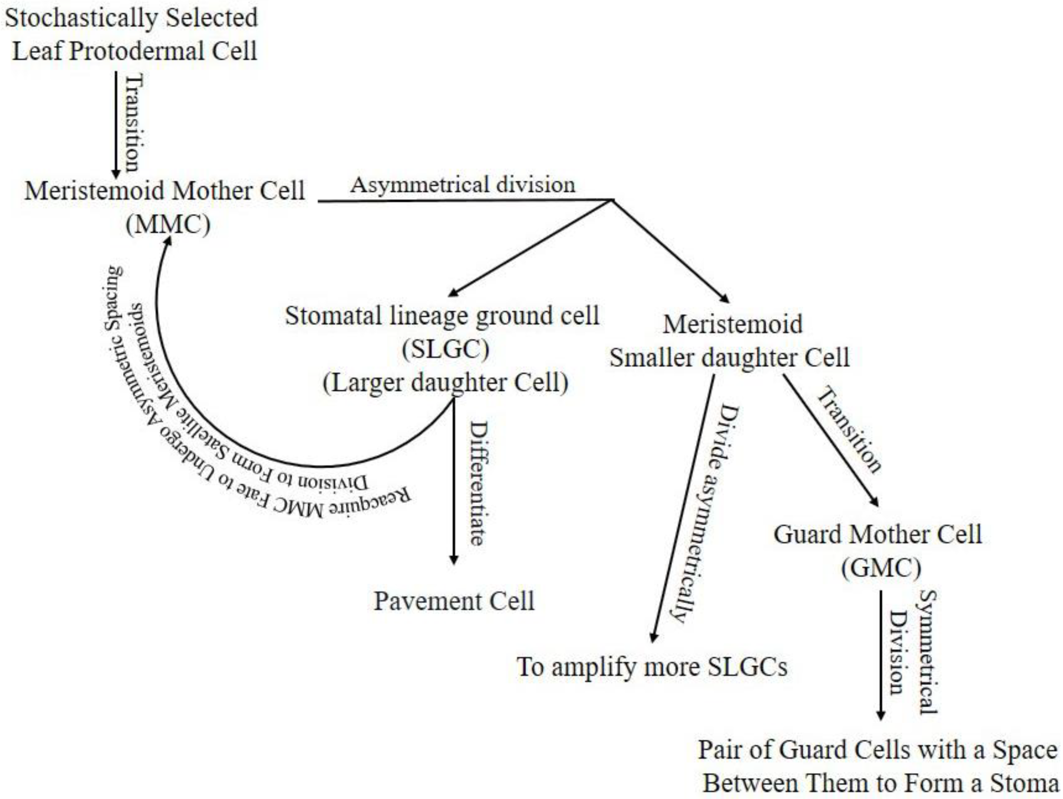
Stomatal lineage starts with stochastically selected leaf protodermal cell transition into the meristemoid mother cell (MMC). The MMC divides asymmetrically into a large daughter cell called a stomatal lineage ground cell (SLGC) and a meristemoid small daughter cell [5]. The SLGC in the daughter cells pair can differentiate into a pavement cell or reacquire MMC fate to undergo asymmetric spacing division to form satellite meristemoids [9]. Meristemoids can divide asymmetrically to amplify more SLGCs, transit into guard mother cells (GMC), or, rarely, exit the lineage [3]. The GMC will divide symmetrically into a pair of GCs with space between them to form a stoma, or its development can be arrested (Figure 1).
2. Role of SPEECHLESS (SPCH), MUTE, and FAMA in Stomata Development
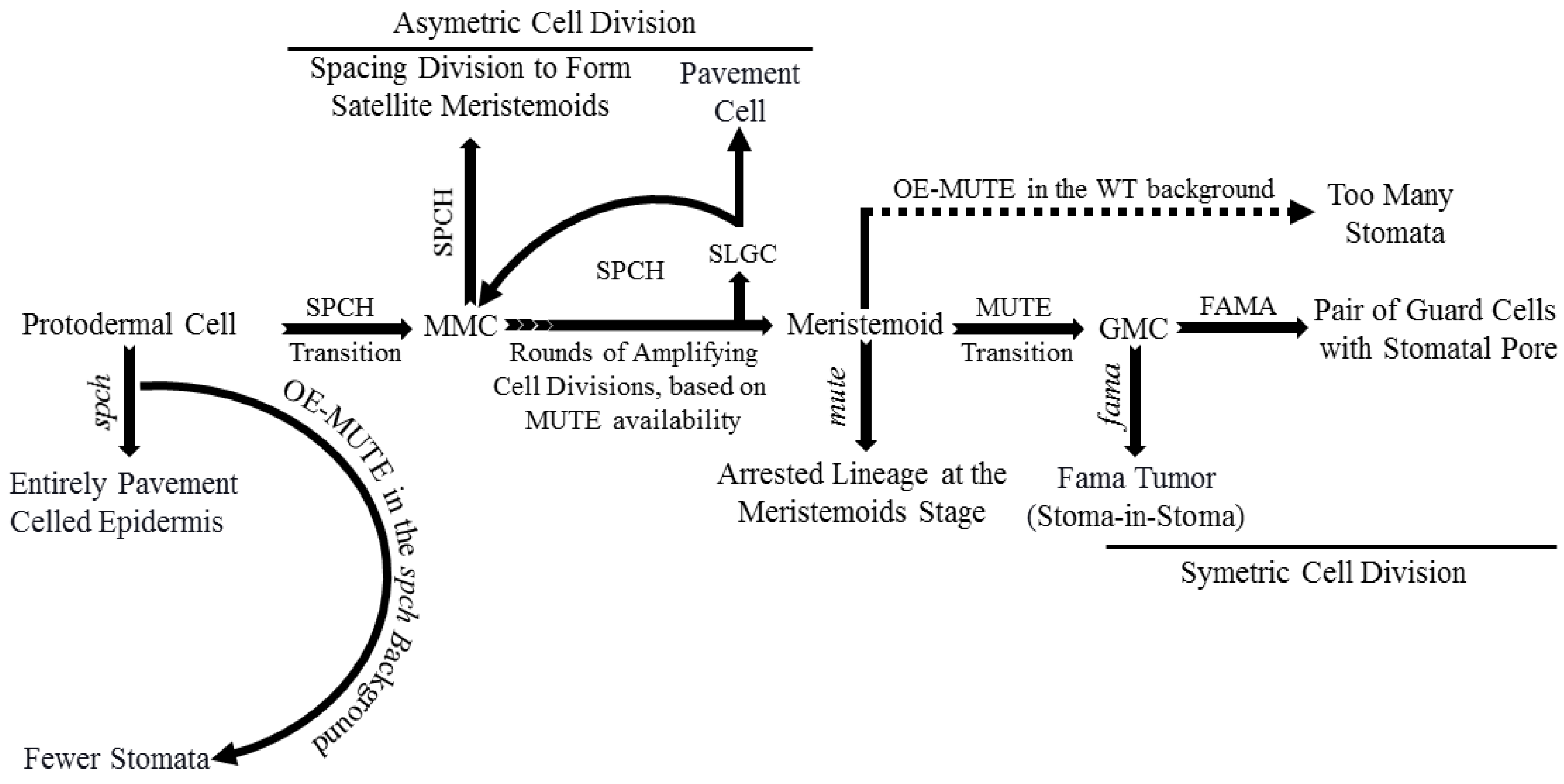
Basic helix-loop-helix (bHLH) transcription factors (TFs) such as SPEECHLESS (SPCH), MUTE, and FAMA are critical for stomatal development. Mutations in any one of these TFs result in improperly developed stomata [6,10,11,12]. SPCH regulates the transition of protodermal cells into MMCs and asymmetric cell division subsequently. SPCH-defective mutants were unable to initiate stomatal lineage, with an entirely pavement-celled epidermis phenotype. The different fate decisions of the meristemoid cells (amplifying division, spacing division, or progression down the lineage) depends on the activity and available level of SPCH (Figure 2) [12,13]. MUTE regulates meristemoids’ transition into GMCs, confirmed by arrested lineage at the meristemoid stage in a MUTE-defective mutant. Overexpression of MUTE in the spch background partially rescued spch-phenotype, and in the wild-type, it exhibited a phenotype, an epidermis densely populated with stomata (Figure 2) [10,14]. These results demonstrate that MUTE has no role in SPCH-regulated asymmetric amplification and spacing cell division [14].
FAMA regulates symmetric cell division of each GMC into a pair of GCs with a space between them to form stomata and terminates lineage cell meristematic activity [11,15,16]. Defective FAMA exhibited uncontrolled symmetric cell division of GMCs with a FAMA-tumor phenotype. FAMA-RETROBLASTOMA-RELATED (RBR) interaction facilitates POLYCOMB REPRESSOR COMPLEX-mediated chromatin methylation to switch off SPCH and MUTE expression. Incorrect FAMA expression resulted in a stoma-in-stoma phenotype, where GCs were pushed into lineage by active SPCH (Figure 2) [11,15,16,17]. Furthermore, FAMA-activated genes such as POTASSIUM CHANNEL IN ARABIDOPSIS THALIANA 1 are required for GCs functioning and control of stomatal aperture [17].
3. Ligand, Receptor, MPK Cascade, and Associated Pathways in Stomatal Development
Peptide signaling, a well-known stomatal development regulator, is underpinned by EPIDERMAL PATTERNING FACTORS 1 (EPF1), EPF2, and EPIDERMAL PATTERNING FACTOR LIKE 9 (EPFL9, denoted as STOMAGEN, STOM) [18,19,20,21,22]. EPF1/2, as negative stomatal development regulators, bind/activate leucine-rich repeat receptor kinases (LRR-RKs). At the same time, STOM competes to bind the LRR-RKs with the EPF1/2 to positively regulate stomatal development [23,24,25]. EPF2 mainly regulates SPCH and the subsequent behavior of meristemoids, whereas EPF1 regulates the one-cell-spacing rule and participates in GMC’s autocrine regulation and inhibition of SLGCs from re-entering the stomatal lineage [26,27]. The ERECTA (ER) family, comprising ER, ERECTA-LIKE 1 (ERL1), and ERL2, are extensively studied LRR-RKs [28]. ERLI and ER play a role in the GMC development stage and facilitate the cell-specific MUTE activity, whereas ERL2 regulates SPCH, mainly as shown in Figure 3 [26,29]. The erl1 and erl2 single mutant and erl1 erl2 double mutant showed reduced SLGC, a less severe phenotype as compared to the er single mutant. The less severe phenotype in ERL mutants than ER mutants demonstrates the ER dominance during stomatal development [30]. Increased stomatal density and index in the er erl1 double mutant and elevated SLGCs in er mutant indicate that they are collectively regulating MUTE in the GMC stage. While slightly increased phenotype in the er erl2 double mutants compared to er, the single mutant suggests that erl2 is one of the main SPCH regulators during stomatal development [30].
TOO MANY MOUTHS (TMM) binds to the ER family (ER and ERL1) to form active extracellular complexes to perceive EPF1 and EPF2 for regulating correct stomatal development [31,32]. TMM, which is mainly expressed in young GCs and stomatal precursor cells, is part of the autoregulatory mechanism and a direct target of SPCH transcription regulators [12,33]. SOMATIC EMBRYOGENESIS RECEPTOR KINASES (SERKs), an irrelative LRR-RK family, have been found required for coupling extracellular TMM/ER complexes to perceive peptide signaling for downstream intracellular signaling, as shown in Figure 3 [34]. There are five homologs (SERK1-5) in Arabidopsis, and single mutants of these genes did not alter stomatal development. However, serk1-1/serk2-1/SERK3-4 triple and serk1-1/serk2-1/SERK3-4/SERK3-5 quadruple mutants displayed clustered stomata phenotypes, and the phenotype for the latter was similar to er erl1 erl2 triple mutants [35].
Upon completion of the ER/SERK/TMM/EPF complex, the ER/SERK cytosolic kinase domain is phosphorylated to target mitogen-activated protein kinase kinase kinase (MPKKK), which is also known as YODA (YDA), a class MPK cascade [2,36,37,38]. YDA, in turn, phosphorylates mitogen-activated protein kinase kinases (MPKKs) that phosphorylate mitogen-activated protein kinase (MPKs) that directly phosphorylate the MPK cascade’s final target [3,37,38]. Only four (MPKK4/5/7/9) and three (MPK3/4/6), out of 20 in Arabidopsis, are involved in stomatal development [37,38,39]. MPK3/6 interacted with SPCH and MPK4 with MUTE, but no in vivo or phenotypic evidence for the latter has been reported previously [36,37,40]. The MPK3/6 was unable to interact with truncated SPCH without MAPK target domain (MPKTD) and exhibited clustered stomata, as shown in Figure 3 [41]. Furthermore, signaling peptides CLAVATA3/ESR-RELATED (CLE) family genes CLE9/10 regulate stomatal lineage development perceived by receptor kinase HAESA-LIKE 1 (HSL1) in a different pathway [42]. HSL1 recruits SERKs as co-receptors in the presence of CLE9/10 for different signaling modes during stomatal development, as shown in Figure 3 [42].
4. Feedback Regulation of SPCH, MUTE, and FAMA
SPCH, MUTE, and FAMA form a heterodimer with SCREAM 1 (SCRM1), SCRM2, and bHLH proteins [36,43,44]. INDUCER OF CBP EXPRESSION 1(ICE1)/SCRM2, as a scaffolding partner, facilitates MPK3/6-mediated phosphorylation of SPCH for its activity inhibition and consequent degradation [36,43,45]. MPK3/6-regulates phosphorylation and subsequent degradation of ICE1/SCRM2 during a process that plays a crucial role in stomatal cell fate specification [43]. Furthermore, FOUR LIPS (FLP) and AtMyb88 constrain GMC division, and an outnumbered GCs phenotype was observed in flp myb88-defective mutants [46]. SPCH/SCRM, which is suppressed by ER/SERK/TMM/EPF-induced phosphorylation of MPK cascade, induces expression of EPF2 and TMM in a negative feedback mechanism [12,33]. MUTE induces ERLI and suppresses EPF2, whereas ERL1, upon perceiving EPF1, regulates meristemoid transition into the GMC stage [20,26,32].
Additionally, cell cycle-related genes such as cyclins A and D (CYCA and CYCD, respectively) in association with CYCLIN-DEPENDENT KINASE A 1;1 and D1;1 (CDKA1;1 and CDKB1;1) play a key role in stomatal development [47,48]. CYCD4 participates in hypocotyl stomatal lineage divisions and CDKB1;1- and CDKA1;1-dominant negative forms, and CYCA higher-order mutants inhibit the division of GMCs [49,50,51,52]. MUTE upregulates cell cycle-related genets (CYCs and CDKs) by binding to their promoters to regulate the symmetric cell division of GMCs. FAMA binds to CDKB1;1 promoter regions, and FLP suppresses the expression of CDKB1;1 and CDKA1;1. As well, FAMA and FLP negatively regulate GMC symmetric division by repressing CDKs and CYCs [51,53,54]. Taken together, MUTE locks in the cells in the differentiation program, promotes regulators of the symmetric division of GMCs, and upregulates FLP and FAMA, which suppress regulators of the cell cycle, to control single symmetric cell division, as shown in Figure 3 [55].
5. Cell Cycle Regulators Join bHLH Transcription Factors to Control Stomatal Development
The SPCH, MUTE, and FAMA bHLH transcription factors interact with ICE1/SCRM and SCRM2, and other bHLH transcription factors through helix-loop-helix domains to form heterodimers [44]. Stomatal lineage progression and cell division are regulated via global transcriptional changes by SPCH, MUTE, FAMA, SCRM/ICE1, SCRM2, and closely related FLP and MYB88 [56]. These transcription factors join cell-cycle core regulators (cyclin-dependent kinases, CDKs) to control stomatal cell multiplication and differentiation throughout the lineage and specific developmental stage [52,56,57]. Cyclin-CDK couple regulates G1/S and G2/M transition phases during stomatal development [50,51]. In Arabidopsis thaliana, CDKA;1 and CDKB1s are known to regulate asymmetric and symmetric cell division during stomatal development, respectively [50,53,58]. CDKA;1 binds with D-type cyclins to facilitate cell entry from G1 into S phase by phosphorylating Retinoblastoma Related 1 (RBR1), a G1-S phase transition inhibitor [58]. CDKA;1-CYCD complex-mediated inhibition of RBR1 results in the release of E2F/DF, cell cycle-related transcription factors, to facilitate the expression of genes required for S phase entry [59].
In contrast, CDKB1;1-CYCA2;3 complex synergistically mediates GMC division by promoting G2-M phase transition [47,52]. Consequently, cdkb1;1 cdkb1;2 double- and cyca2;234 triple-defective mutants exhibit the single undivided GCs phenotype, which was further increased in cyca2;234 cdkb1;1 quadruple mutants [52,53]. Furthermore, cdka;1 and MUTE promoter driving dominant-negative mutants displayed few single undivided GCs, which indicates that CDKA;1-CYCD3 complex activity is partially required in GMC symmetric division [51,60]. Consistently, overexpression of TMM promoter-driven CDKA;1 partially rescued the cdkb1;1 cdkb1;2 single undivided GCs phenotype [60].
SPCH-initiated asymmetric MMC division results in unequal SPCH degradation by protein kinase signaling cascade and scaffold polarity proteins peripheral localization, BREAKING OF ASYMMETRY IN THE STOMATAL LINEAGE (BASL) and POLAR LOCALIZATION DURING ASYMMETRIC DIVISION AND REDISTRIBUTION (POLAR), result in a small meristemoid and large SLGC daughter cells that become a stoma and a pavement cell, respectively [61,62,63].
In Arabidopsis, CDKA;1 is critical in G1-S phase entry and maintenance of stem cells; its loss of function mutant displays few GCs with enormously enlarged epidermal cells [58,60]. The depletion of RBR1 rescued the cdka1 phenotype, and the RBR1 knockout displayed improperly divided meristemoids and stomatal lineage cells. These indicate that CDKA;1 and RBR1 act antagonistically during stomatal development [59,60]. Furthermore, CDKA;1 also phosphorylates SPCH on serine 186 residue, and its substitution with phosphomimetic residue resulted in overactive SPCH-mediated clustered stomata [64]. Hence, CDKA;1 acts in an antagonistic manner to a MAPK cascade, which inhibits stomatal development upon receiving extrinsic peptide signals [32,36,65]. Intriguingly, MAPKs also phosphorylates serine 186 of SPCH [36], indicating that cell cycle inhibitors (cell-cell signals and cell cycle machinery) regulate stomatal development at a SPCH posttranslational modification level. CDKA;1-phosphorylates RBR1, which suppresses SPCH and other stomatal lineage-related genes [17,58,60,64].
SPCH induction increases CYCD3;1 and CYCD3;2, which are abundantly expressed in meristemoids and play an important role in the initial stage of stomatal lineage [12,66]. CYCD3 triple loss of function decreased meristemoids, amplifying SLGCs asymmetric spacing divisions, significantly reducing the number of leaf epidermal cells [67,68]. On the other hand, overexpression of CYCD3 resulted in an abnormal increase in cell number and ectopic cell division, demonstrating CYCD3’s role in regulating cell number and division in developing leaves [67]. Molecular intersections, role, and mechanism of cell cycle regulators during stomatal formation have been modeled in Figure 4.
6. Fate Transition and Cell Divisions in Stomatal Lineage
GIGAS CELL1 (GIG1), which negatively regulates anaphase-promoting complex/cyclosome (APC/C), is necessary for cell fate transition and subsequent mitotic cell division. GIG1 loss of function mutants displayed giant GIGAS cells with a mixed-cell fate [69]. GIGAS cells are jigsaw-puzzle-piece-shaped pavement cells but express SPCH, EPF2, TMM, E1728, and KAT1, markers of stomatal lineage and GCs. Interestingly, spch-defective mutants did not form GIGAS or GCs, which confirms their stomatal lineage origin. Originally, gig1-1 and gig1-2 allelic recessive mutants were obtained from a myb3r4 mutant enhancer screen in a forward genetic screen [56,69]. MYB3R4 transcription factor positive regulators are required for G2-M phase transition and mitotic cell division [69]. SPCH regulates the meristemoid phase of stomatal lineage by controlling the expression of thousands of genes, including its own negative regulators such as BASL, EPF2, TMM, SOL1, and SOL2 [12,33,70].
Key stomatal lineage regulator, SPCH, induced SOL1/2 mediate cell fate transition factors and post-SPCH identities [70]. SOL1/2 vanish just before asymmetric and symmetric cell division. Ectopic expression of SOL2-suppressed GMC division resulted in single undivided GCs. Consistently, sol1 sol2 double defective mutants exhibit an increased number of small cells and paired stomata in the lineage’s early and late phase, respectively. Furthermore, inappropriately persistent MUTE expression after cell division in the sol1 sol2 mutant demonstrates that sole MUTE expression in the absence of SOL1/2 is not enough for GMC fate determination [70]. Interestingly, TSO1, a homology of SOL1/2, expresses in the stomatal lineage with the opposite to SOLs activity; simultaneously, it is not a target of either SPCH or MUTE [12,55]. Knockout mutants of TSO1 in the sol1 sol2 background rescues the sol1 sol2 phenotype and decreases the number of small cells and paired GCs [70]. In conclusion, GIG1 and MYB3R4 synergistically specify cell fate during stomatal lineage development, SPCH induced SOL1/2 mediates fate transition, and TSO1 interacts with MYB3R1 and promotes GMC’s symmetric division (Figure 4).
7. Highly Conserved SnRK1 Positively Regulates SPCH-Mediated Stomatal Development
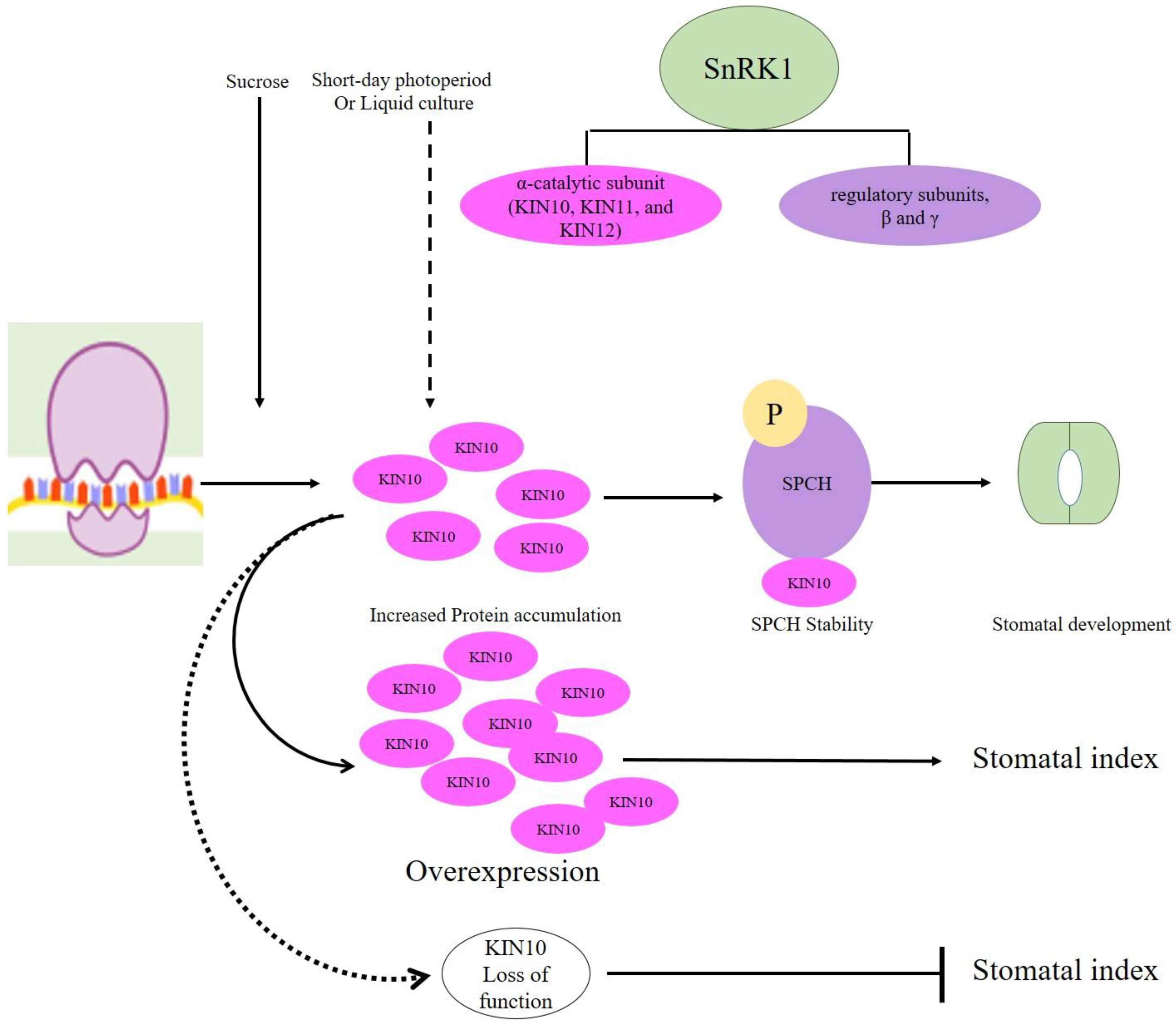
Energy hemostasis during stomatal development is controlled by sucrose non-fermenting-1 (SNF1)-related kinase 1 (SnRK1), which is a central energy sensor kinase in plants [71]. In Arabidopsis, SnRK1 functions as heterotrimeric complexes composed of one α-catalytic subunit (KIN10, KIN11, and KIN12) and two regulatory subunits, β and γ [72,73]. Sucrose-induced KIN10 expresses in all epidermal cells but exhibits cell-specific subcellular localization, accumulation in liquid culture conditions [73]. Furthermore, Han et al. (2020) report that stomatal lineage cells are highly enriched with nuclear-localized KIN10 that phosphorylates SPCH to increase stability and subsequent stomatal development under stress conditions. Overexpression of KIN10 increased, whereas KIN10 and KIN11 loss of function reduced stomatal index under short-day light or in liquid medium with 1% sucrose. However, kin10 mutants displayed increased stomatal index on the solid medium (without sucrose) and low light conditions. Moreover, SPCH stability was greatly reduced by a mutation in KIN10 phosphorylation sites [73]. To sum up, SnRK1 α-catalytic subunit (KIN10) stabilizes SPCH, positively regulating stomatal development (Figure 5).
8. Protein Phosphatase 2A-Mediates SPCH Stability by Dephosphorylating It
Protein Phosphatase 2A (PP2A) heterotrimeric complexes comprise scaffolding subunit A, regulatory subunit B, and catalytic subunit C, with multiple isoforms for each subunit and differentially assembled complexes in Arabidopsis [74,75]. These complexes, differentially assembled from various isoforms of these A, B, and C subunits, regulate development, growth, metabolism, and stress responses in plants [75,76]. PP2A controls blue light-mediated stomatal movement, and in association with SnRK2, it regulates ABA-triggered stomatal closure [77,78]. PP2A function is required for the prophase band formation in Arabidopsis and the orientation of cell division in maize during stomatal development [79,80]. Recently, the role of PP2A-mediated positive regulation SPCH stability by direct interaction between SPCH and A subunit of PP2A to promote stomatal development has been reported [81].
Brassinosteroid (BR) promotes SPCH function by suppressing glycogen synthase kinase 3 (GSK3)/BRASSINOSTEROID insensitive 2 (BIN2), which are phosphorylating SPCH for subsequent degradation [36,82]. On the other hand, CYCLIN-DEPENDENT KINASE A;1 (CDKA;1)-mediated SPCH phosphorylation positively regulates SPCH function [64]. Bian et al. (2020) reported that PP2A increases SPCH accumulation and stabilization by direct interaction between A subunit and SPCH. PP2A dephosphorylates SPCH at a specific site. However, overexpression of PP2A-A did not exhibit a visible phenotype, which indicates that the other two subunits are also required for SPCH stabilization and subsequent consequences, as shown in Figure 6 [81].
9. IDD16 Represses SPCH-Induced Stomatal Initiation
The function of a C2H2 zinc finger transcription factor of the INDETERMINATE DOMAIN (IDD) family, encoded by AT1G25250, plays an essential role in organ-morphogenesis and gravitropic responses [83,84]. In Arabidopsis, C2H2 transcription factor IDD16 plays a critical regulatory role in stomatal development. IDD16 by trans-repression of SPCH negatively regulates stomatal initiation. In a dose-dependent manner, the overexpression of IDD16 reduced abaxial stomatal density in Arabidopsis. The initiation of stomatal lineage was significantly inhibited in IDD16 overexpression plants (IDD16-OE). Consistently, IDD16-OE seedlings displayed a severe reduction in SPCH levels. Conversely, the IDD16-RNAi transgenic line exhibited increased stomatal density, demonstrating that IDD16 is an intrinsic stomatal development regulator. Furthermore, ChIP analysis results showed that IDD16 could bind the promoter of SPCH [85]. With respect to the other eight down-regulated genes (MUTE, FAMA, TMM, SDD1, EPF1, EPF2, BASAL, and POLAR), which are expressed explicitly in MMC, meristemoid cells, GMCs, and GCs are attributed to reduced stomatal precursor cells development in IDD16-OE plants [85].
In some of the IDD16 overexpression transgenic lines, stomata could not form on the abaxial epidermis, whereas the adaxial side displayed a considerable number of stomata for survival and life cycle completion. Mutations in many of the stomatal development regulators such as SPCH, TMM, and SDD1 display similar changes in abaxial and adaxial epidermises [6,85]. The tmm and sdd1 mutants displayed stomata in clusters and increased stomatal density and different adaxial-abaxial ratios [86]. A complex network of regulatory genes regulates the maintenance and acquisition of adaxial-abaxial leaf polarity, resulting in the asymmetric distribution of different cell types in mature leaves [87,88]. In Arabidopsis, pavement cell shape and pattern and stomatal density and pattern are different between both epidermises, which may be regulated by IDD16 [85].
10. Conclusions
We can conclude that SPCH promotes EPF2, the negative stomatal regulator, and TMM component stomatal complexes inhibit stomatal development in a negative feedback mechanism. In contrast, MUTE inhibits EPF2 and induces ER-family to promote SPCH-mediated stomatal lineage and development. Moreover, the PP2A A subunit, in association with its B and C subunits, increases SPCH accumulation, stability, and subsequent SPCH-induced stomatal lineage regulations. Similarly, SNRKs also positively regulate stomatal development by positively regulating SPCH, while IDD16 negatively regulates stomatal lineage initiation by suppressing SPCH.
Author Contributions
Conceptualization, A.W. and M.X.; methodology, A.W; software, A.W; validation, A.W. and M.X.; formal analysis, A.W.; investigation, A.W.; resources, A.W.; data curation, A.W.; writing—original draft preparation, A.W.; writing—review and editing, A.W.; visualization, A.W.; supervision, L.W. and M.X.; project administration, A.W.; funding acquisition, L.W. All authors have read and agreed to the published version of the manuscript.
Funding
This research was funded by National Key Research and Development Program of China, grant number (2017YFA0604300, 2018YFA0606500)” and “The APC was funded by Lin Wang.
Institutional Review Board Statement
Not applicable.
Informed Consent Statement
“Not applicable” for studies not involving humans.
Data Availability Statement
“Not applicable” for studies not involving humans.
Acknowledgments
We are thankful to Steven Xu, a native English speaker, for proofreading our manuscript.
Conflicts of Interest
The authors declare that they have no conflict of interest.
References
- Martin, C.; Glover, B.J. Functional aspects of cell patterning in aerial epidermis. Curr. Opin. Plant Biol. 2007, 10, 70–82. [Google Scholar] [CrossRef] [PubMed]
- Bergmann, D.C.; Lukowitz, W.; Somerville, C.R. Stomatal development and pattern controlled by a MAPKK kinase. Science 2004, 304, 1494–1497. [Google Scholar] [CrossRef] [PubMed] [Green Version]
- Zoulias, N.; Harrison, E.L.; Casson, S.A.; Gray, J.E. Molecular control of stomatal development. Biochem. J. 2018, 475, 441–454. [Google Scholar] [CrossRef] [Green Version]
- Papanatsiou, M.; Amtmann, A.; Blatt, M.R. Stomatal spacing safeguards stomatal dynamics by facilitating guard cell ion transport independent of the epidermal solute reservoir. Plant Physiol. 2016, 172, 254–263. [Google Scholar] [CrossRef] [PubMed] [Green Version]
- Larkin, J.C.; Marks, M.D.; Nadeau, J.; Sack, F. Epidermal cell fate and patterning in leaves. Plant Cell 1997, 9, 1109. [Google Scholar] [CrossRef] [Green Version]
- MacAlister, C.A.; Ohashi-Ito, K.; Bergmann, D.C. Transcription factor control of asymmetric cell divisions that establish the stomatal lineage. Nature 2007, 445, 537–540. [Google Scholar] [CrossRef]
- Hu, Z.; Cools, T.; De Veylder, L. Mechanisms Used by Plants to Cope with DNA Damage. Annu. Rev. Plant Biol. 2016, 67, 439–462. [Google Scholar] [CrossRef]
- Akita, K.; Hasezawa, S.; Higaki, T. Breaking of Plant Stomatal One-Cell-Spacing Rule by Sugar Solution Immersion. PLoS ONE 2013, 8, 72456. [Google Scholar] [CrossRef]
- Le, J.; Zou, J.; Yang, K.; Wang, M. Signaling to stomatal initiation and cell division. Front. Plant Sci. 2014, 5, 297. [Google Scholar] [CrossRef] [Green Version]
- Pillitteri, L.J.; Sloan, D.B.; Bogenschutz, N.L.; Torii, K.U. Termination of asymmetric cell division and differentiation of stomata. Nature 2007, 445, 501–505. [Google Scholar] [CrossRef] [PubMed]
- Ohashi-Ito, K.; Bergmann, D.C. Arabidopsis FAMA controls the final proliferation/differentiation switch during stomatal development. Plant Cell 2006, 18, 2493–2505. [Google Scholar] [CrossRef] [PubMed] [Green Version]
- Lau, O.S.; Davies, K.A.; Chang, J.; Adrian, J.; Rowe, M.H.; Ballenger, C.E.; Bergmann, D.C. Direct roles of SPEECHLESS in the specification of stomatal self-renewing cells. Science 2014, 345, 1605–1609. [Google Scholar] [CrossRef] [Green Version]
- Serna, L. Emerging parallels between stomatal and muscle cell lineages. Plant Physiol. 2009, 149, 1625–1631. [Google Scholar] [CrossRef] [Green Version]
- Liu, T.; Ohashi-Ito, K.; Bergmann, D.C. Orthologs of Arabidopsis thaliana stomatal bHLH genes and regulation of stomatal development in grasses. Development 2009, 136, 2265–2276. [Google Scholar] [CrossRef] [Green Version]
- Lee, E.; Lucas, J.R.; Goodrich, J.; Sack, F.D. Arabidopsis guard cell integrity involves the epigenetic stabilization of the FLP and FAMA transcription factor genes. Plant J. 2014, 78, 566–577. [Google Scholar] [CrossRef] [PubMed]
- Lee, E.; Lucas, J.R.; Sack, F.D. Deep functional redundancy between FAMA and FOUR LIPS in stomatal development. Plant J. 2014, 78, 555–565. [Google Scholar] [CrossRef] [PubMed]
- Matos, J.L.; Lau, O.S.; Hachez, C.; Cruz-Ramírez, A.; Scheres, B.; Bergmann, D.C. Irreversible fate commitment in the Arabidopsis stomatal lineage requires a FAMA and RETINOBLASTOMA-RELATED module. Elife 2014, 3, e03271. [Google Scholar] [CrossRef] [PubMed]
- Hara, K.; Yokoo, T.; Kajita, R.; Onishi, T.; Yahata, S.; Peterson, K.M.; Torii, K.U.; Kakimoto, T. Epidermal cell density is autoregulated via a secretory peptide, EPIDERMAL PATTERNING FACTOR 2 in Arabidopsis leaves. Plant Cell Physiol. 2009, 50, 1019–1031. [Google Scholar] [CrossRef] [PubMed] [Green Version]
- Hunt, L.; Gray, J.E. The signaling peptide EPF2 controls asymmetric cell divisions during stomatal development. Curr. Biol. 2009, 19, 864–869. [Google Scholar] [CrossRef] [Green Version]
- Hara, K.; Kajita, R.; Torii, K.U.; Bergmann, D.C.; Kakimoto, T. The secretory peptide gene EPF1 enforces the stomatal one-cell-spacing rule. Genes Dev. 2007, 21, 1720–1725. [Google Scholar] [CrossRef] [Green Version]
- Sugano, S.S.; Shimada, T.; Imai, Y.; Okawa, K.; Tamai, A.; Mori, M.; Hara-Nishimura, I. Stomagen positively regulates stomatal density in Arabidopsis. Nature 2010, 463, 241–244. [Google Scholar] [CrossRef] [PubMed] [Green Version]
- Hunt, L.; Bailey, K.J.; Gray, J.E. The signalling peptide EPFL9 is a positive regulator of stomatal development. New Phytol. 2010, 186, 609–614. [Google Scholar] [CrossRef]
- Ohki, S.; Takeuchi, M.; Mori, M. The NMR structure of stomagen reveals the basis of stomatal density regulation by plant peptide hormones. Nat. Commun. 2011, 2, 512. [Google Scholar] [CrossRef] [Green Version]
- Lin, G.; Zhang, L.; Han, Z.; Yang, X.; Liu, W.; Li, E.; Chang, J.; Qi, Y.; Shpak, E.D.; Chai, J. A receptor-like protein acts as a specificity switch for the regulation of stomatal development. Genes Dev. 2017, 31, 927–938. [Google Scholar] [CrossRef] [PubMed]
- Lee, J.S.; Hnilova, M.; Maes, M.; Lin, Y.-C.L.; Putarjunan, A.; Han, S.-K.; Avila, J.; Torii, K.U. Competitive binding of antagonistic peptides fine-tunes stomatal patterning. Nature 2015, 522, 439–443. [Google Scholar] [CrossRef] [PubMed] [Green Version]
- Qi, X.; Han, S.-K.; Dang, J.H.; Garrick, J.M.; Ito, M.; Hofstetter, A.K.; Torii, K.U. Autocrine regulation of stomatal differentiation potential by EPF1 and ERECTA-LIKE1 ligand-receptor signaling. Elife 2017, 6, e24102. [Google Scholar] [CrossRef]
- Jewaria, P.K.; Hara, T.; Tanaka, H.; Kondo, T.; Betsuyaku, S.; Sawa, S.; Sakagami, Y.; Aimoto, S.; Kakimoto, T. Differential effects of the peptides Stomagen, EPF1 and EPF2 on activation of MAP kinase MPK6 and the SPCH protein level. Plant Cell Physiol. 2013, 54, 1253–1262. [Google Scholar] [CrossRef] [Green Version]
- Shpak, E.D.; Berthiaume, C.T.; Hill, E.J.; Torii, K.U. Synergistic interaction of three ERECTA-family receptor-like kinases controls Arabidopsis organ growth and flower development by promoting cell proliferation. Development 2004, 131, 1491–1501. [Google Scholar] [CrossRef] [Green Version]
- Ho, C.-M.K.; Paciorek, T.; Abrash, E.; Bergmann, D.C. Modulators of stomatal lineage signal transduction alter membrane contact sites and reveal specialization among ERECTA kinases. Dev. Cell 2016, 38, 345–357. [Google Scholar] [CrossRef] [Green Version]
- Shpak, E.D.; McAbee, J.M.; Pillitteri, L.J.; Torii, K.U. Stomatal patterning and differentiation by synergistic interactions of receptor kinases. Science 2005, 309, 290–293. [Google Scholar] [CrossRef]
- Geisler, M.; Nadeau, J.; Sack, F.D. Oriented asymmetric divisions that generate the stomatal spacing pattern in Arabidopsis are disrupted by the too many mouths mutation. Plant Cell 2000, 12, 2075–2086. [Google Scholar] [CrossRef] [PubMed] [Green Version]
- Lee, J.S.; Kuroha, T.; Hnilova, M.; Khatayevich, D.; Kanaoka, M.M.; McAbee, J.M.; Sarikaya, M.; Tamerler, C.; Torii, K.U. Direct interaction of ligand–receptor pairs specifying stomatal patterning. Genes Dev. 2012, 26, 126–136. [Google Scholar] [CrossRef] [PubMed] [Green Version]
- Horst, R.J.; Fujita, H.; Lee, J.S.; Rychel, A.L.; Garrick, J.M.; Kawaguchi, M.; Peterson, K.M.; Torii, K.U. Molecular framework of a regulatory circuit initiating two-dimensional spatial patterning of stomatal lineage. PLoS Genet. 2015, 11, e1005374. [Google Scholar] [CrossRef] [Green Version]
- Aan den Toorn, M.; Albrecht, C.; de Vries, S. On the origin of SERKs: Bioinformatics analysis of the somatic embryogenesis receptor kinases. Mol. Plant 2015, 8, 762–782. [Google Scholar] [CrossRef] [PubMed] [Green Version]
- Meng, X.; Chen, X.; Mang, H.; Liu, C.; Yu, X.; Gao, X.; Torii, K.U.; He, P.; Shan, L. Differential function of Arabidopsis SERK family receptor-like kinases in stomatal patterning. Curr. Biol. 2015, 25, 2361–2372. [Google Scholar] [CrossRef] [PubMed] [Green Version]
- Lampard, G.R.; MacAlister, C.A.; Bergmann, D.C. Arabidopsis stomatal initiation is controlled by MAPK-mediated regulation of the bHLH SPEECHLESS. Science 2008, 322, 1113–1116. [Google Scholar] [CrossRef] [PubMed] [Green Version]
- Lampard, G.R.; Lukowitz, W.; Ellis, B.E.; Bergmann, D.C. Novel and expanded roles for MAPK signaling in Arabidopsis stomatal cell fate revealed by cell type–specific manipulations. Plant Cell 2009, 21, 3506–3517. [Google Scholar] [CrossRef] [Green Version]
- Lampard, G.R.; Wengier, D.L.; Bergmann, D.C. Manipulation of mitogen-activated protein kinase kinase signaling in the Arabidopsis stomatal lineage reveals motifs that contribute to protein localization and signaling specificity. Plant Cell 2014, 26, 3358–3371. [Google Scholar] [CrossRef] [Green Version]
- Hsu, L.-C.; Liu, Y.-T.; Tzou, Y.-M. Comparison of the spectroscopic speciation and chemical fractionation of chromium in contaminated paddy soils. J. Hazard. Mater. 2015, 296, 230–238. [Google Scholar] [CrossRef]
- Wang, H.; Ngwenyama, N.; Liu, Y.; Walker, J.C.; Zhang, S. Stomatal development and patterning are regulated by environmentally responsive mitogen-activated protein kinases in Arabidopsis. Plant Cell 2007, 19, 63–73. [Google Scholar] [CrossRef] [Green Version]
- Dong, J.; MacAlister, C.A.; Bergmann, D.C. BASL controls asymmetric cell division in Arabidopsis. Cell 2009, 137, 1320–1330. [Google Scholar] [CrossRef] [PubMed] [Green Version]
- Qian, P.; Song, W.; Yokoo, T.; Minobe, A.; Wang, G.; Ishida, T.; Sawa, S.; Chai, J.; Kakimoto, T. The CLE9/10 secretory peptide regulates stomatal and vascular development through distinct receptors. Nat. Plants 2018, 4, 1071–1081. [Google Scholar] [CrossRef] [PubMed]
- Putarjunan, A.; Ruble, J.; Srivastava, A.; Zhao, C.; Rychel, A.L.; Hofstetter, A.K.; Tang, X.; Zhu, J.-K.; Tama, F.; Zheng, N. Bipartite anchoring of SCREAM enforces stomatal initiation by coupling MAP kinases to SPEECHLESS. Nat. Plants 2019, 5, 742–754. [Google Scholar] [CrossRef]
- Kanaoka, M.M.; Pillitteri, L.J.; Fujii, H.; Yoshida, Y.; Bogenschutz, N.L.; Takabayashi, J.; Zhu, J.-K.; Torii, K.U. SCREAM/ICE1 and SCREAM2 specify three cell-state transitional steps leading to Arabidopsis stomatal differentiation. Plant Cell 2008, 20, 1775–1785. [Google Scholar] [CrossRef] [PubMed] [Green Version]
- Chen, L.; Wu, Z.; Hou, S. SPEECHLESS Speaks Loudly in Stomatal Development. Front. Plant Sci. 2020, 11. [Google Scholar] [CrossRef]
- Lai, L.B.; Nadeau, J.A.; Lucas, J.; Lee, E.-K.; Nakagawa, T.; Zhao, L.; Geisler, M.; Sack, F.D. The Arabidopsis R2R3 MYB proteins FOUR LIPS and MYB88 restrict divisions late in the stomatal cell lineage. Plant Cell 2005, 17, 2754–2767. [Google Scholar] [CrossRef] [Green Version]
- Boudolf, V.; Lammens, T.; Boruc, J.; Van Leene, J.; Van Den Daele, H.; Maes, S.; Van Isterdael, G.; Russinova, E.; Kondorosi, E.; Witters, E. CDKB1; 1 forms a functional complex with CYCA2; 3 to suppress endocycle onset. Plant Physiol. 2009, 150, 1482–1493. [Google Scholar] [CrossRef] [Green Version]
- De Veylder, L.; Beeckman, T.; Inzé, D. The ins and outs of the plant cell cycle. Nat. Rev. Mol. Cell Biol. 2007, 8, 655–665. [Google Scholar] [CrossRef]
- Kono, A.; Umeda-Hara, C.; Adachi, S.; Nagata, N.; Konomi, M.; Nakagawa, T.; Uchimiya, H.; Umeda, M. The Arabidopsis D-type cyclin CYCD4 controls cell division in the stomatal lineage of the hypocotyl epidermis. Plant Cell 2007, 19, 1265–1277. [Google Scholar] [CrossRef] [Green Version]
- Boudolf, V.; Barrôco, R.; de Almeida Engler, J.; Verkest, A.; Beeckman, T.; Naudts, M.; Inzé, D.; De Veylder, L. B1-type cyclin-dependent kinases are essential for the formation of stomatal complexes in Arabidopsis thaliana. Plant Cell 2004, 16, 945–955. [Google Scholar] [CrossRef] [Green Version]
- Yang, K.; Wang, H.; Xue, S.; Qu, X.; Zou, J.; Le, J. Requirement for A-type cyclin-dependent kinase and cyclins for the terminal division in the stomatal lineage of Arabidopsis. J. Exp. Bot. 2014, 65, 2449–2461. [Google Scholar] [CrossRef] [PubMed] [Green Version]
- Vanneste, S.; Coppens, F.; Lee, E.; Donner, T.J.; Xie, Z.; Van Isterdael, G.; Dhondt, S.; De Winter, F.; De Rybel, B.; Vuylsteke, M. Developmental regulation of CYCA2s contributes to tissue-specific proliferation in Arabidopsis. EMBO J. 2011, 30, 3430–3441. [Google Scholar] [CrossRef] [PubMed] [Green Version]
- Xie, Z.; Lee, E.; Lucas, J.R.; Morohashi, K.; Li, D.; Murray, J.A.; Sack, F.D.; Grotewold, E. Regulation of cell proliferation in the stomatal lineage by the Arabidopsis MYB FOUR LIPS via direct targeting of core cell cycle genes. Plant Cell 2010, 22, 2306–2321. [Google Scholar] [CrossRef] [PubMed] [Green Version]
- Hachez, C.; Ohashi-Ito, K.; Dong, J.; Bergmann, D.C. Differentiation of Arabidopsis guard cells: Analysis of the networks incorporating the basic helix-loop-helix transcription factor, FAMA. Plant Physiol. 2011, 155, 1458–1472. [Google Scholar] [CrossRef] [Green Version]
- Han, S.-K.; Qi, X.; Sugihara, K.; Dang, J.H.; Endo, T.A.; Miller, K.L.; Kim, E.-D.; Miura, T.; Torii, K.U. MUTE directly orchestrates cell-state switch and the single symmetric division to create stomata. Dev. Cell 2018, 45, 303–315. e305. [Google Scholar] [CrossRef] [Green Version]
- Han, S.-K.; Torii, K.U. Linking cell cycle to stomatal differentiation. Curr. Opin. Plant Biol. 2019, 51, 66–73. [Google Scholar] [CrossRef]
- Blomme, J.; Inzé, D.; Gonzalez, N. The cell-cycle interactome: A source of growth regulators? J. Exp. Bot. 2014, 65, 2715–2730. [Google Scholar] [CrossRef] [Green Version]
- Nowack, M.K.; Harashima, H.; Dissmeyer, N.; Bouyer, D.; Weimer, A.K.; De Winter, F.; Yang, F.; Schnittger, A. Genetic framework of cyclin-dependent kinase function in Arabidopsis. Dev. Cell 2012, 22, 1030–1040. [Google Scholar] [CrossRef] [Green Version]
- Borghi, L.; Gutzat, R.; Fütterer, J.; Laizet, Y.H.; Hennig, L.; Gruissem, W. Arabidopsis RETINOBLASTOMA-RELATED is required for stem cell maintenance, cell differentiation, and lateral organ production. Plant Cell 2010, 22, 1792–1811. [Google Scholar] [CrossRef] [Green Version]
- Weimer, A.K.; Nowack, M.K.; Bouyer, D.; Harashima, H.; Naseer, S.; De Winter, F.; Dissmeyer, N.; Geldner, N.; Schnittger, A. Retinoblastoma related1 regulates asymmetric cell divisions in Arabidopsis. Plant Cell 2012, 24, 4083–4095. [Google Scholar] [CrossRef] [Green Version]
- Zhang, Y.; Wang, P.; Shao, W.; Zhu, J.-K.; Dong, J. The BASL polarity protein controls a MAPK signaling feedback loop in asymmetric cell division. Dev. Cell 2015, 33, 136–149. [Google Scholar] [CrossRef] [PubMed] [Green Version]
- Pillitteri, L.J.; Peterson, K.M.; Horst, R.J.; Torii, K.U. Molecular profiling of stomatal meristemoids reveals new component of asymmetric cell division and commonalities among stem cell populations in Arabidopsis. Plant Cell 2011, 23, 3260–3275. [Google Scholar] [CrossRef] [Green Version]
- Houbaert, A.; Zhang, C.; Tiwari, M.; Wang, K.; de Marcos Serrano, A.; Savatin, D.V.; Urs, M.J.; Zhiponova, M.K.; Gudesblat, G.E.; Vanhoutte, I. POLAR-guided signalling complex assembly and localization drive asymmetric cell division. Nature 2018, 563, 574–578. [Google Scholar] [CrossRef] [PubMed]
- Yang, K.-Z.; Jiang, M.; Wang, M.; Xue, S.; Zhu, L.-L.; Wang, H.-Z.; Zou, J.-J.; Lee, E.-K.; Sack, F.; Le, J. Phosphorylation of serine 186 of bHLH transcription factor SPEECHLESS promotes stomatal development in Arabidopsis. Mol. Plant 2015, 8, 783–795. [Google Scholar] [CrossRef] [PubMed] [Green Version]
- Wang, H.; Liu, Y.; Bruffett, K.; Lee, J.; Hause, G.; Walker, J.C.; Zhang, S. Haplo-insufficiency of MPK3 in MPK6 mutant background uncovers a novel function of these two MAPKs in Arabidopsis ovule development. Plant Cell 2008, 20, 602–613. [Google Scholar] [CrossRef] [Green Version]
- Adrian, J.; Chang, J.; Ballenger, C.E.; Bargmann, B.O.; Alassimone, J.; Davies, K.A.; Lau, O.S.; Matos, J.L.; Hachez, C.; Lanctot, A. Transcriptome dynamics of the stomatal lineage: Birth, amplification, and termination of a self-renewing population. Dev. Cell 2015, 33, 107–118. [Google Scholar] [CrossRef] [Green Version]
- Dewitte, W.; Scofield, S.; Alcasabas, A.A.; Maughan, S.C.; Menges, M.; Braun, N.; Collins, C.; Nieuwland, J.; Prinsen, E.; Sundaresan, V. Arabidopsis CYCD3 D-type cyclins link cell proliferation and endocycles and are rate-limiting for cytokinin responses. Proc. Natl. Acad. Sci. USA 2007, 104, 14537–14542. [Google Scholar] [CrossRef] [Green Version]
- Elsner, J.; Michalski, M.; Kwiatkowska, D. Spatiotemporal variation of leaf epidermal cell growth: A quantitative analysis of Arabidopsis thaliana wild-type and triple cyclinD3 mutant plants. Ann. Bot. 2012, 109, 897–910. [Google Scholar] [CrossRef] [Green Version]
- Iwata, E.; Ikeda, S.; Matsunaga, S.; Kurata, M.; Yoshioka, Y.; Criqui, M.-C.; Genschik, P.; Ito, M. GIGAS CELL1, a novel negative regulator of the anaphase-promoting complex/cyclosome, is required for proper mitotic progression and cell fate determination in Arabidopsis. Plant Cell 2011, 23, 4382–4393. [Google Scholar] [CrossRef] [Green Version]
- Simmons, A.R.; Davies, K.A.; Wang, W.; Liu, Z.; Bergmann, D.C. SOL1 and SOL2 regulate fate transition and cell divisions in the Arabidopsis stomatal lineage. Development 2019, 146. [Google Scholar] [CrossRef] [Green Version]
- Coello, P.; Hey, S.J.; Halford, N.G. The sucrose non-fermenting-1-related (SnRK) family of protein kinases: Potential for manipulation to improve stress tolerance and increase yield. J. Exp. Bot. 2011, 62, 883–893. [Google Scholar] [CrossRef] [Green Version]
- Broeckx, T.; Hulsmans, S.; Rolland, F. The plant energy sensor: Evolutionary conservation and divergence of SnRK1 structure, regulation, and function. J. Exp. Bot. 2016, 67, 6215–6252. [Google Scholar] [CrossRef] [PubMed]
- Han, C.; Liu, Y.; Shi, W.; Qiao, Y.; Wang, L.; Tian, Y.; Fan, M.; Deng, Z.; Lau, O.S.; De Jaeger, G. KIN10 promotes stomatal development through stabilization of the SPEECHLESS transcription factor. Nat. Commun. 2020, 11, 4214. [Google Scholar] [CrossRef] [PubMed]
- Janssens, V. A highly regulated family of serine/threonine phosphatases implicated in cell growth and signalling. Biochem. J. 2001, 353, 417–439. [Google Scholar] [CrossRef] [PubMed]
- Uhrig, R.G.; Labandera, A.-M.; Moorhead, G.B. Arabidopsis PPP family of serine/threonine protein phosphatases: Many targets but few engines. Trends Plant Sci. 2013, 18, 505–513. [Google Scholar] [CrossRef]
- Durian, G.; Rahikainen, M.; Alegre, S.; Brosché, M.; Kangasjärvi, S. Protein phosphatase 2A in the regulatory network underlying biotic stress resistance in plants. Front. Plant Sci. 2016, 7, 812. [Google Scholar] [CrossRef] [Green Version]
- Waadt, R.; Manalansan, B.; Rauniyar, N.; Munemasa, S.; Booker, M.A.; Brandt, B.; Waadt, C.; Nusinow, D.A.; Kay, S.A.; Kunz, H.-H. Identification of open stomata1-interacting proteins reveals interactions with sucrose non-fermenting1-related protein kinases2 and with type 2A protein phosphatases that function in abscisic acid responses. Plant Physiol. 2015, 169, 760–779. [Google Scholar] [CrossRef]
- Tseng, T.-S.; Briggs, W.R. The Arabidopsis rcn1-1 mutation impairs dephosphorylation of Phot2, resulting in enhanced blue light responses. Plant Cell 2010, 22, 392–402. [Google Scholar] [CrossRef] [Green Version]
- Wright, A.J.; Gallagher, K.; Smith, L.G. discordia1 and alternative discordia1 function redundantly at the cortical division site to promote preprophase band formation and orient division planes in maize. Plant Cell 2009, 21, 234–247. [Google Scholar] [CrossRef] [Green Version]
- Spinner, L.; Gadeyne, A.; Belcram, K.; Goussot, M.; Moison, M.; Duroc, Y.; Eeckhout, D.; De Winne, N.; Schaefer, E.; Van De Slijke, E. A protein phosphatase 2A complex spatially controls plant cell division. Nat. Commun. 2013, 4, 1863. [Google Scholar] [CrossRef] [Green Version]
- Bian, C.; Guo, X.; Zhang, Y.; Wang, L.; Xu, T.; DeLong, A.; Dong, J. Protein phosphatase 2A promotes stomatal development by stabilizing SPEECHLESS in Arabidopsis. Proc. Natl. Acad. Sci. USA 2020, 117, 13127–13137. [Google Scholar] [CrossRef]
- Gudesblat, G.E.; Schneider-Pizoń, J.; Betti, C.; Mayerhofer, J.; Vanhoutte, I.; Van Dongen, W.; Boeren, S.; Zhiponova, M.; De Vries, S.; Jonak, C. SPEECHLESS integrates brassinosteroid and stomata signalling pathways. Nat. Cell Biol. 2012, 14, 548–554. [Google Scholar] [CrossRef] [PubMed]
- Colasanti, J.; Tremblay, R.; Wong, A.Y.; Coneva, V.; Kozaki, A.; Mable, B.K. The maize INDETERMINATE1 flowering time regulator defines a highly conserved zinc finger protein family in higher plants. Bmc Genom. 2006, 7, 158. [Google Scholar] [CrossRef] [Green Version]
- Cui, D.; Zhao, J.; Jing, Y.; Fan, M.; Liu, J.; Wang, Z.; Xin, W.; Hu, Y. The Arabidopsis IDD14, IDD15, and IDD16 cooperatively regulate lateral organ morphogenesis and gravitropism by promoting auxin biosynthesis and transport. PLoS Genet. 2013, 9, e1003759. [Google Scholar] [CrossRef] [PubMed] [Green Version]
- Qi, S.L.; Lin, Q.F.; Feng, X.J.; Han, H.L.; Liu, J.; Zhang, L.; Wu, S.; Le, J.; Blumwald, E.; Hua, X.J. IDD 16 negatively regulates stomatal initiation via trans-repression of SPCH in Arabidopsis. Plant Biotechnol. J. 2019, 17, 1446–1457. [Google Scholar] [CrossRef] [Green Version]
- Vráblová, M.; Vrábl, D.; Hronková, M.; Kubásek, J.; Šantrůček, J. Stomatal function, density and pattern, and CO2 assimilation in Arabidopsis thaliana tmm1 and sdd1-1 mutants. Plant Biol. 2017, 19, 689–701. [Google Scholar] [CrossRef]
- Husbands, A.Y.; Chitwood, D.H.; Plavskin, Y.; Timmermans, M.C. Signals and prepatterns: New insights into organ polarity in plants. Genes Dev. 2009, 23, 1986–1997. [Google Scholar] [CrossRef] [PubMed] [Green Version]
- Moon, J.; Hake, S. How a leaf gets its shape. Curr. Opin. Plant Biol. 2011, 14, 24–30. [Google Scholar] [CrossRef] [PubMed]
Figure 1.
Stomatal lineage, starting from protodermal cell to pair of GCs enclosing stomata. Arrow lines indicate the progression of cells in the lineage. Hypothetically, the stomatal lineage ground cell (SLGC) will differentiate into a pavement cell if its neighboring cell is a meristemoid from another cell division. If the SLGC neighbor cell is an SLGC from another cell division, it will undergo a spacing division. Hypothetically, the meristemoid will progress into guard mother cells (GMC) if it is surrounded by SLGCs or pavement cells; otherwise, it will continue amplifying to attain enough SLGCs for a single-celled space rule. Cells may exit lineage in any stage due to unfavorable intrinsic or extrinsic conditions.

Figure 2.
Role of basic helix-loop-helix (bHLH) transcription factors: SPCH, MUTE, and FAMA in stomatal development. SPCH regulates protodermal cells’ transition to meristemoid mother cell (MMC), rounds of amplifying cell division, and spacing division to form satellite meristemoids. The defective SPCH mutant exhibits pavement-celled epidermis entirely, whereas overexpression of MUTE in the SPCH background exhibits epidermis with the fewer-stomata phenotype. MUTE facilitates the meristemoid transition to GMC. The defective MUTE mutant arrests cell lineage at the meristemoid stage, whereas the overexpression of MUTE in the wild-type (WT) background shows a phenotype of too many stomata. FAMA plays a vital role in GMC’s symmetric division into a pair of GCs with a stomatal opening between them. The defective FAMA mutant presents a stomata-in-stomata or FAMA-tumor phenotype.

Figure 3.
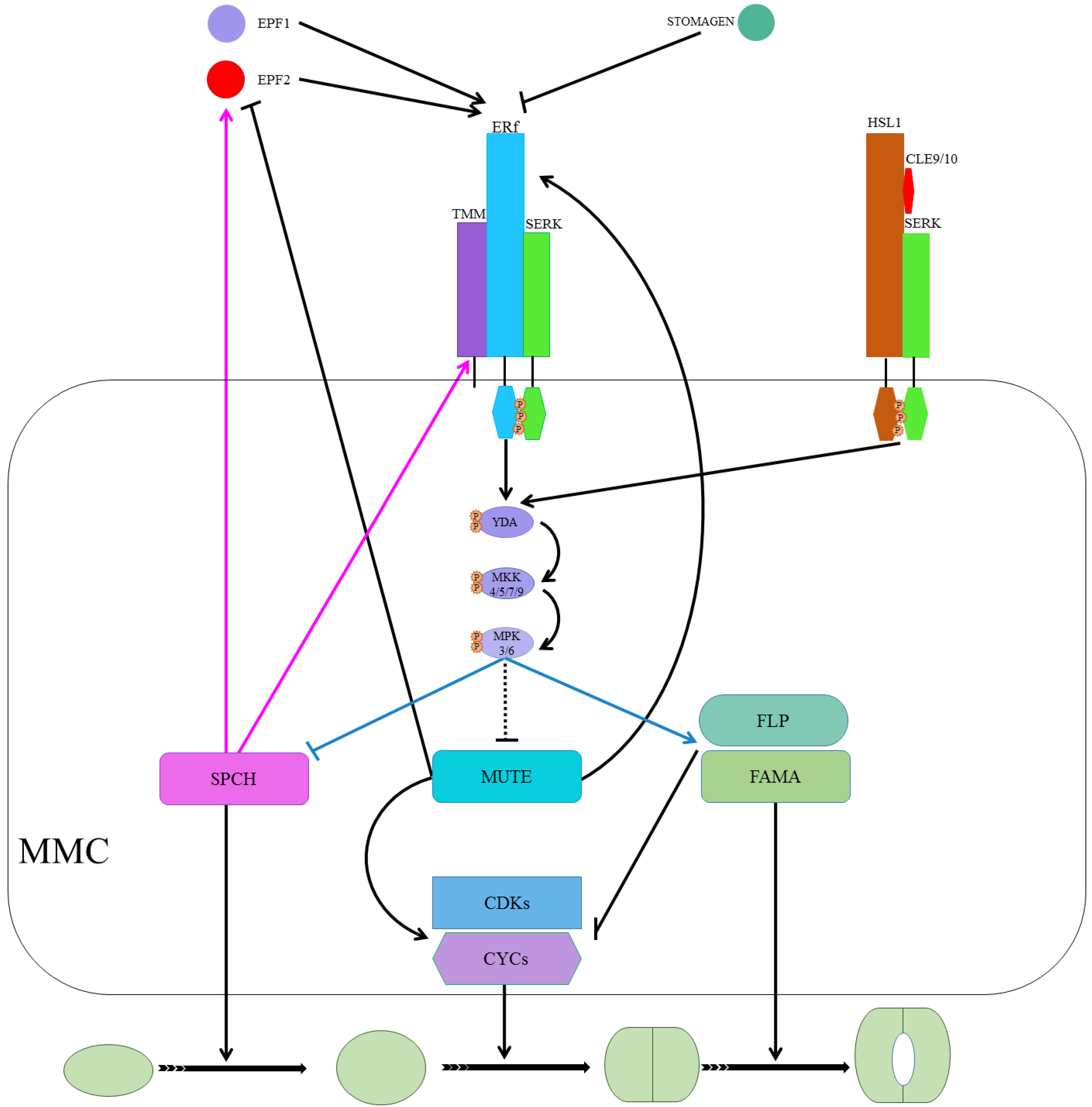
The ligand-receptor interactions are regulating SPCH, MUTE, and FAMA. Ligand (EPF1 and EPF2) binds to the receptor complex (ERf/TMM/SERK). It activates the MPK cascade (YDA-MKK-MPK) that suppresses SPCH, probably suppresses MUTE, and promotes FAMA for respective action during stomatal development. SPCH induces the expression of EPF2 and TMM in a negative feedback mechanism. MUTE induces ERLI and suppresses EPF2. ERL1 regulates meristemoid transition into GMC upon perceiving EPF1. MUTE upregulates CYC and CDK symmetric cell divisions of GMCs. MUTE locks in the cells and upregulates FLP and FAMA in the differentiation program that suppresses the cell cycle’s regulators to control single symmetric cell division. On the right, CLE9/10 is perceived by HSL1 and SERK complex, and signals from this activated complex result in SPCH phosphorylation and destabilization.
Figure 3.
The ligand-receptor interactions are regulating SPCH, MUTE, and FAMA. Ligand (EPF1 and EPF2) binds to the receptor complex (ERf/TMM/SERK). It activates the MPK cascade (YDA-MKK-MPK) that suppresses SPCH, probably suppresses MUTE, and promotes FAMA for respective action during stomatal development. SPCH induces the expression of EPF2 and TMM in a negative feedback mechanism. MUTE induces ERLI and suppresses EPF2. ERL1 regulates meristemoid transition into GMC upon perceiving EPF1. MUTE upregulates CYC and CDK symmetric cell divisions of GMCs. MUTE locks in the cells and upregulates FLP and FAMA in the differentiation program that suppresses the cell cycle’s regulators to control single symmetric cell division. On the right, CLE9/10 is perceived by HSL1 and SERK complex, and signals from this activated complex result in SPCH phosphorylation and destabilization.

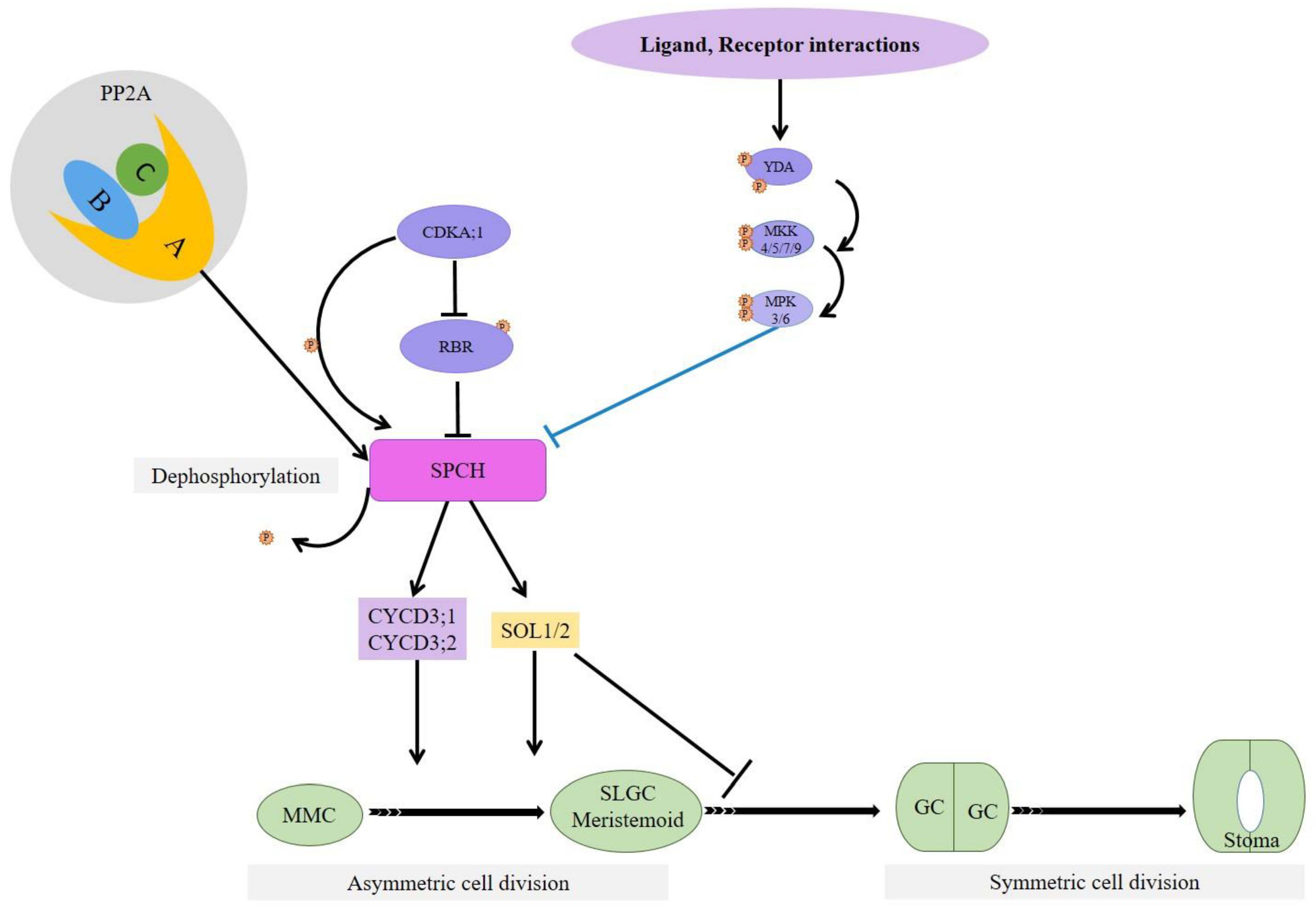
Figure 4.
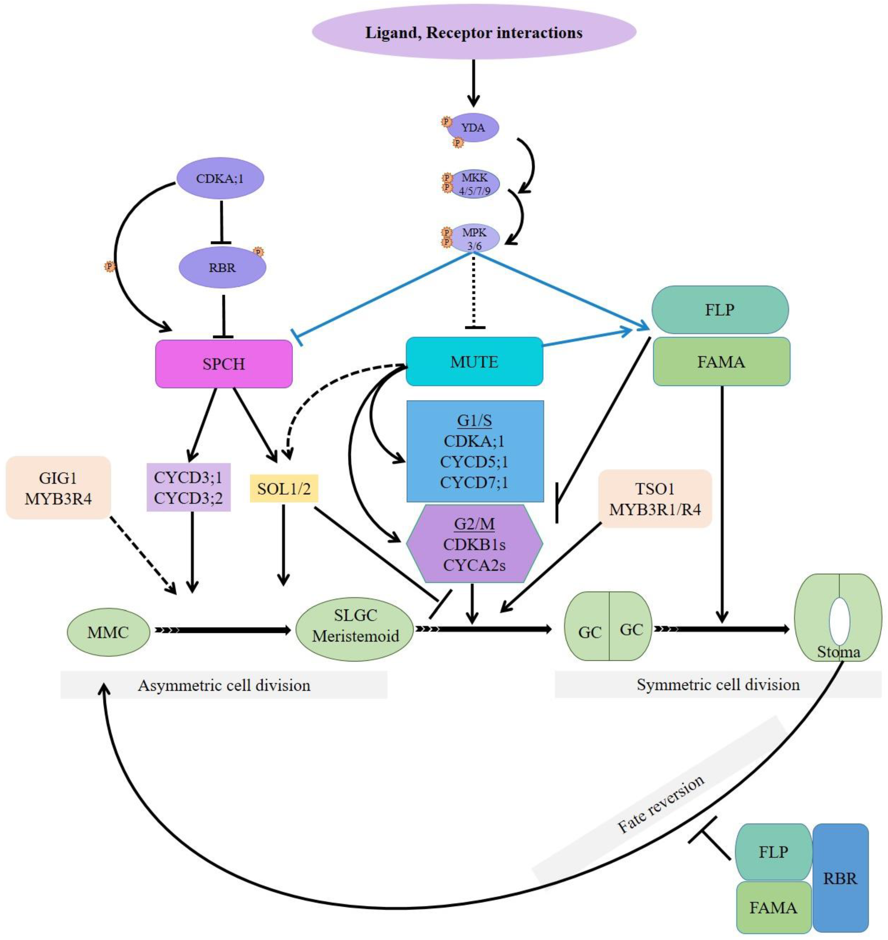
Molecular intersections, role, and mechanism of cell cycle regulators during stomatal formation. Sarcastically selected protodermal cells divide asymmetrically into large SLGC and small meristemoid compartments. Meristemoids, after several amplifying divisions, become a GMC, which divides into a pair of GCs with an opening in between them to form stomata. SPCH, MUTE, FAMA, and FLP/MYB88 regulate all these steps. CDKA;1 can phosphorylate both RBR1 and SPCH; CYCD3 cyclins are the direct targets of SPCH; SPCH expresses SOL1/2, which participates in meristemoid-to-GMC and subsequent symmetric cell division. GIG1 and MYB3R4 synergistically specify cell fate during stomatal lineage development. MUTE controls cell cycle-related core genes and their transcriptional suppressors. TSO1 expression is independent of SPCH or MUTE, physically interacts with MYB3R1, and promotes GMC’s symmetric division. Fate reversion of GCs to MMC requires FAMA/FLP-RBR1 interaction. Black arrows and solid lines mean the place where the factors execute. Dashed lines represent a potential role. T-ended lines indicate suppression.
Figure 4.
Molecular intersections, role, and mechanism of cell cycle regulators during stomatal formation. Sarcastically selected protodermal cells divide asymmetrically into large SLGC and small meristemoid compartments. Meristemoids, after several amplifying divisions, become a GMC, which divides into a pair of GCs with an opening in between them to form stomata. SPCH, MUTE, FAMA, and FLP/MYB88 regulate all these steps. CDKA;1 can phosphorylate both RBR1 and SPCH; CYCD3 cyclins are the direct targets of SPCH; SPCH expresses SOL1/2, which participates in meristemoid-to-GMC and subsequent symmetric cell division. GIG1 and MYB3R4 synergistically specify cell fate during stomatal lineage development. MUTE controls cell cycle-related core genes and their transcriptional suppressors. TSO1 expression is independent of SPCH or MUTE, physically interacts with MYB3R1, and promotes GMC’s symmetric division. Fate reversion of GCs to MMC requires FAMA/FLP-RBR1 interaction. Black arrows and solid lines mean the place where the factors execute. Dashed lines represent a potential role. T-ended lines indicate suppression.

Figure 5.
Sucrose non-fermenting-1 (SNF1)-related kinase 1 (SnRK1), especially subunit KIN10, promoted phosphorylation and stabilization of SPCH-mediates stomatal development under short photoperiod or liquid cultures (mild energy starvation of plants). Sucrose induces the accumulation of KIN10 protein by increasing its translation. Overexpression of KIN10 increases the stomatal index, whereas the loss of function of KIN10-decreases the stomatal index.

Figure 6.
PP2A’s A subunit, which directly interacts with SPCH, in association with B and C subunits and SPCH dephosphorylates SPCH at a specific site. PP2A-induced dephosphorylation increases SPCH accumulation and stability.
Figure 6.
PP2A’s A subunit, which directly interacts with SPCH, in association with B and C subunits and SPCH dephosphorylates SPCH at a specific site. PP2A-induced dephosphorylation increases SPCH accumulation and stability.

Publisher’s Note: MDPI stays neutral with regard to jurisdictional claims in published maps and institutional affiliations. |
© 2021 by the authors. Licensee MDPI, Basel, Switzerland. This article is an open access article distributed under the terms and conditions of the Creative Commons Attribution (CC BY) license (http://creativecommons.org/licenses/by/4.0/).
Share and Cite
MDPI and ACS Style
Wakeel, A.; Wang, L.; Xu, M. SPEECHLESS and MUTE Mediate Feedback Regulation of Signal Transduction during Stomatal Development. Plants 2021, 10, 432. https://doi.org/10.3390/plants10030432
AMA Style
Wakeel A, Wang L, Xu M. SPEECHLESS and MUTE Mediate Feedback Regulation of Signal Transduction during Stomatal Development. Plants. 2021; 10(3):432. https://doi.org/10.3390/plants10030432
Chicago/Turabian StyleWakeel, Abdul, Lin Wang, and Ming Xu. 2021. "SPEECHLESS and MUTE Mediate Feedback Regulation of Signal Transduction during Stomatal Development" Plants 10, no. 3: 432. https://doi.org/10.3390/plants10030432
Note that from the first issue of 2016, this journal uses article numbers instead of page numbers. See further details here.