
Fluorescein-Labeled Thiacalix[4]arenes as Potential Theranostic Molecules: Synthesis, Self-Association, and Antitumor Activity
 , , ,
, , ,  and
and 
Abstract
:1. Introduction
2. Materials and Methods
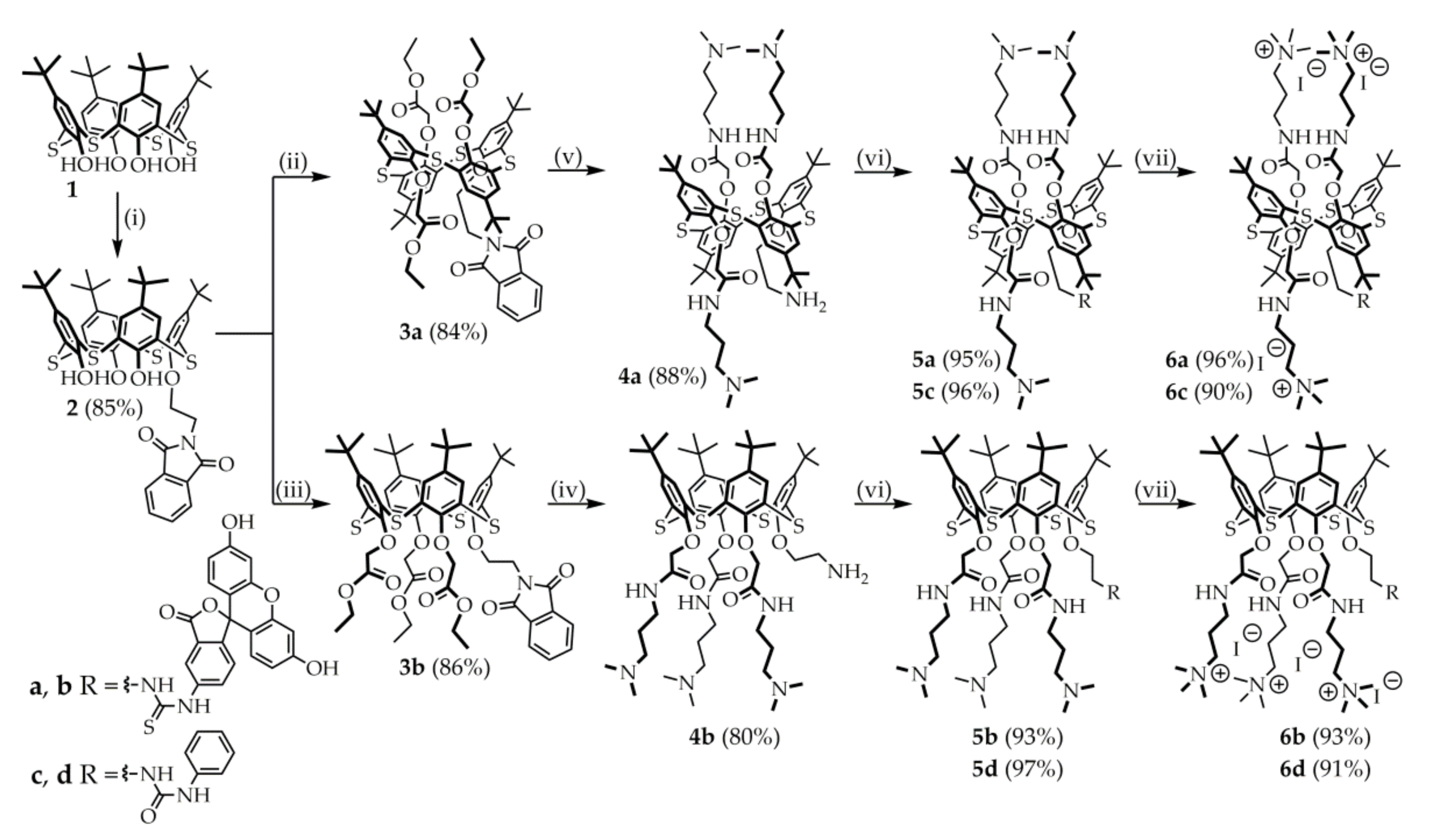
2.1. Chemistry
2.2. Synthesis of Compounds 4a–b, 5a–d, 6a–d
2.2.1. General Synthesis Procedure 4a–b
2.2.2. General Synthesis Procedure 5a–d
2.2.3. General Synthesis Procedure 6a–d
2.3. Determination of the Hydrodynamic Particle Size by Dynamic Light Scattering
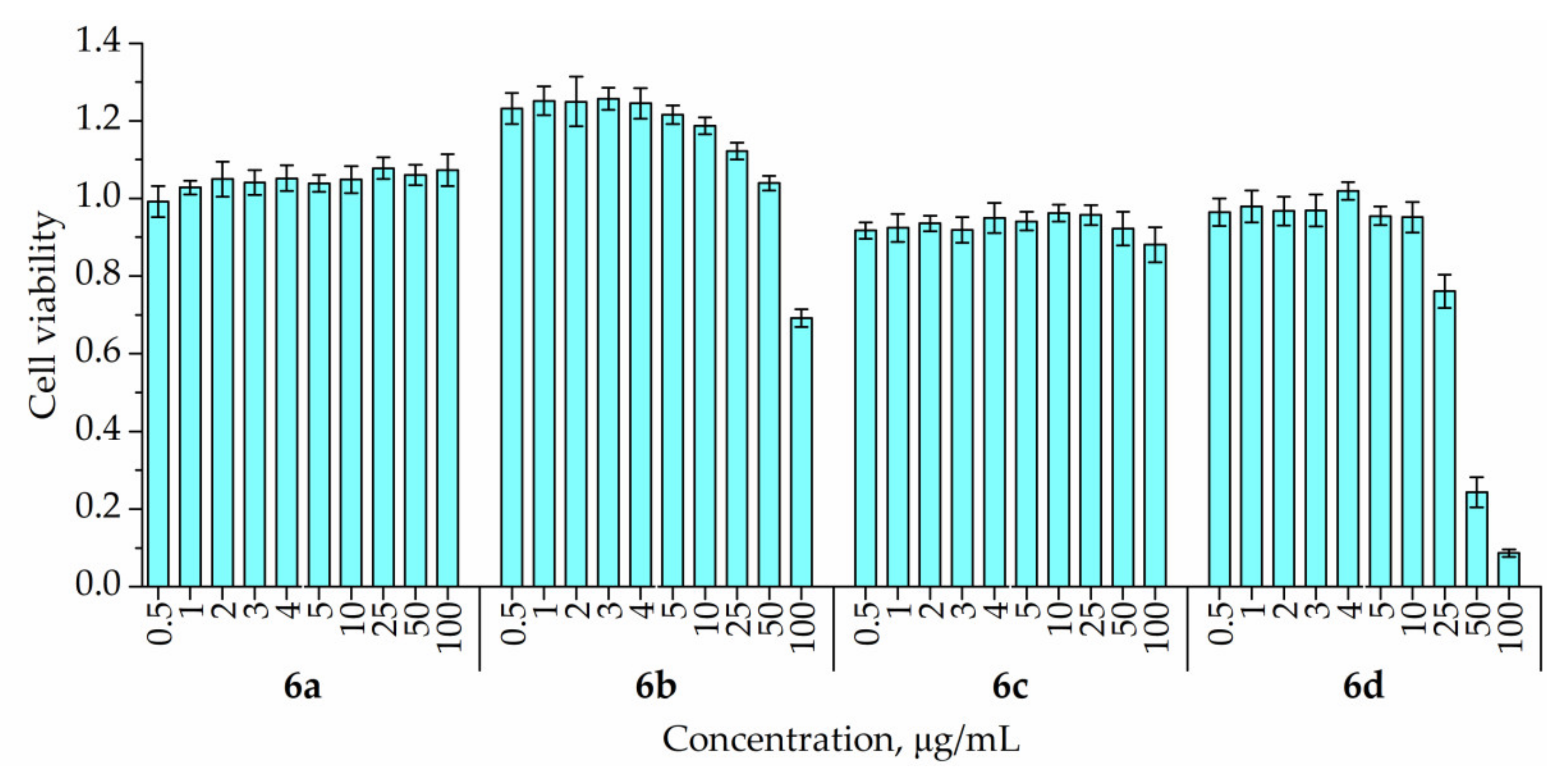
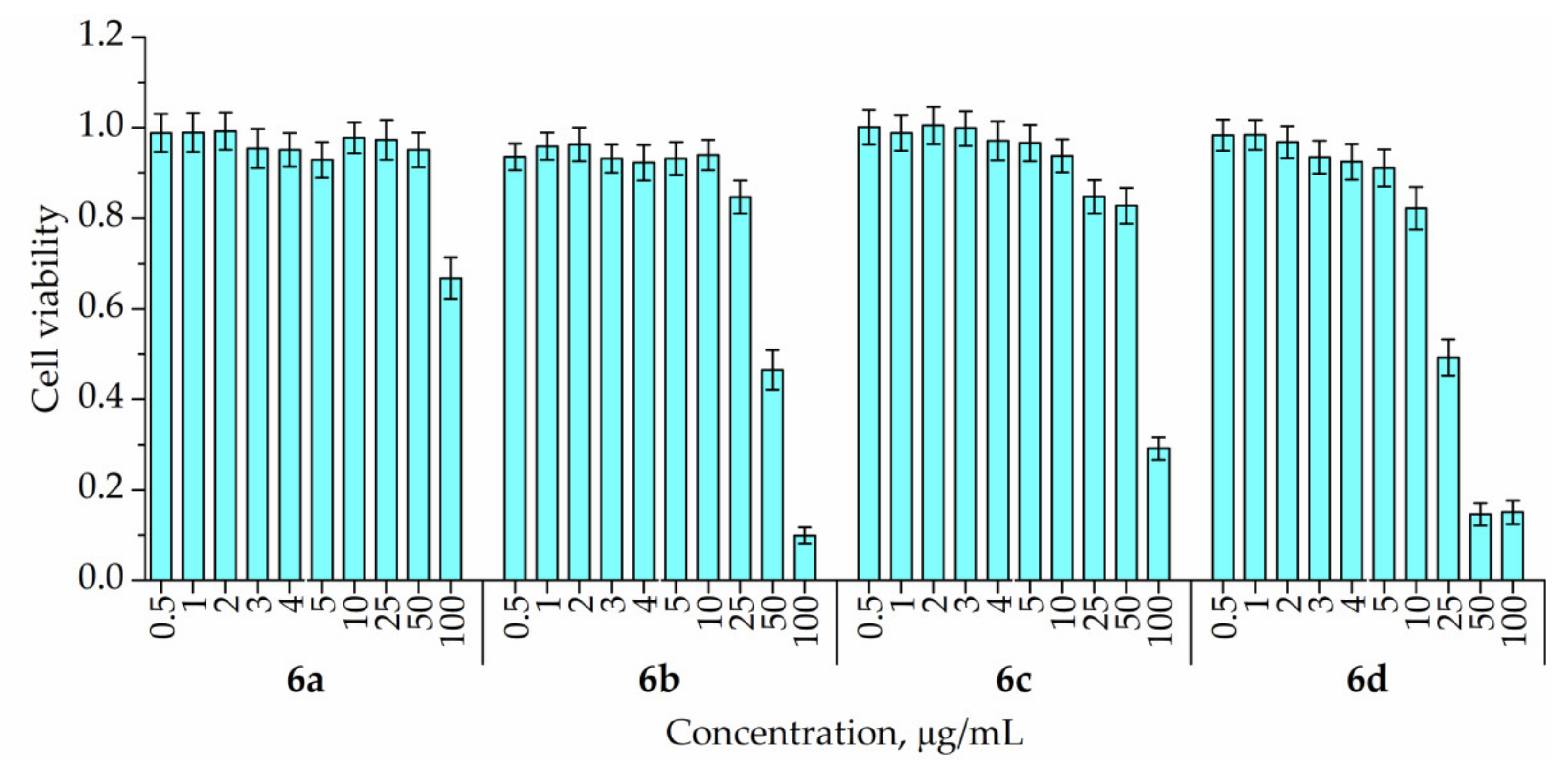
2.4. Cytotoxicity of 6a–d on A549 and HuTu-80 Cell Lines
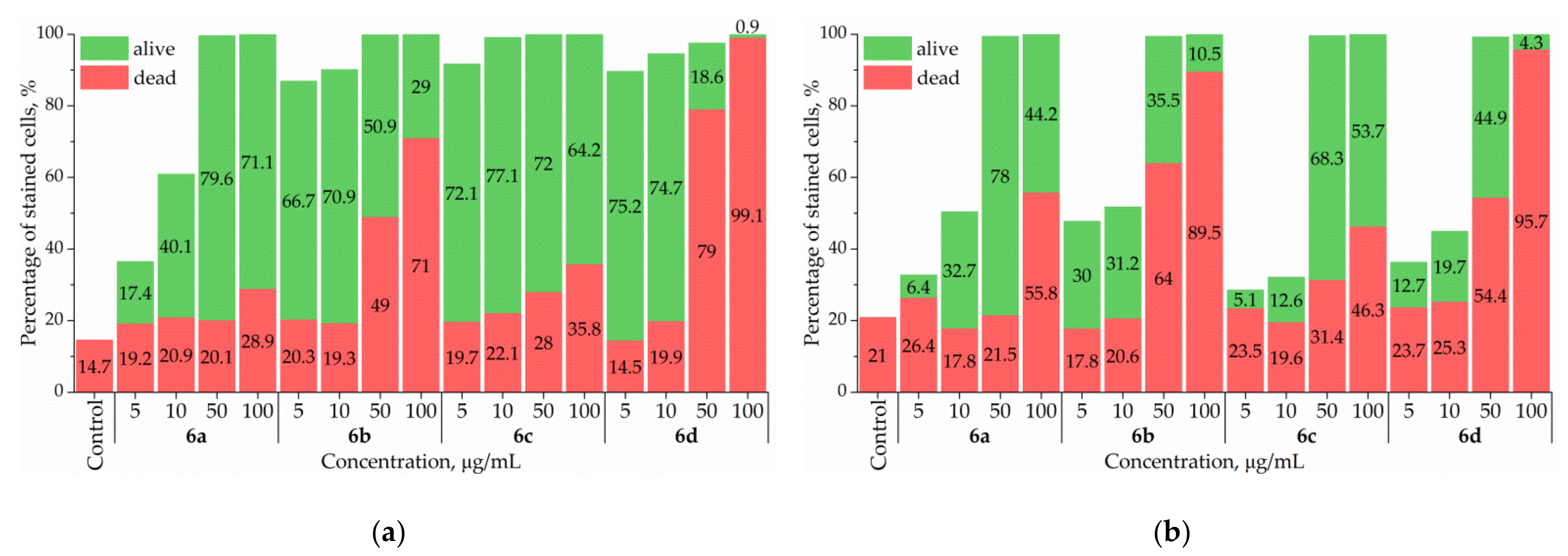
2.5. Characterization of 6a–d Penetration into A549 and HuTu-80 Cells by Flow Cytometry
3. Results and Discussion
3.1. Synthesis of Fluorescein- and Phenyl-Labeled thiacalix[4]arenes
3.2. Aggregation Properties of Fluorescein- and Phenyl-Labeled Thiacalix[4]arenes
3.3. Cytotoxicity of Synthesized Macrocycles
3.4. Penetration into A549 and HuTu-80 Cells of Synthesized Macrocycles
4. Conclusions
Supplementary Materials
Author Contributions
Funding
Institutional Review Board Statement
Informed Consent Statement
Data Availability Statement
Acknowledgments
Conflicts of Interest
References
- Sung, H.; Ferlay, J.; Siegel, R.L.; Laversanne, M.; Soerjomataram, I.; Jemal, A.; Bray, F. Global Cancer Statistics 2020: GLOBOCAN Estimates of Incidence and Mortality Worldwide for 36 Cancers in 185 Countries. CA A Cancer J. Clin. 2021, 71, 209–249. [Google Scholar] [CrossRef] [PubMed]
- Ling, A.; Lundberg, I.V.; Eklöf, V.; Wikberg, M.L.; Öberg, Å.; Edin, S.; Palmqvist, R. The Infiltration, and Prognostic Importance, of Th1 Lymphocytes Vary in Molecular Subgroups of Colorectal Cancer. J. Path. Clin. Res. 2015, 2, 21–31. [Google Scholar] [CrossRef] [PubMed]
- Amicarella, F.; Muraro, M.G.; Hirt, C.; Cremonesi, E.; Padovan, E.; Mele, V.; Governa, V.; Han, J.; Huber, X.; Droeser, R.A.; et al. Dual Role of Tumour-Infiltrating T Helper 17 Cells in Human Colorectal Cancer. Gut 2015, 66, 692–704. [Google Scholar] [CrossRef] [PubMed] [Green Version]
- Thijssen, V.L.; Heusschen, R.; Caers, J.; Griffioen, A.W. Galectin Expression in Cancer Diagnosis and Prognosis: A Systematic Review. Biochim. Et Biophys. Acta (BBA) Rev. Cancer 2015, 1855, 235–247. [Google Scholar] [CrossRef]
- Rabinovich, G.A.; Conejo-García, J.R. Shaping the Immune Landscape in Cancer by Galectin-Driven Regulatory Pathways. J. Mol. Biol. 2016, 428, 3266–3281. [Google Scholar] [CrossRef] [Green Version]
- Chang, W.; Tsai, M.; Kuo, P.; Hung, J. Role of Galectins in Lung Cancer (Review). Oncol. Lett. 2017, 14, 5077–5084. [Google Scholar] [CrossRef] [Green Version]
- Chou, F.-C.; Chen, H.-Y.; Kuo, C.-C.; Sytwu, H.-K. Role of Galectins in Tumors and in Clinical Immunotherapy. Int. J. Mol. Sci. 2018, 19, 430. [Google Scholar] [CrossRef] [Green Version]
- Huang, Y.; Wang, H.-C.; Zhao, J.; Wu, M.-H.; Shih, T.-C. Immunosuppressive Roles of Galectin-1 in the Tumor Microenvironment. Biomolecules 2021, 11, 1398. [Google Scholar] [CrossRef]
- Sanjuan, X.; Fernandez, P.; Castells, A.; Castronovo, V.; van den Brule, F.; Liu, F.; Cardesa, A.; Campo, E. Differential Expression of Galectin 3 and Galectin 1 in Colorectal Cancer Progression. Gastroenterology 1997, 113, 1906–1915. [Google Scholar] [CrossRef]
- Blair, B.B.; Funkhouser, A.T.; Goodwin, J.L.; Strigenz, A.M.; Chaballout, B.H.; Martin, J.C.; Arthur, C.M.; Funk, C.R.; Edenfield, W.J.; Blenda, A.V. Increased Circulating Levels of Galectin Proteins in Patients with Breast, Colon, and Lung Cancer. Cancers 2021, 13, 4819. [Google Scholar] [CrossRef]
- Spano, D.; Russo, R.; Di Maso, V.; Rosso, N.; Terracciano, L.M.; Roncalli, M.; Tornillo, L.; Capasso, M.; Tiribelli, C.; Iolascon, A. Galectin-1 and Its Involvement in Hepatocellular Carcinoma Aggressiveness. Mol. Med. 2009, 16, 102–115. [Google Scholar] [CrossRef] [PubMed]
- Fanfone, D.; Stanicki, D.; Nonclercq, D.; Port, M.; Vander Elst, L.; Laurent, S.; Muller, R.N.; Saussez, S.; Burtea, C. Molecular Imaging of Galectin-1 Expression as a Biomarker of Papillary Thyroid Cancer by Using Peptide-Functionalized Imaging Probes. Biology 2020, 9, 53. [Google Scholar] [CrossRef] [Green Version]
- Chen, R.; Pan, S.; Ottenhof, N.A.; de Wilde, R.F.; Wolfgang, C.L.; Lane, Z.; Post, J.; Bronner, M.P.; Willmann, J.K.; Maitra, A.; et al. Stromal Galectin-1 Expression Is Associated with Long-Term Survival in Resectable Pancreatic Ductal Adenocarcinoma. Cancer Biol. Ther. 2012, 13, 899–907. [Google Scholar] [CrossRef] [PubMed] [Green Version]
- Kuo, P.-L.; Hung, J.-Y.; Huang, S.-K.; Chou, S.-H.; Cheng, D.-E.; Jong, Y.-J.; Hung, C.-H.; Yang, C.-J.; Tsai, Y.-M.; Hsu, Y.-L.; et al. Lung Cancer-Derived Galectin-1 Mediates Dendritic Cell Anergy through Inhibitor of DNA Binding 3/IL-10 Signaling Pathway. J. Immunol. 2010, 186, 1521–1530. [Google Scholar] [CrossRef] [PubMed] [Green Version]
- Abroun, S.; Otsuyama, K.; Shamsasenjan, K.; Islam, A.; Amin, J.; Iqbal, M.S.; Gondo, T.; Asaoku, H.; Kawano, M.M. Galectin-1 Supports the Survival of CD45RA(−) Primary Myeloma Cellsin Vitro. Br. J. Haematol. 2008, 142, 754–765. [Google Scholar] [CrossRef]
- Croci, D.O.; Cerliani, J.P.; Pinto, N.A.; Morosi, L.G.; Rabinovich, G.A. Regulatory Role of Glycans in the Control of Hypoxia-Driven Angiogenesis and Sensitivity to Anti-Angiogenic Treatment. Glycobiology 2014, 24, 1283–1290. [Google Scholar] [CrossRef] [Green Version]
- Astorgues-Xerri, L.; Riveiro, M.E.; Tijeras-Raballand, A.; Serova, M.; Neuzillet, C.; Albert, S.; Raymond, E.; Faivre, S. Unraveling Galectin-1 as a Novel Therapeutic Target for Cancer. Cancer Treat. Rev. 2014, 40, 307–319. [Google Scholar] [CrossRef]
- Blanchard, H.; Bum-Erdene, K.; Bohari, M.H.; Yu, X. Galectin-1 Inhibitors and Their Potential Therapeutic Applications: A Patent Review. Expert Opin. Ther. Pat. 2016, 26, 537–554. [Google Scholar] [CrossRef]
- Berzenji, L.; Van Schil, P. Multimodality Treatment of Malignant Pleural Mesothelioma. F1000Research 2018, 7, 1681. [Google Scholar] [CrossRef] [Green Version]
- Mayo, K.H.; van der Schaft, D.W.J.; Griffioen, A.W. Designed β-sheet peptides that inhibit proliferation and induce apoptosis in endothelial cells. Angiogenesis 2001, 4, 45–51. [Google Scholar] [CrossRef]
- Dings, R.P.M.; Chen, X.; Hellebrekers, D.M.E.I.; van Eijk, L.I.; Zhang, Y.; Hoye, T.R.; Griffioen, A.W.; Mayo, K.H. Design of Nonpeptidic Topomimetics of Antiangiogenic Proteins With Antitumor Activities. JNCI J. Natl. Cancer Inst. 2006, 98, 932–936. [Google Scholar] [CrossRef] [PubMed]
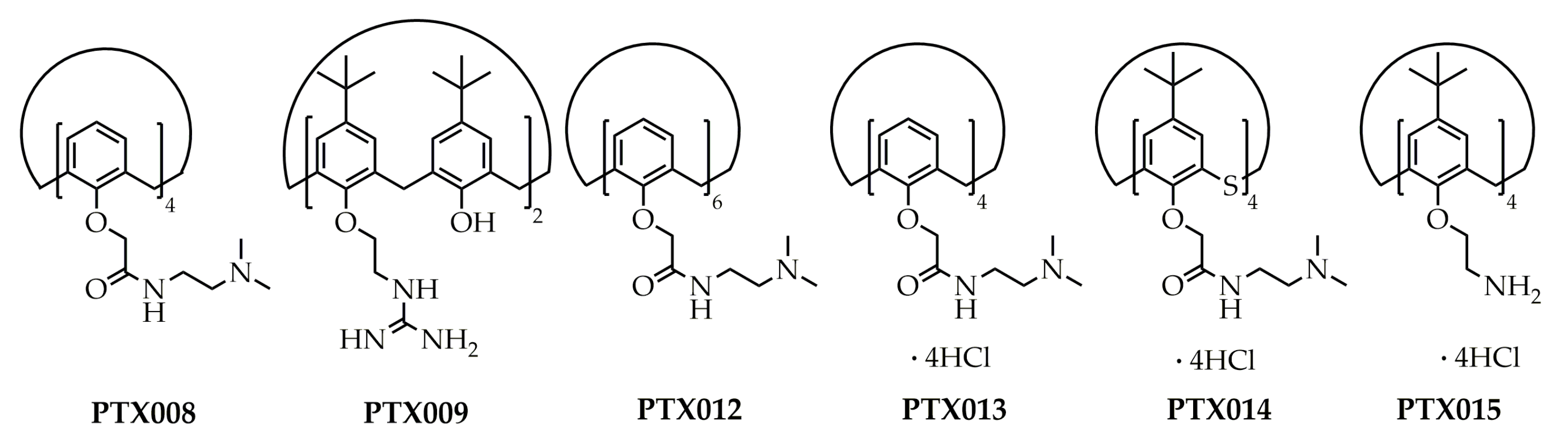
- Dings, R.P.M.; Levine, J.I.; Brown, S.G.; Astorgues-Xerri, L.; MacDonald, J.R.; Hoye, T.R.; Raymond, E.; Mayo, K.H. Polycationic Calixarene PTX013, a Potent Cytotoxic Agent against Tumors and Drug Resistant Cancer. Invest. New Drugs 2013, 31, 1142–1150. [Google Scholar] [CrossRef] [PubMed] [Green Version]
- Kevin, H.M.; Thomas, R.H.; Chen, X. Calixarene-Based Peptide Conformation Mimetics, Methods of Use, and Methods of Making. U.S. Patent 8716343B2, 6 December 2012. International filing date: 24 April 2012. [Google Scholar]
- Dings, R.P.M.; Miller, M.C.; Nesmelova, I.; Astorgues-Xerri, L.; Kumar, N.; Serova, M.; Chen, X.; Raymond, E.; Hoye, T.R.; Mayo, K.H. Antitumor Agent Calixarene 0118 Targets Human Galectin-1 as an Allosteric Inhibitor of Carbohydrate Binding. J. Med. Chem. 2012, 55, 5121–5129. [Google Scholar] [CrossRef] [PubMed] [Green Version]
- Yu, G.; Chen, X. Host-Guest Chemistry in Supramolecular Theranostics. Theranostics 2019, 9, 3041–3074. [Google Scholar] [CrossRef]
- Shurpik, D.N.; Padnya, P.L.; Stoikov, I.I.; Cragg, P.J. Antimicrobial Activity of Calixarenes and Related Macrocycles. Molecules 2020, 25, 5145. [Google Scholar] [CrossRef]
- Padnya, P.L.; Terenteva, O.S.; Akhmedov, A.A.; Iksanova, A.G.; Shtyrlin, N.V.; Nikitina, E.V.; Krylova, E.S.; Shtyrlin, Y.G.; Stoikov, I.I. Thiacalixarene Based Quaternary Ammonium Salts as Promising Antibacterial Agents. Bioorganic Med. Chem. 2021, 29, 115905. [Google Scholar] [CrossRef]
- Shurpik, D.N.; Sevastyanov, D.A.; Zelenikhin, P.V.; Padnya, P.L.; Evtugyn, V.G.; Osin, Y.N.; Stoikov, I.I. Nanoparticles Based on the Zwitterionic Pillardoi:5Arene and Ag+: Synthesis, Self-Assembly and Cytotoxicity in the Human Lung Cancer Cell Line A549. Beilstein J. Nanotechnol 2020, 11, 421–431. [Google Scholar] [CrossRef] [Green Version]
- Akhmedov, A.A.; Shurpik, D.N.; Padnya, P.L.; Khadieva, A.I.; Gamirov, R.R.; Panina, Y.V.; Gazizova, A.F.; Grishaev, D.Y.; Plemenkov, V.V.; Stoikov, I.I. Supramolecular Amphiphiles Based on Pillardoi:5Arene and Meroterpenoids: Synthesis, Self-Association and Interaction with Floxuridine. Int. J. Mol. Sci. 2021, 22, 7950. [Google Scholar] [CrossRef]
- Shurpik, D.N.; Akhmedov, A.A.; Cragg, P.J.; Plemenkov, V.V.; Stoikov, I.I. Progress in the Chemistry of Macrocyclic Meroterpenoids. Plants 2020, 9, 1582. [Google Scholar] [CrossRef]
- Shurpik, D.N.; Aleksandrova, Y.I.; Mostovaya, O.A.; Nazmutdinova, V.A.; Tazieva, R.E.; Murzakhanov, F.F.; Gafurov, M.R.; Zelenikhin, P.V.; Subakaeva, E.V.; Sokolova, E.A.; et al. Self-Healing Thiolated Pillardoi:5Arene Films Containing Moxifloxacin Suppress the Development of Bacterial Biofilms. Nanomaterials 2022, 12, 1604. [Google Scholar] [CrossRef]
- Shurpik, D.N.; Aleksandrova, Y.I.; Rodionov, A.A.; Razina, E.A.; Gafurov, M.R.; Vakhitov, I.R.; Evtugyn, V.G.; Gerasimov, A.V.; Kuzin, Y.I.; Evtugyn, G.A.; et al. Metallo-Supramolecular Coordination Polymers Based on Amidopyridine Derivatives of Pillardoi:5Arene and Cu(II) and Pd(II) Cations: Synthesis and Recognition of Nitroaromatic Compounds. Langmuir 2021, 37, 2942–2953. [Google Scholar] [CrossRef] [PubMed]
- Akhmedov, A.; Shurpik, D.; Latypova, Z.; Gamirov, R.; Plemenkov, V.; Stoikov, I. A Synthetic Analogue of Archaea Lipids Based on Aminoglycerin and Geraniol: Synthesis and Membranotropic Properties. In Proceedings of the International Scientific Conference Actual Problems of Organic Chemistry and Biotechnology, Ekaterinburg, Russia, 18–21 November 2020; p. 69473. [Google Scholar] [CrossRef]
- Lebrón, J.A.; López-López, M.; Moyá, M.L.; Deasy, M.; Muñoz-Wic, A.; García-Calderón, C.B.; Valle Rosado, I.; López-Cornejo, P.; Bernal, E.; Ostos, F.J. Fluorescent Calixarene-Schiff as a Nanovehicle with Biomedical Purposes. Chemosensors 2022, 10, 281. [Google Scholar] [CrossRef]
- Bahojb Noruzi, E.; Shaabani, B.; Geremia, S.; Hickey, N.; Nitti, P.; Kafil, H.S. Synthesis, Crystal Structure, and Biological Activity of a Multidentate Calix[4]Arene Ligand Doubly Functionalized by 2-Hydroxybenzeledene-Thiosemicarbazone. Molecules 2020, 25, 370. [Google Scholar] [CrossRef] [PubMed] [Green Version]
- Oguz, M.; Yildirim, A.; Durmus, I.M.; Karakurt, S.; Yilmaz, M. Synthesis of New Calix[4]Arene Derivatives and Evaluation of Their Cytotoxic Activity. Med. Chem. Res. 2021, 31, 52–59. [Google Scholar] [CrossRef]
- Oguz, M. Synthesis and Anticancer Activity of New P-Tertbutylcalix[4]Arenes Integrated with Trifluoromethyl Aniline Groups against Several Cell Lines. Tetrahedron 2022, 116, 132816. [Google Scholar] [CrossRef]
- Sargazi, S.; Hajinezhad, M.R.; Rahdar, A.; Zafar, M.N.; Awan, A.; Baino, F. Assessment of SnFe2O4 Nanoparticles for Potential Application in Theranostics: Synthesis, Characterization, In Vitro, and In Vivo Toxicity. Materials 2021, 14, 825. [Google Scholar] [CrossRef]
- Redrado, M.; Fernández-Moreira, V.; Gimeno, M.C. Theranostics Through the Synergistic Cooperation of Heterometallic Complexes. ChemMedChem 2021, 16, 932–941. [Google Scholar] [CrossRef]
- Bildziukevich, U.; Kvasnicová, M.; Šaman, D.; Rárová, L.; Wimmer, Z. Novel Oleanolic Acid-Tryptamine and -Fluorotryptamine Amides: From Adaptogens to Agents Targeting In Vitro Cell Apoptosis. Plants 2021, 10, 2082. [Google Scholar] [CrossRef]
- Isik, A.; Oguz, M.; Kocak, A.; Yilmaz, M. Calixarenes: Recent Progress in Supramolecular Chemistry for Application in Cancer Therapy. J. Incl. Phenom. Macrocycl. Chem. 2022, 102, 439–449. [Google Scholar] [CrossRef]
- Pan, Y.; Hu, X.; Guo, D. Biomedical Applications of Calixarenes: State of the Art and Perspectives. Angew. Chem. Int. Ed. 2020, 60, 2768–2794. [Google Scholar] [CrossRef]
- Wang, Y.; Zhang, Z.; Zhao, X.; Xu, L.; Zheng, Y.; Li, H.-B.; Guo, D.-S.; Shi, L.; Liu, Y. Calixarene-Modified Albumin for Stoichiometric Delivery of Multiple Drugs in Combination-Chemotherapy. Theranostics 2022, 12, 3747–3757. [Google Scholar] [CrossRef] [PubMed]
- Nosov, R.V.; Stoikov, I.I. Pentakis-Amidothiacalix[4]Arene Stereoisomers: Synthesis and Effect of Central Core Conformation on Their Aggregation Properties. MHC 2015, 8, 120–127. [Google Scholar] [CrossRef] [Green Version]
- Stoikov, I.I.; Galukhin, A.V.; Zaikov, E.N.; Antipin, I.S. Synthesis and Complexation Properties of 1,3-Alternate Stereoisomers of p-Tert-Butylthiacalix[4]Arenes Tetrasubstituted at the Lower Rim by the Phthalimide Group. Mendeleev Commun. 2009, 19, 193–195. [Google Scholar] [CrossRef]
- Kumagai, H.; Hasegawa, M.; Miyanari, S.; Sugawa, Y.; Sato, Y.; Hori, T.; Ueda, S.; Kamiyama, H.; Miyano, S. Facile Synthesis of P-Tert-Butylthiacalix[4]Arene by the Reaction of p-Tert-Butylphenol with Elemental Sulfur in the Presence of a Base. Tetrahedron Lett. 1997, 38, 3971–3972. [Google Scholar] [CrossRef]
- Andreyko, E.A.; Puplampu, J.B.; Ignacio-De Leon, P.A.; Zharov, I.; Stoikov, I.I. P-Tert-Butylthiacalix[4]Arenes Containing Guanidinium Groups: Synthesis and Self-Assembly into Nanoscale Aggregates. Supramol. Chem. 2019, 31, 473–483. [Google Scholar] [CrossRef]
- Mosmann, T. Rapid Colorimetric Assay for Cellular Growth and Survival: Application to Proliferation and Cytotoxicity Assays. J. Immunol. Methods 1983, 65, 55–63. [Google Scholar] [CrossRef]
- Iki, N.; Narumi, F.; Fujimoto, T.; Morohashi, N.; Miyano, S. Selective Synthesis of Three Conformational Isomers of Tetrakisdoi:(Ethoxycarbonyl)MethoxyThiacalix[4]Arene and Their Complexation Properties towards Alkali Metal Ions†. J. Chem. Soc. Perkin Trans. 1998, 2, 2745–2750. [Google Scholar] [CrossRef]
- Baumann, S.; Schoof, S.; Bolten, M.; Haering, C.; Takagi, M.; Shin-ya, K.; Arndt, H.-D. Molecular Determinants of Microbial Resistance to Thiopeptide Antibiotics. J. Am. Chem. Soc. 2010, 132, 6973–6981. [Google Scholar] [CrossRef]
- Miao, S.; Liang, Y.; Marathe, I.; Mao, J.; DeSantis, C.; Bong, D. Duplex Stem Replacement with BPNA+ Triplex Hybrid Stems Enables Reporting on Tertiary Interactions of Internal RNA Domains. J. Am. Chem. Soc. 2019, 141, 9365–9372. [Google Scholar] [CrossRef]
- Shurpik, D.N.; Aleksandrova, Y.I.; Mostovaya, O.A.; Nazmutdinova, V.A.; Zelenikhin, P.V.; Subakaeva, E.V.; Mukhametzyanov, T.A.; Cragg, P.J.; Stoikov, I.I. Water-Soluble Pillardoi:5Arene Sulfo-Derivatives Self-Assemble into Biocompatible Nanosystems to Stabilize Therapeutic Proteins. Bioorganic Chem. 2021, 117, 105415. [Google Scholar] [CrossRef]
- Sharma, N.; Reja, S.I.; Bhalla, V.; Kumar, M. A New Thiacalix[4]Arene-Fluorescein Based Probe for Detection of CN− and Cu2+ Ions and Construction of a Sequential Logic Circuit. Dalton Trans. 2014, 43, 15929–15936. [Google Scholar] [CrossRef] [PubMed]
- Kumar, R.; Sharma, A.; Singh, H.; Suating, P.; Kim, H.S.; Sunwoo, K.; Shim, I.; Gibb, B.C.; Kim, J.S. Revisiting Fluorescent Calixarenes: From Molecular Sensors to Smart Materials. Chem. Rev. 2019, 119, 9657–9721. [Google Scholar] [CrossRef] [PubMed]
- Zhang, L.; Liu, X.; Tang, Z.; Li, X.; Wang, G. Reversal of Galectin-1 Gene Silencing on Resistance to Cisplatin in Human Lung Adenocarcinoma A549 Cells. Biomed. Pharmacother. 2016, 83, 265–270. [Google Scholar] [CrossRef] [PubMed]


| Concentration, M | FITC Derivatives | PhIC Derivatives | ||||||
|---|---|---|---|---|---|---|---|---|
| 6a | 6b | 6c | 6d | |||||
| D, nm | PDI | D, nm | PDI | D, nm | PDI | D, nm | PDI | |
| 1 × 10⁻³ | 587 ± 35 | 0.43 | 329 ± 59 | 0.57 | – | – | – | – |
| 1 × 10⁻⁴ | 433 ± 43 | 0.51 | 353 ± 93 | 0.64 | 323 ± 16 | 0.28 | 262 ± 38 | 0.49 |
| 1 × 10⁻⁵ | 285 ± 44 | 0.44 | 199 ± 45 | 0.48 | 298 ± 32 | 0.59 | 185 ± 16 | 0.29 |
| 1 × 10⁻⁶ | – | – | – | – | 277 ± 34 | 0.59 | 186 ± 38 | 0.42 |
| Compounds | IC50, μg/mL | |
|---|---|---|
| HuTu-80 | A549 | |
| 6a (1,3-alternate) | >100 a | >100 a |
| 6b (cone) | 49.11 | >100 a |
| 6c (1,3-alternate) | >100 a | >100 a |
| 6d (cone) | 21.83 | 37.55 |
| PTX008 (cone) | nd | 1.87 b |
| PTX013 (cone) | nd | 0.87 b |
| PTX014 (cone) | nd | 8.28 b |
Publisher’s Note: MDPI stays neutral with regard to jurisdictional claims in published maps and institutional affiliations. |
© 2022 by the authors. Licensee MDPI, Basel, Switzerland. This article is an open access article distributed under the terms and conditions of the Creative Commons Attribution (CC BY) license (https://creativecommons.org/licenses/by/4.0/).
Share and Cite
Akhmedov, A.; Terenteva, O.; Subakaeva, E.; Zelenikhin, P.; Shurpik, R.; Shurpik, D.; Padnya, P.; Stoikov, I. Fluorescein-Labeled Thiacalix[4]arenes as Potential Theranostic Molecules: Synthesis, Self-Association, and Antitumor Activity. Pharmaceutics 2022, 14, 2340. https://doi.org/10.3390/pharmaceutics14112340
Akhmedov A, Terenteva O, Subakaeva E, Zelenikhin P, Shurpik R, Shurpik D, Padnya P, Stoikov I. Fluorescein-Labeled Thiacalix[4]arenes as Potential Theranostic Molecules: Synthesis, Self-Association, and Antitumor Activity. Pharmaceutics. 2022; 14(11):2340. https://doi.org/10.3390/pharmaceutics14112340
Chicago/Turabian StyleAkhmedov, Alan, Olga Terenteva, Evgenia Subakaeva, Pavel Zelenikhin, Ramilia Shurpik, Dmitriy Shurpik, Pavel Padnya, and Ivan Stoikov. 2022. "Fluorescein-Labeled Thiacalix[4]arenes as Potential Theranostic Molecules: Synthesis, Self-Association, and Antitumor Activity" Pharmaceutics 14, no. 11: 2340. https://doi.org/10.3390/pharmaceutics14112340