
3D Printing of Dapagliflozin Containing Self-Nanoemulsifying Tablets: Formulation Design and In Vitro Characterization

 , ,
, ,  ,
,
Abstract
:
1. Introduction
2. Materials and Method
2.1. Materials
2.2. Choice of Material
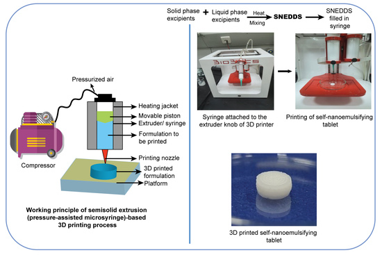
2.3. Preparation and Optimization of Semisolid Extrudable Paste for 3D Printing of Solid-SNEDDS
2.4. 3D Printing of Solid-SNEDDS as a Tablet
2.5. Characterization of the 3D Printed Self-Nanoemulsifying Tablet
2.5.1. Determination of Size and Weight
2.5.2. Solid State Characterization
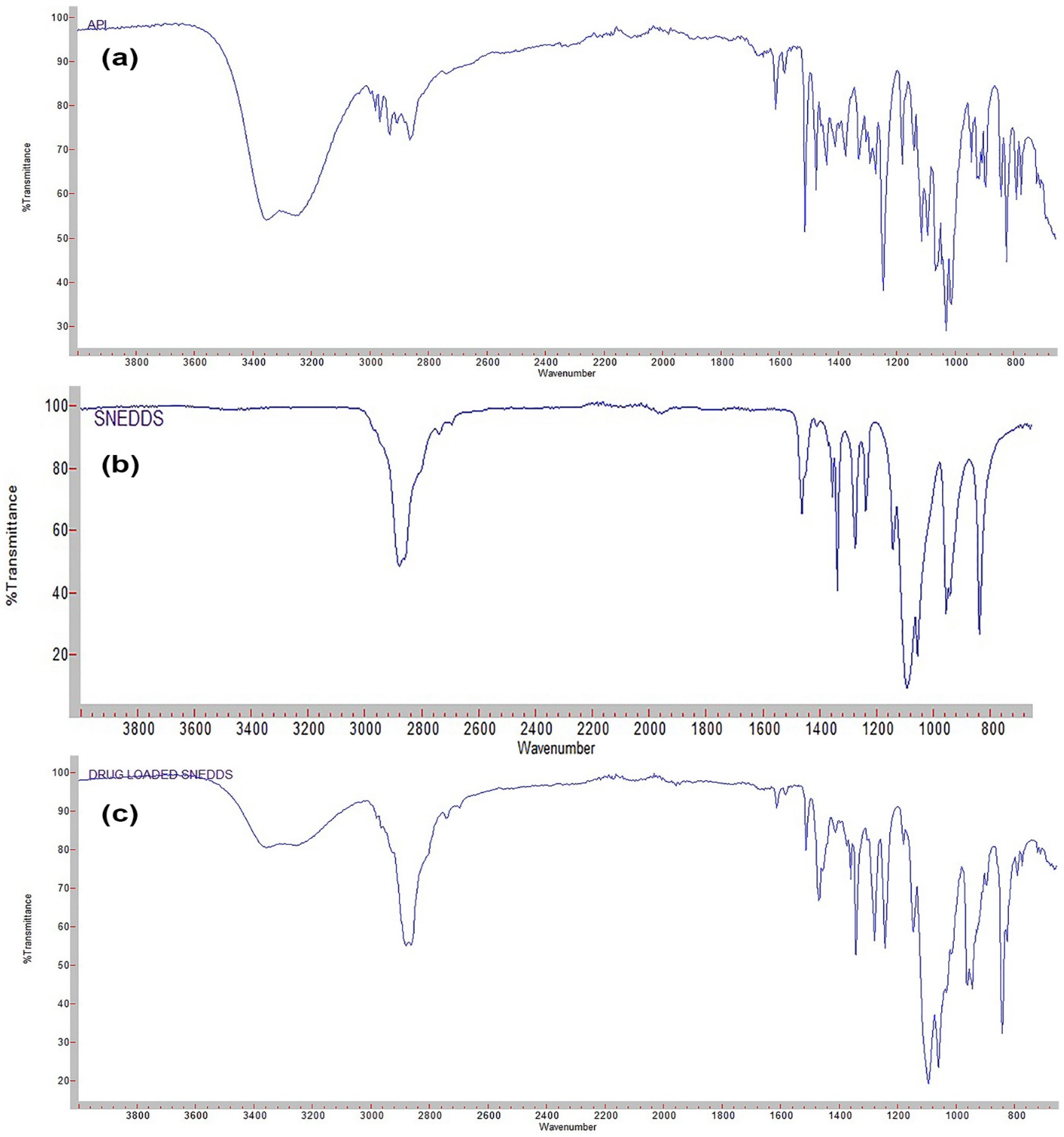
Attenuated Total Reflection-Fourier Transforms Infrared Spectroscopy (ATR-FTIR)
Powder X-ray Diffractometry (XRD)
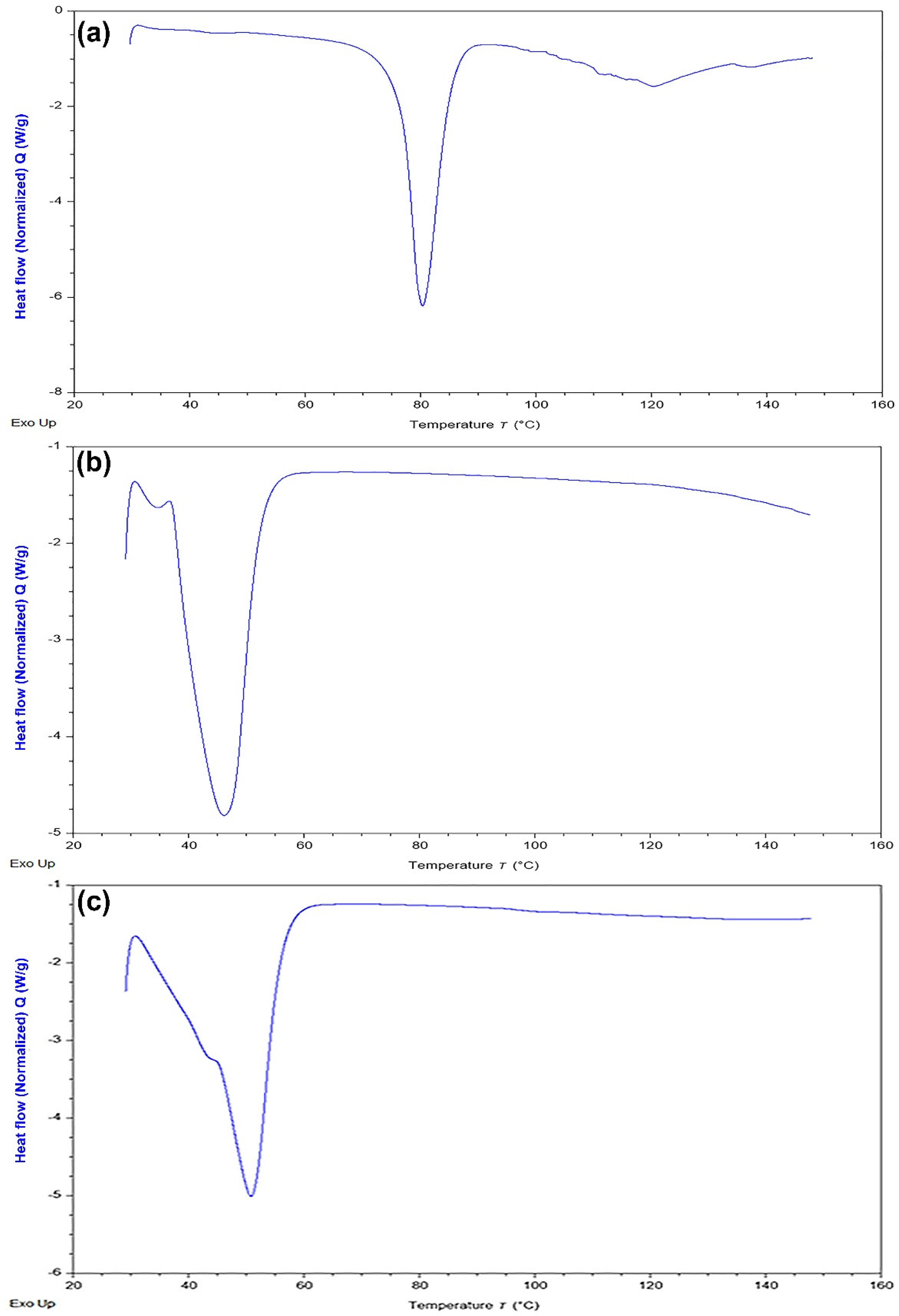
Differential Scanning Calorimetry (DSC)
2.5.3. Surface Morphology by Scanning Electron Microscopy (SEM)
2.5.4. Determination of Droplet Size, Polydispersity Index (PdI), and Zeta Potential (ξ)
2.5.5. Determination of Percentage of Drug Content
2.6. In Vitro Dissolution Study
3. Results and Discussion
3.1. Choice of Materials
3.1.1. Selection of Liquid Phase
3.1.2. Selection of Solid Phase
3.2. Preparation and Optimization of Semisolid Extrudable Paste for 3D Printing of Solid-SNEDDS
3.2.1. Preparation of the Paste
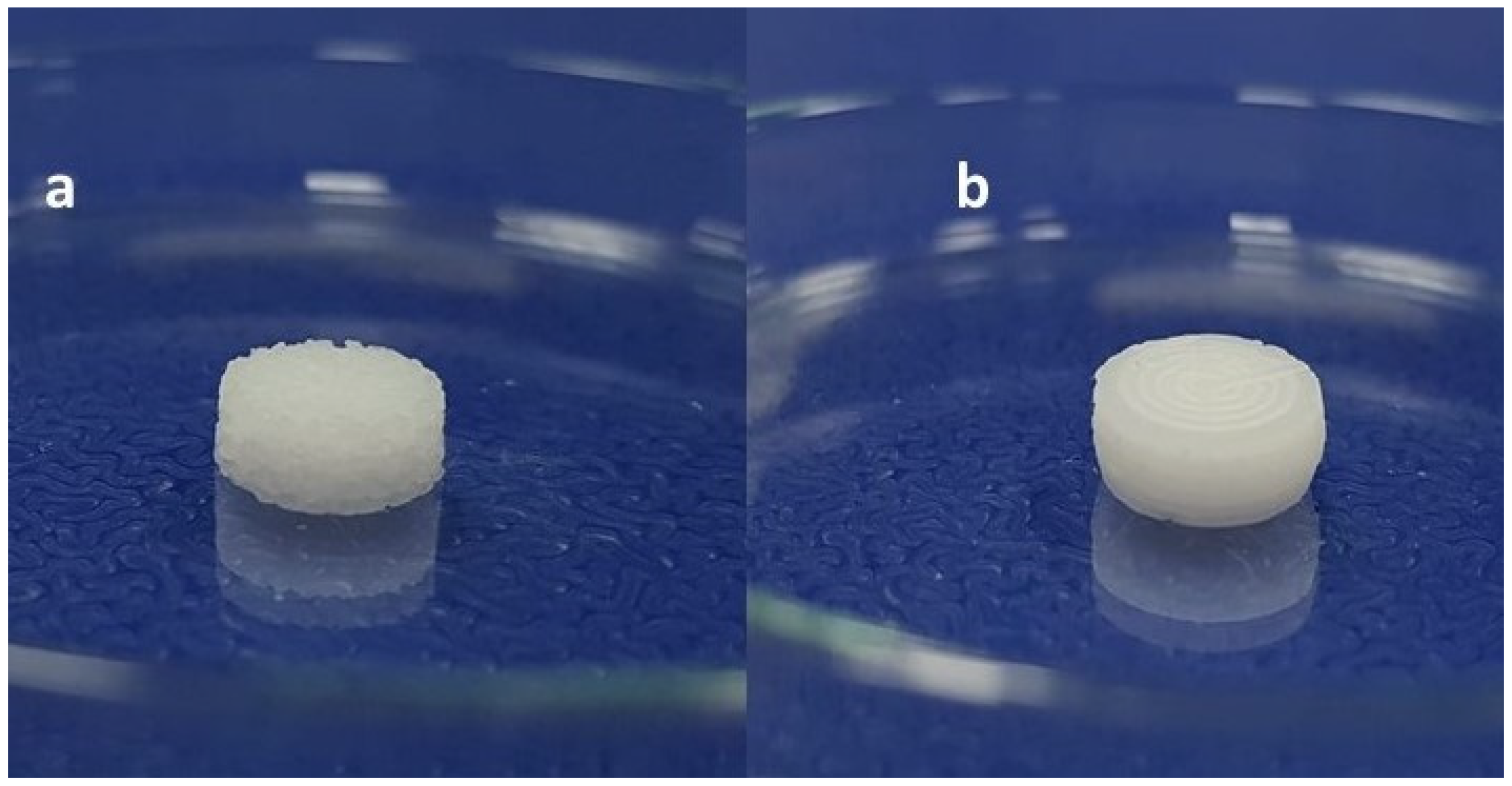
3.2.2. Optimization of Printing Process Parameters and Design of Printed Geometry
3.3. 3D Printing of Solid-SNEDDS as the Self-Nanoemulsifying Tablet
3.4. Characterization of the 3D Printed Self-Nanoemulsifying Tablet
3.4.1. Determination of Size, Weight Variation, and % Drug Content
3.4.2. Solid State Characterization
Attenuated Total Reflection-Fourier Transforms Infrared Spectroscopy (ATR-FTIR)
Differential Scanning Calorimetry (DSC)
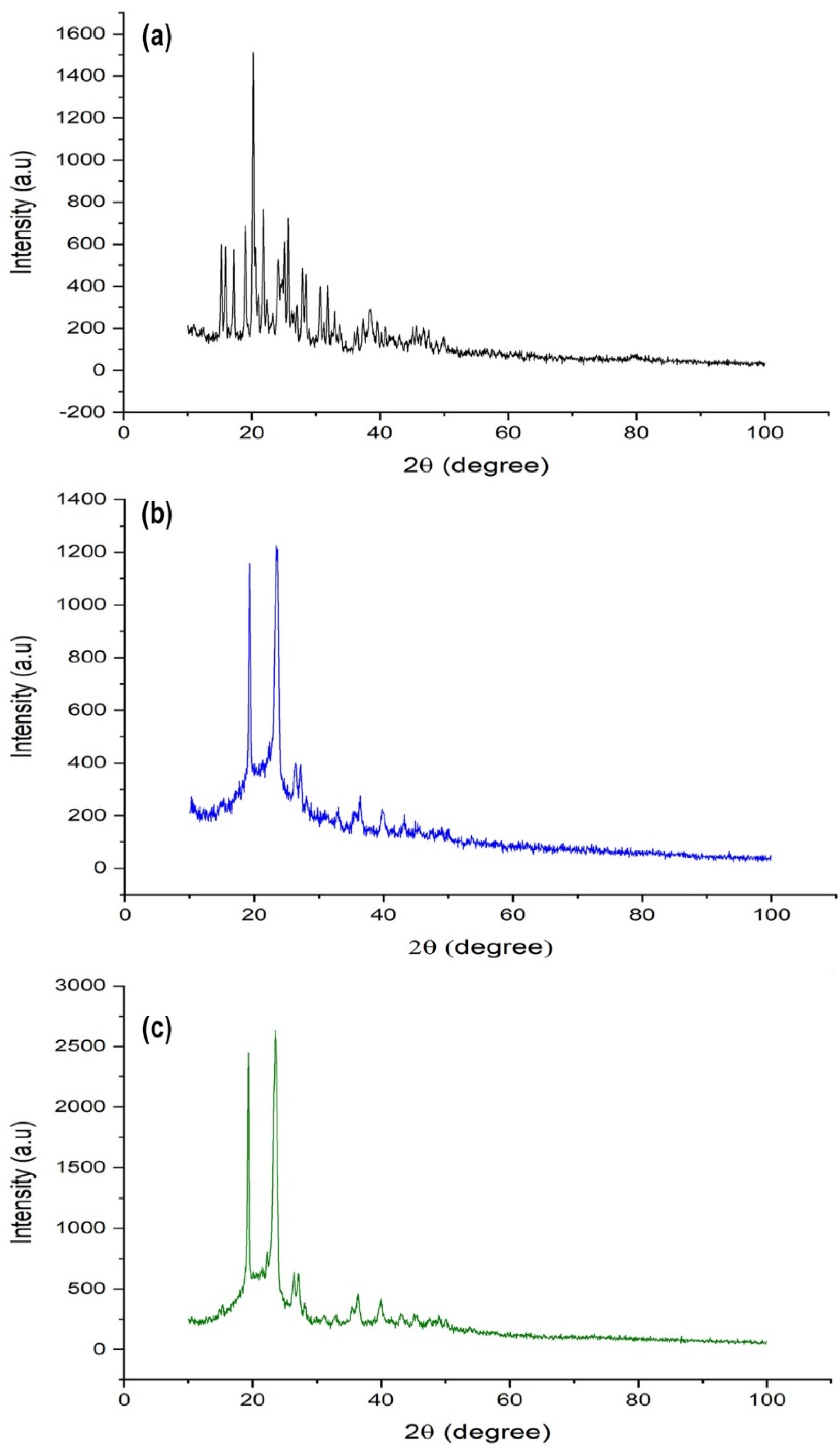
Powder X-ray Diffractometry (XRD)
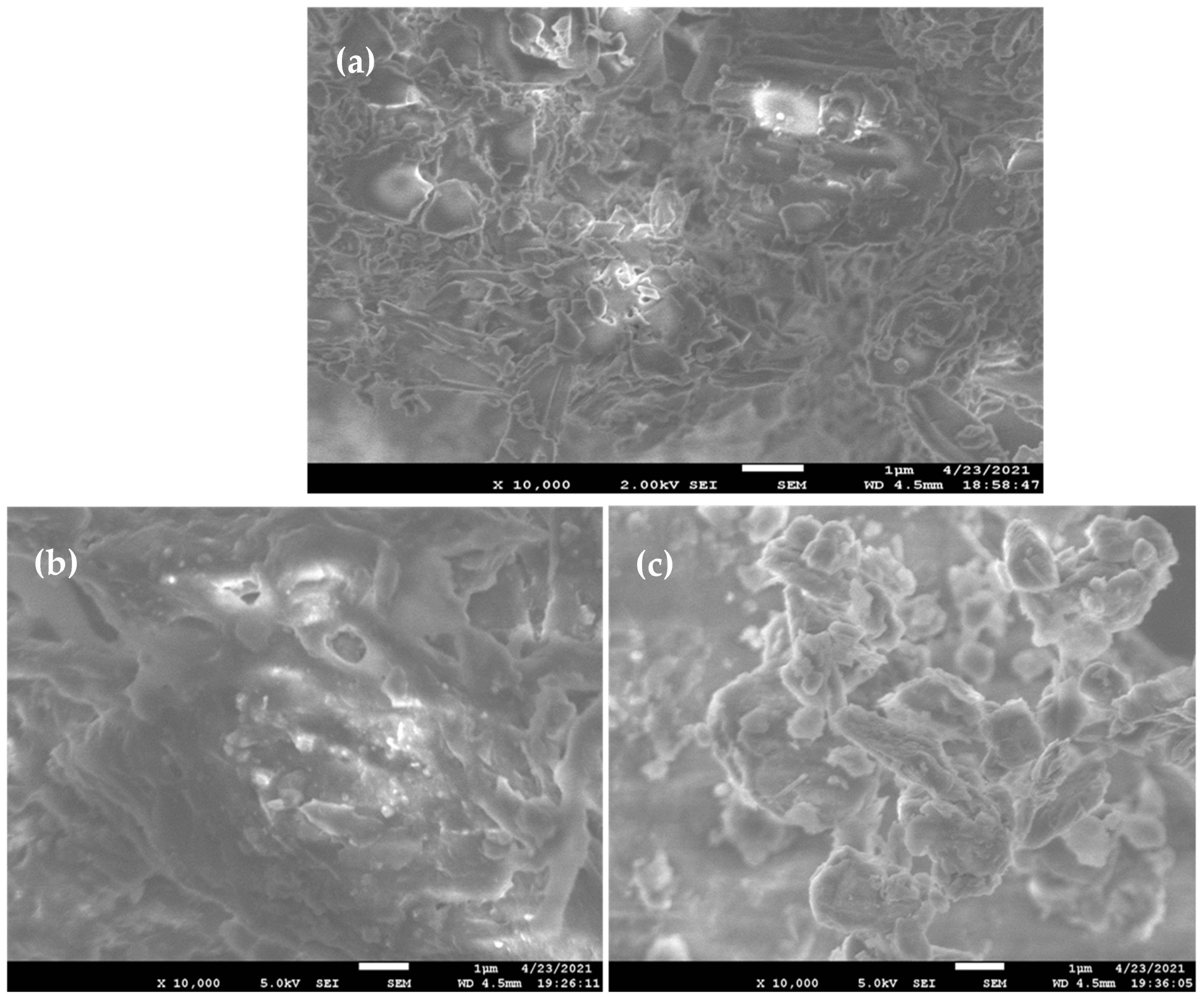
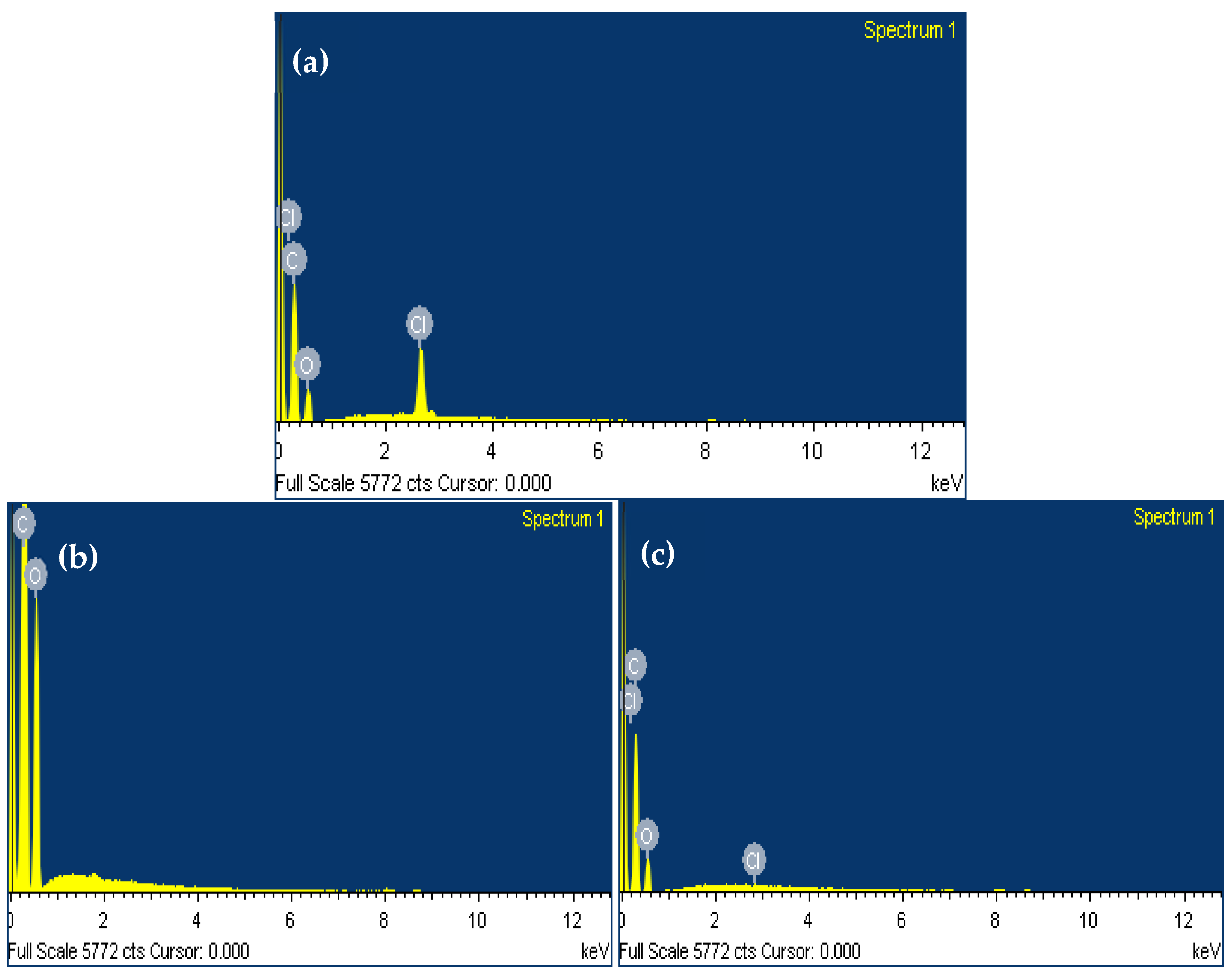
3.4.3. Surface Morphology by Scanning Electron Microscopy (SEM)
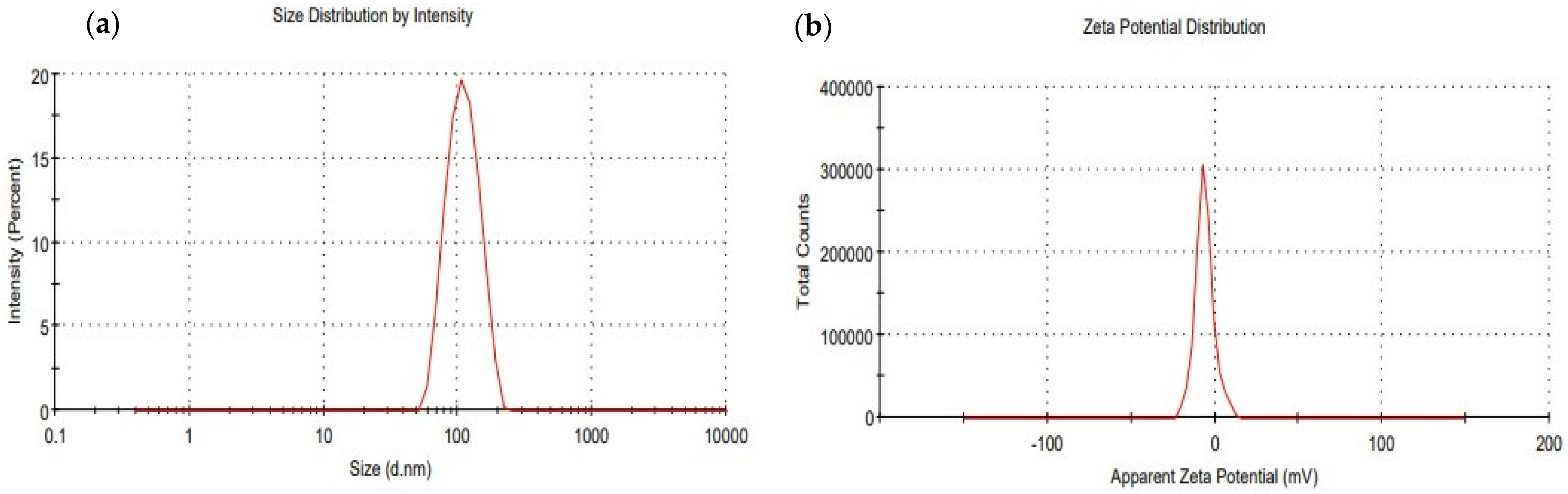
3.4.4. Determination of Droplet Size, Polydispersibility Index (PdI), and Zeta Potential (ξ)
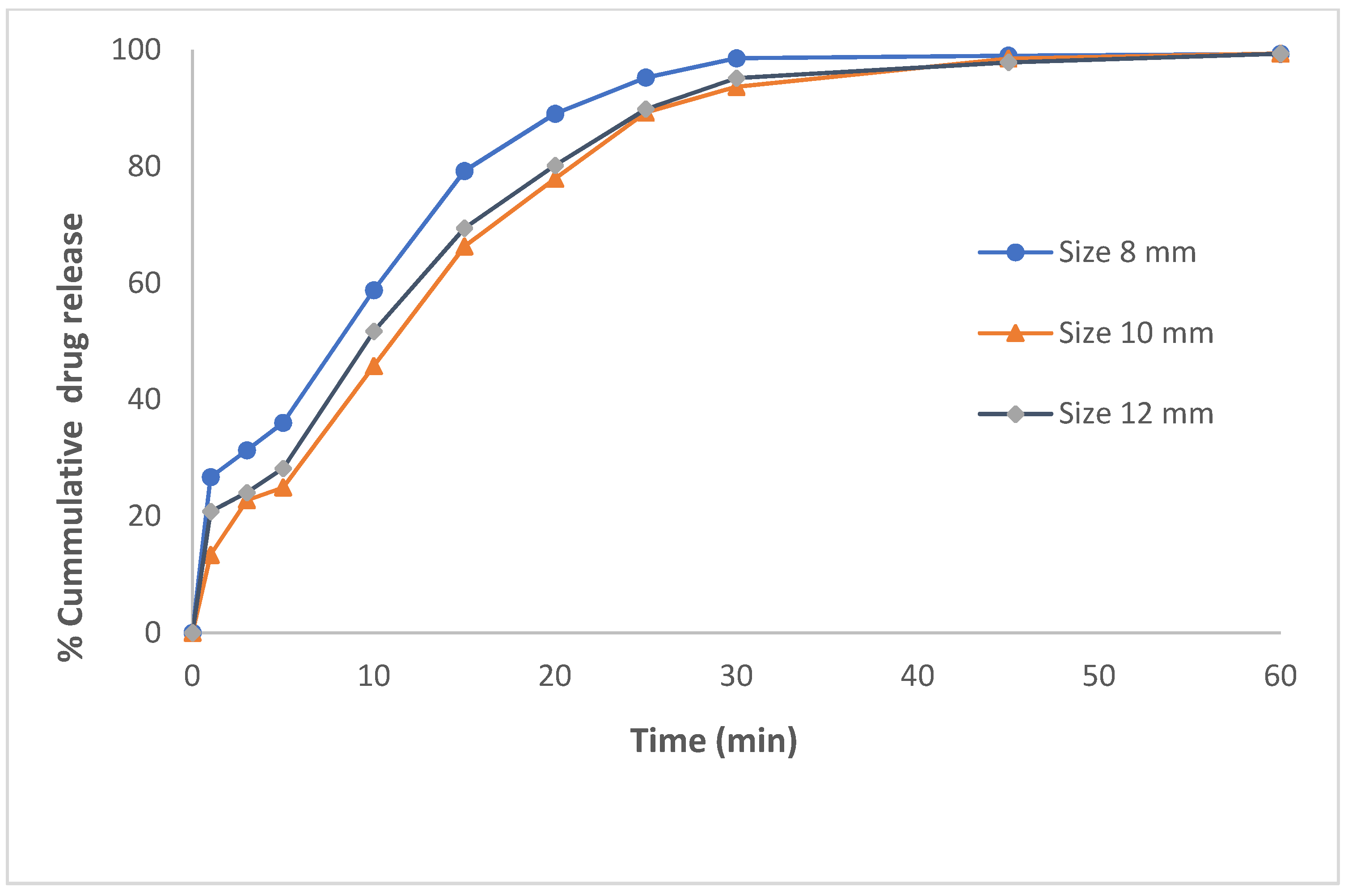
3.5. In Vitro Dissolution Study and Drug Release Kinetics
4. Conclusions
Supplementary Materials
Author Contributions
Funding
Institutional Review Board Statement
Informed Consent Statement
Data Availability Statement
Acknowledgments
Conflicts of Interest
References
- Humberstone, A.J.; Charman, W.N. Lipid-based vehicles for the oral delivery of poorly water soluble drugs. Adv. Drug Deliv. Rev. 1997, 25, 103–128. [Google Scholar] [CrossRef]
- Porter, C.J.H.; Pouton, C.W.; Cuine, J.F.; Charman, W.N. Enhancing intestinal drug solubilisation using lipid-based delivery systems. Adv. Drug Deliv. Rev. 2008, 60, 673–691. [Google Scholar] [CrossRef]
- Lee, M.K. Liposomes for Enhanced Bioavailability of Water-Insoluble Drugs: In Vivo Evidence and Recent Approaches. Pharmaceutics 2020, 12, 264. [Google Scholar] [CrossRef] [Green Version]
- Subongkot, T.; Ngawhirunpat, T. Development of a novel microemulsion for oral absorption enhancement of all-trans retinoic acid. Int. J. Nanomed. 2017, 12, 5585. [Google Scholar] [CrossRef] [Green Version]
- Rizwanullah, M.; Amin, S.; Ahmad, J. Improved pharmacokinetics and antihyperlipidemic efficacy of rosuvastatin-loaded nanostructured lipid carriers. J. Drug Target 2017, 25, 58–74. [Google Scholar] [CrossRef]
- Rehman, F.U.; Shah, K.U.; Shah, S.U.; Khan, I.U.; Khan, G.M.; Khan, A. From nanoemulsions to self-nanoemulsions, with recent advances in self-nanoemulsifying drug delivery systems (SNEDDS). Expert Opin. Drug Deliv. 2017, 14, 1325–1340. [Google Scholar] [CrossRef]
- Vithani, K.; Hawley, A.; Jannin, V.; Pouton, C.; Boyd, B.J. Solubilisation behaviour of poorly water soluble drugs during digestion of solid SMEDDS. Eur. J. Pharm. Biopharm. 2018, 130, 236–246. [Google Scholar] [CrossRef] [PubMed]
- Kalepu, S.; Manthina, M.; Padavala, V. Oral lipid-based drug delivery systems—An overview. Acta Pharm. Sin. B 2013, 3, 361–372. [Google Scholar] [CrossRef] [Green Version]
- Oh, D.H.; Kang, J.H.; Kim, D.W.; Lee, B.J.; Kim, J.O.; Yong, C.S.; Choi, H.G. Comparison of solid selfmicroemulsifying drug delivery system (solid SMEDDS) prepared with hydrophilic and hydrophobic solid carrier. Int. J. Pharm. 2011, 420, 412–418. [Google Scholar] [CrossRef]
- Passerini, N.; Albertini, B.; Perissutti, B.; Rodriguez, L. Evaluation of melt granulation and ultrasonic spray congealing as techniques to enhance the dissolution of praziquantel. Int. J. Pharm. 2006, 318, 92–102. [Google Scholar] [CrossRef]
- Yi, T.; Wan, J.; Xu, H.; Yang, X. A new solid self-microemulsifying formulation prepared by spray-drying to improve the oral bioavailability of poorly water soluble drugs. Eur. J. Pharm. Biopharm. 2008, 70, 439–444. [Google Scholar] [CrossRef]
- Newton, M.; Petersson, J.; Podczeck, F.; Clarke, A.; Booth, S. The influence of formulation variables on the properties of pellets containing a self-emulsifying mixture. J. Pharm. Sci. 2001, 90, 987–995. [Google Scholar] [CrossRef] [PubMed]
- Nazzal, S.; Khan, M.A. Controlled release of a self-emulsifying formulation from a tablet dosage form: Stability assessment and optimization of some processing parameters. Int. J. Pharm. 2006, 315, 110–121. [Google Scholar] [CrossRef]
- Agarwal, V.; Siddiqui, A.; Ali, H.; Nazzal, S. Dissolution and powder flow characterization of solid self-emulsified drug delivery system (SEDDS). Int. J. Pharm. 2009, 366, 44–52. [Google Scholar] [CrossRef]
- Mandic, J.; Pobirk, A.Z.; Vrecer, F.; Gasperlin, M. Overview of solidification techniques for self-emulsifying drug delivery systems from industrial perspective. Int. J. Pharm. 2017, 533, 335–345. [Google Scholar] [CrossRef]
- United States Food and Drug Administration, Highlights of Prescribing Information Spritam. 2015. Available online: http://www.accessdata.fda.gov/drugsatfda_docs/label/2015/207958s000lbl.pdf (accessed on 5 May 2021).
- Available online: https://3dprintingindustry.com/news/triastek-receives-fda-ind-clearance-for-3d-printed-drug-to-treat-rheumatoid-arthritis-184159/ (accessed on 5 May 2021).
- Chatzitaki, A.T.; Tsongas, K.; Tzimtzimis, E.; Tzetzis, D.; Bouropoulos, N.; Barmpalexis, P.; Eleftheriadis, G.; Fatouros, D. 3D printing of patient-tailored SNEDDS-based suppositories of lidocaine. J. Drug Deliv. Sci. Technol. 2021, 61, 102292. [Google Scholar] [CrossRef]
- Vithani, K.; Goyanes, A.; Jannin, V.; Basit, A.W.; Gaisford, S.; Boyd, B.J. A Proof of Concept for 3D Printing of Solid Lipid-Based Formulations of Poorly Water-Soluble Drugs to Control Formulation Dispersion Kinetics. Pharm. Res. 2019, 36, 102. [Google Scholar] [CrossRef]
- Johannesson, J.; Khan, J.; Hubert, M.; Teleki, A.; Bergström, C.A.S. 3D-printing of solid lipid tablets from emulsion gels. Int. J. Pharm. 2021, 597, 120304. [Google Scholar] [CrossRef]
- Plosker, G.L. Dapagliflozin: A review of its use in patients with type 2 diabetes. Drugs. 2014, 74, 2191–2209. [Google Scholar] [CrossRef]
- European Pharmacopoeia. 2.9.5 Uniformity of Mass of Single-Dose Preparations; European Pharmacopoeia Comission, European Pharmacopoeia, European Directorate for the Quality of Medicines & Healthcare (EDQM): Strasbourg, France, 2017. [Google Scholar]
- Singh, S.; Singh, S.K.; Vuddanda, P.R.; Srivastava, A.K. A comparison between use of spray and freeze-drying techniques for preparation of solid self-microemulsifying formulation of valsartan and in vitro and in vivo evaluation. BioMed Res. Int. 2013, 2013, 909045. [Google Scholar] [CrossRef] [Green Version]
- Abdullah, M.M.; Siddiqui, S.A.; Al-Abbas, S.M. Physio-Chemical Properties and Dielectric Behavior of As-Grown Manganese Oxide (γ-Mn2 O3) Nanoparticles. J. Electron. Mater. 2020, 49, 4410–4417. [Google Scholar] [CrossRef]
- Ahmad, J.; Kohli, K.; Mir, S.R.; Amin, S. Formulation of self-nanoemulsifying drug delivery system for telmisartan with improved dissolution and oral bioavailability. J. Dispers. Sci. Technol. 2011, 32, 958–968. [Google Scholar] [CrossRef]
- European Pharmacopoeia. 2.9.6 Uniformity of Content of Single-Dose Preparations; European Pharmacopoeia Comission, European Pharmacopoeia, European Directorate for the Quality of Medicines & Healthcare (EDQM): Strasbourg, France, 2017. [Google Scholar]
- De Meira, R.Z.C.; Maciel, A.B.; Murakami, F.S.; de Oliveira, P.R.; Bernardi, L.S. In Vitro Dissolution Profile of Dapagliflozin: Development, Method Validation, and Analysis of Commercial Tablets. Int. J. Anal. Chem. 2017, 2017, 2951529. [Google Scholar] [CrossRef] [PubMed] [Green Version]
- Date, A.A.; Nagarsenker, M.S. Design and evaluation of self-nanoemulsifying drug delivery systems (SNEDDS) for cefpodoxime proxetil. Int. J. Pharm. 2007, 329, 166–172. [Google Scholar] [CrossRef] [PubMed]
- Kommuru, T.R.; Gurley, B.; Khan, M.A.; Reddy, I.K. Self-emulsifying drug delivery systems (SEDDS) of coenzyme Q10: Formulation development and bioavailability assessment. Int. J. Pharm. 2001, 212, 233–246. [Google Scholar] [CrossRef]
- Brüsewitz, C.; Schendler, A.; Funke, A.; Wagner, T.; Lipp, R. Novel poloxamer-based nanoemulsions to enhance the intestinal absorption of active compounds. Int. J. Pharm. 2007, 329, 173–181. [Google Scholar] [CrossRef]
- Rowe, R.C.; Sheskey, P.J.; Weller, P.J. Handbook of Pharmaceutical Excipients; Libros Digitales-Pharmaceutical Press: London, UK, 2006; pp. 447–450. [Google Scholar]
- Li, P.; Hynes, S.R.; Haefele, T.F.; Pudipeddi, M.; Royce, A.E.; Serajuddin, A.T.M. Development of clinical dosage forms for a poorly water– soluble drug II: Formulation and characterization of a novel solid microemulsion preconcentrate system for oral delivery of a poorly water–soluble drug. J. Pharm. Sci. 2009, 98, 1750–1764. [Google Scholar] [CrossRef] [PubMed]
- Algahtani, M.S.; Mohammed, A.A.; Ahmad, J.; Saleh, E. Development of a 3D printed coating shell to control the drug release of encapsulated immediate-release tablets. Polymers 2020, 12, 1395. [Google Scholar] [CrossRef]
- Zidan, A.; Alayoubi, A.; Coburn, J.; Asfari, S.; Ghammraoui, B.; Cruz, C.N.; Ashraf, M. Extrudability analysis of drug loaded pastes for 3D printing of modified release tablets. Int. J. Pharm. 2019, 554, 292–301. [Google Scholar] [CrossRef] [PubMed]
- Mohammed, A.A.; Algahtani, M.S.; Ahmad, M.Z.; Ahmad, J. Optimization of semisolid extrusion (pressure-assisted microsyringe)-based 3D printing process for advanced drug delivery application. Ann. 3D Print Med. 2021, 2, 100008. [Google Scholar] [CrossRef]
- Seo, Y.G.; Kim, D.H.; Ramasamy, T.; Kim, J.H.; Marasini, N.; Oh, Y.K.; Kim, D.W.; Kim, J.K.; Yong, C.S.; Kim, J.O.; et al. Development of docetaxel-loaded solid self-nanoemulsifying drug delivery system (SNEDDS) for enhanced chemotherapeutic effect. Int. J. Pharm. 2013, 452, 412–420. [Google Scholar] [CrossRef]
- Balakumar, K.; Raghavan, C.V.; Abdu, S. Self-nanoemulsifying drug delivery system (SNEDDS) of rosuvastatin calcium: Design, formulation, bioavailability and pharmacokinetic evaluation. Colloids Surfaces B Biointerfaces 2013, 112, 337–343. [Google Scholar] [CrossRef]
- Zhao, Y.; Wang, C.; Chow, A.H.L.; Ren, K.; Gong, T.; Zhang, Z.; Zhang, Y. Self-nanoemulsifying drug delivery system (SNEDDS) for oral delivery of Zedoary essential oil: Formulation and bioavailability studies. Int. J. Pharm. 2010, 383, 170–177. [Google Scholar] [CrossRef]
- Badran, M.M.; Taha, E.I.; Tayel, M.M.; Al-Suwayeh, S.A. Ultra-fine self nanoemulsifying drug delivery system for transdermal delivery of meloxicam: Dependency on the type of surfactants. J. Mol. Liq. 2014, 190, 16–22. [Google Scholar] [CrossRef]
- Kyobula, M.; Adedeji, A.; Alexander, M.R.; Saleh, E.; Wildman, R.; Ashcroft, I.; Gellert, P.R.; Roberts, C.J. 3D inkjet printing of tablets exploiting bespoke complex geometries for controlled and tuneable drug release. J. Control Release 2017, 261, 207–215. [Google Scholar] [CrossRef]
- Ito, Y.; Kusawake, T.; Ishida, M.; Tawa, R.; Shibata, N.; Takada, K. Oral solid gentamicin preparation using emulsifier and adsorbent. J. Control Release 2005, 105, 23–31. [Google Scholar] [CrossRef]
- Wang, Z.; Sun, J.; Wang, Y.; Liu, X.; Liu, Y.; Fu, Q.; Meng, P.; He, Z. Solid self-emulsifying nitrendipine pellets: Preparation and in vitro/in vivo evaluation. Int. J. Pharm. 2010, 383, 1–6. [Google Scholar] [CrossRef]
- Ansen, T.; Holm, P.; Schultz, K. Process characteristics and compaction of spray-dried emulsions containing a drug dissolved in lipid. Int. J. Pharm. 2004, 287, 55–66. [Google Scholar] [CrossRef]
- Buya, A.B.; Beloqui, A.; Memvanga, P.B.; Preat, V. Self-Nano-Emulsifying Drug-Delivery Systems: From the Development to the Current Applications and Challenges in Oral Drug Delivery. Pharmaceutics 2020, 12, 1194. [Google Scholar] [CrossRef]
- Pouton, C.W.; Porter, C.J.H. Formulation of lipid-based delivery systems for oral administration: Materials, methods and strategies. Adv. Drug Deliv. Rev. 2008, 60, 625–637. [Google Scholar] [CrossRef]
- Jannin, V.; Musakhanian, J.; Marchaud, D. Approaches for the development of solid and semi-solid lipid-based formulations. Adv. Drug Deliv. Rev. 2008, 60, 734–746. [Google Scholar] [CrossRef] [PubMed]
- Abdelmonem, R.; Azer, M.S.; Makky, A.; Zaghloul, A.; El-Nabarawi, M.; Nada, A. Development, Characterization, and in-vivo Pharmacokinetic Study of Lamotrigine Solid Self-Nanoemulsifying Drug Delivery System. Drug Des. Dev. Ther. 2020, 14, 4343. [Google Scholar] [CrossRef]
- Beg, S.; Katare, O.P.; Saini, S.; Garg, B.; Khurana, R.K.; Singh, B. Solid self-nanoemulsifying systems of olmesartan medoxomil: Formulation development, micromeritic characterization, in vitro and in vivo evaluation. Powder Technol. 2016, 294, 93–104. [Google Scholar] [CrossRef]


| Formulation Ingredients | % Composition (w/v) |
|---|---|
| Liquid phase | |
| Capryol 90 | 16.00% |
| Octanoic acid | 16.00% |
| PEG 400 | 8.00% |
| Solid phase | |
| Poloxamer 188 | 30.00% |
| PEG 6000 | 30.00% |
| 3D Printed SNEDDS Tablet | No. of Horizontally Printed Layers | No. of Circular Printed Layers | Printing Time | Printing Speed (mm/s) | Printing Pressure (PSI) | Nozzle Size (mm) |
|---|---|---|---|---|---|---|
| Tablet A | 6 | 4 | 1 min 24 s | 10 | 60 | 0.84 |
| Tablet B | 6 | 5 | 1 min 43 s | 10 | 60 | 0.84 |
| Tablet C | 6 | 6 | 2 min 17 s | 10 | 60 | 0.84 |
| 3D Printed Self-Nanoemulsifying Tablets | Observed Size (Length × Height) in mm | Average Weight (mg) | % Drug Content |
|---|---|---|---|
| Tablet A (8 mm × 3 mm) | (8.108 ± 0.152) × (3.116 ± 0.103) | 193.36 ± 4.2 | 99.46 ± 0.47 |
| Tablet B (10 mm × 3 mm) | (10.052 ± 0.046) × (3.083 ± 0.100) | 277.96 ± 3.6 | 99.12 ± 0.24 |
| Tablet C (12 mm × 3 mm) | (11.983 ± 0.275) × (3.039 ± 0.075) | 439.76 ± 6.2 | 99.4 ± 0.09 |
Publisher’s Note: MDPI stays neutral with regard to jurisdictional claims in published maps and institutional affiliations. |
© 2021 by the authors. Licensee MDPI, Basel, Switzerland. This article is an open access article distributed under the terms and conditions of the Creative Commons Attribution (CC BY) license (https://creativecommons.org/licenses/by/4.0/).
Share and Cite
Algahtani, M.S.; Mohammed, A.A.; Ahmad, J.; Abdullah, M.M.; Saleh, E. 3D Printing of Dapagliflozin Containing Self-Nanoemulsifying Tablets: Formulation Design and In Vitro Characterization. Pharmaceutics 2021, 13, 993. https://doi.org/10.3390/pharmaceutics13070993
Algahtani MS, Mohammed AA, Ahmad J, Abdullah MM, Saleh E. 3D Printing of Dapagliflozin Containing Self-Nanoemulsifying Tablets: Formulation Design and In Vitro Characterization. Pharmaceutics. 2021; 13(7):993. https://doi.org/10.3390/pharmaceutics13070993
Chicago/Turabian StyleAlgahtani, Mohammed S., Abdul Aleem Mohammed, Javed Ahmad, M. M. Abdullah, and Ehab Saleh. 2021. "3D Printing of Dapagliflozin Containing Self-Nanoemulsifying Tablets: Formulation Design and In Vitro Characterization" Pharmaceutics 13, no. 7: 993. https://doi.org/10.3390/pharmaceutics13070993