Review

Therapeutic Approaches to Treat Mitochondrial Diseases: “One-Size-Fits-All” and “Precision Medicine” Strategies

1
Department of Diagnostics and Public Health, Section of Pharmacology, University of Verona, 37134 Verona, Italy
2
Medical Genetics and Neurogenetics Unit, Fondazione IRCCS Istituto Neurologico C. Besta, 20126 Milan, Italy
3
Department of General Pediatrics, Neonatology, and Pediatric Cardiology, University Clinic Düsseldorf (UKD), Heinrich Heine University (HHU), 40225 Dusseldorf, Germany
4
Department of Clinical Science and Community Health, University of Milan, 20122 Milan, Italy
5
Fetal Medicine and Surgery Service, Fondazione IRCCS Ca’ Granda, Ospedale Maggiore Policlinico, 20122 Milan, Italy
6
Department of Medical Biotechnology and Translational Medicine, University of Milan, 20129 Milan, Italy
*
Authors to whom correspondence should be addressed.
Pharmaceutics 2020, 12(11), 1083; https://doi.org/10.3390/pharmaceutics12111083
Submission received: 9 October 2020 / Revised: 8 November 2020 / Accepted: 9 November 2020 / Published: 11 November 2020
(This article belongs to the Special Issue Mitochondria-targeted drug delivery)

Abstract

:
Primary mitochondrial diseases (PMD) refer to a group of severe, often inherited genetic conditions due to mutations in the mitochondrial genome or in the nuclear genes encoding for proteins involved in oxidative phosphorylation (OXPHOS). The mutations hamper the last step of aerobic metabolism, affecting the primary source of cellular ATP synthesis. Mitochondrial diseases are characterized by extremely heterogeneous symptoms, ranging from organ-specific to multisystemic dysfunction with different clinical courses. The limited information of the natural history, the limitations of currently available preclinical models, coupled with the large variability of phenotypical presentations of PMD patients, have strongly penalized the development of effective therapies. However, new therapeutic strategies have been emerging, often with promising preclinical and clinical results. Here we review the state of the art on experimental treatments for mitochondrial diseases, presenting “one-size-fits-all” approaches and precision medicine strategies. Finally, we propose novel perspective therapeutic plans, either based on preclinical studies or currently used for other genetic or metabolic diseases that could be transferred to PMD.

1. Genetics of Mitochondrial Diseases

Primary mitochondrial disorders (PMD) are a group of rare diseases affecting approximately 1 in 4300 live birth and causing progressive, incurable defects often resulting in premature death. PMD are characterized by a high genetic, biochemical, and clinical complexity that arise from the dysfunction of the oxidative phosphorylation (OXPHOS), the essential, final pathway for aerobic metabolism [1]. Such impairment is caused by mutations in genes encoding for proteins involved in mitochondrial respiratory chain (MRC) biogenesis, (i.e., subunits of MRC complexes, assembly factors, or post-assembly quality controllers), or by mutations in genes involved in other mitochondrial functions, including fission and fusion machinery, mitochondrial DNA (mtDNA) maintenance, heme biosynthesis, and iron/sulfur metabolism, among others [2].
Each mitochondrion contains several mitochondrial DNA (mtDNA) molecules, the abundance of which spans from hundreds to thousands of copies per cell, depending on the tissue type. For years, it was trusted that in physiological conditions, all mtDNA molecules have the same sequence, a condition known as homoplasmy. When mutations in mtDNA occur, wild type and mutant molecules coexist in the same cell/organ, a condition known as heteroplasmy. However, recent deep resequencing analysis has shown that low-level mtDNA heteroplasmy is extremely common in humans, and, at very low levels (0.5–1%), heteroplasmy seems to be a universal finding [3].
MtDNA mutations could affect any gene encoding the 13 core subunits of the MRC complexes, the 22 mitochondrial tRNAs, or the two rRNAs. They can include point mutations, either homo- or heteroplasmic, and (invariably heteroplasmic) large-scale rearrangements. Heteroplasmic mutations lead to different clinical phenotypes, such as Leigh syndrome (LS) [4], myoclonic epilepsy with ragged red fibers (MERRF) [5], mitochondrial encephalomyopathy with lactic acidosis and stroke-like episodes (MELAS) [6], and neurogenic weakness, ataxia, and retinitis pigmentosa (NARP) [7]. The central disease entity associated with homoplasmic mtDNA mutations is Leber’s hereditary optic neuropathy (LHON) [8]. Mutations in the same gene can lead to different clinical presentation; for instance, mitochondrial phenotypes described in patients with MT-ATP6 mutations span from maternally inherited Leigh syndrome and neurogenic muscle weakness, ataxia, and retinitis pigmentosa (NARP)[9], to Charcot–Marie–Tooth disease [10], late-onset hereditary spastic paraplegia-like disorder [11], and MERRF-like phenotype [12]. Rearrangements (single deletions or duplications) of mtDNA are responsible for sporadic progressive external ophthalmoplegia (PEO), Kearns–Sayre syndrome (KSS) [13], and Pearson’s syndrome [14,15].
There are approximately 1500 predicted mitochondrial genes, that if mutated could lead to mitochondrial dysfunction [16], and so far, more than 300 have been linked to mitochondrial disorders [17,18]. Advanced diagnostic technologies, like next-generation sequencing, are leading to a rapid escalation in the discoveries of novel disease-causing genes. Therefore, a further, widely accepted, genetic classification of PMD is based on the function of the protein products encoded by the mutated genes and includes (i) structural subunits of complexes I-IV, and ATP synthase complex F0F1; (ii) assembly factors of complexes I-IV, and ATP synthase complex F0F1; (iii) factors performing or regulating replication, expression, and stability of mtDNA; (iv) proteins related to mitochondrial biogenesis or indirectly associated to OXPHOS; (v) proteins of the execution pathways, such as fission/fusion and apoptosis; (vi) proteins involved in the biosynthesis and metabolism of cofactors; or (vii) proteins involved in the biosynthesis and metabolism of mitochondrial membrane lipids.
The mitochondrial membrane potential (ΔΨm) generated by proton-pumping Complexes I, III, and IV, is essential not only for the energy storage but also for the elimination of disabled mitochondria [19]. Genetic defects affecting these respiratory complexes often lead to dysfunctional ΔΨm with significant consequences to the viability of the cells. However, a comprehensive description of clinical features and genetic causes of PMD is beyond the scope of this manuscript; please refer to [20,21] for an exhaustive list.
Despite significant advances in understanding the pathophysiology, the extremely varied phenotype–genotype relationship of PMD [1] (Figure 1) has strongly limited the development of effective therapies. Currently, no approved cure exists for most of them, and existing treatments are focused on relieving complications. However, in recent decades, significant signs of progress have been reported. In this manuscript, we will review the most substantial advances in the treatment of PMD and discuss future therapeutic perspectives.

2. Therapeutic Approaches to Treat Mitochondrial Disorders

In this manuscript, we choose to classify the current treatments as (i) “one-size-fits-all” strategies, which could, in principle, be used to treat different PMD, regardless the underlying genetic mutation; and (ii) “precision medicine” approaches, aimed at treating a specific PMD with a specific mutation or a peculiar metabolic hallmark. The reader should note, however, that such categorization does not act as unconditional rule, since some exceptions are indeed possible.
The ‘one-size-fits-all’ strategies include symptomatic interventions, mainly diet, exercise, exposure to hypoxia, and pharmacological therapy, which is based on drugs aiming at (i) inducing mitochondrial biogenesis; (ii) stimulating the pathway of nitric oxide synthase; (iii) increasing ATP synthesis; (iv) improving antioxidant defense; (v) enhancing the mitochondrial quality control pathway by stimulating dynamics (fission/fusion events) and degradation of damaged mitochondria (autophagy); (vi) targeting cardiolipin. Recent advances in understanding the underlying pathophysiology of several PMD have made possible the development of a ‘precision medicine’ approach in some cases. Specialized therapies include: (i) supplementation of nucleotides; (ii) replacing defective mtDNA in the oocyte; (iii) supplementation of exogenous mitochondria; (iv) gene- and cell-replacement therapies; (v) scavenging of poisoning metabolites; (vi) organ transplantation; (vii) mtDNA editing. Appendix Table A1 summarizes the most relevant discussed approaches, distinguishing those that are currently only at preclinical level from therapies advanced in clinical trials or exploited in compassionate use.

3. “One-Size-Fits-All” Approaches

3.1. Physical Exercise

Although mitochondrial patients’ clinical features are highly variable, neurological involvement is often present, best described as neuromuscular defect, including muscle weakness, exercise intolerance, and fatigue. The molecular defect in patients with mitochondrial myopathies (MM) commonly involves the mitochondrial genome, with either single, large-scale deletions, or point mutations, resulting in mosaicism of MRC-competent and MRC-affected muscle fibers [22]. Given the well-established positive effects of physical exercise on healthy subjects, exercise training has been suggested as an approach to improve physical capacity and quality of life. The rationale of endurance training is based on three fundamental aspects: (i) to counteract adverse physiological effects of deconditioning caused by habitual avoidance of activities, provoking the symptoms of fatigue; (ii) to ameliorate the disease process by increasing mitochondrial biogenesis and MRC activity in skeletal muscle; and, in case of mtDNA mutations/deletions; (iii) to stimulate the induction of muscle satellite cells, which have low or undetectable levels of mtDNA mutations, shifting the wild-type mtDNA templates to mature muscle.
Endurance exercise is a potent inducer of mitochondrial biogenesis [23], not only in skeletal muscle but also in the brain [24]. Exercise triggers mitochondrial proliferation through inducing the peroxisome proliferator-activated receptor-γ (PPAR-γ) coactivator 1α (PGC-1α), which is the master transcriptional regulator modulating mitochondrial biogenesis [25]. Endurance training also activates PGC-1β, AMP-dependent kinase (AMPK), p38γ MAPK, and hypoxia-inducible factors (HIFs) [26], contributing to the extensive metabolic and molecular remodeling that leads to the preservation of aerobic fitness and muscle strength. Also, physical activity upregulates endothelial nitric oxide synthase (eNOS) gene expression with the consequent increase of nitric oxide (NO) production, which in turn induces mitochondrial biogenesis and cell glucose uptake in skeletal and cardiac muscle [27,28]. Please refer to Section 3.6 for an extensive description of the molecular mechanisms.
Muscle satellite cells are dormant, committed myogenic cells reactivated as needed for muscle growth and repair. Since mutant mtDNA molecules are often undetectable in satellite cells cultured from affected muscles of MM patients [29,30], the stimulations of the proliferation and incorporation of satellite cells into existing myofibers through exercise training have been proposed as a method for normalizing the skeletal muscle mtDNA genotype in MM patients, with encouraging results [31,32]. Favorable effects of physical exercise have been demonstrated in preclinical models of PMD. Prolonged endurance exercise conferred muscular protection and prevented early mortality in the transgenic PolG mouse model, harboring a defect in the proofreading-exonuclease activity of mitochondrial polymerase gamma [33]. Significantly, such effects were not limited to skeletal muscle but also involved other organs, including brain, blood, and heart. Exercise training significantly improved aerobic fitness, OXPHOS activity, and muscle strength in the Harlequin mutant mouse, a model of complex I (CI) deficiency due to a proviral insertion in the apoptosis-inducing factor (Aif) gene. Significant activation of the mTORC1-mediated anabolic pathway in skeletal muscle was reported upon training [34]. Exercise training also remodeled MRC complexes organizations in skeletal muscle of healthy humans, increasing the amount of MRC complexes organized into supercomplexes (SC) and promoting the redistribution of CI from SC I+III2 to SC I+III2+IVn and of complex III (CIII) and complex IV (CIV) from free forms or SC I+III2 into more functional SC species, such as the fully assembled SC I+III2+IVn [35].
These and other findings [36,37] supported endurance training as a therapeutic strategy for patients affected by MM, and beneficial effects have been reported in open-label clinical studies. Twelve-week supervised rehabilitation endurance training increased maximal oxygen uptake, work output, minute ventilation, endurance performance, walking distance in shuttle walking test, peripheral muscle strength, and improved clinical symptoms in patients with MM [38]. Prolonged physical exercise increased VO2MAX, citrate synthase activity, and mtDNA quantity in muscle biopsies of MM patients; these beneficial effects partially reverted after deconditioning [39]. Taivassalo and co-workers obtained similar training and detraining results, but they did not see an effect on mtDNA amount [40]. Endurance training also promoted heteroplasmic shifting, reducing the relative proportion of mutant to wild-type mtDNA in patients with heteroplasmic mtDNA deletions and point mutations [41,42]. However, other studies reported a trend toward the preferential proliferation of mutant genomes in MM patients with heteroplasmic mtDNA mutations following prolonged aerobic training, despite enhanced muscle OXPHOS, raising some concern proposing endurance training as a treatment option [43]. Finally, other studies suggested that muscle from MM patients may be exposed to greater levels of oxidative stress during the training, given to the reduced expression of DNA repair machinery, and reduced aconitase activity, despite the induction of the antioxidant enzyme Mn-superoxide dismutase (MnSOD) [44].
Therefore, physical exercise could ameliorate clinical conditions and OXPHOS activity in MM patients; however, further studies are needed to investigate whether a link between heteroplasmic shift towards mutant mtDNA and the level of physical activity may exist.

3.2. Dietary Approaches

Epilepsy is a common feature of PMD. The ketogenic diet (KD) is a high-fat (≈90%), low-carbohydrate diet that allows the generation of ketone bodies (KB) in the liver through mitochondrial β-oxidation of fatty acids. KB are then metabolized to acetyl-CoA, which feeds the tricarboxylic acid (TCA) cycle, thus serving as an alternative energy source for brain, heart, and skeletal muscle. KD can control seizures with an unclear mechanism [45], and for this reason, it has been proposed for PMD patients who have epilepsy. A comparative study reported that 7 out of 14 children with intractable epilepsy and various MRC complex defects treated with KD became seizure-free, while others had important seizure reduction, spanning 50% and 90% [46]. In another study, KD produced clinical progress, including seizure reduction and global functional improvement in 75% of the treated patients [47]. No severe side effects were reported in both studies.
Besides the curative effects on seizure, KD has also been proposed in patients suffering from inborn errors of pyruvate dehydrogenase complex (PDC) [48], given the alternative production of acetyl-CoA from KB rather than pyruvate. Patients treated with KD showed increased longevity and improved mental development [48]. KD was also proposed to treat CI defects, as it could promote the mitochondrial respiration through complex II (CII) activity and the oxidation of FADH2, therefore bypassing the inactive CI [49,50].
KB also increased OXPHOS genes expression through a starvation-like response, resulting in the activation of many transcription factors and cofactors (including AMPK, SIRT1, and PGC-1α), with consequent increase of mitochondrial biogenesis [51]. Additional exciting observations have been reported in cellular models of PMD: KB alleviated mitochondrial dysfunction by restoring CI assembly in cybrid model of MELAS [52], and reduced the mutation load of a heteroplasmic mtDNA deletion in a cybrid cell line from a Kearns–Sayre syndrome patient [53], although the mechanisms of such improvements still need to be deciphered. Similarly, a high-fat diet (HFD) protected fibroblasts with CI deficiency and delayed the neurological phenotype of the Harlequin mouse [54]. A preclinical trial in the Deletor mouse, overexpressing a mutant replicative helicase Twinkle, revealed slowing of mitochondrial myopathy progression in mice treated with the KD [55]. BCS1L-mutated mice with CIII deficiency and progressive hepatopathy fed with KD significantly attenuated liver disease [56]. However, other studies reported that KD could worsen the mitochondrial defect in the Mpv17 knockout (ko) mouse, characterized by profound mtDNA depletion in the liver [57], in the Pank2 ko mouse model [58] or in the astrocyte-specific ko mouse of the replicative mtDNA helicase Twinkle (TwKOastro), a model of spongiotic mitochondrial encephalopathy with mtDNA depletion [59].
A modified ketogenic Atkins diet (mAD) was recently tested in patients with MM and progressive external ophthalmoplegia with single or multiple deletions [60]. All patients developed progressive muscle pain and rhabdomyolysis within two weeks; muscle ultrastructure analysis revealed selective fiber damage [60]. These adverse events determined the interruption of the trial. Incredibly, a two-year follow-up showed an increase in muscle strength, suggesting that, following the acute damage, an injury-induced muscle repair by satellite cells—which do not carry deleted mtDNA molecules—was stimulated. [60].
Other dietary approaches include the use of anaplerotic compounds to treat mitochondrial fat oxidation disorders. An example is the odd-chain fatty acid triheptanoin, an anaplerotic compound inducing a rapid increase of plasmatic C4- and C5-ketone bodies, the latter being a precursor of propionyl-CoA, which is then converted into succinyl-CoA. Treatment with triheptanoin permanently abolished chronic cardiomyopathy, rhabdomyolysis, and muscle weakness in patients with very-long-chain acyl-CoA dehydrogenase (VLCAD) deficiency [61].
In conclusion, in vitro and in vivo shreds of evidence suggest that dietary manipulation promotes different responses in different tissues and cell types, highlighting the need for disease-specific treatments based on their molecular pathophysiology knowledge.

3.3. Exposure to Hypoxia

Hypoxia response, a mechanism that helps cells adapting when oxygen is limited, was identified in 2016 as a potent suppressor of mitochondrial dysfunction, and therefore proposed as a therapeutic approach for mitochondrial diseases [62]. The authors identified the inhibition of the Von Hippel-Lindau (VHL) factor as the most effective suppressor of the mitochondrial dysfunction. VHL factor is a critical protein in cellular responses to oxygen availability, being required for the oxygen-dependent proteolysis of α subunits of hypoxia-inducible factor-1 (HIF). VHL factor negatively regulates HIFs, so downregulation of VHL activates the HIF transcriptional response, which induced the partial shift of cellular bioenergetic reliance on mitochondrial OXPHOS [62]. Genetic or pharmacological activation of the HIF pathway in cellular and zebrafish models, as well as chronic normobaric hypoxic treatment in the murine Ndufs4 ko model of Leigh syndrome, prevented the development of the disease. Hypoxia markedly improved lifespan (from 58 to 270 days), body weight, body temperature, behavior, neuropathology, and disease biomarkers in Ndufs4 ko mouse, a model of severe infantile Leigh syndrome; on the contrary, hyperoxia (55% O2) worsened all the parameters analyzed [62]. Alternate hypoxia/normoxia and moderate hypoxic conditions (17% O2) failed to improve the clinical phenotype in Ndufs4 ko mice [63]. Confirmation of hypoxia’s therapeutic effects in additional models of PMD and an in-depth explanation of the still unclear mechanistic details would open the possibility of pharmacological treatment.

3.4. Strategies to Increase ATP Levels

The observation that combined oral administration of febuxostat—an inhibitor of xanthine oxidoreductase (XOR) used to treat gout and hyperuricemia—and inosine elevated both hypoxanthine and ATP levels in peripheral blood of healthy subjects [64], let Kamatani and colleagues hypothesize that PMD patients may potentially benefit from such treatment.
Purine nucleoside phosphorylase catalyzes the conversion of inosine to hypoxanthine, that is further processed to urate by XOR. The concomitant administration of febuxostat and inosine caused a significant increase of the serum hypoxanthine levels. Hypoxanthine is then converted to IMP, while elevated levels of ATP were observed [64,65]. Two PMD patients—one with homoplasmic mutation (m.12192G>A) in the tRNA histidine (MT-TH) and mitochondrial cardiomyopathy, and the other one with mitochondrial diabetes, carrying a heteroplasmic mutation in tRNA leucine 1 (MT-TL1)—received concurrent administrations of inosine and febuxostat. In the first case, the specific marker of heart failure brain natriuretic peptide (BNP), was decreased by 31%, and in the second case, the insulinogenic index increased 3.1 times, suggesting a favorable action of the treatment [65]. However, further studies are needed to confirm these findings.

3.5. Pharmacological Stimulation of Mitochondrial Biogenesis

Standard features of OXPHOS-related PMD include reduced ATP production and subsequent energy failure. It has been well established that the onset of the clinical phenotype may appear once the residual mitochondrial activity drops below a critical threshold [66]. In this context, the activation of mitochondrial biogenesis could favor the cells residing in affected tissues to improve the ‘mitochondrial energy units’ and ameliorate their energy metabolism. However, debates exist regarding potential harmful consequences of increasing a mitochondrial mass composed mainly of damaged mitochondria, variably characterized by enhanced ROS production and mtDNA damage [67]. Nevertheless, several molecules pharmacologically targeting the mitochondrial biogenesis pathway have been tested in the last decades.
The mitochondrial biogenesis is regulated by a complex signaling cascade requiring coordinated transcription of several proteins encoded by the nuclear and mitochondrial genome. The mitochondrial biogenesis pathway has been extensively investigated in brown adipose tissue and skeletal muscle. The so-called master gene of mitochondrial biogenesis is the Peroxisome proliferator-activated receptor gamma coactivator 1-alpha (PGC-). This transcriptional coactivator physiologically acts as a sensor of various external and internal stimuli (i.e., cold, exercise, nutritional status) to modulate the mitochondrial mass to meet the energy requirements of the cells.
PGC-1α is a transcriptional coactivator of many transcription factors, including the nuclear respiratory factors (NRF1 and NRF2) that control the transcript levels of OXPHOS-related genes [68], the peroxisomal proliferator activator receptors (PPARs) that modulate fatty acid oxidation, as well as estrogen-related receptor α (ERRα) [69], thyroid hormone receptor [70], that modulate thermogenesis but also mitochondrial respiration [71]; transcription factor Yin-Yang 1 (YY1) implicated in respiratory chain expression [72].
Genetic manipulations of preclinical models have confirmed the beneficial effects of the activation of mitochondrial biogenesis pathway in MM. Transgenic mice overexpressing PGC- in skeletal muscle had enhanced endurance performance and a fiber type conversion from type II to type I, with concurrent activation of genes related to mitochondrial oxidative metabolism [73]. When PGC- is overexpressed in the skeletal muscle of Surf1 ko [74] and other OXPHOS-deficient mice, including Acta-Cox15 ko and Sco2 knockin-knockout (kiko) mice, a significant amelioration of the phenotypic and molecular aspects of MM occurs (Bottani E, personal observations). On the contrary, selective depletion of PGC- leads to a blunting of exercise-induced increase of MRC proteins in muscle [75,76].
PGC- gene expression is modulated by many stimuli, which share some common molecular pathways despite the proven tissue-specificity of these mechanisms. Some of them include the PKA/CREB pathway, the calmodulin-dependent protein kinase IV (CaMKIV) and calcineurin A (CnA) pathway, and the NOS/cGMP/PGK pathway (for an extensive review, see [25]). Post-translational modifications modulate PGC-1α activity; in particular, it is activated by phosphorylation triggered by AMPK [77] or by deacetylation operated by the nuclear deacetylase Sirtuin 1 (SIRT1) [78]. The pharmacological modulation of both AMPK and SIRT1 activities is possible, and it has been exploited to activate PGC-1α [25]. AMPK is a highly conserved sensor of intracellular adenosine nucleotide levels activated when even modest decreases in ATP production result in relative increases in AMP or ADP. In response, AMPK promotes catabolic pathways to generate more ATP and inhibits anabolic pathways [79]. SIRT1 is an NAD+-dependent deacetylase that acts on various substrates and is involved in an extensive assortment of physiological functions, comprising control of gene expression, metabolism, and aging [80].
In the last decade, the PGC-1α signaling cascade has become an attractive therapeutic target to manipulate mitochondrial function, and several drugs acting on the PGC-1α pathway have been tested in preclinical models of PMD [74,81]. A schematic representation of PGC-1α pathway and modulating factors are exemplified in Figure 2.

3.5.1. 5-Aminoimidazole-4-Carboxamide Ribonucleoside (AICAR)

The AMP analogue 5-aminoimidazole-4-carboxamide ribonucleoside (AICAR) has been used to induce PGC-1α-dependent mitochondriogenesis via the activation of the AMPK. We reported a robust induction of OXPHOS-related gene transcription with consequent increase of MRC complex activities in three preclinical models of COX deficiency, a Surf1 ko mouse, a Sco2 kiko mouse, and a muscle-specific Cox15 ko mouse [74]. The increase in the MRC activities was paralleled by a significant improvement in the kiko mouse’s motor performance, which has a mild MM, but not in Acta-Cox15 models. This difference is likely due to the severity of the Acta-Cox15 ko mouse model’s clinical phenotype, which could not be corrected despite a clear, although incomplete, rescue of the CIV activity. We also generated an Acta-Cox15 ko mouse model overexpressing PGC-1α in the skeletal muscle. This mouse (Acta-Cox15koPGC-) also showed improved motor performance compared to naive Acta-Cox15 ko littermates, but this effect was transient, and, at six months of age, both Acta-Cox15 ko and Acta-Cox15koPGC- displayed comparable motor performance, suggesting that the overexpression of PGC-1α in the skeletal muscle delayed, but did not arrest, the clinical course of the disease. Intriguingly, chronic (three months) AICAR administration improved CIV activity, rescued the motor phenotype, and delayed the onset of the myopathy in a mouse model of slowly progressing MM (Cox10-Mef2c-Cre), either in pre-symptomatic or post-symptomatic administration protocol [82]. However, the authors attributed the effects of AICAR in promoting the regeneration of muscle fibers rather than activation of mitochondrial biogenesis [82]. Interestingly, Golubitzky and co-workers identified AICAR as the most effective compound able to induce mitochondrial biogenesis without altering mitochondrial membrane potential (Δψ). AICAR also improved growth and ATP content while decreasing ROS production in CI deficient cells [83].
Nevertheless, the use of AICAR to treat CNS disease is limited by its low blood–brain barrier penetrance. Conversely, peripheral stimulation of AMPK and mitochondrial biogenesis may have some beneficial effects on MM.

3.5.2. Bezafibrate and Other PPAR Agonists

Bezafibrate is a fibrate drug, pan-agonist of the isoform alpha of the peroxisome proliferator-activated receptor (PPARα), displaying anti-lipidemic activity. The mechanism of action of fibric acid derivatives was elucidated in the 1990s, after identifying PPARs as targets of these drugs. Upon activation, PPARs bind as obligate heterodimers with the retinoid X receptor (RXR) to specific recognition sequences, called PPAR-response elements (PPRE), in the regulatory region of target genes, leading to cis-activation of gene transcription. There are three isoforms of PPARs (α, β/δ, and γ) characterized by different physiological functions and tissue specificity with high expression in liver, heart, and skeletal muscle, where they promote upregulation of genes encoding enzymes of the oxidation pathway [84,85] and fatty acid catabolism [86]. Initial experiments performed by Bastin et al. revealed that bezafibrate administration exerted positive effects on MRC activities. In particular, both control and MRC-deficient patients′ fibroblasts with some residual respiratory chain function level upregulated the expression of several nuclear genes encoding subunits of CI, CIII, or CIV and augmented the enzymatic activities of MRC complexes [87]. These effects were accompanied by the increased expression of PGC-1α, NRF1/2, and Transcription Factor A, Mitochondrial (TFAM), the latter controlling the transcription and replication of the mitochondrial genome. Encouraging results were also obtained in SCO2 mutant fibroblasts, in which bezafibrate rescued the cytochrome c oxidase (COX) defect [88]. Bezafibrate was also tested in fibroblasts of patients with a de novo heterozygous c.1084G>A (p.G362S) DNM1L mutation [89]. In this case, Bezafibrate effectively normalized growth on glucose-free medium, ATP production and oxygen consumption, and improved mitochondrial morphology, although it caused a mild increase in ROS production at the same time [89].
However, controversial preclinical results in the activation of PGC-1α were obtained by using bezafibrate [74,90]. Bezafibrate failed to induce mitochondrial biogenesis in vivo in two different mouse models of COX deficiency [74]. Bezafibrate administration significantly delayed the accumulation of COX-negative fibers and multiple mtDNA deletions in the Deletor mouse without inducing mitochondrial biogenesis. On the contrary, mtDNA copy number, transcript, and MRC protein amounts decreased in both Deletor and wild-type mice. Furthermore, bezafibrate induced severe lipid oxidation effects, with hepatomegaly and loss of adipose tissue, through a mechanism involving lipid mobilization by high hepatic expression of fibroblast growth factor 21 (FGF21) cytokine [90]. An 8-month bezafibrate treatment of the Mutator mouse—a premature aging model that harbors a proofreading-deficient mtDNA polymerase γ—delayed hair loss and improved skin and spleen aging-like phenotypes, without a generalized increase in mitochondrial markers, or improvements in muscle function or lifespan [91].
Recently, the results of an open-label observational experimental medicine study of six patients with MM caused by the m.3243A>G MTTL1 mutation were published [92]. The aim of this study was to establish preliminary safety and efficacy evidence of bezafibrate on mitochondrial metabolism. No clinically adverse events were reported after the administration of 600–1200 mg bezafibrate daily for 12 weeks to the enrolled patients [92]. A reduction in the number of COX-immunodeficient muscle fibers and improved cardiac function were observed in treated patients. Curiously, some biomarkers, comprising the level of m.3243A>G heteroplasmy in urinary sediments or the exercise physiology, were extremely erratic, explaining why these do not always correlate with clinical severity. Moreover, the known serological biomarkers for PMD, FGF-21, and growth and differentiation factor 15 (GDF-15), were significantly elevated and paralleled by a strong imbalance in amino acid and fatty acid metabolism [92]. This alteration of mitochondrial disease patients’ metabolomic signature following bezafibrate administration suggests being cautious with eventually possible adverse events in long-term treatment.
Thiazolidinediones are a class of heterocyclic compounds used to treat type 2 diabetes mellitus that display a high affinity for the PPARγ receptor. Once activated, the PPARγ binds to DNA in conjunction with the RXR receptor, and this heterodimer interacts with transcriptional coactivators, including PGC-1α [93]. Several lines of evidence support the mitochondriogenic effects of thiazolidinediones compounds, both in in vitro and in vivo models [94,95,96]. Interestingly, Rosiglitazone (a member of this family) simulated mitochondrial biogenesis in mouse brain through an apolipoprotein (Apo) E isozyme-independent manner. Rosiglitazone induced both mtDNA and estrogen-stimulated related receptor alpha (ESRRA) mRNA, a key regulator of mitochondrial biogenesis. PPARγ agonism induced neuronal mitochondrial biogenesis and glucose utilization, leading to progressed cellular function [97]. However, studies on preclinical models of PMD are lacking.

3.5.3. Modulating NAD+ Pool

Another strategy to stimulate mitochondrial biogenesis is based on pharmacological activation of SIRT1, a nuclear deacetylase that utilizes the NAD+ moiety to deacetylate acetyl-lysine residues of proteins. Downstream targets of SIRT1 are the Forkhead box O (FOXO), PGC-1α, the myocyte-specific enhancer factor 2 (MEF2), and the tumor suppressor p53 [98], which are involved in the transcriptional regulation of mitochondrial function. As SIRT1 activity is directly regulated by NAD+ availability, NAD+’s intracellular regulation may represent a strategy to promote SIRT1 activity and its downstream cascade. Various approaches can increase intracellular NAD+ concentrations, including (I) supplementation with NAD+ precursor [99,100]; (II) pharmacological inhibition of poly (ADP) ribosyl polymerase 1 (Parp1), an NAD+ consumer, and SIRT1 competitor [101,102]; or (III) by inhibition of aminocarboxymuconate semialdehyde decarboxylase [103], which results in increased de novo synthesis of NAD+ from tryptophan.
Vitamin B3 is a NAD+ precursor and exists in several forms: nicotinic acid (niacin), nicotinamide (NAM), and nicotinamide riboside (NR) [100,104,105]. Supplementation with NR and reduction of NAD+ consumption by inhibiting the Parp enzymes were tested in Sco2 kiko mice leading to activation of SIRT1, and induction of OXPHOS genes via the PGC-1α axis, resulting in clinical improvement of the motor performance of treated mice up to normal values [81]. Similarly, NR’s administration activated mitochondrial biogenesis, improved mitochondrial ultrastructure, prevented the generation of multiple mtDNA rearrangements, and delayed the MM in the Deletor mouse model [106]. It was also reported that NR administration improved mitochondrial function in iPSC-derived neurons from Parkinson’s Disease (PD) patients and rescued neuronal loss and motor deficits in GBA-PD Drosophila melanogaster [107]. Nicotinamide mononucleotide (NMN), another NAD+ precursor, significantly extended the lifespan of the Ndufs4 ko mice by approximately 2-fold [108]. NMN also attenuated NAD+ redox imbalance, protein hyperacetylation, and suppressed lactate levels in the skeletal muscle, while brain was not responsive [108].
A recent paper from Katsyuba and co-workers demonstrated that cellular NAD+ levels are also controlled by α-amino-β-carboxymuconate-ε-semialdehyde decarboxylase (ACMSD), the enzyme that limits the proportion of ACMS able to undergo spontaneous cyclization in the de novo NAD+ synthesis pathway, through a conserved evolutionary mechanism from C. elegans to the mouse [103]. RNAi of ACMDS led to increasing mitochondrial mass and respiration, and ultimately lifespan of worms through the activation of the mitochondrial stress response. Moreover, ACMSD inhibitors effectively modulate NAD+ levels and mitochondrial function in vitro and in vivo, particularly in mouse models of liver and kidney injury [103].
A systemic NAD+ deficiency has recently been reported in patients with an adult-onset type of MM. An elevated dose of niacin (to 750–1000 mg/day) was administered in 416 patients and their matched controls, for 10 or 4 months, respectively (ClinicalTrials.gov identifier NCT03973203). Blood NAD+ grew to 8-fold in all participants, and the patients’ muscular NAD+ achieved the control level. Mitochondrial mass and muscle strength increased in all patients; furthermore, muscle metabolism normalized, and liver fat dropped by as much as 50% in patients. Lessened concentration of hemoglobin and erythrocytes and increased muscle glycogen have been identified as potential adverse reactions which need focus and follow-up. Similar increases in circulating NAD+ were reported with NR treatment in healthy subjects [109]. These data point to a possible interference of NAD+ precursors with erythropoiesis and/or iron metabolism, which require an appropriate supervising in the context of B3 supplementation [105]. However, these data suggest niacin as a good candidate to treat MM with NAD+ deficiency, whereas possible curative effects on other PMD are still unclear.

3.5.4. I-BET 525762A

Through a high-throughput chemical screen, Barrow and co-workers identified I-BET 525762A, a bromodomain inhibitor, as a top hit that augments COX5a protein levels in CI-mutant cybrid cells. In parallel, bromodomain-containing protein 4 (BRD4), a target of I-BET 525762A, was identified using a genome-wide CRISPR screening to search for genes whose loss of function rescues the death of CI-impaired cybrids. Furthermore, I-BET525762A administration, or loss-of-Brd4, remodeled the mitochondrial proteome and increased the levels and activity of OXPHOS protein complexes, rescuing the CI defects and cell death [110]. BRD4 is a chromatin-bound transcriptional regulator linked to the expression of genes associated with different biological processes, including tumor progression or inflammation [111]. These findings suggest that these programs integrate with mitochondrial energetics and metabolic control, although the precise mechanism(s) needs still to be deciphered.

3.5.5. Polyphenols and Other Pharmacognostic Products

Resveratrol (2,3,4′-trihydroxystilbene) is a polyphenol that received attention for a range of potentially beneficial effects, including mitochondrial function improvement, anti-inflammatory properties, and protection against metabolic diseases and neuronal dysfunction. In 2003, resveratrol (RSV) was identified as the most potent SIRT1 activator molecule in a drug screening [112]. Since then, many studies have pointed to the ability of RSV to upregulate the Sirt1-mediated mitochondrial biogenesis and the functions of other key players as AMPK and PGC-1α. Although the effects of RSV in inducing mitochondrial biogenesis are well established and reproducible [113,114], concerns exist on the molecular mechanism(s) of action. Some authors reported that RSV acts primarily on AMPK activation [115,116], and that the activation of SIRT1 by RSV is an artifact [117,118,119]. In line with this hypothesis, Jee-Hyun Um et al. showed that RSV was unable to exert its pharmacological effects in AMPK deficient mice, proving that AMPK is a crucial target of RSV [120]. Other authors instead reported that RSV might first activate SIRT1 in vivo, leading to AMPK activation [121,122], and deacetylation of PGC-1α [114]. However, different RSV dosages preferentially activate SIRT1 or AMPK in vivo, further increasing the complexity of these pathways [113]. Besides the molecular mechanisms, studies based on preclinical neurodegenerative disease models reported positive effects of RSV in improving mitochondrial function and neurological symptoms [123,124]. More recently, the effects of RSV administration on mitochondrial respiration of skin fibroblasts from PMD-patients have been reviewed [125]. It appeared clear that responses to RSV are not uniform but highly patient- or mutation-dependent. Further studies on proper cellular models are needed to evaluate the effects of RSV on PMD.
Quercetin is a potent antioxidant flavonoid, more specifically a flavonol, with a reported ability to activate SIRT1 and PGC-1α and increases mtDNA and cytochrome c content in skeletal muscle and brain [126,127]. The use of quercetin for the treatment of neurodegenerative disorders with mitochondrial involvement has been exploited in preclinical models of Alzheimer’s Disease [128] and Parkinson’s disease [129].
Hydroxytyrosol (HT) is a polyphenol which activated PGC-1α through SIRT1 de-acetylation and induced mitochondrial biogenesis in vitro [130] and in skeletal muscle in vivo [131]. Furthermore, HT exerted dose-dependent effects on SC assembly in exercised animals through enhancing mitochondrial function [132]. Prolonged HT administration significantly activated AMPK, SIRT1, and PGC-1α, and increased the MRC complexes’ levels in the brain of db/db mice. Likewise, targets of the antioxidative transcription factor nuclear factor erythroid 2 related factor 2 (NRF2), including p62 (sequestosome-1), heme oxygenase 1 (HO-1), and superoxide dismutase 1 and 2 increased, and protein oxidation significantly decreased upon treatment [133]. Recently, by using three different C. elegans models of PD, it was shown that HT enhanced locomotion in worms suffering from α-synuclein-expression in muscles or rotenone exposure, reduced α-synuclein accumulation in muscle cells, and prevent neurodegeneration in α-synuclein-containing dopaminergic neurons [134].
Curcumin—a dietary polyphenol derived from turmeric—also stimulated different mitochondrial biogenesis markers in vivo when administered to the senescence-accelerated mouse-prone 8 (SAMP8) strain. In particular, curcumin upregulated PGC-1α protein expression in the brain, improving MMP and ATP levels and restoring mitochondrial fusion [135]. In another study, curcumin dietary supplementation increased the expression of TFAM and PGC-1α, and ATP levels in mouse brains [136]. Curcumin also showed antioxidant effects in patients affected by β-Thalassemia [137,138]. Therefore, curcumin was proposed to treat LHON patients in phase 3 clinical trial (ClinicalTrials.gov identifier NCT00528151). Seventy patients with 11,778 LHON mutation were randomly treated with oral curcumin (500 mg/day) or placebo for one year. The visual acuity, computerized visual field, electrophysiologic parameters, and oxidative stress enzymes in plasma were compared before and after treatment at 3-, 6-, and 12-month intervals. Although the study was completed in 2007, results have not been published to date.
Despite the wide choice of molecules with a potential effect on mitochondrial biogenesis, more effort is needed to clarify which drug is most effective in patients affected by PMD. Bezafibrate gave highly variable results; AICAR presents several limitations for chronic use [139,140]; furthermore, the potential mutagenic effects of PARP inhibitors are still to be thoroughly evaluated, although data collected in patients treated with Olaparib (AZD-2281) suggest low mutational toxicity [141]. On the contrary, the possibility of translating into clinical practice supplementation with NAD+ precursors seems to be more realistic, based on the high tolerability and substantial lack of adverse effects [142,143,144].

3.6. Pharmacological Modulation of the NO/cGMP/PKG Pathway

Nitric oxide (NO) is an intra- and extra-cellular gaseous second messenger that acts on various signaling pathways in target cells and orchestrates a plethora of physiological processes, including neuronal signaling, modulation of ion channels, immune response, inflammation, and cardiovascular homeostasis, among others. NO is catalytically produced from L-arginine and L-citrulline—the latter is converted to L-arginine via argininosuccinate synthase and argininosuccinate lyase—by the three isoforms of the enzyme nitric oxide synthase (NOS): the neuronal NOS (nNOS, or NOS1), inducible NOS (iNOS or NOS2), and endothelial NOS (eNOS or NOS3). Since mitochondria also produce NO, the existence of a putative mitochondrial NOS (mtNOS) is feasible, yet still controversial [145,146,147]. The enzymatic reaction generating NO involves the transfer of electrons from NADPH, via the flavins in the C-terminal reductase domain, to the heme in the N-terminal oxidase domain of NOS, where the substrate L-arginine is oxidized to L-citrulline and NO [148]. Stimulation of NOS leads to the generation and release of NO, which causes the activation of soluble guanylate cyclase (sGC) and cGMP production. The biological effects of cGMP are mediated by three major groups of cellular targets: cGMP-dependent protein kinases (PKGs), cGMP-gated ion channels, and phosphodiesterases (PDEs) [149]. Once activated by cGMP, PKGs initiates a cascade of phosphorylation events on various target proteins, resulting in modification of physiological processes, including calcium homeostasis, smooth muscle contraction, and cardiac function [149]. cGMP-gated channels are non-selective ion channels that function in response to cGMP binding and have important signal transduction roles in retinal photoreceptors and olfactory receptor neurons [150]. However, it should be noted that, other than cGMP-gated channels, many other ion channels are indirectly regulated by cGMP through PKG-consensus motifs on their sequence [151,152]. Lastly, the cGMP level is determined by the balance between sGC and PDEs activities, the latter breaking down cGMP molecules [149]. Notably, it has been reported that the NOS/NO/sGC/cGMP signaling upregulates PGC-1α [153] in diverse cell types, including neurons [154,155]. The mechanisms by which cGMP activate PGC-1α may involve the PKG-driven modulation of the CREB signaling pathway, which has been recently reviewed elsewhere [156].
NO deficiency occurs in PMD and may be due to multiple factors, although not fully elucidated. The first hypothesis points to a generalized impairment of endothelial function, as observed in PMD [157]. Flow-mediated vasodilation (FMD), which is a function of NO synthesized by endothelial cells in response to reperfusion, is impaired in individuals with MM, MELAS, MERRF, MIDD (maternally inherited diabetes and deafness), and CPEO [158]. As often reported, abnormal mitochondrial proliferation may cause NO sequestration by CI and CIV binding [159]. Finally, reduced levels of NO precursors [160,161,162] and of sarcoplasmic NOS activity in COX-negative fibers from patients with PEO, MM, and MELAS syndrome were reported [163]. Beyond the cause(s), NO depletion may play a significant role in the onset of several observed complications, including stroke-like episodes, myopathy, diabetes, and lactic acidosis [164]. Subjects with MELAS syndrome have lower concentrations of NO metabolites (nitrite and nitrate) during stroke-like episodes [165] and low L-citrulline levels, suggesting that MELAS strokes may be caused by unstable NO homeostasis that leads to vascular endothelial dysfunction [161].

3.6.1. L-Arginine and L-Citrulline

As L-arginine and L-citrulline are NO precursors, their supplementation was proposed to treat NO deficiency-related manifestations of PMD [161,166,167]. L-Arginine supplementation increased the NO production rate [164] and improved FMD in MELAS patients [162]. An open-label trial showed that intravenous L-arginine administration to MELAS patients during stroke-like episodes led to an improvement in the clinical symptoms associated with these episodes, and oral L-arginine supplementation at the interictal phase decreased their frequency and severity [165]. A series of open-label studies confirmed these findings in MELAS patients with the common m.3243A>G mutation [162,168]. Interestingly, the NO synthesis rate effectively increased upon L-citrulline supplementation, rather than L-arginine, indicating that L-citrulline is a more powerful NO precursor than L-arginine [164,169]. Moreover, L-arginine and L-citrulline administration reduced plasmatic alanine and lactate concentrations, suggesting that such supplementation may improve lactic acidemia in MELAS syndrome by improving NO-mediated perfusion and oxygen delivery in all microvasculature compartment [167]. So, the L-citrulline and L-arginine supplementation may also be extended to treat other clinical features of PMD, e.g., lactic acidosis, muscle weakness, exercise intolerance, and diabetes. As such, a randomized crossover study (ClinicalTrials.gov identifier NCT02809170 was performed to evaluate the impact of L-citrulline and L-arginine supplementation on endothelial dysfunction in pediatric PMD patients. The primary outcomes were the changes in reactive hyperemic index, which reflects endothelial function, but results are not yet available. Currently, a Phase-1 clinical trial is recruiting patients to establish dose and safety of L-citrulline treatment of NO deficiency in MELAS (ClinicalTrials.gov identifier NCT03952234). Placebo-controlled randomized clinical trials are necessary before L-arginine and L-citrulline can be definitively recommended to ameliorate or treat stroke-like episodes in MELAS and other PMD.

3.6.2. Natriuretic Peptides and Cyclic Guanosine Monophosphate

Natriuretic peptides (NPs) induce natriuresis (i.e., the excretion of sodium by the kidney). NPs regulate vascular tone via GC, cGMP, and PKG [170]. The polypeptide hormones Atrial natriuretic peptide (ANP) and brain natriuretic peptide (BNP) regulate the vascular tone and natriuresis. ANP and BNP stimulate the production of cGMP via a selective binding to their receptors, the natriuretic peptide receptors A and B (NPRA/GC-A and NPRB/GC-B, respectively) which so activate their intracellular guanylate cyclase domains [170]. Transgenic mice overexpressing BNP or PKG increased the mitochondrial muscle content and fat oxidation through upregulation of PGC-1α and PPARδ, preventing obesity and glucose intolerance; moreover, treatment of myotubes with ANP and BNP stimulates mitochondrial biogenesis and mitochondrial respiration [171]. Exercise induced expression of NPRA/GC-A and was correlated with the expression of PGC-1α-dependent genes in muscle [172]. Whitaker et al. showed that phosphodiesterase type-3 (PDE3) inhibitors cilostamide and trequinsin increased PGC-1α levels, mRNA expression of mitochondrial genes, and mtDNA copy number both in renal proximal tubular cells and in the renal cortex [173]. However, these compounds have not been tested on PMD models yet, and therefore, future studies are necessary to exploit their potential therapeutic effects.

3.6.3. PDE5 Inhibitors

Sildenafil is the first specific phosphodiesterase type-5 (PDE5) inhibitor (PDE5i) marketed to treat erectile dysfunction. PDE5 is expressed in many tissues where it hydrolyzes intracellular cGMP; thus, PDE5i potentiates the endogenous increase of cGMP by inhibiting its breakdown [174]. Sildenafil restored mitochondrial biogenesis and favored renal recovery in mice after folic acid-induced acute kidney injury [173]. A recent study showed that sildenafil treatment induced mitochondrial biogenesis, increased UCP-1 expression, and promoted subcutaneous white adipose tissue browning in healthy mice [175]. Moreover, PDE5is have emerged from drug screening on MILS-neuronal progenitor cells (NPCs) as the most effective drug to ameliorate mitochondrial function. NPCs derived from patients carrying a deleterious homoplasmic mutation (m.9185T>C) in the mitochondrial gene MT-ATP6 showed defective ATP production and abnormally high mitochondrial membrane potential (MMP), with altered calcium homeostasis [176].
Avanafil, a PDE5i, rescued the calcium defect in patient NPCs and differentiated neurons [176]. However, the NO pathway was not evaluated in this study; instead, a possible link with the activation of Ca2+-activated potassium channels mediated by cGMP was speculated [176]. Nevertheless, the beneficial effects of PDE5i to treat PMD are still poorly understood, since preclinical studies [177,178] and case reports [179] gave controversial results; further investigations on the mechanisms need to be implemented.

3.7. Antioxidants

Reactive oxygen species (ROS) are unstable molecules containing oxygen that can quickly react with other molecules within cells. They are generated primarily as by-products of the enzymatic activities of the mitochondrial electron transport chain. ROS molecules comprise superoxide, hydrogen peroxide, hydroxyl radical, and hydroxyl ion. Hydrogen peroxide is not as reactive as the hydroxyl radical, yet the latter is readily generated by the former in the presence of Fe3+, through the so-called Fenton reaction. Mitochondria are the primary site of ROS production within the cell. In physiological conditions, ROS act as signaling molecules through a tightly regulated process in cell proliferation [180], development, immunity, apoptosis, among others [181], while are scavenged by different antioxidant enzymes that include various isoforms of glutathione peroxidase (GP), superoxide dismutase (SOD), and peroxiredoxin (Prx) [182]. In pathological conditions due to mutations in genes involved in the OXPHOS system, the inefficient transfer of electrons among the four respiratory chain complexes causes an accumulation of electrons that react with molecular oxygen to form superoxide anions (O2) [183], superoxide dismutase enzymes then convert that to H2O2, which can be further reduced to hydroxyl radical (OH), the most potent oxidizing agent among the ROS [183]. Therefore, ROS generation is enhanced, leading to ROS-mediated, irreversible cellular damage, including lipid peroxidation, DNA modifications, and cell death [182]. Moreover, ROS further damage MRC complexes, including NADH dehydrogenase, cytochrome c oxidase, and ATP synthase, and alter mitochondrial membrane permeability and structure, resulting in a complete shutdown of mitochondrial energy production (for a detailed review, see Guo et al. [184]).
Therefore, using antioxidant drugs in mitochondrial disease treatment is mainly related to the mitigation of such toxic effects. Antioxidant drugs do not target any specific biochemical pathways directly but help improve cellular energy metabolism regulation. Due to their non-specific mechanism, these drugs can be used in various PMD with an accumulation of mitochondrial ROS. Several antioxidant drugs have variable degrees of efficacy in terms of longevity and mitigation of oxidative stress in preclinical models of CI defects, pointing at the importance of such treatments in the therapy of PMD [185]. The currently used antioxidant drugs, their clinical uses in MRC diseases, and clinical trials results are discussed below.

3.7.1. Glutathione

Glutathione (GSH; γ-glutamyl-cysteinyl-glycine) is a tripeptide that contains an unusual γ-amide bond; it is a critical intracellular antioxidant agent that is the substrate of several peroxidases, helping to destroy peroxides generated by oxidases. Reduced blood GSH and redox imbalance have been reported in various PMD-patients [186,187]; therefore, supplementation of glutathione precursors may counteract ROS-driven damage. Cysteine donors have received increasing attention as cysteine is the rate-limiting substrate for glutathione biogenesis. However, a 30-day, double-blind, cross-over study providing an oral supplement with a glutathione precursor significantly reduced the oxidative stress biomarkers yet did not modify lactate concentration, clinical scale, or quality of life of the individuals [188]. Beyond its role of GSH precursor, cysteine is required for the 2-thiomodification of mitochondrial tRNAs, which is therefore useful for treating mtDNA mutations affecting mitochondrial transfer tRNA. Supplementation with cysteine, but not N-acetyl-cysteine, partially rescued the mitochondrial translation defect in fibroblasts of patients carrying the m.3243A>G and m.8344A>G mutations, suggesting a possible benefit in a subgroup of patients with impaired mitochondrial translation [189].

3.7.2. Cysteamine

Cysteamine is an amino thiol that is synthesized in mammals, including humans, through the breakdown of Coenzyme A. Cysteamine is an FDA-approved drug for the treatment of cystinosis. This lysosomal storage disease results from defects in the lysosomal cystine transporter (cystinosis), leading to a pathological accumulation of cystine-crystals in lysosomes [190]. Cysteamine exerts its function by entering into the lysosomes where it converts cystine into cysteine and cysteine-cysteamine disulfide, both of which can exit the lysosome [191]. Therefore, cysteamine increases the glutathione precursor cysteine availability, raising the possibility of its repositioning as a drug for PMD. A recent study evaluated cysteamine bitartrate’s therapeutic potential in three different models of mitochondrial disorders: C. elegans model of CI defect, FBXL4 mutant human fibroblast, and zebrafish models of pharmacologically-induced CI and CIV defects [192]. Although a therapeutic potential has been observed, no evident modulation of total glutathione levels was reported, raising concerns about its application in MRC diseases [192].
The microsphere formulation of Cysteamine bitartrate delayed-release (RP103) [193] has been used in a clinical trial. An open-label, dose-escalating study assessing safety, tolerability, efficacy, pharmacokinetics, and pharmacodynamics of RP103 in children affected by inherited PMD was completed in November 2017. RP103 was administered up to 1.3 g/m2/day in two divided doses, every 12 h, for up to 6 months. The primary outcome measured focused on changes from baseline in Newcastle Paediatric Mitochondrial Disease Scale Score (NPMDS). Secondary outcomes focused on the measurement of glutathione, lactate, glutathione disulfide, lactate, and evaluation of myopathy by 6 Minute Walk Test. The data analysis is ongoing (ClinicalTrials.gov Identifier NCT02023866).

3.7.3. N-Acetylcysteine

N-acetylcysteine (NAC) also increases glutathione synthesis by increasing cysteine availability, which is, as mentioned above, a rate-limiting substrate for GSH biosynthesis [194]. NAC has been successfully used in a mouse model of ethylmalonic encephalopathy [195]. Ethylmalonic encephalopathy is a severe, fatal disorder caused by mutations in the ETHE1 gene which encodes a mitochondrial sulfur dioxygenase necessary for the detoxification of sulfide [196]; therefore, mutations in ETHE1 gene lead to the accumulation of hydrogen sulfide, that is a potent inhibitor of cytochrome c oxidase [197]. Since the supplementation of NAC replenishes the intracellular pool of reduced glutathione, the sulfide is effectively buffered. NAC supplementation is currently used in patients with ethylmalonic encephalopathy [198,199,200], with encouraging results.

3.7.4. Lipoic Acid

Lipoic acid (also called α-lipoic acid) is an essential cofactor covalently bound to several mitochondrial multi-enzymatic complexes, including the ketoglutarate dehydrogenase and pyruvate dehydrogenase [201], involved in energy metabolism. Lipoic acid is also a potent ROS scavenger [202] and antioxidant regenerator in vitro (mainly of CoQ10, vitamin C, and glutathione) [203]. However, any increase in radical scavenging activity in vivo is unlikely to be sustained [204], due to the rapid elimination of its free form from cells. Nevertheless, lipoic acid is often administered with other antioxidants to PMD patients [205]. A randomized, double-blind, placebo-controlled, crossover study with 16 patients with mitochondrial diseases demonstrated that the supplementation of lipoic acid combined with creatine monohydrate and CoQ10 was able to decrease the levels of oxidative stress markers measured in urine, with parallel amelioration of clinical symptoms [206].

3.7.5. Vitamin C

Limited cases are documenting some improvements with Vitamin C administration, alone or in combination with other drugs. Progressive spasticity in a patient with familial spastic paraparesis and multiple MRC defects was arrested by combined treatment with CoQ10, carnitine, vitamin C, and K [207]. Other patients with CIII defect showed mild recovery of some clinical symptoms by combining vitamin C and vitamin K administration [208,209]. However, other patients failed to respond to similar treatment [210].

3.7.6. Vitamin E

The vitamin E-derivative Trolox has been successfully used as ROS scavenger in fibroblasts from patients with CI defect, ameliorating the enzymatic activity’s deficit, supporting evidence that CI expression may be controlled by the cell’s oxidative balance [211]. Moreover, chronic Trolox administration in patients’ fibroblasts with CI defects did restore mitochondrial membrane potential and normalized ER Ca2+ uptake without affecting control cell lines [212].

3.7.7. Coenzyme Q10

Coenzyme Q10 (CoQ10, or ubiquinone) is an endogenous, small lipophilic redox-active benzoquinone derivative with an isoprenoid side chain synthesized in every cell apart of erythrocytes. CoQ10 is an essential mobile electron carrier, which transfers electrons to mitochondrial respiratory chain CIII from CI and II and the oxidation of fatty acids and branched-chain amino acids. Moreover, CoQ10, in its reduced form (ubiquinol), is an effective lipophilic antioxidant that protects cellular membranes from ROS-mediated oxidation and maintains the vitamin E and vitamin C in their reduced form [213]. CoQ10 supplementation may be expected to benefit patients with disorders of the mitochondrial respiratory chain by several mechanisms that are not mutually exclusive. First, it would be useful in patients affected by primary or secondary CoQ10 deficiencies, in which there is a pathological reduction of CoQ10 due to mutations in genes directly or indirectly involved in the CoQ10 biosynthetic pathway, that are, therefore, clinically heterogeneous [214,215]. Second, for the electron carrier properties mentioned above, CoQ10 could facilitate electron transport by circumventing a block in the electron transport chain, similar to what has been demonstrated for CIII defect treated with high doses of vitamin C and vitamin K3 [216]. Third, because of its antioxidant properties, CoQ10 may accept electrons from disrupted electron transport and reduce ROS formation risk that might cause various cellular damage [217,218]. This is the most general mechanism, potentially applicable to any defect of electron transport [219]. CoQ10 is the most common supplement used in PMD patients because it is well tolerated and lacks any chronic side effects. Recent work also provided evidence that CoQ10 may act as an enhancer of Parkin-mediated mitophagy flux in trans-mitochondrial cybrids, fibroblasts, and mutant-induced neurons derived from a MERRF patient, with partial improvement of the cellular bioenergetics and pathophysiology [220].
The first paper reporting beneficial effects of CoQ10 administration in vivo was published in 1986 and described the effects in five patients with Kearns–Sayre syndrome (KSS). The administration of CoQ10 as monotherapy improved abnormal metabolism of pyruvate, as seen by pyruvate/lactate ratio in the cerebrospinal fluid, and NADH oxidation in skeletal muscle, with concomitant amelioration of neurologic symptoms [221]. Since then, many studies have assessed the therapeutic potential of CoQ10 administration in patients with mitochondrial respiratory chain disorders. One-year treatment with 120 mg/day of CoQ10 in seven patients with KSS and other mitochondrial myopathies with CPEO demonstrated a progressive reduction of serum lactate and pyruvate levels following standard muscle exercise and generally improved neurological functions. Consistent findings on the normalization of pyruvate and lactate levels after exercise have been reported in many clinical studies [222,223,224]. A patient with mitochondrial encephalomyopathy with COX deficiency was treated for two years with a high dose of CoQ10 with beneficial effects on pyruvate metabolism and neurological function [225]. Another chronic, 2-year treatment with CoQ10 in oral doses of 150–100 mg/day in a patient with KSS syndrome and significantly reduced levels of CoQ10 in serum and skeletal muscle biopsy resulted in a marked physical and behavioral improvement. Tremor and ataxia disappeared, but external ophthalmoplegia, retinal degeneration, and cardiac function were unchanged [226]. Treatment with CoQ10 improves mitochondrial respiration in skeletal muscle and brain. One study reported that 6 months of treatment with CoQ10 (150 mg/day) in 10 patients with mitochondrial cytopathies remarkably improved all brain MRS-measurable variables and muscle rate mitochondrial respiration in all subjects [227]. Supplementation of CoQ10 and succinate resulted in clinical improvement of the respiratory function of a patient with Kearns-Sayre and chronic external ophthalmoplegia plus (KS/CEOP). In this case, the patient had virtually no CI activity as a consequence of 4.9 kDa mtDNA deletion; thus, the rationale of the combined treatment was a bypass of the CI defect by feeding the electron transport chain with succinate, plus the electron shuttle CoQ10. A direct association between treatment regime and improved clinical status of the patient was documented [228].
In contrast, other studies failed to demonstrate any significant, reproducible, objective clinical improvement following CoQ10 administration in a variety of PMD patients [229]. However, the authors reported only a short treatment (2 months). CoQ10 treatment also failed to improve ptosis and CPEO [230]. Clinical trials also reported little if no benefit in patients with PMD: a study that enrolled 12 patients with different OXPHOS defects failed to demonstrate any clinical improvement upon CoQ10 treatment, regardless of its ability to promote ATP synthetic capacity in peripheral lymphocytes [231]. A randomized, double-blind, cross-over trial was performed in 30 patients with mitochondrial disorders, who received 1200 mg/day CoQ10 for 60 days. Although the treatment benefited from aerobic capacity and post-exercise lactate, it did not affect other clinically relevant variables [232]. In a multicenter study, eight patients with different PMD and documented CoQ10 defect received 300 mg/day of ubiquinone for 12 months; only subjective improvements on exercise intolerance, fatigue, and stiffness were reported, without any other significant amelioration of other clinical signs [233]. In the same study, CoQ10 was also administered to 15 patients with myopathy and normal CoQ10 levels in muscle. Only one patient, presenting with encephalomyopathy and an unknown genetic defect, reported subjective improvement of fatigue [233]. A phase 3 trial of CoQ10 (ClinicalTrials.gov identifier: NCT00432744) in children with PMD has been designed and implemented; the future outcomes will highlight any therapeutic effects [234].

3.7.8. Idebenone

Since the CoQ10 is lipophilic, water insoluble, and poorly absorbed in the gut, novel formulations with improved bioavailability have been developed. Idebenone is an organic molecule of the quinone family, with hydrophilic and redox-active properties, that increases the ATP production, reduces free radicals, inhibits lipid peroxidation, and consequently protects the lipid membranes and mitochondria from oxidative damage [235]. Its pharmacokinetic profile is more favorable than that of its analogue CoQ10 [236]. In rats and dogs, the idebenone plasma plateau is reached after 15 min from the administration, with a variable decline of half-life; moreover, idebenone is quickly and homogeneously distributed in the body, but the brain tends to lose its drug content very rapidly [235].
Idebenone is the only EU approved drug for the treatment of LHON. Treatment of fibroblasts from LHON patients with idebenone gave rise to increased CI activity, but yielded contradictory results on mitochondrial respiration, leading to impairment in some cases and stimulation in others [237]. Another study on LHON fibroblasts displayed metabolic alterations that were reversed by idebenone treatments, together with a significant rescue of CI activity [238]. The pharmacological effects of idebenone in retinal ganglion cells (RGC, which are inactive but viable in LHON patients) and in a mouse model of LHON were protective on retinal toxicity and visual impairment induced by CI dysfunction [239]. The first complete randomized, placebo-controlled, double-blind clinical trial in LHON (Rescue of Hereditary Optic Disease Outpatient Study “RHODOS”, ClinicalTrials.gov identifier: NCT00747487) was conducted in 85 LHON patients with m.3460G>A, m.11778G>A, and m.14484T>C mutations. This study demonstrated the safety and well tolerability of idebenone (900 mg/day for 24 weeks) and reported amelioration of the visual outcome in a subgroup of patients [240]. Another randomized, double-blind placebo-controlled intervention study investigated the red–green (protan) and blue–yellow (tritan) color contrast sensitivity in 39 LHON patients, demonstrating significant protection from loss of color vision in subjects receiving idebenone for 6 months [241]. A clinical trial consisting of a single visit follow-up observational study in a subset of patients enrolled in the RHODOS study (RHODOS-OFU, ClinicalTrials.gov identifier: NCT01421381) demonstrated that the beneficial effect of idebenone treatment persisted despite discontinuation of therapy [242]. Additional studies are required to confirm these initial observations.
While use for LHON patients is well described, the exact mechanism is still undeciphered. However, beneficial effect of idebenone administration was described in an old adolescent patient suffering from an infantile-onset neurodegenerative disorder with severe cerebellar atrophy, epilepsy, dystonia, optic atrophy, and peripheral neuropathy, diagnosed with an homozygous stop mutation in Thioredoxin 2 (TXN2). TXN2 is a small mitochondrial redox protein essential for controlling the homeostasis of mitochondrial reactive oxygen species; based on the established defect in ROS regulation, TXN2 patient was treated with Idebenone (900 mg/day) in a compassionate use. During the 4 months follow-up period the, patient showed an improvement of feeding behavior (less tube feeding required), a considerable weight gain and increased physical capacity [243].
Idebenone has also been used to treat OPA1-dependent Dominant Optic Atrophy. Dominant optic atrophy (DOA) arises from heterozygous mutations in the OPA1 gene that promotes fusion of the inner mitochondrial membrane and plays a role in maintaining ATP levels. Patients display optic disc pallor, RGC loss, and bilaterally reduced vision [244]. A randomized, placebo-controlled trial of idebenone at 2000 mg/kg/day in Opa1 mutant mice with visual loss revealed limited therapeutic effects on RGC dendropathy and visual functions and showed a detrimental effect of idebenone in wild-type mice [245]. Nevertheless, patients’ results are more encouraging: a pilot study on seven DOA patients documented encouraging results after 1-year of idebenone administration, with some improvement of visual function [246]. A recent retrospective cohort study investigated the effect of off-label idebenone administration on visual outcome in a DOA group of 87 patients, demonstrating that the treatment was significantly associated with stabilization/recovery of visual acuity [247].

3.7.9. MitoQ

MitoQ is a CoQ10 analogue that contains the antioxidant quinone moiety covalently attached to a lipophilic triphenylphosphonium cation (TPP+), specifically designed to be accumulated by mitochondria in vivo, driven by the plasma- and mitochondrial-membrane potential [248]. To enter mitochondria, alkyl triphenylphosphonium cations first bind to the inner membrane’s outer surface, then permeate the phospholipid bilayer’s hydrophobic potential energy barrier, before binding to the inner surface of the membrane [249]. Once imported into mitochondria, nearly all the molecule is adsorbed into the IMM matrix surface, where it is continuously recycled to the antioxidant quinol form by the succinate-CoQ reductase [249,250]. However, MitoQ does not work as an electron carrier because it is a poor substrate for CI, CIII, and electron-transferring flavoprotein (ETF): quinone oxidoreductase (ETF-QOR) [250]. The selective accumulation of MitoQ prevents mitochondrial oxidative damage far more efficiently than untargeted antioxidants, although an intact mitochondrial membrane potential is required for its efficacy [251]. In vivo studies assessed that MitoQ can be safely administered for long term treatments [252,253]. Therefore, it has been developed as a pharmaceutical compound by Antipodean Pharmaceuticals Inc. and tested in few clinical trials to evaluate the beneficial effect of its antioxidant properties. The PROTECT study (ClinicalTrials.gov identifier: NCT00329056) evaluated the effect of MitoQ administration on the progression of Parkinson’s Disease, which showed no significant improvement compared to the placebo group [254]. Some encouraging results have instead been obtained in age-related vascular dysfunction [255]. Although MitoQ is the most extensively studied mitochondria-targeted antioxidant in several disease contexts ranging from diabetes [256] to ageing [257] and heart failure [258] among others, MitoQ efficacy has never been evaluated in patients with PMD.

3.8. Redox-Active Molecules

3.8.1. EPI-Molecules

Modifications of the redox head and lipid tail of the CoQ10 molecule accomplished by Enns and co-workers [259] led to new experimental, redox-active molecules. Such new drugs, including the EPI-743, EPI-A0001, EPI-589 work as pro-oxidant, electron shuttles, and also display antioxidant properties. Importantly, the chemical modifications of the quinone ring, i.e., the substitution of the two methoxy groups with two methyl groups, significantly increased the redox properties of EPI-743, which undergo oxidation-reduction at a redox potential offset by −75 mV compared to CoQ10 and idebenone [259]. The changes at the isoprene tail significantly reduce these three molecules’ lipophilicity, thus raising their bioavailability.
EPI-743 (Vatiquinone) is a drug belonging to the class of para-benzoquinones, a group of potent cellular oxidative stress protectants. EPI-743 targets the enzyme NADPH quinone oxidoreductase 1 (NQO1), increasing the biosynthesis of glutathione and modulating the redox control of metabolism [259]. It is an orally bioavailable molecule that can efficiently cross the blood–brain barrier [259]. The first clinical trial in 2011 enrolled 14 participants, who were selected based on two criteria: (I) genetically confirmed mitochondrial disease; and (II) possibility of end-of-life care starting within 90 days. All but one patient had an encephalomyopathy phenotype. Subjects were treated with EPI-743 orally or via gastrostomy tube for 12 weeks in a subject controlled, open-label study. Two patients died; the twelve survivors showed a modified disease progression, with a significant improvement of quality of life, brain imaging parameters, and clinical in >90% of the cases [259]. A prospective single-arm subject-controlled trial of EPI-743 was conducted in 2012 in children with genetically confirmed Leigh syndrome, at least moderately severe disease and MRI confirmation of necrotizing encephalopathy [260]. Subjects were treated for six months, with 100 mg of EPI-743 three times daily orally or via a gastrostomy tube. The clinical outcome showed that all children demonstrated arrested of the disease progression and/or reversal [260]. Analysis of blood samples in other children with mitochondrial encephalopathy showed EPI-743 administration’s ability to restore reduced glutathione pools [261]. A recent case report documented the visible improvement of a pediatric patient with Leigh syndrome due to a mutation in the mitochondrially encoded ND3 gene treated with EPI-743. She was the only child surviving after four years of age, suggesting that EPI-743 could modify the natural course of the syndrome and contribute to the patient’s long-term survival [262]. In a small open-label trial, EPI-743 arrested disease progression and reversed vision loss in most treated patients with LHON, suggesting that the previously described irreversible priming to retinal ganglion cell loss may be reversed by EPI-743 administration [263]. Other clinical trials evaluating the efficacy of EPI-743 in mitochondrial disease are still ongoing: one study has recruited 31 patients with Leigh syndrome to evaluate the long-term safety and neurodevelopmental effects of EPI-743 administration the dose of 15 mg/kg, up to a total 200 mg three times daily. The estimated primary completion date is December 2021 (ClinicalTrials.gov Identifier: NCT02352896). Another non-randomized, double-blind, placebo-controlled, cross-over study has finished recruiting children aged 2–11 with PMD in 2019. The primary outcome measures the effects of EPI-743 on quality of life. Secondary outcome measures include various biochemical, imaging, and clinical abnormalities (ClinicalTrials.gov Identifier: NCT01642056). Other molecules of the EPI series could be applied in the treatment of PMD due to OXPHOS defect, such as EPI-A0001 and EPI-589 although they have not been tested yet. EPI-A0001 (α-tocopheryl quinone) is a potent antioxidant, that has been tested for the treatment of Friedreich ataxia [264]. Only one double-blind, randomized, placebo-controlled, 28-days trial of two doses of EPI-A0001 in 31 patients reported encouraging results in terms of improvement of neurological function (ClinicalTrials.gov Identifier: NCT01035671). However, no other further studies have been reported. EPI-589, also known as (R)-troloxamide quinone, is expected to increase the reserves of antioxidant molecules, but to date, there are no published data regarding its mechanism of action. It is currently used for the treatment of ALS and in a clinical trial for Parkinson’s Disease.

3.8.2. JP4-039

The affinity of the antibiotic Gramicidin S for the bacterial membrane has inspired the chemical structure of the JP4-039 molecule, a new, mitochondrial-targeted antioxidant drug [265]. JP4-039 displayed electron scavenger properties in animal models and in several tumor cell lines, as well as to improve mitochondrial respiration and scavenge ROS in ACAD9- [266] and in Very Long-Chain Acyl-CoA Dehydrogenase (VLCAD)- mutant fibroblasts [267]. Similar results have been reported in ETHE1 and MOCS1 mutant cell lines, in which JP4-039 treatment did increase the oxygen consumption rate, ATP production, and decrease superoxide levels. Preliminary pharmacokinetics after intravenous administration suggested fair tissue distribution, including in the brain, opening future perspectives for mitochondrial neurological disease therapies.

3.8.3. KH176

The ROS-Redox modulator KH176 was developed by the optimization of the Trolox-derivatives molecules [268]. KH176 has a dual effect: (I) it successfully reduces cellular ROS levels, and (II) it protects against redox perturbation by targeting the thioredoxin/peroxiredoxin system. The mechanism of action of KH176 requires its conversion into the quinone metabolite KH176m [268]. KH176 could counteract the ROS production and mitigate the altered cellular redox state in cellular models of CI defects [268]. The therapeutic efficacy of KH176 was tested in preclinical models of PMD. Long-term KH176 treatment ameliorated the clinical phenotype and the brain microstructural coherence of the CI-deficient Ndufs4 ko mouse model [269,270]; however, no further improvement was observed with combined treatment with the PPAR agonist clofibrate [270]. A Phase 1 clinical trial in healthy adult male volunteers deemed that KH176 is well tolerated up to single doses of 800 mg and multiple doses of 400 mg b.i.d. and has a pharmacokinetic profile supportive for a twice-daily dose (ClinicalTrials.gov Identifier NCT02544217) [271]. Phase 2, double-blind, randomized, placebo-controlled, single-center, two-way cross-over trial has also been performed [272] (The KHENERGY STUDY - ClinicalTrials.gov Identifier NCT02909400). This study recruited patients with m.3242A>G mutation and aimed to explore the effects of treatment with KH176 for 4 weeks on clinical signs and symptoms and biomarkers of PMD and evaluate the KH176-related safety and pharmacokinetics. Results confirmed that KH176 was well tolerated and appeared safe at the 100-mg twice a day dose regimen; a significant improvement of the patients’ overall mental health status was also documented [272]. Recently, KH176 (Sonlicromanol, developed by the biopharmaceutical company Khondrion, The Netherlands), received a rare pediatric disease (RPD) designation from the United States Food and Drug Administration (FDA), for the treatment of patients with MELAS syndrome [273]. Sonlicromanol is currently in Phase IIb clinical development (The KHENERGYZE Study, ClinicalTrials.gov Identifier: NCT04165239).

3.8.4. SKQ1

The mitochondria-targeted antioxidant 10-(6′-plastoquinonyl)-decyl-triphenyl-phosphonium (SKQ1) is a cationic plastoquinone derivative containing a positively charged phosphonium connected to plastoquinone by a decane linker. The antioxidant activities of mitochondria-targeted cationic plastoquinone derivatives (SKQs) are accomplished in two different ways: (I) by preventing peroxidation of cardiolipin [274] (mediated by quinol moieties) and (II) by fatty acid cycling, resulting in mild uncoupling that inhibits the formation of ROS in mitochondrial State IV (mediated by cation moieties) [275]. SKQ1 can effectively mitigate the oxidation induced either by hydrogen peroxide or by organic hydroperoxide in vitro [276]. SKQ1 has mainly been tested in several pathological cellular and pre-clinical models in which ROS-mediated mitochondrial dysfunction and cell death play a crucial role, such as Alzheimer’s Disease [277,278], multiple sclerosis [279], and Parkinson’s Disease [280]. In contrast, only one work tested its efficacy in a PMD model [281]. Shabalina and co-workers reported that chronic administration of SKQ1 to the Mutator mouse ameliorated mitochondrial ultrastructure in several tissues and significantly improved age-related phenotypic features, including the occurrence of hair loss, kyphosis, loss of estrus cycle, body weight loss, reduced lipid stores, hypothermia, immobility, and torpor-like states. Most importantly, SKQ1 administration significantly increased the lifespan of the Mutator mice [281]. However, increased oxidative damage has not been observed in the mtDNA Mutator mice (as reviewed by Edgar and Trifunovic [282]).

3.9. Pharmacological Modulation of Mitochondrial Dynamics

Mitochondria are highly dynamic organelles that undergo coordinated cycles of fission and fusion, referred to as “mitochondrial dynamics”, to maintain their shape, distribution, and size [283]. Mitochondrial shape and mass are finely tuned by the activity of the pro-fusion proteins Mitofusin 1 (MFN1) and Mitofusin 2 (MFN2)—acting on the outer mitochondrial membrane (OMM)—and optic atrophy protein 1 (OPA1)—acting on the inner mitochondrial membrane (IMM)—plus the antagonist action of pro-fission proteins, such as dynamin-related protein 1 (DRP1) and mitochondrial fission 1 protein (FIS1) [284]. OPA1 is a multitasking GTPase with a total of eight long and short isoforms, which are involved in two independent mechanisms: (I) tighten of mitochondrial cristae of the IMM [285], that favors MRC supercomplexes assembly and optimizes mitochondrial respiration [286] (II) elongation of the mitochondrial network, promoting mitochondrial fusion [287]. Modifying these processes may benefit different PMD [98].
Genetic disorders of mitochondrial dynamics comprise defects of mitochondrial fusion triggered by mutations in MFN2 or OPA1, exhibiting as Charcot–Marie–Tooth type 2A and autosomal dominant optic atrophy, respectively [288,289,290], and impaired mitochondrial fission caused by mutations in DRP1 [291] and MFF [292]. The observation that the overexpression of OPA1 increased respiratory efficiency by stabilizing the respiratory chain SC [287] suggested that moderate overexpression of OPA1 could be beneficial in MRC defects models. Significant amelioration of mitochondrial encephalopathy and myopathy was obtained in a mouse model of COX defect crossed with a transgenic Opa1 mouse model [293]. Furthermore, Opa1 overexpression also prevented kidney focal glomerulosclerosis in the Mpv17 ko mouse [294]. The recent discovery of chemical modulators of mitochondrial fusion (M-hydrazone) and fission (MDIVI-1 and P110) may represent a therapeutic option for OXPHOS defect [295,296,297].
Mitochondrial dynamics could be indirectly modulated, targeting the cytoskeleton organization, which has a significant role in supporting the mitochondrial network [298]. Recently, the Escherichia coli protein toxin called Cytotoxic Necrotizing Factor 1 (CNF1), which acts on the Rho GTPases regulators of the actin cytoskeleton [299], was tested in OXPHOS deficient patients‘cells [300]. CNF1 effectively induced mitochondrial elongation, rescuing the wild-type-like mitochondrial morphology and increasing the ATP content in fibroblasts derived from a MERRF patient with m.8344A>G mutation [300]. Further studies are needed to assess the potential use of these drugs on preclinical models of PMDs.

3.10. Pharmacological Protection of Cardiolipin

Among experimentally new drugs for PMD, there is Elamipretide (also known as MTP-131, SS-31, and Bendavia), a small aromatic-cationic tetrapeptide (D-Arg-dimethylTyr-Lys-Phe-NH2) that readily penetrates cell membranes in a non-energy requiring and non-saturable manner, and transiently localizes to the IMM, where reversibly binds to cardiolipin [301]. Cardiolipin is a unique phospholipid that is only located on the IMM and plays essential structural roles in modulating the IMM curvature leading to cristae formation and organizing the electron transport chain complexes into SC to facilitate optimal electron transfer and energy production. Cardiolipin also plays a role in anchoring cytochrome c to the inner membrane facilitating electron transfer from CIII to CIV [302]. When oxidized, cardiolipin participates in cell death. It is highly vulnerable to oxidative damage because it contains many unsaturated fatty acids and is located close to reactive oxygen species’ production site. Oxidation of cardiolipin leads to the disruption of intimal microregions and the loss of membrane curvature and cristae [303]. Elamipretide binds selectively to cardiolipin via electrostatic and hydrophobic interactions and protects it from oxidation, keeping mitochondrial cristae, promoting oxidative phosphorylation, and inhibiting mitochondrial permeability transition pore opening [205]. The idea is that elamipretide restores energy production, reduces the production of reactive oxygen species, and ultimately increases the energy supply to affected cells and organs. It has been shown that elamipretide consistently improves mitochondrial, cellular, and organ function in both in vitro and in vivo disease models for which mitochondrial dysfunction is understood to be an essential component, including cardiovascular, renal, metabolic, skeletal muscle, neurodegenerative, and mitochondrial genetic disease [304,305,306,307,308].
Elamipretide is metabolized via sequential C-terminal degradation to the tripeptide M1 and the dipeptide M2. The apparent plasma half-life (t½) of M1 was comparable to that of elamipretide, whereas t½ of M2 was longer than that of elamipretide. elamipretide and its metabolites are excreted primarily through the kidneys. Elamipretide was initially used in preclinical models to treat ischemia/reperfusion injury, a common complication of interventional procedures for acute myocardial infarction and coronary bypass surgery [309].
As of November 2017, the U.S. FDA Office of Orphan Products Development has granted Orphan Drug Designation to Stealth’s investigational drug candidate, elamipretide, to treat patients with primary mitochondrial myopathy (MM). SPIMM-301 was a Phase 3, multicentered, double-blind, parallel-group, placebo-controlled trial followed by an open-label treatment extension (ClinicalTrials.gov Identifier: NCT03323749). It evaluated the efficacy and safety of elamipretide over 32 weeks in 218 patients, ages 16 to 80, with MM. Enrolled subjects received in Part 1 single daily 40 mg/mL subcutaneous injections of fixed doses of elamipretide/placebo for up to 24 weeks; in Part 2 received single daily 40 mg/mL subcutaneous injections of fixed doses of elamipretide for up to 144 weeks. The trial was conducted at 28 clinical sites across North America, Europe, and Australia. The primary endpoints assessed efficacies were the 6-min walk test (6MWT) and the Primary Mitochondrial Myopathy Symptom Assessment (PMMSA) Total Fatigue Score. The 6MWT measures the distance an individual can walk over a total of 6 min on a hard, flat surface. PMMSA is a patient-reported outcome tool developed by Stealth in which individuals with PMM report their fatigue, muscle weakness, and other symptoms on a scale from 1 (least severe) to 4 (most severe). Safety results showed that treatment with elamipretide was well-tolerated with most adverse events mild to moderate in severity, but it did not produce significant improvements in 6MWT and PMMSA assessments [310].
Elamipretide is now in late-stage clinical studies in other three PMD: Barth syndrome (ClinicalTrials.gov Identifier: NCT03098797), Leber’s hereditary optic neuropathy (ClinicalTrials.gov Identifier: NCT02693119), as well as a clinical study in dry age-related macular degeneration (AMD) with non-central geographic atrophy (ClinicalTrials.gov Identifier: NCT03891875).

3.11. Pharmacological Modulation of Autophagy

Autophagy is an evolutionarily conserved process that degrades cargoes-like aggregate-prone proteins, pathogens, damaged organelles, and macromolecules via delivery to lysosomes, to warrant cellular quality control. Targets for degradation are first encircled into specific, double-membrane structures termed autophagosomes, whose formation (phagophore), elongation, and closure are controlled by autophagy-related (ATG) proteins [311]. Autophagosomes are fused with lysosomes to generate autophagolysosomes that carry out the degradation of the substrates. Stimulation of autophagy has been proposed as a therapeutic approach to target and eliminate dysfunctional mitochondria. The most widely used inhibitor of (macro) autophagy is rapamycin, which acts by blocking the target of rapamycin (mTOR) complex 1 (mTORC1) [312]. Johnson and co-workers first published the results of a chronic treatment of a PMD mouse model with the mTOR inhibitor Rapamycin in 2013. The authors reported a significant delay in the disease progression and fatal outcome of the Ndufs4 ko mouse [313]. These results were further confirmed in other preclinical models of OXPHOS, including (I) a muscle-specific Cox15 ko mouse [314], (II) an ND2-deficient Drosophila model of LS [315], (III) iPSCs-derived neurons carrying a mutation in the MT-ATP6 gene [316], and (IV) the gas-1 (fc21) nematodes [317]. These encouraging results led to developing a clinical study (ClinicalTrials.gov Identifier: NCT03747328) in four MELAS patients treated with Everolimus, a rapamycin analogue. Patients’ derived primary fibroblasts showed improvement of mitochondrial morphology, membrane potential, and replicative capacity [318]. Recently, Everolimus was used to treat two children affected by Leigh disease or MELAS. The latter failed to respond and died of progressive disease 10 months after starting the treatment [319], whereas the child with Leigh syndrome improved health status. Brain MRI reduced the bilateral signal hyperintensity in thalami and brainstem after 6 months of treatment. Further improvements were documented after 19 months of treatment, being the patient able to walk independently with a slightly ataxic gait, and with no longer required of tracheostomy and gastrostomy. However, although these data support the idea that rapamycin may be useful in several PMD, others recently reported that rapamycin treatment exacerbated the disease progression in mice with CoQ10 deficiency [320] and failed to rescue the cerebral pathological features of TwKOastro mice [59], indicating that not all metabolic defects may benefit from rapamycin therapy. Moreover, mTORC1 inhibitors are linked to immunosuppressive outcomes [321], and it is currently unknown whether this effect could be detrimental for PMD patients in the long-term.

3.12. Bypassing cI-cIII-cIV Defects with Alternative Enzymes

A recent therapeutic strategy concerns the possibility to by-pass OXPHOS defects by using the alternative enzymes NADH dehydrogenase/CoQ10 reductase (NDI1), plant alternative NADH dehydrogenases (NDH-2), and CoQ10/O2 alternative oxidase (AOX). AOX and NDI1 are single-peptide enzymes present in yeast, plants, and lower eukaryotes where they act as alternative components of the respiratory chain. NDI1 substitutes CI in yeast, where it transfers electrons to CoQ10 and regenerates the NAD+ pool, while AOX bypasses CIII and CIV by accepting electrons from CoQ10. In contrast to Saccharomyces Cerevisiae NDI1, which enzymatically competes with endogenous CI [322], plant alternative NDH-2 naturally coexists with endogenous CI and supports the oxidation of NADH only in specific physiological conditions [323], e.g., when CI is metabolically inactive, or the concentration of matrix NADH exceeds a certain threshold. It should be noted that such alternatives electron transfer activities are not linked to a proton pumping across the inner mitochondrial membrane. Expression of these enzymes has been used to bypass CI deficiency in Drosophila melanogaster [324], combined CI-III-IV deficiencies in ρ0 mouse cells [325], CIII-IV deficiencies in human cells [326] and Drosophila melanogaster [327], raising the possibility to use these genes to treat OXPHOS-related disorders. Adeno-associated virus (AAV) expressing NDI1 (AAV-NDI1) was shown to protect retinal ganglion cells (RGCs) in a rotenone-induced murine model of LHON, significantly reducing RGC death by 1.5-fold and optic nerve atrophy by 1.4-fold and considerably preserving retinal function [328]. Recently, the effects of the expression of NDI1 in vivo have been tested in a mouse model of Leigh syndrome due to the lack of the 18-KDa complex I subunit Ndufs4 [329]. McElroy and co-workers generated a mouse that conditionally expressed the yeast NDI1, while the Ndufs4 was lost, specifically in the brain. NDI1 expression was sufficient to dramatically prolong lifespan without significantly ameliorate the ataxic phenotype [329].
Interestingly, the authors demonstrated that mitochondrial CI activity in the brain supports organismal survival through its NAD+ regeneration capacity, while optimal motor control requires the bioenergetic function of mitochondrial CI [329]. When transgenic Ciona intestinalis AOX mice were crossed with CIII-deficient Bcs1lp.S78G knock-in mice—a model of GRACILE syndrome (growth retardation, aminoaciduria, cholestasis, iron overload, lactic acidosis, and early death) [330,331], with multiple visceral manifestations and premature death—AOX expression was able to increase lifespan, prevent lethal cardiomyopathy, and ameliorate renal and cerebral manifestations. On the contrary, when the transgenic AOX mouse was crossed with the Acta-Cox15 ko model, the double ko-AOX mutants showed a decreased lifespan and a substantial worsening of the myopathy compared to the ko alone. Decreased ROS production in ko-AOX versus ko mice led to impaired AMPK/PGC-1α signaling and PAX7/MYOD-dependent muscle regeneration, blunting compensatory responses [332].
Recently, the two different mitochondrially-targeted NDH-2 (AtNDA2 and AtNDB4) from Arabidopsis thaliana (At) were used to bypass the OXPHOS defect in human CI deficient fibroblasts and reduce oxidative stress [333].However, a competition between AtNDA2 and endogenous CI for NADH oxidation was reported in control cell lines [333], raising some concern about its potential therapeutic application for human PMD.

4. Precision Medicine Approaches for PMD Caused by mtDNA Defects

4.1. Pre-Implantation Therapies to Prevent the Transmission of mtDNA Mutations

Each mammalian cell contains numerous copies of mtDNA. The coexistence of mutated and wild-type mtDNA molecules is called heteroplasmy, the percentage of which can range from negligible values to 100%. Heteroplasmy allows detrimental mutations to persist, and most importantly, to be transferred to the next generation. MtDNA molecules segregate unequally during primordial germline development. Upon oocyte maturation, such segregated pools of mtDNA expand within each egg cell. In the case of an asymptomatic mother carrying heteroplasmic mutations in her germline, the heterogeneous population of oocytes could develop offspring with vastly varying levels of heteroplasmy [334]. Therefore, pathogenic mutations of mtDNA are maternally transmitted [335] although a rare, paternal inheritance was debated in the last years, and recently reviewed by Wei and Chinnery [336]. Heteroplasmy is well tolerated until the percentage of the mutation (i.e., mutational load) exceeds a certain threshold, often greater than 60% mutated mtDNA, beyond which bioenergetic defect manifests, mainly in high energy demanding tissues. No effective therapies for mtDNA-linked disease exist, and several techniques can prevent the transmission of pathogenic mutations.

4.1.1. Pre-Implantation Genetic Diagnosis

Pre-implantation genetic diagnosis is a preventive approach that still represents families’ best option with a known story of mtDNA mutations [337]. Pre-implantation diagnosis is an in vitro fertilization (IVF)-based approach in which the fertilized egg harboring the pathogenic mtDNA mutation is cultured until the stage of 6–8 cells [338] or blastocyst [339] and then biopsied for genetic analysis before implantation. However, the pre-implantation diagnosis has some limitations: (I) it will only benefit women who have low levels of mtDNA mutations in oocytes [340] and, (II) it assumes that the diagnosed heteroplasmy level is representative of that of the entire embryo and would not change over time.

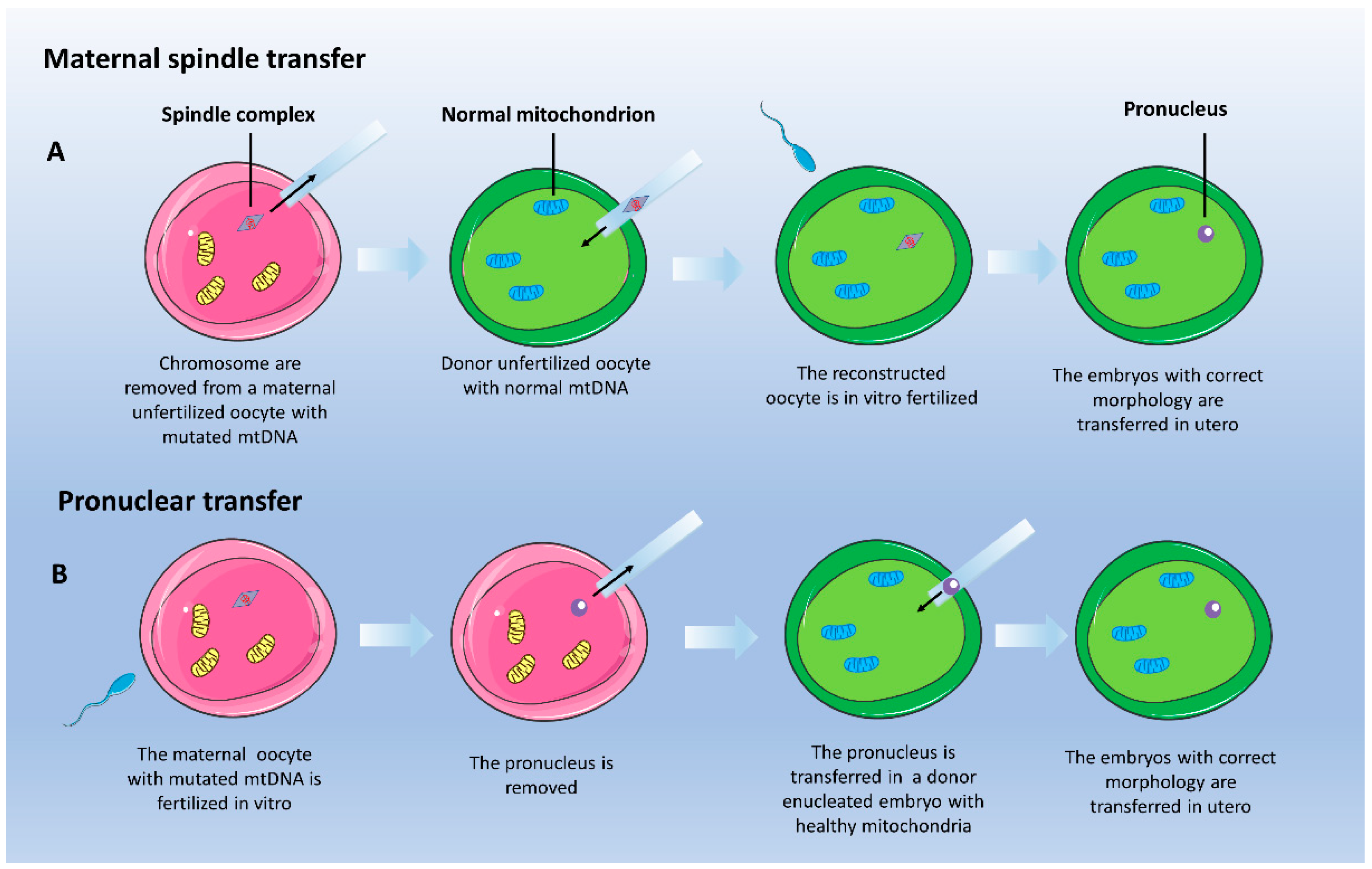
4.1.2. Mitochondrial Donation: Maternal Spindle Transfer

Recently, mitochondrial replacement or mitochondrial donation (MD) has been proposed as a potential method for preventing transmission of mutated mtDNA from the mother to the offspring, by replacing the mitochondria in the oocytes of carrier women. The most recently exploited MD reproductive technologies include maternal spindle transfer (MST) [341] and pronuclear transfer (PNT) [342]. This technology is legally approved for use in the U.K., but the governments of Australia and Singapore are in the process of formal discussions aimed at MD legalization. However, the current state of MD-relevant activity and regulation remains largely elusive in many countries; please refer to [343] for a detailed overview. MST is a complex technique that involves the transfer of nuclear genetic material between a patient’s egg with mutated mtDNA to an enucleated donor’s unfertilized metaphase II oocyte with healthy mitochondria. MST generates an oocyte with a patient’s nuclear DNA devoid of mutated mtDNA. MST is then followed by intracytoplasmic sperm injection (ICSI) and in vitro embryo culture (Figure 3A). A proof-of-principle use of MST to prevent the transmission of mutated mtDNA molecules was first reported in rhesus macaques by the group of Mitalipov in 2009 [344]. In that case, MST and subsequent ICSI resulted in the birth of healthy offspring (named Mito and Tracker) with undetectable levels of spindle donor’s associated mtDNA [344]. The same group then translated this technique to human oocytes, and similar fertilization and blastulation rates between MST and control groups were observed, suggesting that embryo development was not compromised [345]. MtDNA analysis revealed that all examined spindle transfer zygotes and cleaving embryos contained more than 99% donor mtDNA. Similar outcomes were observed in 87% of embryonic stem (ES) cell lines established from spindle transfer blastocysts, regardless of donor mtDNA. However, a reversal of mtDNA haplogroup from donor to maternal mtDNA in a limited number of ES clones was reported, for which a mechanistic explanation based on replicative advantages conferred by some D-loops polymorphisms were proposed [345]. It should be noted that a further mtDNA analysis on those and others ES cell lines [346] raised some concerns about the evidence provided by Kang and co-workers [345]. At the American Society for Reproductive Medicine (ASRM) annual meeting 2016, Dr. John Zhang, New Hope Fertility Center of New York City reported the outcome of the use of MST in a woman carrying mtDNA mutation of Leigh syndrome (8993 T>G), which resulted in the birth of an healthy baby with less than 10% mutated mtDNA in tissues tested 2 days after the birth [347]. A case report was published in 2017 [348].

4.1.3. Mitochondrial Donation: Pronuclear Transfer

The pronuclear transfer (PNT) involves a first step of in vitro fertilization of the patient’s oocyte, followed by the removal of the diploid nucleus, which is then transferred into a donor’s enucleated zygote with healthy mitochondria (Figure 3B) [340]. PNT was proposed in 2005 to prevent transmission of mtDNA disease in the mito-mouse, a model that accumulates large-scale mtDNA deletions [349]. In this elegant paper, second polar bodies were used as biopsy samples to diagnose mtDNA genotypes of mito-mouse zygotes. Nuclear transplantation was carried out from mito-mouse zygotes to enucleated normal zygotes and was shown to rescue all of the F(0) progeny from the expression of respiration defects throughout their lives [349]. In a second report published in 2010, abnormally fertilized human zygotes were used, and reconstructed embryos developed following PNT showed the capacity to reach the blastocyst stage [340]. In 2016, the first preclinical evaluation of PNT using normally fertilized human embryos was reported [350]. Since the techniques used these studies [340] were not tolerated by normally fertilized zygotes, an alternative approach was developed, that significantly improved the efficient development to the blastocyst stage. This was based on performing the pronuclear transplantation immediately after completing meiosis rather than before the first mitotic division. Following this optimization, mtDNA carryover was reduced to less than 2% in the majority (79%) of PNT blastocysts. The study reported low levels of mtDNA carryover in PNT embryos and observed a reversion to the maternal haplogroup in a limited number of hESC clones derived from PNT inner cell mass embryos [350]. In conclusion, although PNT can reduce the risk of mtDNA disease, it may not guarantee prevention [350].

4.2. Personalized Therapies for mtDNA Disorders

4.2.1. Delivery of Nucleic Acids to the Mitochondria

Mutations in mitochondrial genes or mitochondrial tRNAs are associated with a variety of maternally inherited neuromyopathies. An effective therapy would imply delivery therapeutic genes or tRNAs to the mitochondrial matrix, where mtDNA resides.
Nucleic acid delivery into the mitochondrion has been attempted using liposome-based nanocarriers such as Mito-Porter [351,352] and dequalinium-based liposome-like vesicles (DQAsomes)-transfection system [353], or by RNA Import Complex [354]. Mito-Porter is a liposome-based carrier that introduces macromolecular cargos into mitochondria via membrane fusion. The authors provided evidence of nucleic acid delivery into the mitochondrial matrix either in isolated rat liver mitochondria and in living, intact cells [351]. As proof of principle, this method was further exploited to deliver wild-type mitochondrial pre-tRNAPhe to decrease the mutation rate of tRNAPhe in mitochondria of the patient’s cell with a G625A heteroplasmic mutation in the tRNAPhe of mtDNA, with a significant correction of the mutation rate [355]. Similarly, a therapeutic correction of ND3 mutant fibroblasts’ mitochondrial respiration was obtained by reintroducing the wt mRNA of ND3 via Mito-Porter [356].
DQAsomes are the prototype for all mitochondria-targeted vesicular pharmaceutical nanocarrier systems. First described in 1998, they have been successfully explored to deliver DNA and low-molecular-weight molecules to mitochondria within living mammalian cells [357].
The RNA Import Complex (RIC) is a multi-subunit protein complex from the mitochondria of the Kinetoplastid protozoon Leishmania tropica that induces the transport of tRNA across natural and artificial membranes [358]. Observing that Kinetoplastid protozoa have evolved specialized systems for importing nucleus-encoded tRNAs into mitochondria, the RIC was used to import endogenous cytosolic tRNAs, including tRNALys, and restored mitochondrial function in wild-type, MERFF, and KSS cybrids [354].
However, no effective treatment was translated into animal models despite this evidence of delivering the exogenous nucleic acids into the cellular mitochondria.

4.2.2. Heteroplasmic Shift

A second strategy to correct mtDNA mutations is based on the disruption of mutant molecules using selective nucleases to shift the heteroplasmy level below the critical, pathological threshold. To this aim, several approaches based on the following tools have been developed in the past years and are discussed below: (I) restriction enzymes; (II) antisense oligonucleotides; (III) molecular scissors; (IV) DddA.
The very first work carried out by Tanaka and co-workers in 2002 [359] implied the delivery of the restriction endonuclease SmaI into the mitochondria of cybrids to specifically degrade mtDNA in which the pathogenic 8993T>G mutation was present, creating a specific SmaI-restriction site [359]. SmaI specifically degraded mutant mtDNA, with consequent repopulation of mitochondrial genome by wild-type molecules, and subsequent restoration of intracellular ATP level and mitochondrial membrane potential [359].
Heteroplasmy shifting has also been achieved using antisense oligonucleotides in cybrids containing a heteroplasmic mtDNA deletion [360]. In this case, anti-replicative RNA molecules were designed and transfected into a cybrid cell line derived by a KSS patient’s fibroblasts carrying an mtDNA deletion involving 65% of the mtDNA molecules. The anti-replicative effect of the RNA oligoribonucleotides complementary to the mutant mtDNA region specifically reduced the proportion of pathological mtDNA population, shifting the heteroplasmy level from 65% to 50% [360].
The Clustered Regularly Interspaced Short Palindromic Repeats (CRISPR)/Cas9 technology has been largely used for nuclear gene editing; however its application for the editing of the mitochondrial genome appears highly challenging mainly due to sub-efficient delivery of guide RNA and Cas9 enzyme complexes into mitochondria. However, in 2015, Jo and colleagues successfully manipulated the mtDNA with the CRISPR/Cas9 technology [361]. Despite the lack of MTS peptides, the authors showed that the flagged Cas9 localized into the mitochondria, while the gRNAs allowed specific depletion of targeted portions of mtDNA but not degradation of the entire molecule. This point was difficult to reconcile with the fact that mtDNA behaves as a unit [362].
A recent preprint study reported the editing of the MT-ND4 gene accomplished by targeting the guide RNA to an RNA transport derived stem loop element (RP-loop), while expressing the Cas9 enzyme with a mitochondrial localization sequence [363]. However, due to the controversial nature of mammalian mitochondrial RNA import [364], the use of the CRISPR/Cas9 application for mtDNA editing is still debated.
Very recently, adeno-associated viral vectors (AAVs) were used to deliver molecular scissors (i.e., ZFNs, mitochondrial-targeted (mt) TALENs and mtZFNs) in vivo, to destroy mutated mtDNA selectively. Using the first available mouse model of heteroplasmic mitochondrial disease, bearing the point mutation m.5024C>T in mitochondrial tRNAALA (mt-tRNAALA) [365], both TALENs and mtZFNs were able to reduce the mtDNA heteroplasmy with the concomitant rescue of molecular and biochemical phenotypes [366,367].
However, the approaches described so far cannot introduce specific nucleotide changes in mtDNA and cannot be applied to homoplasmic mtDNA mutations because they would destroy all mtDNA copies. To overcome those issues, Mok et al. has recently set up a precision genome editing strategy. They obtained outbreaking results using the interbacterial toxin “double-stranded DNA deaminase toxin A”, or DddA, encoded by Burkholderia cenocepacia, which catalyzes the deamination of cytidines within dsDNA [368]. Engineered split-DddA halves are inactive until simultaneously carried on the target DNA by adjacently bound programmable DNA-binding proteins. The fusions of the split-DddA halves, transcription activator-like effector (TALE) array proteins, and an uracil glycosylase inhibitor generated the RNA-free DddA-derived cytosine base editors (DdCBEs), which allows guided CG-to-TA changes in mtDNA without the need of double-strand breaks. This technique can potentially correct pathogenic mutations on mtDNA with high levels of purity and specificity [368]. Despite these findings looking extremely innovative, additional research aimed to improve the delivery of DdCBEs in vitro and in vivo is essential for exploring their therapeutic potential.

4.2.3. Allotopic Gene Expression

Allotopic gene expression is a method to overcome the mtDNA mutations by re-expressing the missing mtDNA-encoded protein from the nucleus. In this case, an engineered nuclear version of a mitochondrial gene encodes a protein that can be imported into the mitochondria due to an MTS presence. This approach was used to deliver the protein product of the MT-ATP6 gene to the mitochondria in cybrids containing the m.8993T>G mutation [369]. Such allotopic expression significantly improved the cell growth in selective medium and the ATP synthesis [369]. Similarly, allotopic expression of the MT-ND4 gene effectively prevented blindness in a rat model of LHON [370]. Consequently, a human clinical trial of gene therapy to treat LHON has been carried out. Although there was weak evidence of allotopic expression, Guy et al. reported the amelioration of visual acuity in the injected eyes [371]. Another clinical trial (ClinicalTrials.gov identifier NTC01267422) in which nine patients affected by LHON carrying the G11778A mutation were treated with a single dose (5 × 10⁹ vg/0.05 mL) of rAAV2-ND4 reported no adverse effects. In six out of nine patients, the injected eyes’ visual acuity improved by at least 0.3 log MAR after nine months of follow-up. The visual field was enlarged, but the retinal nerve fiber layer remained relatively stable. The visual acuity improved, and the visual field was enlarged nine months after treatment, while other parameters were unchanged [372]. A third open-label phase I/II clinical trial (ClinicalTrials.gov identifier NCT02064569) investigated both safety and preliminary efficacy of a rAAV2/2-ND4 in four dose-escalation cohorts. The treatment resulted in safe with mild adverse effects. Six out of 14 patients manifested a clinically relevant improvement in the best-corrected visual acuity. Taken together, these results suggest the possible use of gene therapy for LHON.

4.2.4. Mitochondrial Augmentation Therapy

Recently, Minovia Therapeutics developed a novel form of cellular therapy called Mitochondrial Augmentation Therapy (MAT). Its rationale is based on the capability of exogenous mitochondria to enter cells in culture [373,374], bringing their own genetic material, augmenting endogenous mitochondrial function, and content and fusing with the endogenous mitochondrial network. The mitochondrial uptake process was termed mitochondrial augmentation and is currently proposed for Kearns–Sayre syndrome (KSS) and Pearson Syndrome. KSS is a progressive retinitis pigmentosa and external ophthalmoplegia occurring at childhood due to de-novo mtDNA deletions [13]. Other systems may be involved over time, including hearing, heart, skeletal muscles, central nervous system, endocrine glands, and kidneys [13]. Pearson Syndrome is a rare disorder affecting the bone marrow and exocrine pancreas [14], also characterized by de-novo mtDNA deletion [15]. MAT approach enriches hematopoietic stem cells (HSCs) with healthy mitochondria before transplantation in patients. MAT involves a series of complex steps: first, mitochondria are extracted from white blood cells derived from the patient’s mother (confirmed non-deleted); in turn, stem cells are collected from the patient, who receives treatment with Neupogen (filgrastim, by Amgen) to boost the production of white blood cells in the bone marrow, and with Mozobil (plerixafor, by Sanofi-Aventis) to mobilize the hematopoietic stem cells containing the CD34 protein marker into the bloodstream. The healthy mitochondria are then introduced into patient-derived stem cells and given back to the patient by intravenous infusion. A first human study was performed on three children with Pearson syndrome under a compassionate use program. Results were reported at the “Targeting Mitochondria Conference 2019” and at “Mitochondrial Medicine 2019 Meeting”. Mitochondrial augmentation therapy improved the in vitro PS-derived HSC function, the metabolic determinants, the aerobic capacity, and the quality of life of the treated patients. The same company also promoted an open-label study (ClinicalTrials.gov identifier NCT03384420) to assess the MAT’s safety and therapeutic effects. Recruitment of seven children is now in progress to analyze any treatment-related adverse events at one year, and any improvement in the quality of life, as assessed by the International Pediatric Mitochondrial Disease Scale.
Moreover, promising results of the first MAT treatment on a KSS patient were recently reported. The 14-year-old patient underwent leukapheresis, and positively selected CD34+ cells were augmented with maternal mitochondria before infusion (2 × 10⁶ cells/kg). Then, the patient was followed for clinical and metabolic parameters. Before MAT, she weighed only 19 kg, she could not sit, walk, express words, and experienced 1 or 2 seizures a week. Seven months after treatment, she gained weight, she could reach objects, sit independently, walk with assistance, and express herself in short sentences. Seizures were resolved 4 months after treatment. Her normalized functional score on the International Pediatric Mitochondrial Disease Score improved from 91% to 57%. Also, the ATP content of the peripheral blood lymphocytes was increased. Those impressive improvements of her physiological and metabolic status make MAT a potential therapy for KSS [375].

5. Precision Medicine Approaches for PMD Caused by Nuclear Defects

5.1. Gene Therapy Approaches

Gene therapy is the most straightforward option for treating disease caused by a single recessive genetic defect. Re-expressing the wild-type form of a mutated gene, or other therapeutic genes, using appropriate viral vectors is an attractive strategy, currently exploited for disorders affecting a single organ. In fact, while the expression of an ectopic gene in the whole body is still unfeasible, specific critical cells or tissue can be targeted with currently available technologies. AAVs are emerging as a suitable delivery method because they are not associated with any disease in humans or animals and remain episomic in the cells for a prolonged time, thus reducing the risk of insertional mutagenesis [376]. Moreover, the availability of several serotypes allows tissue-specific targeting [377]. Potential pitfalls concern the limited cloning capacity, the difficulty in achieving therapeutic expression levels in several tissues—especially skeletal muscle, due to is abundance and distribution- and the brain, due to the low blood–brain-barrier (BBB) penetrance. One of the first preclinical gene therapy studies applied to a mitochondrial myopathy was performed using the AAV2 carrying the ANT1 cDNA in the ANT1 ko mouse model. ANT1 encodes the mitochondrial adenine nucleotide translocator, an integral IMM protein that forms homodimers or tetramers [378], acting as electrogenic pumps that export ATP out across the IMM in exchange for cytosolic ADP [379]. Mutations in ANT1 lead to MM with progressive external ophthalmoplegia (PEO), caused by paralysis of the extraocular eye muscles [380]. AAV-ANT1 transduction resulted in long-term stable expression in muscle precursor cells and differentiated muscle fibers. The transgenic ANT1 protein was targeted into the IMM, formed a functional ADP/ATP carrier, increased the mitochondrial export of ATP, and reversed the histopathological changes associated with the MM [381]. Furthermore, the efficacy of AAV-mediated gene therapy has been confirmed in several mouse models of mitochondrial disorders, including the models for hepato-cerebral forms of severe mtDNA depletion syndrome [57] and Leigh syndrome [382].

5.2. Liver Transplantation

Liver transplantation (LT) is a feasible approach to treat PMD, mainly affecting the single organ [383]. LT has been performed in individuals with hepatocerebral forms of mtDNA depletion syndromes, which frequently progress to liver failure, as those due to mutations in MPV17 gene [384]. However, LT outcome has not been satisfactory, since almost fifty percent of the transplanted patients died in the post-transplantation period due to multiorgan failure. For this and other reasons, LT in mitochondrial hepatopathies remains controversial. LT is not recommended in patients with disorders characterized by a rapid progression of neurological manifestations, such as Alpers–Huttenlocher syndrome, but it may be beneficial in patients with an acceptable quality of life. LT may also differentially affect survival, e.g., patients with mtDNA depletion syndromes caused by mutations in DGUOK gene—encoding the mitochondrial deoxyguanosine kinase, which phosphorylates purines to the corresponding nucleotides in the mitochondrial nucleotides salvage pathway—show lower survival than those of patients with other PMD. Although a recent retrospective study on 12 PMD patients receiving LT confirmed these findings [385], others suggest that, even in the presence of neurological MRI findings, but in the absence of significant neurological symptoms, LT represents a viable option in DGUOK-deficient patients [386].

5.3. PMD Characterised by Systemic Accumulation of Toxic Compounds

5.3.1. Application of Gene Therapy Protocol

AAV therapy has been successfully achieved by targeting the missing gene to the liver when the underlying disease mechanism is linked to toxic compounds’ systemic accumulation. This is the case of the ethylmalonic encephalopathy caused by mutations in the ETHE1 gene encoding for a mitochondrial sulfur dioxygenase involved in the detoxification pathway of hydrogen sulfide (H2S) [387]. Mutations in ETHE1 lead to systemic accumulation of H2S, which acts as a potent inhibitor of complex IV [197], leading to the onset of the phenotype (for a detailed description of the H2S pathway, see Viscomi, Bottani et al., 2015 [388]). The re-expression of the ETHE1 gene by a hepatotropic AAV2/8 serotype restored the missing enzymatic activity in the liver, with a significant clearance of H2S from the bloodstream, a major amelioration of the phenotype and prolonged lifespan of the Ethe1 ko mice [389]. Similarly, systemic accumulations of thymidine and deoxy uridine, which interfere with mtDNA replication and lead to mitochondrial dysfunction were corrected by hepatotropic AAV2/8 vector carrying the human TYMP in a mouse model of MNGIE [390]. The nucleoside reduction achieved by this treatment prevented deoxycytidine triphosphate (dCTP) depletion, which is the limiting factor affecting mtDNA replication in this disease [390].
AAV treatment may be useful also in mitochondrial neurodegenerative disorders: it was reported that the administration of AAV9 carrying the human NDUFS4 partially rescued the phenotype of Ndufs4 ko mice only when simultaneously administered systemically and intracranially. AAV9 serotype did not efficiently cross the BBB, and mainly targeted glial cells when injected intracranially in new-borns. Interestingly, newly engineered serotypes AAV-PHP.eB and AAV-PHP.S showed great promises in their efficiency to transduce the central and peripheral nervous systems, crossing the BBB [391].

5.3.2. Application of Liver Transplantation Protocol

Like the gene therapy approach, the liver is an attractive target for pathologies triggered by systemic accumulation of toxic compounds. LT is proposed as an alternative approach to treat mitochondrial neurogastrointestinal encephalopathy (MNGIE) disease. MNGIE is caused by a deficiency of thymidine phosphorylase (TP) due to mutations in the nuclear gene TYMP [392]. TP is a cytosolic enzyme catalyzing the first step of thymidine (dThd) and deoxy uridine (dUrd) catabolism. Mutations in TYMP lead to the accumulation of dThd and dUrd systemically, which induce an imbalance of the cytosolic nucleotide pool. Because the mitochondrial nucleotide pool relies, in part, on nucleotides imported from the cytosol, an imbalanced cytosolic nucleotide pool lead to an imbalanced mitochondrial nucleotide pool, which ultimately has mutagenic effects on mtDNA, resulting in depletion, multiple deletions, and point mutations causing progressive mitochondrial deficiency and organ failure [392,393,394]. LT rapidly normalized serum levels of toxic nucleosides in a 25-year-old MNGIE patient, and his conditions were stable after 400 days of follow-up [395]. LT also resulted in an effective option to treat ethylmalonic encephalopathy due to mutation in ETHE1 gene [396,397], since the replaced organ can substitute the deficient ETHE1 enzyme and clear the toxic H2S that accumulate in this disorder, constituting a feasible therapeutic option in patients. Patients showed progressive improvement of the neurological functions and normalization or amelioration of the biochemical abnormalities [396,397]. However, the decision to perform LT remains difficult because neurological manifestations may worsen despite their absence before the transplant [398].

5.3.3. Cell Replacement

Cell therapy consists of using cells or cell-based products to replace dead or defective cells to restore the function of the affected tissue(s) [399]. Again, this approach has been proposed to treat MNGIE. While TP is not expressed in all tissues, cellular and plasmatic dThd and dUrd levels are in equilibrium among all body compartments. Therefore, replacing the lost enzyme in a circulating form—i.e., in the blood cells—should favor the catabolism of the toxic metabolites in plasma, thus clearing these freely diffusible substrates from the tissue compartments, normalizing the cellular nucleotide pools, and preventing further mtDNA damage.
Cell replacement therapies may offer a permanent cure to MNGIE. They comprise (I) Allogeneic Hematopoietic Stem Cell Transplantation (AHSCT) and (II) erythrocyte-encapsulated thymidine phosphorylase (EE-TP). Clinical and biochemical improvements following AHSCT have been reported in MNGIE patients [400,401]. Although AHSCT corrects biochemical abnormalities and improves gastrointestinal symptoms, the procedure is risky for subjects in poor medical conditions, as many MNGIE patients are. Since transplant-related morbidity and mortality increases with the progression of the disease and the number of comorbidities, MNGIE patients should be submitted to AHSCT when they are still relatively healthy, to minimize the complications of the procedure [401].
EE-TP consists of the ex-vivo encapsulation of Escherichia coli TP within the patient’s autologous erythrocytes using a reversible hypo-osmotic dialysis process. Once inside the erythrocyte, the encapsulated enzyme catalyzes the deoxyribonucleosides’ metabolism to the specific products thymine and uracil, which then exit the erythrocyte and flow into their physiological metabolic pathways. Recently, three adult MNGIE patients received escalating intravenous doses of EE-TP. EE-TP was well tolerated, and reductions in the disease-associated plasma metabolites, thymidine, and deoxy uridine were observed. Clinical ameliorations, including weight gain and improved disease scores, were observed in two patients, suggesting that EE-TP can reverse some aspects of the disease [401]. Advantages of the EE-TP are the prolonged circulatory half-life of the enzyme and the minimization of the immunogenic reactions compared to those frequently observed in enzyme replacement therapies administered by the conventional route [401].

5.4. Molecular Bypass Therapy in Disorders of mtDNA Instability

Syndromes characterized by mtDNA instability are usually due to mutations in nuclear genes involved either in the mtDNA replication machinery or deoxynucleotide triphosphate (dNTP) metabolism consequently affecting OXPHOS activities.
From the clinical perspective, these diseases are characterized by disorders that range from severe infantile hepatocerebral encephalopathy to childhood-onset myopathy or adult-onset PEO [402]. The TK2 gene provides an example. The protein product of this gene is a deoxyribonucleoside kinase with mitochondrial localization that specifically phosphorylates thymidine, deoxycytidine, and deoxyuridine. This enzyme is required for mtDNA synthesis. Recessive mutations in the human TK2 gene are responsible for the myopathic form of the mitochondrial depletion/multiple deletions syndrome [403]. Mitochondrial dNTPs pools are supplied either by de novo synthesis and import from the cytosol or by the mitochondrial deoxyribonucleoside salvage pathway [404]. Supplementation of the missing or insufficient dNTP may bypass the block restoring the deoxynucleotides triphosphate (dNTP) pools. Molecular bypass therapy (MBP) with deoxypyrimidine monophosphates (dCMP and dTMP) or substrate enhancement therapy with deoxypyrimidine nucleosides (dC and dT) were tested on the Tk2 knock-in (ki) mouse model in the early pre-symptomatic, but biochemically affected, stage [405]. Treatment with dCMP and dTMP raised dTTP concentrations, increase levels of mtDNA, ameliorated the defects of MRC enzymes, and significantly prolonged the lifespan (from 13 to 34 days) in a dose-dependent manner [406,407]. Late treatment of 29-day-old mice was ineffective.
Similar strategies were then extended in different models of PMD. The mtDNA depletion in human fibroblasts mutated in the DGUOK gene was ameliorated by the supplementation of deoxyguanosine [404], and similarly, pyrimidine and purine nucleoside administration, but not the corresponding monophosphate nucleotides, adjusted the mtDNA depletion induced by ethidium bromide in human RRM2B-mutant cells [408]. Similarly, deoxycytidine and tetrahydro uridine were also able to prevent mtDNA depletion in a cell model of the same syndrome [408]. Recently a mutant dguok zebrafish line was developed using CRISPR/Cas9 mediated mutagenesis; dguok−/− fishes have significantly reduced mtDNA levels compared to the wild-type counterpart. In contrast with previous cell culture studies, when supplemented with only one purine nucleoside (dGuo), mtDNA copy number in both mutant and wt juvenile animals was significantly reduced, possibly because of nucleotide pool imbalance. However, a significant increase in liver mtDNA was documented in adult dguok−/− zebrafish when supplemented with both purine nucleosides. [409].
Recently, an open-label study showed the results of deoxynucleoside monophosphates and deoxynucleoside administration under a compassionate program to 16 early-onset TK2-patients. Prolonged survival and improvement of motor abilities compared to untreated patients were recorded. Four of 5 patients who required enteric nutrition were able to discontinue using the feeding tube; and 1 of 9 patients who required mechanical ventilation became able to breathe independently. Out of 8, 3 non-ambulatory patients recovered the ability to walk. Out of 5, 4 patients with enteric nutrition discontinued the use of the feeding tube. Out of 9, 1 patient who required mechanical ventilation became able to breathe independently. Although diarrhea was the most common side effect manifested, discontinuation of the therapy was not necessary. Among 12 other TK2 patients treated with deoxynucleoside, two adults developed elevated liver enzymes normalized following discontinuation of therapy [410].

6. Future Perspectives

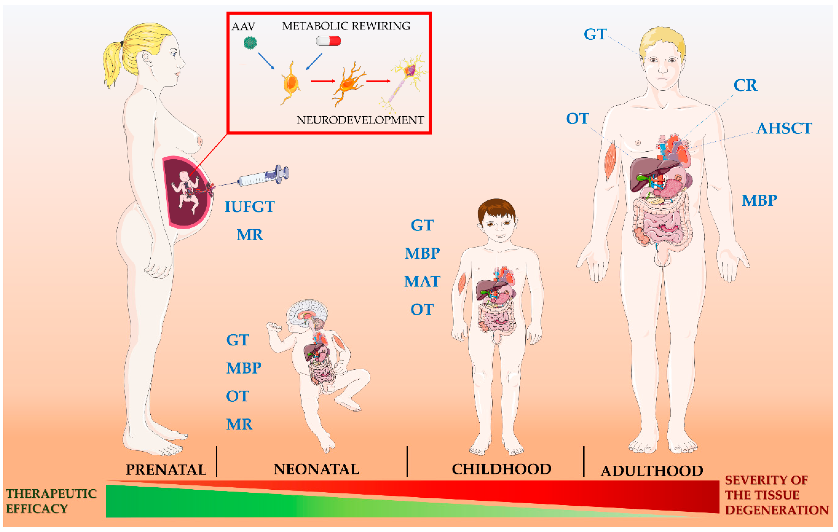
6.1. Fetal Gene Therapy

Current prenatal genetic technology can diagnose rare genetic diseases as early as the 12th week of pregnancy by chorionic villous sampling or from the 16th week by amniocentesis. Advances in fetal imaging and minimally invasive surgical equipment have also led to the development of interventional techniques for prenatal treatment of several fetal structural defects, such as congenital diaphragmatic hernia, myelomeningocele, pulmonary sequestration, hydrothorax, urinary tract obstruction, fetal tumours and others [411]. Also, intrauterine access to the fetal circulation through the umbilical vein is a well-described procedure, used to perform ultrasound-guided fetal blood or platelet transfusions, which are currently the standard of care in cases of fetal anemia and thrombocytopenia [412]. The option of fetal intervention is offered in specialized centers to reduce infant mortality and/or morbidity compared to postnatal treatment and, for each type of procedure, parents are extensively informed about the potential benefits balanced against the risks, which are mainly fetal death, miscarriage or preterm delivery.
For inherited genetic diseases, In-Utero Gene Therapy (IUGT) offers the potential of prophylaxis against early, irreversible, and lethal pathological change [413]. From a technical perspective, potential genetic therapeutic agents can be delivered to the fetus through infusion into the umbilical vein or via direct injection into the fetal organs. The rationale for IUGT is essentially based on the hypothesis that anticipating the treatment during fetal life could prevent or mitigate irreversible pathological changes associated with rare genetic disorders and improve clinical outcomes compared to postnatal therapy. Possible advantages of IUGT that may overcome some of the limitations encountered in postnatal gene therapy include: (i) the small fetal size (i.e., smaller area to be treated), (ii) the tolerogenic fetal immune system, (iii) the presence of highly proliferative and accessible stem/progenitor cells in multiple organs and, (iv) the ability to treat diseases in which irreversible pathological molecular and metabolic changes begin before birth [414].
Fetal therapeutic interventions could be especially useful for early-onset PMD in which mitochondrial dysfunction begins before birth, as the GRACILE syndrome. GRACILE syndrome initially develops with intrauterine growth retardation. A fatal lactic acidosis arises in the new-borns, often accompanied by nonspecific aminoaciduria, cholestasis, iron overload, and liver dysfunction [415].
Similarly, mitochondrial dysfunction likely starts before birth in SURF1-associated Leigh syndrome. Shreds of evidence suggest that SURF1 mutations lead to metabolic impairments in neural progenitor cells (NPCs), which cannot switch from glycolytic to OXPHOS metabolism, with subsequent aberrant proliferation and insufficient support for neuronal morphogenesis [416]. A similar neuronal impairment was reported in a SURF1 ko pig model [417]. Cerebral organoids from LS patients carrying MT-ATP6 mutations also showed defective corticogenesis and suggest pre-natal impairment [418]. These findings suggest that OXPHOS defect could impair the NSC cellular metabolism in the early phase of the development leading to the onset of the neurological phenotypes; so prenatal intervention for pediatric PMD may be crucial for amelioration of the clinical course of the disease.
Fetal gene therapy may provide an alternative therapeutic approach for inherited diseases leading to early death or lifelong irreversible damage. Due to the lack of PMD-specific, fetal therapeutic approaches, curative strategies proposed for other genetic diseases should be considered. A recent study investigated the efficacy of human survival motor neuron (hSMN) gene expression after IU delivery in SMA mouse embryos. In the first part of the research report, authors showed that IU-intracerebroventricular injection of adeno-associated virus serotype-9 (AAV9)-EGFP led to an extensive expression of EGFP protein in different parts of the CNS with a significant number of transduced NSCs. SMA mouse fetuses receiving a single i.c.v. injection of a single-stranded or self-complementary AAV9-SMN vector extended their lifespan of 93 (median of 63) or 171 (median 105) days. Both muscle pathology and motor neuron survival improved upon treatments, with slightly better results from scAAV administration [413].
Additional evidence on the safety and efficacy of IUGT in a mouse model of neuronopathic Gaucher’s disease were recently provided [419]. Fetal intracranial injection of an AAV9 carrying the curative gene improved neuronal inflammation and spectacularly increased the mice’s overall survival. Of note, neonatal treatment did not achieve the same results of fetal therapy.
Maternal safety is a critical consideration in any fetal therapy. Possible maternal exposure to the viral vectors infused into the fetus should be considered in IUGT. Contact with the viral particles may result in maternal immune responses to the capsid protein or the recombinant protein, although the latter is unlikely, as the mother should already be producing—and therefore be tolerant to—the protein missing in the fetus.
Therapeutic viruses, including the AAV vectors, may undergo random integration in fetuses after IUGT; however, do evidences of germline integration have been reported. New technologies with more specific gene editing will possibly minimize off-target events of IUGT in future.
Despite promising preliminary results of IUGT, more experimental evidence on animal models is needed to demonstrate a significant improvement in the pathological hallmarks and the clinical course of the disease. Clinical trials involving human pregnancies would subsequently need to be setup, ensuring accurate monitoring of adverse events and long-term postnatal clinical follow-up. Randomized controlled trials on IUGT versus postnatal treatment may not be realistic due to the rarity of the investigated diseases and ethical concerns that may arise from offering randomization for conditions associated with a very severe if not lethal outcome. Also, rare genetic disorders currently lack any recommended screening policy in the general low-risk population. Therefore, most of the patients currently receiving a prenatal diagnosis of PMD have experienced a previously affected child’s birth. In these cases, prenatal treatment parents could be offered within an experimental trial, after extensive counselling on the uncertainties regarding the clinical results and after discussion of other management options, which would be termination of pregnancy or postnatal therapy. It could be anticipated that the recruitment rate in such a context is likely to be extremely low. However, multicenter collaborative efforts may allow to collect and analyze a meaningful number of cases using a pre-defined shared protocol, to provide reliable information on the effects of IUGT on long-term postnatal development of infants affected by rare genetic disorders.
Ethical issues of fetal gene therapy have been reviewed by MacKenzie and collaborators [420]. Fetal treatment pivots on the concept of non-directive advising, in which the choices of no treatment and exploratory therapy—with all the conceivable risks and benefits—are rationalized without the physician’s individual inclination. Rigorous preclinical trials and multidisciplinary debates will continue to advance the frontiers of fetal therapy, while these and other concerns deserve a continuous discussion [420].

6.2. Metabolic Rewiring

Pharmacological modulations of neuronal morphogenesis and neuronal maturation of the immature precursors, the neural stem cells (NSCs) have been proposed as an attractive therapeutic opportunity to treat several neurological diseases. NSCs require a metabolic shift towards oxidative phosphorylation during the process of neuronal differentiation [421]. Therefore, OXPHOS defects may inhibit this shift impairing neuronal differentiation and driving neural stem cells (NSCs) to a proliferative and less differentiated state [416]. We recently tested inductors of oxidative metabolism (developed and patented by Professional Dietetics, IT) composed of TCA cycle intermediates, specific amino acids, and co-factors that helped enhance mitochondrial function in different in vitro and in vivo models. Supplementing wild-type NSC-culture medium with these compounds during the differentiation phase enhanced the metabolic shift towards OXPHOS and mitochondrial function of mouse and human NSCs, improving their full differentiation capacity [422]. Neurons derived from treated-NSC changed the fission–fusion processing resulting in a mitochondrial elongated phenotype; moreover, the activation of the mTORC1 pathway with subsequent significant increase of ATP production was reported. Also, the antioxidant defense system was also triggered by the increase of the NRF2 gene expression [422]. We observed similar metabolic and molecular changes in vivo, where they counteracted the pathological mitochondrial dysfunction occurring during the aging process. Three-month oral administration of metabolic inductor PD-0E7 (Professional Dietetics, IT) to the Senescence Accelerated Mouse-Prone 8 (SAMP8) mice significantly improved the sarcopenia and cognitive decline, enhancing oxidative metabolism by inducing mitochondrial biogenesis and increasing respiratory efficiency [423]. In particular, we documented a strong shift toward oxidative, COX-positive fibers and a general increase of the MRC enzymatic activities in the skeletal muscle of the 12-month-old PD-0E7-supplemented mice, which may explain the preserved physical endurance of the treated SAMP8 mice [423]. Also, the Opa1 isoforms were significantly increased in the skeletal muscle, as shown by western blot analysis, and this might support the improved stabilization of the CIII holocomplex into SCs that was detected by blue native gel electrophoresis (BNGE) analysis [423]. To note, the preserved cognitive function observed in the treated mice correlate with enhancement of the hippocampal mitochondrial proteostasis and with the upregulation of PITRM1, a mitochondrial matrix enzyme that digests the mitochondrial targeting sequences (MTS) and the mitochondrial fraction of amyloid beta [424,425].
Although the molecular mechanisms at the basis of the improvement of mitochondrial functions by such metabolic modulators were not fully elucidated, it is plausible that supplementation of TCA cycle intermediates and amino acids would feed the anaplerotic flux sustaining mitochondrial energy production. In support of this, it has recently been demonstrated that anaplerosis is protective in OXPHOS-deficient neurons with disruption of the MFN2 gene, and that genetic blockage of the anaplerotic pathway further exacerbated the neuronal degeneration [426]. To date, triheptanoin is the only reported example of anaplerotic treatment in patients with very-long-chain acyl-CoA dehydrogenase (VLCAD) deficiency [61]. Studies of the effects of anaplerotic substrates in PMD should be encouraged.

7. Conclusions

The extreme clinical, genetic, and biochemical variability of PMD coupled with the low number of patients and the frequent lack of adequate preclinical models have limited identifying useful clinical outcomes and the development of effective therapy.
The enhancement of mitochondrial function and ATP production through the pharmacological stimulation of mitochondrial biogenesis, mitophagy, dynamics, and ROS detoxification using antioxidants may represent a general strategy to alleviate or at least partially corrected different clinical outcomes. Although these strategies do not solve the problem at the root, these are, in principle, adaptable to a large group of mitochondrial diseases and could help improve patients’ everyday quality of life.
Conclusive cure for mitochondrial disease could be achieved by a precision medicine strategy that considers individual variability in genes, age, sex, stage of the disease, and tissues compromised for each patient (Figure 4). Although organ transplantation was already used successfully, cell replacement and gene therapy are still far to become routine for mitochondrial disease due to technical and regulatory reasons. However, given the recent exciting progress in gene editing and fetal surgery, we expect steps forward in the coming decades.

Author Contributions

Conceptualization, E.B. and D.B.; methodology, D.B., E.B., C.L., N.P., A.P., and V.T. writing—original draft preparation, D.B., E.B., C.L., N.P., A.P., and V.T.; writing—review and editing, D.B. and E.B.; visualization, C.L., N.P., A.P., and V.T.; supervision D.B. and E.B.; funding acquisition, D.B. and C.L. All authors have read and agreed to the published version of the manuscript.

Funding

This study was supported by the Italian Ministry of Health (grants RF-2016-02361495, and “Ricerca Corrente”).

Acknowledgments

D.B. acknowledge University of Milan, Grant number: BIOMETRA PSR2019-BRUNETTI; C.L. is members of the European Reference Network for Rare Neuromuscular Diseases (ERN EURO-NMD). This project was carried out in the Center for the Study of Mitochondrial Pediatric Diseases (http://www.mitopedia.org) funded by the Mariani Foundation.

Conflicts of Interest

The authors declare no conflict of interest.

Appendix A

Table A1. Summary of the therapeutic approaches discussed in this manuscript, with a distinction between those that have been tested in pre-clinical models and those that have already been used in clinical practice. This table is restricted to therapies used to treat PMD and does not include others that, although cited in the text, have not been tested in PMD models/patients.
TherapyModelStudyRef.
Strategies to increase ATP levels
Febuxostat plus inosinepatients with homoplasmic mtDNA mutationclinical[65]
patient with mitochondrial diabetes with heteroplasmic mutation in tRNA leucine 1clinical[65]
Pharmacological stimulation of mitochondrial biogenesis
AICARmouse models of COX defectspre-clinical[74]
mouse model Cox10-Mef2c-Crepre-clinical[82]
CI deficient cells (NDUFS2, NDUFS4, NDUFAF4, C20ORF7, FOXRED1, NDUFA12L)pre-clinical[83]
Bezafibrate and other PPAR agonistsMRC-deficient patients’ fibroblastspre-clinical[87]
SCO2 mutant fibroblastspre-clinical[88]
DNM1L mutant cellspre-clinical[89]
mouse models of COX defectspre-clinical[74]
Deletor mousepre-clinical[90]
Mutator mousepre-clinical[91]
patients with MMclinical[92]
NAD+ precursorsSco2 ko mousepre-clinical[81]
Deletor mousepre-clinical[106]
GBA-PD Drosophila melanogasterpre-clinical[107]
Ndufs4 ko micepre-clinical[108]
Niacinpatients with MMclinical (NCT03973203)[109]
I-BET 525762Acybrids carrying 3796A>G mutationpre-clinical[110]
Polyphenols and other Pharmacognostic Products
Resveratrolfibroblasts from MT-TL1, MT-TK, MT-ATP6-patientspre-clinical[125]
CurcuminLHON patientsclinical (NCT00528151)unpublished
Pharmacological modulation of the NO/cGMP/PKG pathway
L-arginine and L-citrullineMELAS patientsclinical (open-label trial)[162,164,168]
MELAS patientsclinical (open-label trial)[165]
various PMD patientsclinical (NCT02809170)unpublished
MELAS patientsPhase-1 clinical trial NCT03952234unpublished
PDE5 inhibitorsNPCs with homoplasmic mutation in MT-ATP6pre-clinical[176]
LHONclinical (case report)[179]
Antioxidant
GlutathioneMM patientsclinical (double-blind cross-over study)[188]
fibroblasts of patients carrying the m.3243A>G and m.8344A>G mutationspre-clinical[189]
CysteamineC. elegans model of CI defect, FBXL4 mutant human fibroblast, and zebrafish models of pharmacologically induced CI and CIV defectspre-clinical[192]
Cysteamine bitartrate delayed-release (RP103)PMD paediatric patientsclinical (NCT02023866)unpublished
N-acetylcysteineEthe1 mouse modelpre-clinical[195]
ETHE1 patientsclinical (compassionate use)[198,199,200]
Lipoic acid and CoQ10PMD patientsclinical (randomized, double-blind, placebo-controlled, crossover study)[206]
Vitamin C and KPMD patientsClinical[208,209,210]
Vitamin Efibroblasts from patients with CI defectpre-clinical[211,212]
Coenzyme Q10MERRF cellspre-clinical[220]
KSS patientsclinical[221]
KSS patents and other MM with CPEOclinical[222,223,224,226,228,230]
patient with mitochondrial encephalomyopathy with COX deficiencyclinical[225]
patients with mitochondrial cytopathiesclinical[227]
PMD patientsclinical[229]
patients with different OXPHOS defectsclinical[231]
PMD patientsrandomized, double-blind, cross-over trial[232]
patients with different PMD; 15 patients with MMclinical (multicenter study)[233]
PMD paediatric patientsclinical (NCT00432744)[234]
Idebenonefibroblasts from LHON patientspre-clinical[237,238]
mouse model of LHON pre-clinical[239]
85 LHON patients with to m.3460G>A, m.11778G>A, and m.14484T>C mutationsclinical (“RHODOS” study, NCT00747487)[241]
subset of LHON patients from RHODOS studyclinical (RHODOS-OFU, NCT01421381)[242]
patient with TXN2 mutation [243]
Opa1 mutant mice pre-clinical[245]
seven DOA patients clinial[246]
87 DOA patientsclinical (retrospective cohort study)[247]
Redox-Active Molecules
EPI-743PMD patientsclinical[259]
patients with Leigh syndromeclinical (prospective single-arm subject-controlled trial)[260]
children with mitochondrial encephalopathyClinical[261]
one patient with Leigh syndrome due to ND3 mutationClinical[262]
patients with LHONclinical (open-label trial)[263]
patients with Leigh syndromeclinical (NCT02352896)unpublished
PMD paediatric patientsclinical (NCT01642056)unpublished
JP4-039ACAD9- VLCAD-, ETHE1 and MOCS1 mutant fibroblastspre-clinical[266,267]
KH176cellular models of CI defects pre-clinical[268]
Ndufs4 ko mouse model pre-clinical[269,270]
patients with m.3242A>G mutation clinical (KHENERGY STUDY, NCT02909400)[272]
patients with MELASclinical (KHENERGYZE Study, NCT04165239)[273]
SKQ1Mutator mousepre-clinical[281]
Pharmacological modulation of mitochondrial dynamics
Cytotoxic Necrotizing Factor 1 (CNF1)MERRF fibroblastspre-clinical[300]
Pharmacological protection of cardiolipin
Elamipretidepatients with primary mitochondrial myopathy (MM)clinical (NCT03323749)[310]
Barth syndromeclinical (NCT03098797)unpublished
LHON patientsclinical (NCT02693119)unpublished
age-related macular degeneration (AMD) with non-central geographic atrophyclinical (NCT03891875)unpublished
Pharmacological modulation of autophagy
RapamycinNdufs4 ko mousepre-clinical[313]
muscle-specific Cox15 ko mousepre-clinical[314]
ND2-deficient Drosophila model of LSpre-clinical[315]
MT-ATP6-mutant, iPSCs-derived neuronspre-clinical[316]
gas-1 (fc21) nematodespre-clinical[317]
mice with CoQ10 deficiencypre-clinical[320]
TwKOastro micepre-clinical[59]
Everolimus (rapamycin analogue)MELAS patientsclinical (NCT03747328)[318]
children affected by Leigh disease or MELASClinical[319]
Bypassing cI-cIII-cIV defects with alternative enzymes
NDI1CI deficiency in Drosophila melanogasterpre-clinical[324]
mouse model of LHONpre-clinical[238]
mouse model of Leigh syndromepre-clinical[239]
AOX, NDI1ρ0 mouse cellspre-clinical[325]
AOXCIII-IV deficiencies in human cellspre-clinical[326]
CIV deficient Drosophila melanogasterpre-clinical[327]
Bcs1lp.S78G knock-in micepre-clinical[330,331]
Acta-Cox15 ko modelpre-clinical[332]
NDH-2human CI deficient fibroblastspre-clinical[333]
Personalized therapies for mtDNA disorders
Mitochondrial donation: maternal spindle transferlegally approved for use in the U.Kclinical[343]
rhesus macaquespre-clinical[344]
woman carrying mtDNA mutation of Leigh syndrome (8993 T>G)clinical[347,348]
Mitochondrial donation: pronuclear transfermito-mousepre-clinical[349]
Delivery of nucleic acids to the mitochondriapatient’s cell with a G625A heteroplasmic mutation in the tRNAPhepre-clinical[355]
ND3 mutant fibroblastspre-clinical[356]
MERFF and KSS cybridspre-clinical[354]
Heteroplasmic shiftvarious cells with heteroplasmic mutationpre-clinical[359,360,361]
mouse model of heteroplasmic PMDpre-clinical[366,367]
Allotopic gene expressioncybrids with m.8993T>G mutationpre-clinical[369]
rat model of LHONpre-clinical[370]
LHON patientsclinical (NTC01267422; NCT02064569)[371,372]
Mitochondrial augmentation therapychildren with Pearson syndromeclinicalUnpublished
children with KSS or Pearson Syndromeclinical (NCT03384420)unpublished
KSS patientclinical[375]
Precision medicine approaches for PMD caused by nuclear defects
Gene therapy approachesAnt1 ko mouse modelpre-clinical[381]
Tymp ko mouse modelpre-clinical[390]
Ethe1 ko mouse modelpre-clinical[389]
Ndufs4 mouse modelpre-clinical[391]
Mpv17 ko mouse modelpre-clinical[58]
mouse model of Leigh syndromepre-clinical[382]
Liver transplantationPMD patientsclinical[385]
DGUOK-deficient patientsclinical[386]
25-year-old MNGIE patientclinical[395]
ETHE1 patientsclinical[396,397]
Cell replacementMNGIE patientsclinical[400,401]
Molecular bypass therapy in disorders of mtDNA instabilityTk2 mouse modelpre-clinical[405,406,407]
DGUOK mutant fibroblastspre-clinical[404]
RRM2B mutant fibroblastspre-clinical[408]
dguok−/− zebrafishpre-clinical[409]
early-onset TK2-patientsclinical (open-label study)[410]

References

  1. Gorman, G.S.; Chinnery, P.F.; DiMauro, S.; Hirano, M.; Koga, Y.; McFarland, R.; Suomalainen, A.; Thorburn, D.R.; Zeviani, M.; Turnbull, D.M. Mitochondrial diseases. Nat. Rev. Dis. Primers 2016, 2, 16080. [Google Scholar] [CrossRef]
  2. Wallace, D.C. A Mitochondrial Paradigm of Metabolic and Degenerative Diseases, Aging, and Cancer: A Dawn for Evolutionary Medicine. Annu. Rev. Genet. 2005, 39, 359–407. [Google Scholar] [CrossRef] [PubMed] [Green Version]
  3. Stewart, J.B.; Chinnery, P.F. Extreme heterogeneity of human mitochondrial DNA from organelles to populations. Nat. Rev. Genet. 2020, 1–13. [Google Scholar] [CrossRef] [PubMed]
  4. Rahman, S.; Blok, R.B.; Dahl, H.-H.M.; Danks, D.M.; Kirby, D.M.; Chow, C.W.; Christodoulou, J.; Thorburn, D.R. Leigh syndrome: Clinical features and biochemical and DNA abnormalities. Ann. Neurol. 1996, 39, 343–351. [Google Scholar] [CrossRef] [PubMed]
  5. Shoffner, J.M.; Lott, M.T.; Lezza, A.M.; Seibel, P.; Ballinger, S.W.; Wallace, D.C. Myoclonic epilepsy and ragged-red fiber disease (MERRF) is associated with a mitochondrial DNA tRNALys mutation. Cell 1990, 61, 931–937. [Google Scholar] [CrossRef]
  6. Goto, Y.-I.; Nonaka, I.; Horai, S. A mutation in the tRNALeu(UUR) gene associated with the MELAS subgroup of mitochondrial encephalomyopathies. Nat. Cell Biol. 1990, 348, 651–653. [Google Scholar] [CrossRef]
  7. Holt, I.J.; Harding, A.E.; Petty, R.K.; Morgan-Hughes, J.A. A new mitochondrial disease associated with mitochondrial DNA heteroplasmy. Am. J. Hum. Genet. 1990, 46, 428–433. [Google Scholar]
  8. Wallace, D.C.; Singh, G.; Lott, M.T.; Hodge, J.A.; Schurr, T.G.; Lezza, A.M.; Elsas, L.J.; Nikoskelainen, E.K. Mitochondrial DNA mutation associated with Leber’s hereditary optic neuropathy. Science 1988, 242, 1427–1430. [Google Scholar] [CrossRef]
  9. Lopez-Gallardo, E.; Solano, A.; Herrero-Martin, M.D.; Martinez-Romero, I.; Castano-Perez, M.D.; Andreu, A.L.; Herrera, A.; Lopez-Perez, M.J.; Ruiz-Pesini, E.; Montoya, J. NARP syndrome in a patient harbouring an insertion in the MT-ATP6 gene that results in a truncated protein. J. Med. Genet. 2008, 46, 64–67. [Google Scholar] [CrossRef]
  10. Pitceathly, R.D.; Murphy, S.M.; Cottenie, E.; Chalasani, A.; Sweeney, M.G.; Woodward, C.; Mudanohwo, E.E.; Hargreaves, I.; Heales, S.; Land, J.; et al. Genetic dysfunction of MT-ATP6 causes axonal Charcot-Marie-Tooth disease. Neurology 2012, 79, 1145–1154. [Google Scholar] [CrossRef] [Green Version]
  11. Verny, C.; Guegen, N.; Desquiret-Dumas, V.; Chevrollier, A.; Prundean, A.; Dubas, F.; Cassereau, J.; Ferre, M.; Amati-Bonneau, P.; Bonneau, D.; et al. Hereditary spastic paraplegia-like disorder due to a mitochondrial ATP6 gene point mutation. Mitochondrion 2011, 11, 70–75. [Google Scholar] [CrossRef] [PubMed] [Green Version]
  12. Bugiardini, E.; Bottani, E.; Marchet, S.; Poole, O.V.; Benincá, C.; Horga, A.; Woodward, C.; Lam, A.; Hargreaves, I.; Chalasani, A.; et al. Expanding the molecular and phenotypic spectrum of truncating MT-ATP6 mutations. Neurol. Genet. 2020, 6, e381. [Google Scholar] [CrossRef] [PubMed] [Green Version]
  13. Moraes, C.T.; DiMauro, S.; Zeviani, M.; Lombes, A.; Shanske, S.; Miranda, A.F.; Nakase, H.; Bonilla, E.; Werneck, L.C.; Servidei, S.; et al. Mitochondrial DNA Deletions in Progressive External Ophthalmoplegia and Kearns-Sayre Syndrome. N. Engl. J. Med. 1989, 320, 1293–1299. [Google Scholar] [CrossRef] [PubMed]
  14. Pearson, H.A.; Lobel, J.S.; Kocoshis, S.A.; Naiman, J.L.; Windmiller, J.; Lammi, A.T.; Hoffman, R.; Marsh, J.C. A new syndrome of refractory sideroblastic anemia with vacuolization of marrow precursors and exocrine pancreatic dysfunction. J. Pediatr. 1979, 95, 976–984. [Google Scholar] [CrossRef]
  15. Baerlocher, K.E.; Feldges, A.; Weissert, M.; Simonsz, H.J.; Rötig, A. Mitochondrial DNA deletion in an 8-year-old boy with pearson syndrome. J. Inherit. Metab. Dis. 1992, 15, 327–330. [Google Scholar] [CrossRef] [PubMed] [Green Version]
  16. Calvo, S.; Jain, M.; Xie, X.; Sheth, S.A.; Chang, B.; Goldberger, O.A.; Spinazzola, A.; Zeviani, M.; Carr, S.A.; Mootha, V.K. Systematic identification of human mitochondrial disease genes through integrative genomics. Nat. Genet. 2006, 38, 576–582. [Google Scholar] [CrossRef]
  17. Vafai, S.B.; Mootha, V.K. Mitochondrial disorders as windows into an ancient organelle. Nat. Cell Biol. 2012, 491, 374–383. [Google Scholar] [CrossRef]
  18. Stenton, S.L.; Prokisch, H. Genetics of mitochondrial diseases: Identifying mutations to help diagnosis. EBioMedicine 2020, 56, 102784. [Google Scholar] [CrossRef]
  19. Zorova, L.D.; Popkov, V.A.; Plotnikov, E.Y.; Silachev, D.N.; Pevzner, I.B.; Jankauskas, S.S.; Babenko, V.A.; Zorov, S.D.; Balakireva, A.V.; Juhaszova, M.; et al. Mitochondrial membrane potential. Anal. Biochem. 2018, 552, 50–59. [Google Scholar] [CrossRef]
  20. La Morgia, C.; Maresca, A.; Caporali, L.; Valentino, M.; Carelli, V. Mitochondrial diseases in adults. J. Intern. Med. 2020, 287, 592–608. [Google Scholar] [CrossRef]
  21. Ghezzi, D.; Zeviani, M. Human diseases associated with defects in assembly of OXPHOS complexes. Essays Biochem. 2018, 62, 271–286. [Google Scholar] [CrossRef] [PubMed]
  22. Taylor, R.W.; Turnbull, D.M. Mitochondrial DNA mutations in human disease. Nat. Rev. Genet. 2005, 6, 389–402. [Google Scholar] [CrossRef] [PubMed] [Green Version]
  23. Freyssenet, D.; Berthon, P.; Denis, C. Mitochondrial Biogenesis in Skeletal Muscle in Response to Endurance Exercises. Arch. Physiol. Biochem. 1996, 104, 129–141. [Google Scholar] [CrossRef] [PubMed]
  24. Steiner, J.L.; Murphy, E.A.; McClellan, J.L.; Carmichael, M.D.; Davis, J.M. Exercise training increases mitochondrial biogenesis in the brain. J. Appl. Physiol. 2011, 111, 1066–1071. [Google Scholar] [CrossRef] [Green Version]
  25. Fernandez-Marcos, P.J.; Auwerx, J. Regulation of PGC-1α, a nodal regulator of mitochondrial biogenesis. Am. J. Clin. Nutr. 2011, 93, 884S–890S. [Google Scholar] [CrossRef] [PubMed] [Green Version]
  26. Egan, B.; Zierath, J.R. Exercise Metabolism and the Molecular Regulation of Skeletal Muscle Adaptation. Cell Metab. 2013, 17, 162–184. [Google Scholar] [CrossRef] [Green Version]
  27. Vettor, R.; Valerio, A.; Ragni, M.; Trevellin, E.; Granzotto, M.; Olivieri, M.; Tedesco, L.; Ruocco, C.; Fossati, A.; Fabris, R.; et al. Exercise training boosts eNOS-dependent mitochondrial biogenesis in mouse heart: Role in adaptation of glucose metabolism. Am. J. Physiol. Metab. 2014, 306, E519–E528. [Google Scholar] [CrossRef] [Green Version]
  28. Miller, M.W.; Knaub, L.A.; Olivera-Fragoso, L.F.; Keller, A.C.; Balasubramaniam, V.; Watson, P.A.; Reusch, J.E. Nitric oxide regulates vascular adaptive mitochondrial dynamics. Am. J. Physiol. Circ. Physiol. 2013, 304, H1624–H1633. [Google Scholar] [CrossRef] [Green Version]
  29. Weber, K.; Wilson, J.N.; Taylor, L.; Brierley, E.; Johnson, M.A.; Turnbull, D.M.; Bindoff, L.A. A new mtDNA mutation showing accumulation with time and restriction to skeletal muscle. Am. J. Hum. Genet. 1997, 60, 373–380. [Google Scholar]
  30. Fu, K.; Hartlen, R.; Johns, T.; Genge, A.; Karpati, G.; Shoubridge, E.A. A novel heteroplasmic tRNAleu(CUN) mtDNA point mutation in a sporadic patient with mitochondrial encephalomyopathy segregates rapidly in skeletal muscle and suggests an approach to therapy. Hum. Mol. Genet. 1996, 5, 1835–1840. [Google Scholar] [CrossRef] [Green Version]
  31. Clark, K.M.; Bindoff, L.A.; Chrzanowska-Lightowlers, Z.; Andrews, R.M.; Griffiths, P.G.; Johnson, M.A.; Brierley, E.J.; Turnbull, D.M. Reversal of a mitochondrial DNA defect in human skeletal muscle. Nat. Genet. 1997, 16, 222–224. [Google Scholar] [CrossRef] [PubMed]
  32. Shoubridge, E.A.; Johns, T.; Karpati, G. Complete restoration of a wild-type mtDNA genotype in regenerating muscle fibres in a patient with a tRNA point mutation and mitochondrial encephalomyopathy. Hum. Mol. Genet. 1997, 6, 2239–2242. [Google Scholar] [CrossRef] [PubMed] [Green Version]
  33. Safdar, A.; Bourgeois, J.M.; Ogborn, D.I.; Little, J.P.; Hettinga, B.P.; Akhtar, M.; Thompson, J.E.; Melov, S.; Mocellin, N.J.; Kujoth, G.C.; et al. Endurance exercise rescues progeroid aging and induces systemic mitochondrial rejuvenation in mtDNA mutator mice. Proc. Natl. Acad. Sci. USA 2011, 108, 4135–4140. [Google Scholar] [CrossRef] [PubMed] [Green Version]
  34. Fiuza-Luces, C.; Valenzuela, P.L.; Laine-Menéndez, S.; La Torre, M.F.-D.; Bermejo-Gómez, V.; Rufián-Vázquez, L.; Arenas, J.; Martín, M.A.; Lucia, A.; Moran, M. Physical Exercise and Mitochondrial Disease: Insights from a Mouse Model. Front. Neurol. 2019, 10. [Google Scholar] [CrossRef] [PubMed]
  35. Greggio, C.; Jha, P.; Kulkarni, S.S.; Lagarrigue, S.; Broskey, N.T.; Boutant, M.; Wang, X.; Alonso, S.C.; Ofori, E.; Auwerx, J.; et al. Enhanced Respiratory Chain Supercomplex Formation in Response to Exercise in Human Skeletal Muscle. Cell Metab. 2017, 25, 301–311. [Google Scholar] [CrossRef] [PubMed] [Green Version]
  36. Voet, N.; Van Der Kooi, E.L.; Van Engelen, B.G.M.; Geurts, A.C.H. Strength training and aerobic exercise training for muscle disease. Cochrane Database Syst. Rev. 2013, 2013, CD003907. [Google Scholar] [CrossRef] [Green Version]
  37. Tarnopolsky, M. Exercise as a Therapeutic Strategy for Primary Mitochondrial Cytopathies. J. Child Neurol. 2014, 29, 1225–1234. [Google Scholar] [CrossRef]
  38. Cejudo, P.; Bautista, J.; Montemayor, T.; Villagómez, R.; Jiménez, L.; Ortega, F.; Campos, Y.; Sánchez, H.; Arenas, J. Exercise training in mitochondrial myopathy: A randomized controlled trial. Muscle Nerve 2005, 32, 342–350. [Google Scholar] [CrossRef]
  39. Jeppesen, T.D.; Schwartz, M.; Olsen, D.B.; Wibrand, F.; Krag, T.; Duno, M.; Hauerslev, S.; Vissing, J. Aerobic training is safe and improves exercise capacity in patients with mitochondrial myopathy. Brain 2006, 129, 3402–3412. [Google Scholar] [CrossRef] [Green Version]
  40. Taivassalo, T.; Gardner, J.L.; Taylor, R.W.; Schaefer, A.M.; Newman, J.; Barron, M.J.; Haller, R.G.; Turnbull, D.M. Endurance training and detraining in mitochondrial myopathies due to single large-scale mtDNA deletions. Brain 2006, 129, 3391–3401. [Google Scholar] [CrossRef] [Green Version]
  41. Taivassalo, T.; Fu, K.; Johns, T.; Arnold, D.; Karpati, G.; Shoubridge, E.A. Gene shifting: A novel therapy for mitochondrial myopathy. Hum. Mol. Genet. 1999, 8, 1047–1052. [Google Scholar] [CrossRef] [PubMed] [Green Version]
  42. Murphy, J.L.; Blakely, E.L.; Schaefer, A.M.; He, L.; Wyrick, P.; Haller, R.G.; Taylor, R.W.; Turnbull, U.M.; Taivassalo, T. Resistance training in patients with single, large-scale deletions of mitochondrial DNA. Brain 2008, 131, 2832–2840. [Google Scholar] [CrossRef] [PubMed] [Green Version]
  43. Taivassalo, T.; Shoubridge, E.A.; Chen, J.; Kennaway, N.G.; DiMauro, S.; Arnold, U.L.; Ørngreen, M.C. Aerobic conditioning in patients with mitochondrial myopathies: Physiological, biochemical, and genetic effects. Ann. Neurol. 2001, 50, 133–141. [Google Scholar] [CrossRef] [PubMed]
  44. Adhihetty, P.J.; Taivassalo, T.; Haller, R.G.; Walkinshaw, D.R.; Hood, D.A. The effect of training on the expression of mitochondrial biogenesis- and apoptosis-related proteins in skeletal muscle of patients with mtDNA defects. Am. J. Physiol. Metab. 2007, 293, E672–E680. [Google Scholar] [CrossRef] [Green Version]
  45. Kossoff, E.; Wang, H.-S.; Eh, K.; Wang, H.-S. Dietary Therapies for Epilepsy. Biomed. J. 2013, 36, 2. [Google Scholar] [CrossRef]
  46. Kang, H.-C.; Lee, Y.M.; Kim, H.D.; Lee, J.S.; Slama, A. Safe and Effective Use of the Ketogenic Diet in Children with Epilepsy and Mitochondrial Respiratory Chain Complex Defects. Epilepsia 2007, 48, 82–88. [Google Scholar] [CrossRef]
  47. Lee, Y.M.; Kang, H.-C.; Lee, J.S.; Kim, S.H.; Kim, E.Y.; Lee, S.-K.; Slama, A.; Kim, H.D. Mitochondrial respiratory chain defects: Underlying etiology in various epileptic conditions. Epilepsia 2008, 49, 685–690. [Google Scholar] [CrossRef]
  48. Wexler, I.D.; Hemalatha, S.G.; McConnell, J.; Buist, N.; Dahl, H.-H.M.; Berry, S.A.; Cederbaum, S.D.; Patel, M.S.; Kerr, D.S. Outcome of pyruvate dehydrogenase deficiency treated with ketogenic diets: Studies in patients with identical mutations. Neurology 1997, 49, 1655–1661. [Google Scholar] [CrossRef]
  49. Rahman, S. Mitochondrial disease and epilepsy. Dev. Med. Child Neurol. 2012, 54, 397–406. [Google Scholar] [CrossRef] [Green Version]
  50. Steriade, C.; Andrade, D.M.; Faghfoury, H.; Tarnopolsky, M.A.; Tai, P. Mitochondrial Encephalopathy With Lactic Acidosis and Stroke-like Episodes (MELAS) May Respond to Adjunctive Ketogenic Diet. Pediatr. Neurol. 2014, 50, 498–502. [Google Scholar] [CrossRef]
  51. Nunnari, J.; Suomalainen, A. Mitochondria: In Sickness and in Health. Cell 2012, 148, 1145–1159. [Google Scholar] [CrossRef] [PubMed] [Green Version]
  52. Frey, S.; Geffroy, G.; Desquiret-Dumas, V.; Gueguen, N.; Bris, C.; Belal, S.; Amati-Bonneau, P.; Chevrollier, A.; Barth, M.; Henrion, D.; et al. The addition of ketone bodies alleviates mitochondrial dysfunction by restoring complex I assembly in a MELAS cellular model. Biochim. Biophys. Acta (BBA) Mol. Basis Dis. 2017, 1863, 284–291. [Google Scholar] [CrossRef] [PubMed]
  53. Santra, S.; Gilkerson, R.W.; Davidson, M.; Schon, E.A. Ketogenic treatment reduces deleted mitochondrial DNAs in cultured human cells. Ann. Neurol. 2004, 56, 662–669. [Google Scholar] [CrossRef] [PubMed]
  54. Schiff, M.; Bénit, P.; El-Khoury, R.; Schlemmer, D.; Benoist, J.-F.; Rustin, P. Mouse Studies to Shape Clinical Trials for Mitochondrial Diseases: High Fat Diet in Harlequin Mice. PLoS ONE 2011, 6, e28823. [Google Scholar] [CrossRef] [PubMed] [Green Version]
  55. Ahola-Erkkilä, S.; Carroll, C.J.; Peltola-Mjösund, K.; Tulkki, V.; Mattila, I.; Seppänen-Laakso, T.; Orešič, M.; Tyynismaa, H.; Suomalainen, A. Ketogenic diet slows down mitochondrial myopathy progression in mice. Hum. Mol. Genet. 2010, 19, 1974–1984. [Google Scholar] [CrossRef] [PubMed] [Green Version]
  56. Purhonen, J.; Rajendran, J.; Mörgelin, M.; Uusi-Rauva, K.; Katayama, S.; Krjutskov, K.; Einarsdottir, E.; Velagapudi, V.; Kere, J.; Jauhiainen, M.; et al. Ketogenic diet attenuates hepatopathy in mouse model of respiratory chain complex III deficiency caused by a Bcs1l mutation. Sci. Rep. 2017, 7, 1–16. [Google Scholar] [CrossRef]
  57. Bottani, E.; Giordano, C.; Civiletto, G.; Di Meo, I.; Auricchio, A.; Ciusani, E.; Marchet, S.; Lamperti, C.; D’Amati, G.; Viscomi, C.; et al. AAV-mediated Liver-specific MPV17 Expression Restores mtDNA Levels and Prevents Diet-induced Liver Failure. Mol. Ther. 2014, 22, 10–17. [Google Scholar] [CrossRef] [Green Version]
  58. Brunetti, D.; Dusi, S.; Giordano, C.; Lamperti, C.; Morbin, M.; Fugnanesi, V.; Marchet, S.; Fagiolari, G.; Sibon, O.; Moggio, M.; et al. Pantethine treatment is effective in recovering the disease phenotype induced by ketogenic diet in a pantothenate kinase-associated neurodegeneration mouse model. Brain 2014, 137, 57–68. [Google Scholar] [CrossRef] [Green Version]
  59. Ignatenko, O.; Nikkanen, J.; Kononov, A.; Zamboni, N.; Ince-Dunn, G.; Suomalainen, A. Mitochondrial spongiotic brain disease: Astrocytic stress and harmful rapamycin and ketosis effect. Life Sci. Alliance 2020, 3, e202000797. [Google Scholar] [CrossRef]
  60. Ahola, S.; Auranen, M.; Isohanni, P.; Niemisalo, S.; Urho, N.; Buzkova, J.; Velagapudi, V.; Lundbom, N.; Hakkarainen, A.; Muurinen, T.; et al. Modified Atkins diet induces subacute selective ragged-red-fiber lysis in mitochondrial myopathy patients. EMBO Mol. Med. 2016, 8, 1234–1247. [Google Scholar] [CrossRef]
  61. Roe, C.R.; Sweetman, L.; Roe, D.S.; David, F.; Brunengraber, H. Treatment of cardiomyopathy and rhabdomyolysis in long-chain fat oxidation disorders using an anaplerotic odd-chain triglyceride. J. Clin. Investig. 2002, 110, 259–269. [Google Scholar] [CrossRef] [PubMed]
  62. Jain, I.H.; Zazzeron, L.; Goli, R.; Alexa, K.; Schatzman-Bone, S.; Dhillon, H.; Goldberger, O.; Peng, J.; Shalem, O.; Sanjana, N.E.; et al. Hypoxia as a therapy for mitochondrial disease. Science 2016, 352, 54–61. [Google Scholar] [CrossRef] [PubMed] [Green Version]
  63. Ferrari, M.; Jain, I.H.; Goldberger, O.; Rezoagli, E.; Thoonen, R.; Cheng, K.-H.; Sosnovik, D.E.; Scherrer-Crosbie, M.; Mootha, V.K.; Zapol, W.M. Hypoxia treatment reverses neurodegenerative disease in a mouse model of Leigh syndrome. Proc. Natl. Acad. Sci. USA 2017, 114, E4241–E4250. [Google Scholar] [CrossRef] [PubMed] [Green Version]
  64. Kamatani, N.; Hashimoto, M.; Sakurai, K.; Gokita, K.; Yoshihara, J.; Sekine, M.; Mochii, M.-A.; Fukuuchi, T.; Yamaoka, N.; Kaneko, K. Clinical studies on changes in purine compounds in blood and urine by the simultaneous administration of febuxostat and inosine, or by single administration of each. Gout Nucleic Acid Metab. 2017, 41, 171–181. [Google Scholar] [CrossRef] [Green Version]
  65. Kamatani, N.; Kushiyama, A.; Toyo-Oka, L.; Toyo-Oka, T. Treatment of two mitochondrial disease patients with a combination of febuxostat and inosine that enhances cellular ATP. J. Hum. Genet. 2019, 64, 351–353. [Google Scholar] [CrossRef]
  66. Rossignol, R.; Faustin, B.; Rocher, C.; Malgat, M.; Mazat, J.-P.; Letellier, T. Mitochondrial threshold effects. Biochem. J. 2003, 370, 751–762. [Google Scholar] [CrossRef] [Green Version]
  67. Lee, H.-C.; Wei, Y.-H. Mitochondrial biogenesis and mitochondrial DNA maintenance of mammalian cells under oxidative stress. Int. J. Biochem. Cell Biol. 2005, 37, 822–834. [Google Scholar] [CrossRef]
  68. Gleyzer, N.; Vercauteren, K.; Scarpulla, R.C. Control of Mitochondrial Transcription Specificity Factors (TFB1M and TFB2M) by Nuclear Respiratory Factors (NRF-1 and NRF-2) and PGC-1 Family Coactivators. Mol. Cell. Biol. 2005, 25, 1354–1366. [Google Scholar] [CrossRef] [Green Version]
  69. Schreiber, S.N.; Emter, R.; Hock, M.B.; Knutti, D.; Cardenas, J.; Podvinec, M.; Oakeley, E.J.; Kralli, A. The estrogen-related receptor (ERR) functions in PPAR coactivator 1 (PGC-1)-induced mitochondrial biogenesis. Proc. Natl. Acad. Sci. USA 2004, 101, 6472–6477. [Google Scholar] [CrossRef] [Green Version]
  70. Wu, Z.; Puigserver, P.; Andersson, U.; Zhang, C.; Adelmant, G.; Mootha, V.; Troy, A.; Cinti, S.; Lowell, B.; Scarpulla, R.C.; et al. Mechanisms Controlling Mitochondrial Biogenesis and Respiration through the Thermogenic Coactivator PGC-1. Cell 1999, 98, 115–124. [Google Scholar] [CrossRef] [Green Version]
  71. Mullur, R.; Liu, Y.-Y.; Brent, G.A. Thyroid Hormone Regulation of Metabolism. Physiol. Rev. 2014, 94, 355–382. [Google Scholar] [CrossRef] [PubMed] [Green Version]
  72. Scarpulla, R.C.; Vega, R.B.; Kelly, D.P. Transcriptional integration of mitochondrial biogenesis. Trends Endocrinol. Metab. 2012, 23, 459–466. [Google Scholar] [CrossRef] [PubMed] [Green Version]
  73. Lin, J.; Wu, H.; Tarr, P.T.; Zhang, C.-Y.; Wu, Z.; Boss, O.; Michael, L.F.; Puigserver, P.; Isotani, E.; Olson, E.N.; et al. Transcriptional co-activator PGC-1α drives the formation of slow-twitch muscle fibres. Nat. Cell Biol. 2002, 418, 797–801. [Google Scholar] [CrossRef] [PubMed]
  74. Viscomi, C.; Bottani, E.; Civiletto, G.; Cerutti, R.; Moggio, M.; Fagiolari, G.; Schon, E.A.; Lamperti, C.; Zeviani, M. In Vivo Correction of COX Deficiency by Activation of the AMPK/PGC-1α Axis. Cell Metab. 2011, 14, 80–90. [Google Scholar] [CrossRef] [PubMed] [Green Version]
  75. Puigserver, P.; Wu, Z.; Park, C.W.; Graves, R.; Wright, M.; Spiegelman, B.M. A Cold-Inducible Coactivator of Nuclear Receptors Linked to Adaptive Thermogenesis. Cell 1998, 92, 829–839. [Google Scholar] [CrossRef] [Green Version]
  76. Geng, T.; Li, P.; Okutsu, M.; Yin, X.; Kwek, J.; Zhang, M.; Yan, Z. PGC-1α plays a functional role in exercise-induced mitochondrial biogenesis and angiogenesis but not fiber-type transformation in mouse skeletal muscle. Am. J. Physiol. Physiol. 2010, 298, C572–C579. [Google Scholar] [CrossRef] [Green Version]
  77. Jäger, S.; Handschin, C.; St.-Pierre, J.; Spiegelman, B.M. AMP-activated protein kinase (AMPK) action in skeletal muscle via direct phosphorylation of PGC-1. Proc. Natl. Acad. Sci. USA 2007, 104, 12017–12022. [Google Scholar] [CrossRef] [Green Version]
  78. Gerhart-Hines, Z.; Rodgers, J.T.; Bare, O.; Lerin, C.; Kim, S.-H.; Mostoslavsky, R.; Alt, F.W.; Wu, Z.; Puigserver, P. Metabolic control of muscle mitochondrial function and fatty acid oxidation through SIRT1/PGC-1α. EMBO J. 2007, 26, 1913–1923. [Google Scholar] [CrossRef]
  79. Hardie, D.G. AMP-activated protein kinase--an energy sensor that regulates all aspects of cell function. Genes Dev. 2011, 25, 1895–1908. [Google Scholar] [CrossRef] [Green Version]
  80. Cantó, C.; Auwerx, J. PGC-1α, SIRT1 and AMPK, an energy sensing network that controls energy expenditure. Curr. Opin. Lipidol. 2009, 20, 98–105. [Google Scholar] [CrossRef] [Green Version]
  81. Cerutti, R.; Pirinen, E.; Lamperti, C.; Marchet, S.; Sauve, A.A.; Li, W.; Leoni, V.; Schon, E.A.; Dantzer, F.; Auwerx, J.; et al. NAD+-Dependent Activation of Sirt1 Corrects the Phenotype in a Mouse Model of Mitochondrial Disease. Cell Metab. 2014, 19, 1042–1049. [Google Scholar] [CrossRef] [PubMed] [Green Version]
  82. Peralta, S.; Garcia, S.; Yin, H.Y.; Arguello, T.; Diaz, F.; Moraes, C.T. Sustained AMPK activation improves muscle function in a mitochondrial myopathy mouse model by promoting muscle fiber regeneration. Hum. Mol. Genet. 2016, 25, 3178–3191. [Google Scholar] [CrossRef] [PubMed] [Green Version]
  83. Saada, A.; Dan, P.; Weissman, S.; Link, G.; Wikstrom, J.D.; Saada, A. Screening for Active Small Molecules in Mitochondrial Complex I Deficient Patient’s Fibroblasts, Reveals AICAR as the Most Beneficial Compound. PLoS ONE 2011, 6, e26883. [Google Scholar] [CrossRef] [Green Version]
  84. Lefebvre, P.; Chinetti, G.; Fruchart, J.-C.; Staels, B. Sorting out the roles of PPAR in energy metabolism and vascular homeostasis. J. Clin. Investig. 2006, 116, 571–580. [Google Scholar] [CrossRef] [Green Version]
  85. Kersten, S. Integrated physiology and systems biology of PPARα. Mol. Metab. 2014, 3, 354–371. [Google Scholar] [CrossRef]
  86. Djouadi, F.; Bastin, J. Mitochondrial Genetic Disorders: Cell Signaling and Pharmacological Therapies. Cells 2019, 8, 289. [Google Scholar] [CrossRef] [Green Version]
  87. Bastin, J.; Aubey, F.; Rötig, A.; Munnich, A.; Djouadi, F. Activation of Peroxisome Proliferator-Activated Receptor Pathway Stimulates the Mitochondrial Respiratory Chain and Can Correct Deficiencies in Patients’ Cells Lacking Its Components. J. Clin. Endocrinol. Metab. 2008, 93, 1433–1441. [Google Scholar] [CrossRef] [Green Version]
  88. Casarin, A.; Giorgi, G.; Pertegato, V.; Siviero, R.; Cerqua, C.; Doimo, M.; Basso, G.; Sacconi, S.; Cassina, M.; Rizzuto, R.; et al. Copper and bezafibrate cooperate to rescue cytochrome c oxidase deficiency in cells of patients with sco2 mutations. Orphanet J. Rare Dis. 2012, 7, 21. [Google Scholar] [CrossRef] [Green Version]
  89. Douiev, L.; Sheffer, R.; Horvath, G.A.; Saada, A. Bezafibrate Improves Mitochondrial Fission and Function in DNM1L-Deficient Patient Cells. Cells 2020, 9, 301. [Google Scholar] [CrossRef] [Green Version]
  90. Yatsuga, S.; Suomalainen, A. Effect of bezafibrate treatment on late-onset mitochondrial myopathy in mice. Hum. Mol. Genet. 2011, 21, 526–535. [Google Scholar] [CrossRef] [Green Version]
  91. Dillon, L.M.; Hida, A.; Garcia, S.; Prolla, T.A.; Moraes, C.T. Long-Term Bezafibrate Treatment Improves Skin and Spleen Phenotypes of the mtDNA Mutator Mouse. PLoS ONE 2012, 7, e44335. [Google Scholar] [CrossRef] [PubMed] [Green Version]
  92. Steele, H.; Gomez-Duran, A.; Pyle, A.; Hopton, S.; Newman, J.; Stefanetti, R.J.; Charman, S.J.; Parikh, J.D.; He, L.; Viscomi, C.; et al. Metabolic effects of bezafibrate in mitochondrial disease. EMBO Mol. Med. 2020, 12, e11589. [Google Scholar] [CrossRef] [PubMed]
  93. Hondares, E.; Mora, O.; Yubero, P.; De La Concepción, M.R.; Iglesias, R.; Giralt, M.; Villarroya, F. Thiazolidinediones and Rexinoids Induce Peroxisome Proliferator-Activated Receptor-Coactivator (PGC)-1α Gene Transcription: An Autoregulatory Loop Controls PGC-1α Expression in Adipocytes via Peroxisome Proliferator-Activated Receptor-γ Coactivation. Endocrinology 2006, 147, 2829–2838. [Google Scholar] [CrossRef] [PubMed] [Green Version]
  94. Miglio, G.; Rosa, A.C.; Rattazzi, L.; Collino, M.; Lombardi, G.; Fantozzi, R. PPARγ stimulation promotes mitochondrial biogenesis and prevents glucose deprivation-induced neuronal cell loss. Neurochem. Int. 2009, 55, 496–504. [Google Scholar] [CrossRef]
  95. Wilson-Fritch, L.; Burkart, A.; Bell, G.; Mendelson, K.; Leszyk, J.D.; Nicoloro, S.M.; Czech, M.P.; Corvera, S. Mitochondrial Biogenesis and Remodeling during Adipogenesis and in Response to the Insulin Sensitizer Rosiglitazone. Mol. Cell. Biol. 2003, 23, 1085–1094. [Google Scholar] [CrossRef] [Green Version]
  96. Rong, J.X.; Klein, J.-L.D.; Qiu, Y.; Xie, M.; Johnson, J.H.; Waters, K.M.; Zhang, V.; Kashatus, J.A.; Remlinger, K.S.; Bing, N.; et al. Rosiglitazone Induces Mitochondrial Biogenesis in Differentiated Murine 3T3-L1 and C3H/10T1/2 Adipocytes. PPAR Res. 2011, 2011, 1–11. [Google Scholar] [CrossRef] [Green Version]
  97. Strum, J.C.; Shehee, R.; Virley, D.; Richardson, J.; Mattie, M.; Selley, P.; Ghosh, S.; Nock, C.; Saunders, A.; Roses, A. Rosiglitazone Induces Mitochondrial Biogenesis in Mouse Brain. J. Alzheimers Dis. 2007, 11, 45–51. [Google Scholar] [CrossRef]
  98. Andreux, P.A.; Houtkooper, R.H.; Auwerx, J. Pharmacological approaches to restore mitochondrial function. Nat. Rev. Drug Discov. 2013, 12, 465–483. [Google Scholar] [CrossRef] [Green Version]
  99. Bieganowski, P.; Brenner, C. Discoveries of Nicotinamide Riboside as a Nutrient and Conserved NRK Genes Establish a Preiss-Handler Independent Route to NAD+ in Fungi and Humans. Cell 2004, 117, 495–502. [Google Scholar] [CrossRef] [Green Version]
  100. Cantó, C.; Menzies, K.J.; Auwerx, J. NAD+ Metabolism and the Control of Energy Homeostasis: A Balancing Act between Mitochondria and the Nucleus. Cell Metab. 2015, 22, 31–53. [Google Scholar] [CrossRef] [Green Version]
  101. Bai, P.; Cantó, C.; Oudart, H.; Brunyánszki, A.; Cen, Y.; Thomas, C.; Yamamoto, H.; Huber, A.; Kiss, B.; Houtkooper, R.H.; et al. PARP-1 Inhibition Increases Mitochondrial Metabolism through SIRT1 Activation. Cell Metab. 2011, 13, 461–468. [Google Scholar] [CrossRef] [PubMed] [Green Version]
  102. Pirinen, E.; Cantó, C.; Jo, Y.S.; Morato, L.; Zhang, H.; Menzies, K.J.; Williams, E.G.; Mouchiroud, L.; Moullan, N.; Hagberg, C.; et al. Pharmacological Inhibition of Poly(ADP-Ribose) Polymerases Improves Fitness and Mitochondrial Function in Skeletal Muscle. Cell Metab. 2014, 19, 1034–1041. [Google Scholar] [CrossRef] [PubMed] [Green Version]
  103. Katsyuba, E.; Mottis, A.; Zietak, M.; De Franco, F.; Van Der Velpen, V.; Gariani, K.; Ryu, D.; Cialabrini, L.; Matilainen, O.; Liscio, P.; et al. De novo NAD+ synthesis enhances mitochondrial function and improves health. Nature 2018, 563, 354–359. [Google Scholar] [CrossRef] [PubMed]
  104. Belenky, P.; Bogan, K.L.; Brenner, C. NAD+ metabolism in health and disease. Trends Biochem. Sci. 2007, 32, 12–19. [Google Scholar] [CrossRef]
  105. Pirinen, E.; Auranen, M.; Khan, N.A.; Brilhante, V.; Urho, N.; Pessia, A.; Hakkarainen, A.; Kuula, J.; Heinonen, U.; Schmidt, M.S.; et al. Niacin Cures Systemic NAD+ Deficiency and Improves Muscle Performance in Adult-Onset Mitochondrial Myopathy. Cell Metab. 2020, 31, 1078–1090.e5. [Google Scholar] [CrossRef]
  106. Khan, N.A.; Auranen, M.; Paetau, I.; Pirinen, E.; Euro, L.; Forsström, S.; Pasila, L.; Velagapudi, V.; Carroll, C.J.; Auwerx, J.; et al. Effective treatment of mitochondrial myopathy by nicotinamide riboside, a vitamin B 3. EMBO Mol. Med. 2014, 6, 721–731. [Google Scholar] [CrossRef]
  107. Schöndorf, D.C.; Ivanyuk, D.; Baden, P.; Sanchez-Martinez, A.; De Cicco, S.; Yu, C.; Giunta, I.; Schwarz, L.K.; Di Napoli, G.; Panagiotakopoulou, V.; et al. The NAD+ Precursor Nicotinamide Riboside Rescues Mitochondrial Defects and Neuronal Loss in iPSC and Fly Models of Parkinson’s Disease. Cell Rep. 2018, 23, 2976–2988. [Google Scholar] [CrossRef]
  108. Lee, C.F.; Caudal, A.; Abell, L.; Gowda, G.A.N.; Tian, R. Targeting NAD+ Metabolism as Interventions for Mitochondrial Disease. Sci. Rep. 2019, 9, 1–10. [Google Scholar] [CrossRef]
  109. Airhart, S.E.; Shireman, L.M.; Risler, L.J.; Anderson, G.D.; Gowda, G.A.N.; Raftery, D.; Tian, R.; Shen, D.D.; O’Brien, K.D. An open-label, non-randomized study of the pharmacokinetics of the nutritional supplement nicotinamide riboside (NR) and its effects on blood NAD+ levels in healthy volunteers. PLoS ONE 2017, 12, e0186459. [Google Scholar] [CrossRef]
  110. Barrow, J.J.; Balsa, E.; Verdeguer, F.; Tavares, C.D.J.; Soustek, M.S.; Hollingsworth, L.R.; Jedrychowski, M.; Vogel, R.; Paulo, J.A.; Smeitink, J.; et al. Bromodomain Inhibitors Correct Bioenergetic Deficiency Caused by Mitochondrial Disease Complex I Mutations. Mol. Cell 2016, 64, 163–175. [Google Scholar] [CrossRef] [Green Version]
  111. Baratta, M.G.; Schinzel, A.C.; Zwang, Y.; Bandopadhayay, P.; Bowman-Colin, C.; Kutt, J.; Curtis, J.; Piao, H.; Wong, L.C.; Kung, A.L.; et al. An in-tumor genetic screen reveals that the BET bromodomain protein, BRD4, is a potential therapeutic target in ovarian carcinoma. Proc. Natl. Acad. Sci. USA 2015, 112, 232–237. [Google Scholar] [CrossRef] [PubMed] [Green Version]
  112. Howitz, K.T.; Bitterman, K.J.; Cohen, H.Y.; Lamming, D.W.; Lavu, S.; Wood, J.G.; Zipkin, R.E.; Chung, P.; Kisielewski, A.; Zhang, L.-L.; et al. Small molecule activators of sirtuins extend Saccharomyces cerevisiae lifespan. Nat. Cell Biol. 2003, 425, 191–196. [Google Scholar] [CrossRef] [PubMed]
  113. Price, N.L.; Gomes, A.P.; Ling, A.J.; Duarte, F.V.; Martin-Montalvo, A.; North, B.J.; Agarwal, B.; Ye, L.; Ramadori, G.; Teodoro, J.S.; et al. SIRT1 Is Required for AMPK Activation and the Beneficial Effects of Resveratrol on Mitochondrial Function. Cell Metab. 2012, 15, 675–690. [Google Scholar] [CrossRef] [PubMed] [Green Version]
  114. Lagouge, M.; Argmann, C.; Gerhart-Hines, Z.; Meziane, H.; Lerin, C.; Daussin, F.; Messadeq, N.; Milne, J.; Lambert, P.; Elliott, P.; et al. Resveratrol Improves Mitochondrial Function and Protects against Metabolic Disease by Activating SIRT1 and PGC-1α. Cell 2006, 127, 1109–1122. [Google Scholar] [CrossRef] [PubMed]
  115. Cantó, C.; Gerhart-Hines, Z.; Feige, J.N.; Lagouge, M.; Noriega, L.; Milne, J.C.; Elliott, P.J.; Puigserver, P.; Auwerx, J. AMPK regulates energy expenditure by modulating NAD+ metabolism and SIRT1 activity. Nat. Cell Biol. 2009, 458, 1056–1060. [Google Scholar] [CrossRef] [PubMed]
  116. Dasgupta, B.; Milbrandt, J. Resveratrol stimulates AMP kinase activity in neurons. Proc. Natl. Acad. Sci. USA 2007, 104, 7217–7222. [Google Scholar] [CrossRef] [Green Version]
  117. Borra, M.T.; Smith, B.C.; Denu, J.M. Mechanism of Human SIRT1 Activation by Resveratrol. J. Biol. Chem. 2005, 280, 17187–17195. [Google Scholar] [CrossRef] [Green Version]
  118. Beher, D.; Wu, J.; Cumine, S.; Kim, K.W.; Lu, S.-C.; Atangan, L.; Wang, M. Resveratrol is Not a Direct Activator of SIRT1 Enzyme Activity. Chem. Biol. Drug Des. 2009, 74, 619–624. [Google Scholar] [CrossRef]
  119. Kaeberlein, M.; McDonagh, T.; Heltweg, B.; Hixon, J.; Westman, E.A.; Caldwell, S.D.; Napper, A.; Curtis, R.; Distefano, P.S.; Fields, S.; et al. Substrate-specific Activation of Sirtuins by Resveratrol. J. Biol. Chem. 2005, 280, 17038–17045. [Google Scholar] [CrossRef] [Green Version]
  120. Um, J.-H.; Park, S.-J.; Kang, H.; Yang, S.; Foretz, M.; McBurney, M.W.; Kim, M.K.; Viollet, B.; Chung, J.H. AMP-Activated Protein Kinase-Deficient Mice Are Resistant to the Metabolic Effects of Resveratrol. Diabetes 2010, 59, 554–563. [Google Scholar] [CrossRef] [Green Version]
  121. Hou, X.; Xu, S.; Maitland-Toolan, K.A.; Sato, K.; Jiang, B.; Ido, Y.; Lan, F.; Walsh, K.; Wierzbicki, M.; Verbeuren, T.J.; et al. SIRT1 Regulates Hepatocyte Lipid Metabolism through Activating AMP-activated Protein Kinase. J. Biol. Chem. 2008, 283, 20015–20026. [Google Scholar] [CrossRef] [PubMed] [Green Version]
  122. Lan, F.; Cacicedo, J.M.; Ruderman, N.; Ido, Y. SIRT1 Modulation of the Acetylation Status, Cytosolic Localization, and Activity of LKB1. J. Biol. Chem. 2008, 283, 27628–27635. [Google Scholar] [CrossRef] [PubMed] [Green Version]
  123. Akyuva, Y.; Nazıroğlu, M. Resveratrol attenuates hypoxia-induced neuronal cell death, inflammation and mitochondrial oxidative stress by modulation of TRPM2 channel. Sci. Rep. 2020, 10, 1–16. [Google Scholar] [CrossRef] [PubMed] [Green Version]
  124. Revin, V.V.; Pinyaev, S.; Parchaykina, M.V.; Revina, E.S.; Maksimov, G.V.; Kuzmenko, T.P. The Effect of Resveratrol on the Composition and State of Lipids and the Activity of Phospholipase A2 During the Excitation and Regeneration of Somatic Nerves. Front. Physiol. 2019, 10. [Google Scholar] [CrossRef]
  125. De Paepe, B.; Van Coster, R. A Critical Assessment of the Therapeutic Potential of Resveratrol Supplements for Treating Mitochondrial Disorders. Nutrients 2017, 9, 1017. [Google Scholar] [CrossRef] [Green Version]
  126. Davis, J.M.; Murphy, E.A.; Carmichael, M.D.; Davis, B. Quercetin increases brain and muscle mitochondrial biogenesis and exercise tolerance. Am. J. Physiol. Integr. Comp. Physiol. 2009, 296, R1071–R1077. [Google Scholar] [CrossRef] [Green Version]
  127. Koshinaka, K.; Honda, A.; Masuda, H.; Sato, A. Effect of Quercetin Treatment on Mitochondrial Biogenesis and Exercise-Induced AMP-Activated Protein Kinase Activation in Rat Skeletal Muscle. Nutrients 2020, 12, 729. [Google Scholar] [CrossRef] [Green Version]
  128. Wang, D.-M.; Li, S.-Q.; Wu, W.-L.; Zhu, X.-Y.; Wang, Y.; Yuan, H.-Y. Effects of Long-Term Treatment with Quercetin on Cognition and Mitochondrial Function in a Mouse Model of Alzheimer’s Disease. Neurochem. Res. 2014, 39, 1533–1543. [Google Scholar] [CrossRef]
  129. Karuppagounder, S.; Madathil, S.; Pandey, M.; Haobam, R.; Rajamma, U.; Mohanakumar, K. Quercetin up-regulates mitochondrial complex-I activity to protect against programmed cell death in rotenone model of Parkinson’s disease in rats. Neuroscience 2013, 236, 136–148. [Google Scholar] [CrossRef]
  130. Zhu, L.; Liu, Z.; Feng, Z.; Hao, J.; Shen, W.; Li, X.; Sun, L.; Sharman, E.; Wang, Y.; Wertz, K.; et al. Hydroxytyrosol protects against oxidative damage by simultaneous activation of mitochondrial biogenesis and phase II detoxifying enzyme systems in retinal pigment epithelial cells. J. Nutr. Biochem. 2010, 21, 1089–1098. [Google Scholar] [CrossRef]
  131. Feng, Z.; Bai, L.; Yan, J.; Li, Y.; Shen, W.; Wang, Y.; Wertz, K.; Weber, P.; Zhang, Y.; Chen, Y.; et al. Mitochondrial dynamic remodeling in strenuous exercise-induced muscle and mitochondrial dysfunction: Regulatory effects of hydroxytyrosol. Free Radic. Biol. Med. 2011, 50, 1437–1446. [Google Scholar] [CrossRef] [PubMed]
  132. Casuso, R.A.; Al-Fazazi, S.; Hidalgo-Gutierrez, A.; López, L.C.; Plaza-Díaz, J.; Rueda-Robles, A.; Huertas, J.R. Hydroxytyrosol influences exercise-induced mitochondrial respiratory complex assembly into supercomplexes in rats. Free Radic. Biol. Med. 2019, 134, 304–310. [Google Scholar] [CrossRef] [PubMed]
  133. Zheng, A.; Li, H.; Xu, J.; Cao, K.; Li, H.; Pu, W.; Yang, Z.; Peng, Y.; Long, J.; Liu, J.; et al. Hydroxytyrosol improves mitochondrial function and reduces oxidative stress in the brain of db/db mice: Role of AMP-activated protein kinase activation. Br. J. Nutr. 2015, 113, 1667–1676. [Google Scholar] [CrossRef] [PubMed] [Green Version]
  134. Brunetti, G.; Di Rosa, G.; Scuto, M.; Leri, M.; Stefani, M.; Schmitz-Linneweber, C.; Calabrese, V.; Saul, N. Healthspan Maintenance and Prevention of Parkinson’s-like Phenotypes with Hydroxytyrosol and Oleuropein Aglycone in C. elegans. Int. J. Mol. Sci. 2020, 21, 2588. [Google Scholar] [CrossRef] [Green Version]
  135. Eckert, G.P.; Schiborr, C.; Hagl, S.; Abdel-Kader, R.; Müller, W.E.; Rimbach, G.; Frank, J. Curcumin prevents mitochondrial dysfunction in the brain of the senescence-accelerated mouse-prone 8. Neurochem. Int. 2013, 62, 595–602. [Google Scholar] [CrossRef]
  136. Chin, D.; Hagl, S.; Hoehn, A.; Huebbe, P.; Pallauf, K.; Grune, T.; Frank, J.; Eckert, G.P.; Rimbach, G. Adenosine triphosphate concentrations are higher in the brain of APOE3- compared to APOE4-targeted replacement mice and can be modulated by curcumin. Genes Nutr. 2014, 9, 397. [Google Scholar] [CrossRef] [Green Version]
  137. Kalpravidh, R.W.; Siritanaratkul, N.; Insain, P.; Charoensakdi, R.; Panichkul, N.; Hatairaktham, S.; Srichairatanakool, S.; Phisalaphong, C.; Rachmilewitz, E.; Fucharoen, S. Improvement in oxidative stress and antioxidant parameters in β-thalassemia/Hb E patients treated with curcuminoids. Clin. Biochem. 2010, 43, 424–429. [Google Scholar] [CrossRef]
  138. Nasseri, E.; Mohammadi, E.; Tamaddoni, A.; Qujeq, D.; Zayeri, F.; Zand, H. Benefits of Curcumin Supplementation on Antioxidant Status in β-Thalassemia Major Patients: A Double-Blind Randomized Controlled Clinical Trial. Ann. Nutr. Metab. 2017, 71, 136–144. [Google Scholar] [CrossRef]
  139. Musi, N.; Goodyear, L.J. Targeting the AMP-activated protein kinase for the treatment of type 2 diabetes. Curr. Drug Targets Immune Endocr. Metab. Disord. 2002, 2, 119–127. [Google Scholar] [CrossRef]
  140. Goodyear, L.J. The Exercise Pill—Too Good to Be True? N. Engl. J. Med. 2008, 359, 1842–1844. [Google Scholar] [CrossRef]
  141. Bundred, N.J.; Gardovskis, J.; Jaskiewicz, J.; Eglitis, J.; Paramonov, V.; McCormack, P.; Swaisland, H.; Cavallin, M.; Parry, T.; Carmichael, J.; et al. Evaluation of the pharmacodynamics and pharmacokinetics of the PARP inhibitor olaparib: A Phase I multicentre trial in patients scheduled for elective breast cancer surgery. Investig. New Drugs 2013, 31, 949–958. [Google Scholar] [CrossRef] [PubMed]
  142. Conze, D.; Brenner, C.; Kruger, C.L. Safety and Metabolism of Long-term Administration of NIAGEN (Nicotinamide Riboside Chloride) in a Randomized, Double-Blind, Placebo-controlled Clinical Trial of Healthy Overweight Adults. Sci. Rep. 2019, 9, 1–13. [Google Scholar] [CrossRef] [PubMed]
  143. Conze, D.B.; Crespo-Barreto, J.; Kruger, C.L. Safety assessment of nicotinamide riboside, a form of vitamin B3. Hum. Exp. Toxicol. 2016, 35, 1149–1160. [Google Scholar] [CrossRef] [PubMed] [Green Version]
  144. Marinescu, A.G.; Chen, J.; Holmes, H.E.; Guarente, L.; Mendes, O.; Morris, M.; Dellinger, R.W. Safety Assessment of High-Purity, Synthetic Nicotinamide Riboside (NR-E) in a 90-Day Repeated Dose Oral Toxicity Study, With a 28-Day Recovery Arm. Int. J. Toxicol. 2020, 39, 307–320. [Google Scholar] [CrossRef]
  145. Lacza, Z.; Pankotai, E.; Csordás, A.; Gero, D.; Kiss, L.; Horváth, E.M.; Kollai, M.; Busija, D.W.; Szabó, C. Mitochondrial NO and reactive nitrogen species production: Does mtNOS exist? Nitric Oxide 2006, 14, 162–168. [Google Scholar] [CrossRef]
  146. Leite, A.C.R.; Oliveira, H.C.; Utino, F.L.; Garcia, R.; Alberici, L.C.; Fernandes, M.P.; Castilho, R.F.; Vercesi, A.E. Mitochondria generated nitric oxide protects against permeability transition via formation of membrane protein S-nitrosothiols. Biochim. Biophys. Acta (BBA) Bioenerg. 2010, 1797, 1210–1216. [Google Scholar] [CrossRef] [Green Version]
  147. Ghafourifar, P.; Cadenas, E. Mitochondrial nitric oxide synthase. Trends Pharmacol. Sci. 2005, 26, 190–195. [Google Scholar] [CrossRef]
  148. Eqian, J.; Fulton, D.J. Post-translational regulation of endothelial nitric oxide synthase in vascular endothelium. Front. Physiol. 2013, 4, 347. [Google Scholar] [CrossRef] [Green Version]
  149. Francis, S.H.; Busch, J.L.; Corbin, J.D. cGMP-Dependent Protein Kinases and cGMP Phosphodiesterases in Nitric Oxide and cGMP Action. Pharmacol. Rev. 2010, 62, 525–563. [Google Scholar] [CrossRef]
  150. Kaupp, U.B.; Seifert, R. Cyclic Nucleotide-Gated Ion Channels. Physiol. Rev. 2002, 82, 769–824. [Google Scholar] [CrossRef] [Green Version]
  151. Fischmeister, R.; Méry, P.-F. Regulation of cardiac Ca2+ channels by cGMP and NO. In Molecular Physiology and Pharmacology of Cardiac Ion Channels and Transporters; Springer: Dordrecht, The Netherlands, 1996; pp. 93–105. [Google Scholar] [CrossRef]
  152. White, R.E. Cyclic GMP and Ion Channel Regulation. In Adv. Second Messenger and Phosphoprotein Res.; Elsevier: London, UK, 1999; Volume 33, pp. 251–277. [Google Scholar] [CrossRef]
  153. Brown, G.C. CELL BIOLOGY: Enhanced: NO Says Yes to Mitochondria. Science 2003, 299, 838–839. [Google Scholar] [CrossRef] [PubMed]
  154. Gureev, A.P.; Shaforostova, E.A.; Popov, V.N. Regulation of Mitochondrial Biogenesis as a Way for Active Longevity: Interaction Between the Nrf2 and PGC-1α Signaling Pathways. Front. Genet. 2019, 10, 435. [Google Scholar] [CrossRef] [PubMed] [Green Version]
  155. Gutsaeva, D.R.; Carraway, M.S.; Suliman, H.B.; Demchenko, I.T.; Shitara, H.; Yonekawa, H.; Piantadosi, C.A. Transient Hypoxia Stimulates Mitochondrial Biogenesis in Brain Subcortex by a Neuronal Nitric Oxide Synthase-Dependent Mechanism. J. Neurosci. 2008, 28, 2015–2024. [Google Scholar] [CrossRef] [PubMed]
  156. Sanders, O. Sildenafil for the Treatment of Alzheimer’s Disease: A Systematic Review. J. Alzheimers Dis. Rep. 2020, 4, 91–106. [Google Scholar] [CrossRef] [PubMed] [Green Version]
  157. Ohama, E.; Ohara, S.; Ikuta, F.; Tanaka, K.; Nishizawa, M.; Miyatake, T. Mitochondrial angiopathy in cerebral blood vessels of mitochondrial eneephalomyopathy. Acta Neuropathol. 1987, 74, 226–233. [Google Scholar] [CrossRef] [PubMed]
  158. Vattemi, G.; Mechref, Y.; Marini, M.; Tonin, P.; Minuz, P.; Grigoli, L.; Guglielmi, V.; Klouckova, I.; Chiamulera, C.; Meneguzzi, A.; et al. Increased Protein Nitration in Mitochondrial Diseases: Evidence for Vessel Wall Involvement. Mol. Cell. Proteom. 2010, 10, 110 002964. [Google Scholar] [CrossRef] [PubMed] [Green Version]
  159. Sarti, P.; Forte, E.; Giuffrè, A.; Mastronicola, D.; Magnifico, M.C.; Arese, M. The Chemical Interplay between Nitric Oxide and Mitochondrial Cytochrome c Oxidase: Reactions, Effectors and Pathophysiology. Int. J. Cell Biol. 2012, 2012, 1–11. [Google Scholar] [CrossRef] [Green Version]
  160. El-Hattab, A.W.; Hsu, J.W.; Emrick, L.T.; Wong, L.-J.C.; Craigen, W.J.; Jahoor, F.; Scaglia, F. Restoration of impaired nitric oxide production in MELAS syndrome with citrulline and arginine supplementation. Mol. Genet. Metab. 2012, 105, 607–614. [Google Scholar] [CrossRef] [Green Version]
  161. Naini, A.; Kaufmann, P.; Shanske, S.; Engelstad, K.; De Vivo, D.C.; Schon, E.A. Hypocitrullinemia in patients with MELAS: An insight into the “MELAS paradox”. J. Neurol. Sci. 2005, 229, 187–193. [Google Scholar] [CrossRef]
  162. Koga, Y.; Akita, Y.; Junko, N.; Yatsuga, S.; Povalko, N.; Fukiyama, R.; Ishii, M.; Matsuishi, T. Endothelial dysfunction in MELAS improved by l-arginine supplementation. Neurology 2006, 66, 1766–1769. [Google Scholar] [CrossRef]
  163. Tengan, C.H.; Kiyomoto, B.H.; Godinho, R.O.; Gamba, J.; Neves, A.C.; Schmidt, B.; Oliveira, A.S.; Gabbai, A.A. The role of nitric oxide in muscle fibers with oxidative phosphorylation defects. Biochem. Biophys. Res. Commun. 2007, 359, 771–777. [Google Scholar] [CrossRef] [PubMed]
  164. El-Hattab, A.W.; Emrick, L.T.; Craigen, W.J.; Scaglia, F. Citrulline and arginine utility in treating nitric oxide deficiency in mitochondrial disorders. Mol. Genet. Metab. 2012, 107, 247–252. [Google Scholar] [CrossRef] [PubMed]
  165. Koga, Y.; Akita, Y.; Nishioka, J.; Yatsuga, S.; Povalko, N.; Tanabe, Y.; Fujimoto, S.; Matsuishi, T. L-Arginine improves the symptoms of strokelike episodes in MELAS. Neurology 2005, 64, 710–712. [Google Scholar] [CrossRef] [PubMed]
  166. El-Hattab, A.W.; Emrick, L.T.; Chanprasert, S.; Craigen, W.J.; Scaglia, F. Mitochondria: Role of citrulline and arginine supplementation in MELAS syndrome. Int. J. Biochem. Cell Biol. 2014, 48, 85–91. [Google Scholar] [CrossRef] [PubMed]
  167. El-Hattab, A.W.; Emrick, L.T.; Williamson, K.C.; Craigen, W.J.; Scaglia, F. The effect of citrulline and arginine supplementation on lactic acidemia in MELAS syndrome. Meta Gene 2013, 1, 8–14. [Google Scholar] [CrossRef]
  168. Koga, Y.; Povalko, N.; Nishioka, J.; Katayama, K.; Kakimoto, N.; Matsuishi, T. MELAS and l-arginine therapy: Pathophysiology of stroke-like episodes. Ann. N. Y. Acad. Sci. 2010, 1201, 104–110. [Google Scholar] [CrossRef]
  169. El-Hattab, A.W.; Emrick, L.T.; Hsu, J.W.; Chanprasert, S.; Almannai, M.; Craigen, W.J.; Jahoor, F.; Scaglia, F. Impaired nitric oxide production in children with MELAS syndrome and the effect of arginine and citrulline supplementation. Mol. Genet. Metab. 2016, 117, 407–412. [Google Scholar] [CrossRef] [Green Version]
  170. Potter, L.R.; Yoder, A.R.; Flora, D.R.; Antos, L.K.; Dickey, D.M. Natriuretic Peptides: Their Structures, Receptors, Physiologic Functions and Therapeutic Applications. In cGMP: Generators, Effectors and Therapeutic Implications; Schmidt, H.H.H.W., Hofmann, F., Stasch, J.-P., Eds.; Springer: Berlin/Heidelberg, Germany, 2009; pp. 341–366. [Google Scholar]
  171. Miyashita, K.; Itoh, H.; Tsujimoto, H.; Tamura, N.; Fukunaga, Y.; Sone, M.; Yamahara, K.; Taura, D.; Inuzuka, M.; Sonoyama, T.; et al. Natriuretic Peptides/cGMP/cGMP-Dependent Protein Kinase Cascades Promote Muscle Mitochondrial Biogenesis and Prevent Obesity. Diabetes 2009, 58, 2880–2892. [Google Scholar] [CrossRef] [Green Version]
  172. Engeli, S.; Birkenfeld, A.L.; Badin, P.-M.; Bourlier, V.; Louche, K.; Viguerie, N.; Thalamas, C.; Montastier, E.; Larrouy, D.; Harant, I.; et al. Natriuretic peptides enhance the oxidative capacity of human skeletal muscle. J. Clin. Investig. 2012, 122, 4675–4679. [Google Scholar] [CrossRef] [Green Version]
  173. Whitaker, R.M.; Wills, L.P.; Stallons, L.J.; Schnellmann, R.G. cGMP-Selective Phosphodiesterase Inhibitors Stimulate Mitochondrial Biogenesis and Promote Recovery from Acute Kidney Injury. J. Pharmacol. Exp. Ther. 2013, 347, 626–634. [Google Scholar] [CrossRef] [Green Version]
  174. Corbin, J.D. Mechanisms of action of PDE5 inhibition in erectile dysfunction. Int. J. Impot. Res. 2004, 16, S4–S7. [Google Scholar] [CrossRef] [PubMed] [Green Version]
  175. Mitschke, M.M.; Hoffmann, L.S.; Gnad, T.; Scholz, D.; Kruithoff, K.; Mayer, P.; Haas, B.; Sassmann, A.; Pfeifer, A.; Kilić, A. Increased cGMP promotes healthy expansion and browning of white adipose tissue. FASEB J. 2013, 27, 1621–1630. [Google Scholar] [CrossRef] [PubMed]
  176. Lorenz, C.; Lesimple, P.; Bukowiecki, R.; Zink, A.; Inak, G.; Mlody, B.; Singh, M.; Semtner, M.; Mah, N.; Auré, K.; et al. Human iPSC-Derived Neural Progenitors Are an Effective Drug Discovery Model for Neurological mtDNA Disorders. Cell Stem Cell 2017, 20, 659–674.e9. [Google Scholar] [CrossRef] [PubMed] [Green Version]
  177. Percival, J.M.; Siegel, M.P.; Knowels, G.; Marcinek, D.J. Defects in mitochondrial localization and ATP synthesis in the mdx mouse model of Duchenne muscular dystrophy are not alleviated by PDE5 inhibition. Hum. Mol. Genet. 2013, 22, 153–167. [Google Scholar] [CrossRef] [PubMed] [Green Version]
  178. Tetsi, L.; Charles, A.-L.; Georg, I.; Goupilleau, F.; Lejay, A.; Talha, S.; Maumy-Bertrand, M.; Lugnier, C.; Geny, B. Effect of the Phosphodiesterase 5 Inhibitor Sildenafil on Ischemia-Reperfusion-Induced Muscle Mitochondrial Dysfunction and Oxidative Stress. Antioxidants 2019, 8, 93. [Google Scholar] [CrossRef] [PubMed] [Green Version]
  179. Cornish, K.S.; Barras, C. Leber’s Hereditary Optic Neuropathy Precipitated by Tadalafil Use for Erectile Dysfunction. Semin. Ophthalmol. 2011, 26, 7–10. [Google Scholar] [CrossRef]
  180. Choi, M.H.; Lee, I.K.; Kim, G.W.; Kim, B.U.; Han, Y.-H.; Yu, D.-Y.; Park, H.S.; Kim, K.Y.; Lee, J.S.; Choi, C.; et al. Regulation of PDGF signalling and vascular remodelling by peroxiredoxin II. Nat. Cell Biol. 2005, 435, 347–353. [Google Scholar] [CrossRef]
  181. Sena, L.A.; Chandel, N.S. Physiological Roles of Mitochondrial Reactive Oxygen Species. Mol. Cell 2012, 48, 158–167. [Google Scholar] [CrossRef] [Green Version]
  182. Balaban, R.S.; Nemoto, S.; Finkel, T. Mitochondria, Oxidants, and Aging. Cell 2005, 120, 483–495. [Google Scholar] [CrossRef] [Green Version]
  183. Wallace, D.C.; Fan, W. The pathophysiology of mitochondrial disease as modeled in the mouse. Genes Dev. 2009, 23, 1714–1736. [Google Scholar] [CrossRef] [Green Version]
  184. Guo, C.; Sun, L.; Chen, X.; Zhang, D. Oxidative stress, mitochondrial damage and neurodegenerative diseases. Neural Regen. Res. 2013, 8, 2003–2014. [Google Scholar] [PubMed]
  185. Polyak, E.; Ostrovsky, J.; Peng, M.; Dingley, S.D.; Tsukikawa, M.; Kwon, Y.J.; McCormack, S.E.; Bennett, M.; Xiao, R.; Seiler, C.; et al. N-acetylcysteine and vitamin E rescue animal longevity and cellular oxidative stress in pre-clinical models of mitochondrial complex I disease. Mol. Genet. Metab. 2018, 123, 449–462. [Google Scholar] [CrossRef] [PubMed]
  186. Enns, G.M.; Moore, T.; Le, A.; Atkuri, K.; Shah, M.K.; Cusmano-Ozog, K.; Niemi, A.-K.; Cowan, T.M. Degree of Glutathione Deficiency and Redox Imbalance Depend on Subtype of Mitochondrial Disease and Clinical Status. PLoS ONE 2014, 9, e100001. [Google Scholar] [CrossRef] [PubMed] [Green Version]
  187. Salmi, H.; Leonard, J.V.; Rahman, S.; Lapatto, R. Plasma thiol status is altered in children with mitochondrial diseases. Scand. J. Clin. Lab. Investig. 2012, 72, 152–157. [Google Scholar] [CrossRef]
  188. Mancuso, M.; Orsucci, D.; LoGerfo, A.; Rocchi, A.; Petrozzi, L.; Nesti, C.; Galetta, F.; Santoro, G.; Murri, L.; Siciliano, G. Oxidative stress biomarkers in mitochondrial myopathies, basally and after cysteine donor supplementation. J. Neurol. 2009, 257, 774–781. [Google Scholar] [CrossRef] [PubMed]
  189. Bartsakoulia, M.; Mϋller, J.S.; Gomez-Duran, A.; Yu-Wai-Man, P.; Boczonadi, V.; Horváth, H.R. Cysteine Supplementation May be Beneficial in a Subgroup of Mitochondrial Translation Deficiencies. J. Neuromuscul. Dis. 2016, 3, 363–379. [Google Scholar] [CrossRef] [PubMed] [Green Version]
  190. Nesterova, G.; Gahl, W.A. Cystinosis. In GeneReviews®; Adam, M.P., Ardinger, H.H., Pagon, R.A., Wallace, S.E., Bean, L.J., Stephens, K., Amemiya, A., Eds.; University of Washington: Seattle, WA, USA, 1993. [Google Scholar]
  191. Besouw, M.; Masereeuw, R.; Heuvel, L.V.D.; Levtchenko, E. Cysteamine: An old drug with new potential. Drug Discov. Today 2013, 18, 785–792. [Google Scholar] [CrossRef] [PubMed]
  192. Guha, S.; Konkwo, C.; Lavorato, M.; Mathew, N.D.; Peng, M.; Ostrovsky, J.; Kwon, Y.-J.; Polyak, E.; Lightfoot, R.; Seiler, C.; et al. Pre-clinical evaluation of cysteamine bitartrate as a therapeutic agent for mitochondrial respiratory chain disease. Hum. Mol. Genet. 2019, 28, 1837–1852. [Google Scholar] [CrossRef]
  193. Dohil, R.; Rioux, P. Pharmacokinetic Studies of Cysteamine Bitartrate Delayed-Release. Clin. Pharmacol. Drug Dev. 2013, 2, 178–185. [Google Scholar] [CrossRef]
  194. Ferreira, L.F.; Campbell, K.S.; Reid, M.B. N-acetylcysteine in handgrip exercise: Plasma thiols and adverse reactions. Int. J. Sport Nutr. Exerc. Metab. 2011, 21, 146–154. [Google Scholar] [CrossRef] [Green Version]
  195. Viscomi, C.; Burlina, A.B.; Dweikat, I.; Savoiardo, M.; Lamperti, C.; Hildebrandt, T.M.; Tiranti, V.; Zeviani, M. Combined treatment with oral metronidazole and N-acetylcysteine is effective in ethylmalonic encephalopathy. Nat. Med. 2010, 16, 869–871. [Google Scholar] [CrossRef] [PubMed]
  196. Tiranti, V.; Viscomi, C.; Hildebrandt, T.; Di Meo, I.; Mineri, R.; Tiveron, C.; Levitt, M.D.; Prelle, A.; Fagiolari, G.; Rimoldi, M.; et al. Loss of ETHE1, a mitochondrial dioxygenase, causes fatal sulfide toxicity in ethylmalonic encephalopathy. Nat. Med. 2009, 15, 200–205. [Google Scholar] [CrossRef] [PubMed]
  197. Di Meo, I.; Fagiolari, G.; Prelle, A.; Viscomi, C.; Zeviani, M.; Tiranti, V. Chronic Exposure to Sulfide Causes Accelerated Degradation of Cytochrome c Oxidase in Ethylmalonic Encephalopathy. Antioxid. Redox Signal. 2011, 15, 353–362. [Google Scholar] [CrossRef] [PubMed]
  198. Kitzler, T.M.; Gupta, I.R.; Osterman, B.; Poulin, C.; Trakadis, Y.; Waters, P.J.; Buhas, D.C. Acute and Chronic Management in an Atypical Case of Ethylmalonic Encephalopathy. JIMD Rep. 2018, 45, 57–63. [Google Scholar] [CrossRef] [PubMed]
  199. Kılıç, M.; Dedeoğlu, Ö.; Göçmen, R.; Kesici, S.; Yüksel, D. Successful treatment of a patient with ethylmalonic encephalopathy by intravenous N-acetylcysteine. Metab. Brain Dis. 2017, 32, 293–296. [Google Scholar] [CrossRef]
  200. Boyer, M.; Sowa, M.; Di Meo, I.; Eftekharian, S.; Steenari, M.; Tiranti, V.; Abdenur, J. Response to medical and a novel dietary treatment in newborn screen identified patients with ethylmalonic encephalopathy. Mol. Genet. Metab. 2018, 124, 57–63. [Google Scholar] [CrossRef]
  201. Bustamante, J. α-Lipoic Acid in Liver Metabolism and Disease. Free Radic. Biol. Med. 1998, 24, 1023–1039. [Google Scholar] [CrossRef]
  202. Smith, A.R.; Shenvi, S.V.; Widlansky, M.; Suh, J.H.; Hagen, T.M. Lipoic Acid as a Potential Therapy for Chronic Diseases Associated with Oxidative Stress. Curr. Med. Chem. 2004, 11, 1135–1146. [Google Scholar] [CrossRef]
  203. Kozlov, A.V.; Gille, L.; Staniek, K.; Nohl, H. Dihydrolipoic Acid Maintains Ubiquinone in the Antioxidant Active Form by Two-Electron Reduction of Ubiquinone and One-Electron Reduction of Ubisemiquinone. Arch. Biochem. Biophys. 1999, 363, 148–154. [Google Scholar] [CrossRef]
  204. Teichert, J.; Hermann, R.; Ruus, P.; Preiss, R. Plasma Kinetics, Metabolism, and Urinary Excretion of Alpha-Lipoic Acid following Oral Administration in Healthy Volunteers. J. Clin. Pharmacol. 2003, 43, 1257–1267. [Google Scholar] [CrossRef]
  205. El-Hattab, A.W.; Zarante, A.M.; Almannai, M.; Scaglia, F. Therapies for mitochondrial diseases and current clinical trials. Mol. Genet. Metab. 2017, 122, 1–9. [Google Scholar] [CrossRef] [PubMed]
  206. Rodriguez, M.C.; Macdonald, J.R.; Mahoney, D.J.; Parise, G.; Beal, M.F.; Tarnopolsky, M.A. Beneficial effects of creatine, CoQ10, and lipoic acid in mitochondrial disorders. Muscle Nerve 2007, 35, 235–242. [Google Scholar] [CrossRef] [PubMed]
  207. Beltran, R.S.; Coker, S.B. Familial Spastic Paraparesis: A Case of a Mitochondrial Disorder. Pediatr. Neurosurg. 1990, 16, 40–42. [Google Scholar] [CrossRef] [PubMed]
  208. Eleff, S.; Kennaway, N.G.; Buist, N.R.; Darley-Usmar, V.M.; Capaldi, R.A.; Bank, W.J.; Chance, B. 31P NMR study of improvement in oxidative phosphorylation by vitamins K3 and C in a patient with a defect in electron transport at complex III in skeletal muscle. Proc. Natl. Acad. Sci. USA 1984, 81, 3529–3533. [Google Scholar] [CrossRef] [Green Version]
  209. Mowat, D.; Kirby, D.M.; Kamath, K.R.; Kan, A.; Thorburn, D.R.; Christodoulou, J. Respiratory chain complex III deficiency with pruritus: A novel vitamin responsive clinical feature. J. Pediatr. 1999, 134, 352–354. [Google Scholar] [CrossRef]
  210. Andreu, A.L.; Hanna, M.G.; Reichmann, H.; Bruno, C.; Penn, A.S.; Tanji, K.; Pallotti, F.; Iwata, S.; Bonilla, E.; Lach, B.; et al. Exercise Intolerance Due to Mutations in the CytochromebGene of Mitochondrial DNA. N. Engl. J. Med. 1999, 341, 1037–1044. [Google Scholar] [CrossRef]
  211. Koopman, W.J.H.; Verkaart, S.; Vries, S.E.V.E.-D.; Grefte, S.; Smeitink, J.A.; Nijtmans, L.G.; Willems, P.H. Mitigation of NADH: Ubiquinone oxidoreductase deficiency by chronic Trolox treatment. Biochim. Biophys. Acta (BBA) Bioenerg. 2008, 1777, 853–859. [Google Scholar] [CrossRef] [Green Version]
  212. Distelmaier, F.; Visch, H.-J.; Smeitink, J.A.M.; Mayatepek, E.; Koopman, W.J.H.; Willems, P.H.G.M. The antioxidant Trolox restores mitochondrial membrane potential and Ca2+-stimulated ATP production in human complex I deficiency. J. Mol. Med. 2009, 87, 515–522. [Google Scholar] [CrossRef] [Green Version]
  213. Bentinger, M.; Brismar, K.; Dallner, G. The antioxidant role of coenzyme Q. Mitochondrion 2007, 7, S41–S50. [Google Scholar] [CrossRef]
  214. Musumeci, O.; Naini, A.; Slonim, A.E.; Skavin, N.; Hadjigeorgiou, G.L.; Krawiecki, N.; Weissman, B.M.; Tsao, C.-Y.; Mendell, J.R.; Shanske, S.; et al. Familial cerebellar ataxia with muscle coenzyme Q10 deficiency. Neurology 2001, 56, 849–855. [Google Scholar] [CrossRef] [Green Version]
  215. Lamperti, C.; Naini, A.; Hirano, M.; De Vivo, D.; Bertini, E.; Servidei, S.; Valeriani, M.; Lynch, D.; Banwell, B.; Berg, M.; et al. Cerebellar ataxia and coenzyme Q10 deficiency. Neurology 2003, 60, 1206–1208. [Google Scholar] [CrossRef] [PubMed]
  216. Argov, Z.; Bank, W.J.; Maris, J.; Eleff, S.; Kennaway, N.G.; Olson, R.E.; Chance, B. Treatment of mitochondrial myopathy due to complex III deficiency with vitamins K3 and C: A31P-NMR follow-up study. Ann. Neurol. 1986, 19, 598–602. [Google Scholar] [CrossRef] [PubMed]
  217. Turunen, M.; Olsson, J.; Dallner, G. Metabolism and function of coenzyme Q. Biochim. Biophys. Acta (BBA) Biomembr. 2004, 1660, 171–199. [Google Scholar] [CrossRef] [PubMed] [Green Version]
  218. Geromel, V.; Darin, N.; Chrétien, D.; Bénit, P.; Delonlay, P.; Rötig, A.; Munnich, A.; Rustin, P. Coenzyme Q10 and idebenone in the therapy of respiratory chain diseases: Rationale and comparative benefits. Mol. Genet. Metab. 2002, 77, 21–30. [Google Scholar] [CrossRef]
  219. Neergheen, V.; Chalasani, A.; Wainwright, L.; Yubero, D.; Montero, R.; Artuch, R.; Hargreaves, I. Coenzyme Q10 in the Treatment of Mitochondrial Disease. J. Inborn Errors Metab. Screen. 2017, 5, 232640981770777. [Google Scholar] [CrossRef] [Green Version]
  220. Villanueva-Paz, M.; Povea-Cabello, S.; Villalón-García, I.; Álvarez-Córdoba, M.; Suárez-Rivero, J.M.; Talaverón-Rey, M.; Jackson, S.; Falcón-Moya, R.; Rodríguez-Moreno, A.; Sánchez-Alcázar, J.A. Parkin-mediated mitophagy and autophagy flux disruption in cellular models of MERRF syndrome. Biochim. Biophys. Acta (BBA) Mol. Basis Dis. 2020, 1866, 165726. [Google Scholar] [CrossRef]
  221. Ogasahara, S.; Nishikawa, Y.; Yorifuji, S.; Soga, F.; Nakamura, Y.; Takahashi, M.; Hashimoto, S.; Kono, N.; Tarui, S. Treatment of Kearns-Sayre syndrome with coenzyme Q10. Neurology 1986, 36, 45. [Google Scholar] [CrossRef]
  222. Chan, A.; Reichmann, H.; Kögel, A.; Beck, A.; Gold, R. Metabolic changes in patients with mitochondrial myopathies and effects of coenzyme Q10 therapy. J. Neurol. 1998, 245, 681–685. [Google Scholar] [CrossRef]
  223. Bendahan, D.; Desnuelle, C.; Vanuxem, D.; Confort-Gouny, S.; Figarella-Branger, D.; Pellissier, J.-F.; Kozak-Ribbens, G.; Pouget, J.; Serratrice, G.; Cozzone, P.J. 31P NMR spectroscopy and ergometer exercise test as evidence for muscle oxidative performance improvement with coenzyme Q in mitochondrial myopathies. Neurology 1992, 42, 1203. [Google Scholar] [CrossRef]
  224. Goda, S.; Hamada, T.; Ishimoto, S.; Kobayashi, T.; Goto, I.; Kuroiwa, Y. Clinical improvement after administration of coenzyme Q10 in a patient with mitochondrial encephalomyopathy. J. Neurol. 1987, 234, 62–63. [Google Scholar] [CrossRef]
  225. Nishikawa, Y.; Takahashi, M.; Yorifuji, S.; Nakamura, Y.; Ueno, S.; Tarui, S.; Kozuka, T.; Nishimura, T. Long-term coenzyme Q10 therapy for a mitochondrial encephalomyopathy with cytochrome c oxidase deficiency: A 31P NMR study. Neurology 1989, 39, 399. [Google Scholar] [CrossRef] [PubMed]
  226. Zierz, S.; Jahns, G.; Jerusalem, F. Coenzyme Q in serum and muscle of 5 patients with Kearns-Sayre syndrome and 12 patients with ophthalmoplegia plus. J. Neurol. 1989, 236, 97–101. [Google Scholar] [CrossRef] [PubMed]
  227. Barbiroli, B.; Iotti, S.; Lodi, R. Improved brain and muscle mitochondrial respiration with CoQ. Anin vivostudy by31P-MR spectroscopy in patients with mitochondrial cytopathies. BioFactors 1999, 9, 253–260. [Google Scholar] [CrossRef] [PubMed]
  228. Shoffner, J.M.; Lott, M.T.; Voljavec, A.S.; Soueidan, S.A.; Costigan, D.A.; Wallace, D.C. Spontaneous Kearns-Sayre/chronic external ophthalmoplegia plus syndrome associated with a mitochondrial DNA deletion: A slip-replication model and metabolic therapy. Proc. Natl. Acad. Sci. USA 1989, 86, 7952–7956. [Google Scholar] [CrossRef] [Green Version]
  229. Matthews, P.M.; Ford, B.; Dandurand, R.J.; Eidelman, D.H.; O’Connor, D.; Sherwin, A.; Karpati, G.; Andermann, F.; Arnold, D.L. Coenzyme Q10 with multiple vitamins is generally ineffective in treatment of mitochondrial disease. Neurology 1993, 43, 884. [Google Scholar] [CrossRef]
  230. Bresolin, N.; Bet, L.; Binda, A.; Moggio, M.; Comi, G.; Nador, F.; Ferrante, C.; Carenzi, A.; Scarlato, G. Clinical and biochemical correlations in mitochondrial myopathies treated with coenzyme Q10. Neurology 1988, 38, 892. [Google Scholar] [CrossRef]
  231. Marriage, B.; Clandinin, M.; Macdonald, I.M.; Glerum, D. Cofactor treatment improves ATP synthetic capacity in patients with oxidative phosphorylation disorders. Mol. Genet. Metab. 2004, 81, 263–272. [Google Scholar] [CrossRef]
  232. Glover, E.I.; Martin, J.; Maher, A.; Thornhill, R.E.; Moran, G.R.; Tarnopolsky, M.A. A randomized trial of coenzyme Q10in mitochondrial disorders. Muscle Nerve 2010, 42, 739–748. [Google Scholar] [CrossRef]
  233. Sacconi, S.; Trevisson, E.; Salviati, L.; Aymé, S.; Rigal, O.; Redondo, A.G.; Mancuso, M.; Siciliano, G.; Tonin, P.; Angelini, C.; et al. Coenzyme Q10 is frequently reduced in muscle of patients with mitochondrial myopathy. Neuromuscul. Disord. 2010, 20, 44–48. [Google Scholar] [CrossRef]
  234. Stacpoole, P.W.; Degrauw, T.J.; Feigenbaum, A.S.; Hoppel, C.; Kerr, D.S.; McCandless, S.E.; Miles, M.V.; Robinson, B.H.; Tang, P.H. Design and implementation of the first randomized controlled trial of coenzyme Q10 in children with primary mitochondrial diseases. Mitochondrion 2012, 12, 623–629. [Google Scholar] [CrossRef] [Green Version]
  235. Zs.-Nagy, I. Chemistry, toxicology, pharmacology and pharmacokinetics of idebenone: A review. Arch. Gerontol. Geriatr. 1990, 11, 177–186. [Google Scholar] [CrossRef]
  236. Di Prospero, N.A.; Sumner, C.J.; Penzak, S.R.; Ravina, B.; Fischbeck, K.H.; Taylor, J.P. Safety, Tolerability, and Pharmacokinetics of High-Dose Idebenone in Patients With Friedreich Ataxia. Arch. Neurol. 2007, 64, 803–808. [Google Scholar] [CrossRef] [PubMed]
  237. Angebault, C.; Gueguen, N.; Desquiret-Dumas, V.; Chevrollier, A.; Guillet, V.; Verny, C.; Cassereau, J.; Ferré, M.; Milea, D.; Amati-Bonneau, P.; et al. Idebenone increases mitochondrial complex I activity in fibroblasts from LHON patients while producing contradictory effects on respiration. BMC Res. Notes 2011, 4, 557. [Google Scholar] [CrossRef] [PubMed] [Green Version]
  238. Morvan, D.; Demidem, A. NMR metabolomics of fibroblasts with inherited mitochondrial Complex I mutation reveals treatment-reversible lipid and amino acid metabolism alterations. Metabolomics 2018, 14, 1–10. [Google Scholar] [CrossRef]
  239. Heitz, F.D.; Erb, M.; Anklin, C.; Robay, D.; Pernet, V.; Gueven, N. Idebenone Protects against Retinal Damage and Loss of Vision in a Mouse Model of Leber’s Hereditary Optic Neuropathy. PLoS ONE 2012, 7, e45182. [Google Scholar] [CrossRef] [Green Version]
  240. Klopstock, T.; Yu-Wai-Man, P.; Dimitriadis, K.; Rouleau, J.; Heck, S.; Bailie, M.; Atawan, A.; Chattopadhyay, S.; Schubert, M.; Garip, A.; et al. A randomized placebo-controlled trial of idebenone in Leber’s hereditary optic neuropathy. Brain 2011, 134, 2677–2686. [Google Scholar] [CrossRef]
  241. Rudolph, G.; Dimitriadis, K.; Büchner, B.; Heck, S.; Al-Tamami, J.; Seidensticker, F.; Rummey, C.; Leinonen, M.; Meier, T.; Klopstock, T. Effects of Idebenone on Color Vision in Patients With Leber Hereditary Optic Neuropathy. J. Neuro Ophthalmol. 2013, 33, 30–36. [Google Scholar] [CrossRef] [Green Version]
  242. Klopstock, T.; Metz, G.; Yu-Wai-Man, P.; Büchner, B.; Gallenmüller, C.; Bailie, M.; Nwali, N.; Griffiths, P.G.; Von Livonius, B.; Reznicek, L.; et al. Persistence of the treatment effect of idebenone in Leber’s hereditary optic neuropathy. Brain 2013, 136, e230. [Google Scholar] [CrossRef] [Green Version]
  243. Holzerova, E.; Danhauser, K.; Haack, T.B.; Kremer, L.S.; Melcher, M.; Ingold, I.; Kobayashi, S.; Terrile, C.; Wolf, P.; Schaper, J.; et al. Human thioredoxin 2 deficiency impairs mitochondrial redox homeostasis and causes early-onset neurodegeneration. Brain 2015, 139, 346–354. [Google Scholar] [CrossRef] [Green Version]
  244. Alexander, C.; Votruba, M.; Pesch, U.E.; Thiselton, D.L.; Mayer, S.; Moore, A.; Rodriguez, M.; Kellner, U.; Leo-Kottler, B.; Auburger, G.; et al. OPA1, encoding a dynamin-related GTPase, is mutated in autosomal dominant optic atrophy linked to chromosome 3q28. Nat. Genet. 2000, 26, 211–215. [Google Scholar] [CrossRef]
  245. Smith, T.; Seto, S.; Ganne, P.; Votruba, M. A randomized, placebo-controlled trial of the benzoquinone idebenone in a mouse model of OPA1-related dominant optic atrophy reveals a limited therapeutic effect on retinal ganglion cell dendropathy and visual function. Neuroscience 2016, 319, 92–106. [Google Scholar] [CrossRef] [PubMed] [Green Version]
  246. Barboni, P.; Valentino, M.L.; La Morgia, C.; Carbonelli, M.; Savini, G.; De Negri, A.; Simonelli, F.; Sadun, F.; Caporali, L.; Maresca, A.; et al. Idebenone treatment in patients with OPA1-mutant dominant optic atrophy. Brain 2013, 136, e231. [Google Scholar] [CrossRef] [PubMed] [Green Version]
  247. Romagnoli, M.; La Morgia, C.; Carbonelli, M.; Di Vito, L.; Amore, G.; Zenesini, C.; Cascavilla, M.L.; Barboni, P.; Carelli, V. Idebenone increases chance of stabilization/recovery of visual acuity in OPA1-dominant optic atrophy. Ann. Clin. Transl. Neurol. 2020, 7, 590–594. [Google Scholar] [CrossRef] [PubMed]
  248. Smith, R.A.; Murphy, M.P. Animal and human studies with the mitochondria-targeted antioxidant MitoQ. Ann. N. Y. Acad. Sci. USA 2010, 1201, 96–103. [Google Scholar] [CrossRef]
  249. Asin-Cayuela, J.; Manas, A.-R.B.; James, A.M.; Smith, R.A.J.; Murphy, M.P. Fine-tuning the hydrophobicity of a mitochondria-targeted antioxidant. FEBS Lett. 2004, 571, 9–16. [Google Scholar] [CrossRef] [Green Version]
  250. James, A.M.; Sharpley, M.S.; Manas, A.-R.B.; Frerman, F.E.; Hirst, J.; Smith, R.A.J.; Murphy, M.P. Interaction of the Mitochondria-targeted Antioxidant MitoQ with Phospholipid Bilayers and Ubiquinone Oxidoreductases. J. Biol. Chem. 2007, 282, 14708–14718. [Google Scholar] [CrossRef] [Green Version]
  251. Jauslin, M.L.; Meier, T.; Smith, R.A.J.; Murphy, P.M. Mitochondria-targeted antioxidants protect Friedreich Ataxia fibroblasts from endogenous oxidative stress more effectively than untargeted antioxidants. FASEB J. 2003, 17, 1–10. [Google Scholar] [CrossRef]
  252. Rodriguez-Cuenca, S.; Cochemé, H.M.; Logan, A.; Abakumova, I.; Prime, T.A.; Rose, C.; Vidal-Puig, A.; Smith, A.C.; Rubinsztein, D.C.; Fearnley, I.M.; et al. Consequences of long-term oral administration of the mitochondria-targeted antioxidant MitoQ to wild-type mice. Free Radic. Biol. Med. 2010, 48, 161–172. [Google Scholar] [CrossRef]
  253. Smith, R.A.J.; Porteous, C.M.; Gane, A.M.; Murphy, M.P. Delivery of bioactive molecules to mitochondria in vivo. Proc. Natl. Acad. Sci. USA 2003, 100, 5407–5412. [Google Scholar] [CrossRef] [Green Version]
  254. Snow, B.; Rolfe, F.L.; Lockhart, M.M.; Frampton, C.M.; O’Sullivan, J.D.; Fung, V.; Smith, R.A.; Murphy, M.P.; Taylor, K.M. A double-blind, placebo-controlled study to assess the mitochondria-targeted antioxidant MitoQ as a disease-modifying therapy in Parkinson’s disease. Mov. Disord. 2010, 25, 1670–1674. [Google Scholar] [CrossRef]
  255. Rossman, M.J.; Santos-Parker, J.R.; Steward, C.A.; Bispham, N.Z.; Cuevas, L.M.; Rosenberg, H.L.; Woodward, K.A.; Chonchol, M.; Gioscia-Ryan, R.A.; Murphy, M.P.; et al. Chronic Supplementation With a Mitochondrial Antioxidant (MitoQ) Improves Vascular Function in Healthy Older Adults. Hypertension 2018, 71, 1056–1063. [Google Scholar] [CrossRef] [PubMed]
  256. MacKenzie, R.M.; Salt, I.P.; Miller, W.H.; Logan, A.; Ibrahim, H.A.; Degasperi, A.; Dymott, J.A.; Hamilton, C.A.; Murphy, M.P.; Delles, C.; et al. Mitochondrial reactive oxygen species enhance AMP-activated protein kinase activation in the endothelium of patients with coronary artery disease and diabetes. Clin. Sci. 2012, 124, 403–411. [Google Scholar] [CrossRef] [PubMed] [Green Version]
  257. Gioscia-Ryan, R.A.; Battson, M.L.; Cuevas, L.M.; Eng, J.S.; Murphy, M.P.; Seals, D.R. Mitochondria-targeted antioxidant therapy with MitoQ ameliorates aortic stiffening in old mice. J. Appl. Physiol. 2018, 124, 1194–1202. [Google Scholar] [CrossRef] [PubMed]
  258. Junior, R.F.R.; Dabkowski, E.R.; Shekar, K.C.; O’connell, K.A.; Hecker, P.A.; Murphy, M.P. MitoQ improves mitochondrial dysfunction in heart failure induced by pressure overload. Free Radic. Biol. Med. 2018, 117, 18–29. [Google Scholar] [CrossRef]
  259. Enns, G.M.; Kinsman, S.L.; Perlman, S.L.; Spicer, K.M.; Abdenur, J.E.; Cohen, B.H.; Amagata, A.; Barnes, A.; Kheifets, V.; Shrader, W.D.; et al. Initial experience in the treatment of inherited mitochondrial disease with EPI-743. Mol. Genet. Metab. 2012, 105, 91–102. [Google Scholar] [CrossRef]
  260. Martinelli, D.; Catteruccia, M.; Piemonte, F.; Pastore, A.; Tozzi, G.; Dionisi-Vici, C.; Pontrelli, G.; Corsetti, T.; Livadiotti, S.; Kheifets, V.; et al. EPI-743 reverses the progression of the pediatric mitochondrial disease—Genetically defined Leigh Syndrome. Mol. Genet. Metab. 2012, 107, 383–388. [Google Scholar] [CrossRef]
  261. Pastore, A.; Petrillo, S.; Tozzi, G.; Carrozzo, R.; Martinelli, D.; Dionisi-Vici, C.; Di Giovamberardino, G.; Ceravolo, F.; Klein, M.B.; Miller, G.; et al. Glutathione: A redox signature in monitoring EPI-743 therapy in children with mitochondrial encephalomyopathies. Mol. Genet. Metab. 2013, 109, 208–214. [Google Scholar] [CrossRef]
  262. Kouga, T.; Takagi, M.; Miyauchi, A.; Shimbo, H.; Iai, M.; Yamashita, S.; Murayama, K.; Klein, M.B.; Miller, G.; Goto, T.; et al. Japanese Leigh syndrome case treated with EPI-743. Brain Dev. 2018, 40, 145–149. [Google Scholar] [CrossRef]
  263. Sadun, A.A.; Chicani, C.F.; Ross-Cisneros, F.N.; Barboni, P.; Thoolen, M.; Shrader, W.D.; Kubis, K.; Carelli, V.; Miller, G. Effect of EPI-743 on the Clinical Course of the Mitochondrial Disease Leber Hereditary Optic Neuropathy. Arch. Neurol. 2012, 69, 331–338. [Google Scholar] [CrossRef] [Green Version]
  264. Lynch, D.R.; Willi, S.M.; Wilson, R.B.; Cotticelli, M.G.; Brigatti, K.W.; Deutsch, E.C.; Kucheruk, O.; Shrader, W.; Rioux, P.; Miller, G.; et al. A0001 in Friedreich ataxia: Biochemical characterization and effects in a clinical trial. Mov. Disord. 2012, 27, 1026–1033. [Google Scholar] [CrossRef]
  265. Frantz, M.-C.; Skoda, E.M.; Sacher, J.R.; Epperly, M.W.; Goff, J.P.; Greenberger, J.S.; Wipf, P. Synthesis of analogs of the radiation mitigator JP4-039 and visualization of BODIPY derivatives in mitochondria. Org. Biomol. Chem. 2013, 11, 4147–4153. [Google Scholar] [CrossRef] [PubMed] [Green Version]
  266. Leipnitz, G.; Mohsen, A.-W.; Karunanidhi, A.; Seminotti, B.; Roginskaya, V.Y.; Markantone, D.M.; Grings, M.; Mihalik, S.J.; Wipf, P.; Van Houten, B.; et al. Evaluation of mitochondrial bioenergetics, dynamics, endoplasmic reticulum-mitochondria crosstalk, and reactive oxygen species in fibroblasts from patients with complex I deficiency. Sci. Rep. 2018, 8, 1–14. [Google Scholar] [CrossRef] [PubMed] [Green Version]
  267. Seminotti, B.; Leipnitz, G.; Karunanidhi, A.; Kochersperger, C.; Roginskaya, V.Y.; Basu, S.; Wang, Y.; Wipf, P.; Van Houten, B.; Mohsen, A.-W.; et al. Mitochondrial energetics is impaired in very long-chain acyl-CoA dehydrogenase deficiency and can be rescued by treatment with mitochondria-targeted electron scavengers. Hum. Mol. Genet. 2019, 28, 928–941. [Google Scholar] [CrossRef] [PubMed] [Green Version]
  268. Beyrath, J.; Pellegrini, M.; Renkema, H.; Houben, L.; Pecheritsyna, S.; Van Zandvoort, P.; Broek, P.V.D.; Bekel, A.; Eftekhari, P.; Smeitink, J.A.M. KH176 Safeguards Mitochondrial Diseased Cells from Redox Stress-Induced Cell Death by Interacting with the Thioredoxin System/Peroxiredoxin Enzyme Machinery. Sci. Rep. 2018, 8, 1–14. [Google Scholar] [CrossRef]
  269. De Haas, R.; Das, D.; Garanto, A.; Renkema, H.G.; Greupink, R.; Broek, P.V.D.; Pertijs, J.; Collin, R.W.J.; Willems, P.; Beyrath, J.; et al. Therapeutic effects of the mitochondrial ROS-redox modulator KH176 in a mammalian model of Leigh Disease. Sci. Rep. 2017, 7, 1–11. [Google Scholar] [CrossRef]
  270. Frambach, S.J.; Van De Wal, M.A.; Broek, P.H.V.D.; Smeitink, J.A.; Russel, F.G.; De Haas, R.; Schirris, T.J.J. Effects of clofibrate and KH176 on life span and motor function in mitochondrial complex I-deficient mice. Biochim. Biophys. Acta (BBA) Mol. Basis Dis. 2020, 1866, 165727. [Google Scholar] [CrossRef]
  271. Koene, S.; Spaans, E.; Van Bortel, L.M.; Van Lancker, G.; Delafontaine, B.; Badilini, F.; Beyrath, J.; Smeitink, J. KH176 under development for rare mitochondrial disease: A first in man randomized controlled clinical trial in healthy male volunteers. Orphanet J. Rare Dis. 2017, 12, 163. [Google Scholar] [CrossRef]
  272. Janssen, M.C.; Koene, S.; De Laat, P.; Hemelaar, P.; Pickkers, P.; Spaans, E.; Beukema, R.; Beyrath, J.; Groothuis, J.; Verhaak, C.; et al. The KHENERGY Study: Safety and Efficacy of KH 176 in Mitochondrial m.3243A>G Spectrum Disorders. Clin. Pharmacol. Ther. 2019, 105, 101–111. [Google Scholar] [CrossRef] [Green Version]
  273. Khondrion Receives Rare Pediatric Disease Designation for Sonlicromanol from US FDA. GlobeNewswire News Room. 28 September 2020. Available online: http://www.globenewswire.com/news-release/2020/09/28/2099659/0/en/Khondrion-Receives-Rare-Pediatric-Disease-Designation-for-Sonlicromanol-from-US-FDA.html (accessed on 6 October 2020).
  274. Antonenko, Y.N.; Avetisyan, A.V.; Bakeeva, L.E.; Chernyak, B.V.; Chertkov, V.A.; Domnina, L.V.; Ivanova, O.Y.; Izyumov, D.S.; Khailova, L.S.; Klishin, S.S.; et al. Mitochondria-targeted plastoquinone derivatives as tools to interrupt execution of the aging program. 1. Cationic plastoquinone derivatives: Synthesis and in vitro studies. Biochemistry 2008, 73, 1273–1287. [Google Scholar] [CrossRef]
  275. Severin, F.F.; Severina, I.I.; Antonenko, Y.N.; Rokitskaya, T.I.; Cherepanov, D.A.; Mokhova, E.N.; Vyssokikh, M.Y.; Pustovidko, A.V.; Markova, O.V.; Yaguzhinsky, L.S.; et al. Penetrating cation/fatty acid anion pair as a mitochondria-targeted protonophore. Proc. Natl. Acad. Sci. USA 2010, 107, 663–668. [Google Scholar] [CrossRef] [Green Version]
  276. Lyamzaev, K.G.; Panteleeva, A.A.; Karpukhina, A.A.; Galkin, I.I.; Popova, E.N.; Pletjushkina, O.Y.; Rieger, B.; Busch, K.B.; Mulkidjanian, A.Y.; Chernyak, B.V. Novel Fluorescent Mitochondria-Targeted Probe MitoCLox Reports Lipid Peroxidation in Response to Oxidative Stress In Vivo. Oxidative Med. Cell. Longev. 2020, 2020, 1–11. [Google Scholar] [CrossRef] [PubMed]
  277. Stefanova, N.A.; Ershov, N.I.; Kolosova, N.G. Suppression of Alzheimer’s Disease-Like Pathology Progression by Mitochondria-Targeted Antioxidant SkQ1: A Transcriptome Profiling Study. Oxidative Med. Cell. Longev. 2019, 2019, 3984906-17. [Google Scholar] [CrossRef] [PubMed] [Green Version]
  278. Kolosova, N.G.; Tyumentsev, M.A.; Muraleva, N.A.; Kiseleva, E.; Vitovtov, A.O.; Stefanova, N.A. Antioxidant SkQ1 Alleviates Signs of Alzheimer’s Disease-like Pathology in Old OXYS Rats by Reversing Mitochondrial Deterioration. Curr. Alzheimer Res. 2017, 14, 1283–1292. [Google Scholar] [CrossRef] [PubMed]
  279. Fetisova, E.K.; Muntyan, M.S.; Lyamzaev, K.G.; Chernyak, B.V. Therapeutic Effect of the Mitochondria-Targeted Antioxidant SkQ1 on the Culture Model of Multiple Sclerosis. Oxidative Med. Cell. Longev. 2019, 2019, 1–10. [Google Scholar] [CrossRef]
  280. Veretinskaya, A.G.; Podshivalova, L.S.; Frolova, O.Y.; Belopolskaya, M.V.; Averina, O.A.; Kushnir, E.A.; Marmiy, N.V.; Lovat, M.L. Effects of mitochondrial antioxidant SkQ1 on biochemical and behavioral parameters in a Parkinsonism model in mice. Biochemistry 2017, 82, 1513–1520. [Google Scholar] [CrossRef]
  281. Shabalina, I.G.; Vyssokikh, M.Y.; Gibanova, N.; Csikasz, R.I.; Edgar, D.; Hallden-Waldemarson, A.; Rozhdestvenskaya, Z.; Bakeeva, L.E.; Vays, V.B.; Pustovidko, A.V.; et al. Improved health-span and lifespan in mtDNA mutator mice treated with the mitochondrially targeted antioxidant SkQ1. Aging 2017, 9, 315–339. [Google Scholar] [CrossRef] [Green Version]
  282. Edgar, D.; Trifunovic, A. The mtDNA mutator mouse: Dissecting mitochondrial involvement in aging. Aging 2009, 1, 1028–1032. [Google Scholar] [CrossRef] [Green Version]
  283. Tilokani, L.; Nagashima, S.; Paupe, V.; Prudent, J. Mitochondrial dynamics: Overview of molecular mechanisms. Essays Biochem. 2018, 62, 341–360. [Google Scholar] [CrossRef] [Green Version]
  284. Friedman, J.R.; Nunnari, J. Mitochondrial form and function. Nat. Cell Biol. 2014, 505, 335–343. [Google Scholar] [CrossRef] [Green Version]
  285. Frezza, C.; Cipolat, S.; De Brito, O.M.; Micaroni, M.; Beznoussenko, G.V.; Rudka, T.; Bartoli, D.; Polishuck, R.S.; Danial, N.N.; De Strooper, B.; et al. OPA1 Controls Apoptotic Cristae Remodeling Independently from Mitochondrial Fusion. Cell 2006, 126, 177–189. [Google Scholar] [CrossRef] [Green Version]
  286. Lapuente-Brun, E.; Moreno-Loshuertos, R.; Acín-Pérez, R.; Latorre-Pellicer, A.; Colás, C.; Balsa, E.; Perales-Clemente, E.; Quirós, P.M.; Calvo, E.; Rodríguez-Hernández, Á.; et al. Supercomplex Assembly Determines Electron Flux in the Mitochondrial Electron Transport Chain. Science 2013, 340, 1567–1570. [Google Scholar] [CrossRef] [PubMed]
  287. Cogliati, S.; Frezza, C.; Soriano, M.E.; Varanita, T.; Quintana-Cabrera, R.; Corrado, M.; Cipolat, S.; Costa, V.; Casarin, A.; Gomes, L.C.; et al. Mitochondrial Cristae Shape Determines Respiratory Chain Supercomplexes Assembly and Respiratory Efficiency. Cell 2013, 155, 160–171. [Google Scholar] [CrossRef] [PubMed] [Green Version]
  288. Delettre, C.; Lenaers, G.; Pelloquin, L.; Belenguer, P.; Hamel, C.P. OPA1 (Kjer type) dominant optic atrophy: A novel mitochondrial disease. Mol. Genet. Metab. 2002, 75, 97–107. [Google Scholar] [CrossRef] [PubMed]
  289. Delettre, C.; Lenaers, G.; Griffoin, J.-M.; Gigarel, N.; Lorenzo, C.; Belenguer, P.; Pelloquin, L.; Grosgeorge, J.; Turc-Carel, C.; Perret, E.; et al. Nuclear gene OPA1, encoding a mitochondrial dynamin-related protein, is mutated in dominant optic atrophy. Nat. Genet. 2000, 26, 207–210. [Google Scholar] [CrossRef]
  290. Züchner, S.; Mersiyanova, I.V.; Muglia, M.; Bissar-Tadmouri, N.; Rochelle, J.M.; Dadali, E.L.; Zappia, M.; Nelis, E.; Patitucci, A.; Senderek, J.P.; et al. Mutations in the mitochondrial GTPase mitofusin 2 cause Charcot-Marie-Tooth neuropathy type 2A. Nat. Genet. 2004, 36, 449–451. [Google Scholar] [CrossRef]
  291. Waterham, H.R.; Koster, J.; Van Roermund, C.W.T.; Mooyer, P.A.W.; Wanders, R.J.A.; Leonard, J.V. A Lethal Defect of Mitochondrial and Peroxisomal Fission. N. Engl. J. Med. 2007, 356, 1736–1741. [Google Scholar] [CrossRef]
  292. Shamseldin, H.E.; Alshammari, M.; Al-Sheddi, T.; Salih, M.A.; Alkhalidi, H.; Kentab, A.; Repetto, G.M.; Hashem, M.; Alkuraya, F.S. Genomic analysis of mitochondrial diseases in a consanguineous population reveals novel candidate disease genes. J. Med. Genet. 2012, 49, 234–241. [Google Scholar] [CrossRef]
  293. Civiletto, G.; Varanita, T.; Cerutti, R.; Gorletta, T.; Barbaro, S.; Marchet, S.; Lamperti, C.; Viscomi, C.; Scorrano, L.; Zeviani, M. Opa1 Overexpression Ameliorates the Phenotype of Two Mitochondrial Disease Mouse Models. Cell Metab. 2015, 21, 845–854. [Google Scholar] [CrossRef] [Green Version]
  294. Zeviani, M.; Luna-Sanchez, M.; Scorrano, L.; Viscomi, C.; Calvo, G.B. Opa1 overexpression protects from early onset Mpv17-/--related mouse kidney disease. Mol. Ther. 2020. [Google Scholar] [CrossRef] [Green Version]
  295. Cassidy-Stone, A.; Chipuk, J.E.; Ingerman, E.; Song, C.; Yoo, C.; Kuwana, T.; Kurth, M.J.; Shaw, J.T.; Hinshaw, J.E.; Green, D.R.; et al. Chemical Inhibition of the Mitochondrial Division Dynamin Reveals Its Role in Bax/Bak-Dependent Mitochondrial Outer Membrane Permeabilization. Dev. Cell 2008, 14, 193–204. [Google Scholar] [CrossRef] [Green Version]
  296. Wang, D.; Wang, J.; Bonamy, G.M.C.; Meeusen, S.; Brusch, R.G.; Turk, C.; Yang, P.-Y.; Schultz, P.G. A Small Molecule Promotes Mitochondrial Fusion in Mammalian Cells. Angew. Chem. Int. Ed. 2012, 51, 9302–9305. [Google Scholar] [CrossRef]
  297. Qi, X.; Qvit, N.; Su, Y.-C.; Mochly-Rosen, D. A novel Drp1 inhibitor diminishes aberrant mitochondrial fission and neurotoxicity. J. Cell Sci. 2013, 126, 789–802. [Google Scholar] [CrossRef] [PubMed] [Green Version]
  298. Bartolák-Suki, E.; Imsirovic, J.; Nishibori, Y.; Krishnan, R.; Suki, B. Regulation of Mitochondrial Structure and Dynamics by the Cytoskeleton and Mechanical Factors. Int. J. Mol. Sci. 2017, 18, 1812. [Google Scholar] [CrossRef] [PubMed]
  299. Flatau, G.; Lemichez, E.; Gauthier, M.J.; Chardin, P.; Paris, S.; Fiorentini, C.; Boquet, P. Toxin-induced activation of the G protein p21 Rho by deamidation of glutamine. Nat. Cell Biol. 1997, 387, 729–733. [Google Scholar] [CrossRef] [PubMed]
  300. Fabbri, A.; Travaglione, S.; Maroccia, Z.; Guidotti, M.; Pierri, C.; Primiano, G.; Servidei, S.; Loizzo, S.; Fiorentini, C. The Bacterial Protein CNF1 as a Potential Therapeutic Strategy against Mitochondrial Diseases: A Pilot Study. Int. J. Mol. Sci. 2018, 19, 1825. [Google Scholar] [CrossRef] [PubMed] [Green Version]
  301. Karaa, A.; Haas, R.; Goldstein, A.; Vockley, J.; Weaver, W.D.; Cohen, B.H. Randomized dose-escalation trial of elamipretide in adults with primary mitochondrial myopathy. Neurology 2018, 90, e1212–e1221. [Google Scholar] [CrossRef] [PubMed]
  302. Schlame, M.; Ren, M. The role of cardiolipin in the structural organization of mitochondrial membranes. Biochim. Biophys. Acta (BBA) Biomembr. 2009, 1788, 2080–2083. [Google Scholar] [CrossRef] [PubMed] [Green Version]
  303. Zhang, M.; Mileykovskaya, E.; Dowhan, W. Cardiolipin is essential for organization of complexes III and IV into a supercomplex in intact yeast mitochondria. J. Biol. Chem. 2005, 280, 29403–29408. [Google Scholar] [CrossRef] [Green Version]
  304. Manczak, M.; Mao, P.; Calkins, M.J.; Cornea, A.; Reddy, A.P.; Murphy, M.P.; Szeto, H.H.; Park, B.; Reddy, P.H. Mitochondria-Targeted Antioxidants Protect Against Amyloid-β Toxicity in Alzheimer’s Disease Neurons. J. Alzheimers Dis. 2010, 20, S609–S631. [Google Scholar] [CrossRef] [Green Version]
  305. Birk, A.V.; Liu, S.; Soong, Y.; Mills, W.; Singh, P.; Warren, J.D.; Seshan, S.V.; Pardee, J.D.; Szeto, H.H. The Mitochondrial-Targeted Compound SS-31 Re-Energizes Ischemic Mitochondria by Interacting with Cardiolipin. J. Am. Soc. Nephrol. 2013, 24, 1250–1261. [Google Scholar] [CrossRef]
  306. Dai, D.-F.; Hsieh, E.J.; Chen, T.; Menendez, L.G.; Basisty, N.B.; Tsai, L.; Beyer, R.P.; Crispin, D.A.; Shulman, N.J.; Szeto, H.H.; et al. Global Proteomics and Pathway Analysis of Pressure-Overload–Induced Heart Failure and Its Attenuation by Mitochondrial-Targeted Peptides. Circ. Heart Fail. 2013, 6, 1067–1076. [Google Scholar] [CrossRef] [Green Version]
  307. Neil, E.E.; Bisaccia, E.K. Nusinersen: A Novel Antisense Oligonucleotide for the Treatment of Spinal Muscular Atrophy. J. Pediatr. Pharmacol. Ther. 2019, 24, 194–203. [Google Scholar] [CrossRef] [PubMed]
  308. Siegel, M.P.; Kruse, S.E.; Percival, J.M.; Goh, J.; White, C.C.; Hopkins, H.C.; Kavanagh, T.J.; Szeto, H.H.; Rabinovitch, P.S.; Marcinek, D.J. Mitochondrial-targeted peptide rapidly improves mitochondrial energetics and skeletal muscle performance in aged mice. Aging Cell 2013, 12, 763–771. [Google Scholar] [CrossRef] [PubMed] [Green Version]
  309. Kloner, R.A.; Shi, J.; Dai, W. New therapies for reducing post-myocardial left ventricular remodeling. Ann. Transl. Med. 2015, 3. [Google Scholar] [CrossRef]
  310. Karaa, A.; Haas, R.; Goldstein, A.; Vockley, J.; Cohen, B.H. A randomized crossover trial of elamipretide in adults with primary mitochondrial myopathy. J. Cachex Sarcopenia Muscle 2020, 11, 909–918. [Google Scholar] [CrossRef] [PubMed] [Green Version]
  311. Bento, C.F.; Renna, M.; Ghislat, G.; Puri, C.; Ashkenazi, A.; Vicinanza, M.; Menzies, F.M.; Rubinsztein, D.C. Mammalian Autophagy: How Does It Work? Annu. Rev. Biochem. 2016, 85, 685–713. [Google Scholar] [CrossRef]
  312. Saxton, R.A.; Sabatini, D.M. mTOR Signaling in Growth, Metabolism, and Disease. Cell 2017, 168, 960–976. [Google Scholar] [CrossRef] [Green Version]
  313. Johnson, S.C.; Yanos, M.E.; Kayser, E.-B.; Quintana, A.; Sangesland, M.; Castanza, A.; Uhde, L.; Hui, J.; Wall, V.Z.; Gagnidze, A.; et al. mTOR Inhibition Alleviates Mitochondrial Disease in a Mouse Model of Leigh Syndrome. Science 2013, 342, 1524–1528. [Google Scholar] [CrossRef] [Green Version]
  314. Civiletto, G.; Dogan, S.A.; Cerutti, R.; Fagiolari, G.; Moggio, M.; Lamperti, C.; Benincá, C.; Viscomi, C.; Zeviani, M. Rapamycin rescues mitochondrial myopathy via coordinated activation of autophagy and lysosomal biogenesis. EMBO Mol. Med. 2018, 10, e8799. [Google Scholar] [CrossRef]
  315. Wang, A.; Mouser, J.; Pitt, J.; Promislow, D.; Kaeberlein, M. Rapamycin enhances survival in a Drosophila model of mitochondrial disease. Oncotarget 2016, 7, 80131–80139. [Google Scholar] [CrossRef] [Green Version]
  316. Zheng, X.; Boyer, L.; Jin, M.; Kim, Y.; Fan, W.; Bardy, C.; Berggren, T.; Evans, R.M.; Gage, F.H.; Hunter, T. Alleviation of neuronal energy deficiency by mTOR inhibition as a treatment for mitochondria-related neurodegeneration. eLife 2016, 5. [Google Scholar] [CrossRef]
  317. Peng, M.; Ostrovsky, J.; Kwon, Y.J.; Polyak, E.; Licata, J.; Tsukikawa, M.; Marty, E.; Thomas, J.; Felix, C.A.; Xiao, R.; et al. Inhibiting cytosolic translation and autophagy improves health in mitochondrial disease. Hum. Mol. Genet. 2015, 24, 4829–4847. [Google Scholar] [CrossRef] [PubMed] [Green Version]
  318. Johnson, S.C.; Martinez, F.; Bitto, A.; Gonzalez, B.; Tazaerslan, C.; Cohen, C.; Delaval, L.; Timsit, J.; Knebelmann, B.; Terzi, F.; et al. mTOR inhibitors may benefit kidney transplant recipients with mitochondrial diseases. Kidney Int. 2019, 95, 455–466. [Google Scholar] [CrossRef] [PubMed]
  319. Sage-Schwaede, A.; Engelstad, K.; Salazar, R.; Curcio, A.; Khandji, A.; Jr, J.H.G.; De Vivo, D.C. Exploring mTOR inhibition as treatment for mitochondrial disease. Ann. Clin. Transl. Neurol. 2019, 6, 1877–1881. [Google Scholar] [CrossRef] [PubMed] [Green Version]
  320. Barriocanal-Casado, E.; Hidalgo-Gutiérrez, A.; Raimundo, N.; González-García, P.; Acuña-Castroviejo, D.; Escames, G.; López, L.C. Rapamycin administration is not a valid therapeutic strategy for every case of mitochondrial disease. EBioMedicine 2019, 42, 511–523. [Google Scholar] [CrossRef] [PubMed] [Green Version]
  321. Opelz, G.; Unterrainer, C.; Süsal, C.; Döhler, B. Immunosuppression with mammalian target of rapamycin inhibitor and incidence of post-transplant cancer in kidney transplant recipients. Nephrol. Dial. Transplant. 2016, 31, 1360–1367. [Google Scholar] [CrossRef] [Green Version]
  322. Seo, B.B.; Matsuno-Yagi, A.; Yagi, T. Modulation of oxidative phosphorylation of human kidney 293 cells by transfection with the internal rotenone-insensitive NADH-quinone oxidoreductase (NDI1) gene of Saccharomyces cerevisiae. Biochim. Biophys. Acta (BBA) Bioenerg. 1999, 1412, 56–65. [Google Scholar] [CrossRef] [Green Version]
  323. Rasmusson, A.G.; Soole, K.L.; Elthon, T.E. ALTERNATIVE NAD(P)H DEHYDROGENASES OF PLANT MITOCHONDRIA. Annu. Rev. Plant Biol. 2004, 55, 23–39. [Google Scholar] [CrossRef]
  324. Sanz, A.; Soikkeli, M.; Portero-Otín, M.; Wilson, A.; Kemppainen, E.; McIlroy, G.; Ellilä, S.; Kemppainen, K.K.; Tuomela, T.; Lakanmaa, M.; et al. Expression of the yeast NADH dehydrogenase Ndi1 in Drosophila confers increased lifespan independently of dietary restriction. Proc. Natl. Acad. Sci. USA 2010, 107, 9105–9110. [Google Scholar] [CrossRef] [Green Version]
  325. Perales-Clemente, E.; Bayona-Bafaluy, M.P.; Pérez-Martos, A.; Barrientos, A.; Fernández-Silva, P.; Enriquez, J.A. Restoration of electron transport without proton pumping in mammalian mitochondria. Proc. Natl. Acad. Sci. USA 2008, 105, 18735–18739. [Google Scholar] [CrossRef] [Green Version]
  326. Dassa, E.P.; Dufour, E.; Goncalves, S.; Paupe, V.; Hakkaart, G.A.J.; Jacobs, H.T.; Rustin, P. Expression of the alternative oxidase complements cytochrome c oxidase deficiency in human cells. EMBO Mol. Med. 2009, 1, 30–36. [Google Scholar] [CrossRef]
  327. Fernandez-Ayala, D.J.; Sanz, A.; Vartiainen, S.; Kemppainen, K.K.; Babusiak, M.; Mustalahti, E.; Costa, R.; Tuomela, T.; Zeviani, M.; Chung, J.; et al. Expression of the Ciona intestinalis Alternative Oxidase (AOX) in Drosophila Complements Defects in Mitochondrial Oxidative Phosphorylation. Cell Metab. 2009, 9, 449–460. [Google Scholar] [CrossRef] [PubMed]
  328. Chadderton, N.; Palfi, A.; Millington-Ward, S.; Gobbo, O.; Overlack, N.; Carrigan, M.; O’Reilly, M.; Campbell, M.T.; Ehrhardt, C.; Wolfrum, U.; et al. Intravitreal delivery of AAV-NDI1 provides functional benefit in a murine model of Leber hereditary optic neuropathy. Eur. J. Hum. Genet. 2013, 21, 62–68. [Google Scholar] [CrossRef] [PubMed]
  329. McElroy, G.S.; Reczek, C.R.; Reyfman, P.A.; Mithal, D.S.; Horbinski, C.M.; Chandel, N.S. NAD+ Regeneration Rescues Lifespan, but Not Ataxia, in a Mouse Model of Brain Mitochondrial Complex I Dysfunction. Cell Metab. 2020, 32, 301–308.e6. [Google Scholar] [CrossRef] [PubMed]
  330. Rapola, J.; Heikkilä, P.; Fellman, V. Pathology of lethal fetal growth retardation syndrome with aminoaciduria, iron overload, and lactic acidosis (GRACILE). Pediatr. Pathol. Mol. Med. 2002, 21, 183–193. [Google Scholar] [CrossRef] [PubMed]
  331. Fellman, V.; Rapola, J.; Pihko, H.; Varilo, T.; Raivio, K.O. Iron-overload disease in infants involving fetal growth retardation, lactic acidosis, liver haemosiderosis, and aminoaciduria. Lancet 1998, 351, 490–493. [Google Scholar] [CrossRef]
  332. Dogan, S.A.; Cerutti, R.; Benincá, C.; Brea-Calvo, G.; Jacobs, H.T.; Zeviani, M.; Szibor, M.; Viscomi, C. Perturbed Redox Signaling Exacerbates a Mitochondrial Myopathy. Cell Metab. 2018, 28, 764–775.e5. [Google Scholar] [CrossRef] [Green Version]
  333. Catania, A.; Iuso, A.; Bouchereau, J.; Kremer, L.S.; Paviolo, M.; Terrile, C.; Bénit, P.; Rasmusson, A.G.; Schwarzmayr, T.; Tiranti, V.; et al. Arabidopsis thaliana alternative dehydrogenases: A potential therapy for mitochondrial complex I deficiency? Perspectives and pitfalls. Orphanet J. Rare Dis. 2019, 14, 236. [Google Scholar] [CrossRef]
  334. Houshmand, M.; Holme, E.; Oldfors, A.; Holmberg, E. De novo mutation in the mitochondrial ATP synthase subunit 6 gene (T8993G) with rapid segregation resulting in Leigh syndrome in the offspring. Qual. Life Res. 1995, 96, 290–294. [Google Scholar] [CrossRef]
  335. Carling, P.J.; Cree, L.M.; Chinnery, P.F. The implications of mitochondrial DNA copy number regulation during embryogenesis. Mitochondrion 2011, 11, 686–692. [Google Scholar] [CrossRef]
  336. Wei, W.; Chinnery, P.F. Inheritance of mitochondrial DNA in humans: Implications for rare and common diseases. J. Intern. Med. 2020, 287, 634–644. [Google Scholar] [CrossRef] [Green Version]
  337. Rai, P.K.; Craven, L.; Hoogewijs, K.; Russell, O.M.; Chrzanowska-Lightowlers, Z. Advances in methods for reducing mitochondrial DNA disease by replacing or manipulating the mitochondrial genome. Essays Biochem. 2018, 62, 455–465. [Google Scholar] [CrossRef] [PubMed]
  338. Sallevelt, S.C.E.H.; Dreesen, J.C.F.M.; Coonen, E.; Paulussen, A.D.C.; EI Hellebrekers, D.M.; Die-Smulders, C.E.M.D.; Smeets, H.J.M.; Lindsey, P. Preimplantation genetic diagnosis for mitochondrial DNA mutations: Analysis of one blastomere suffices. J. Med. Genet. 2017, 54, 693–697. [Google Scholar] [CrossRef] [PubMed]
  339. Treff, N.R.; Campos, J.; Tao, X.; Levy, B.; Ferry, K.M.; Scott, R.T. Blastocyst preimplantation genetic diagnosis (PGD) of a mitochondrial DNA disorder. Fertil. Steril. 2012, 98, 1236–1240. [Google Scholar] [CrossRef]
  340. Craven, L.; Tuppen, H.A.; Greggains, G.D.; Harbottle, S.J.; Murphy, J.L.; Cree, L.M.; Murdoch, A.P.; Chinnery, P.F.; Taylor, R.W.; Lightowlers, R.N.; et al. Pronuclear transfer in human embryos to prevent transmission of mitochondrial DNA disease. Nat. Cell Biol. 2010, 465, 82–85. [Google Scholar] [CrossRef] [PubMed] [Green Version]
  341. Herbert, M.; Turnbull, D. Progress in mitochondrial replacement therapies. Nat. Rev. Mol. Cell Biol. 2018, 19, 71–72. [Google Scholar] [CrossRef]
  342. Craven, L.; Tang, M.-X.; Gorman, G.S.; De Sutter, P.; Heindryckx, B. Novel reproductive technologies to prevent mitochondrial disease. Hum. Reprod. Update 2017, 23, 501–519. [Google Scholar] [CrossRef] [Green Version]
  343. Ishii, T.; Hibino, Y. Mitochondrial manipulation in fertility clinics: Regulation and responsibility. Reprod. Biomed. Soc. Online 2018, 5, 93–109. [Google Scholar] [CrossRef] [Green Version]
  344. Tachibana, M.; Sparman, M.; Sritanaudomchai, H.; Ma, H.; Clepper, L.; Woodward, J.; Li, Y.; Ramsey, C.; Kolotushkina, O.; Mitalipov, S. Mitochondrial gene replacement in primate offspring and embryonic stem cells. Nat. Cell Biol. 2009, 461, 367–372. [Google Scholar] [CrossRef] [Green Version]
  345. Kang, E.; Wu, J.; Gutierrez, N.M.; Koski, A.; Tippner-Hedges, R.; Agaronyan, K.; Platero-Luengo, A.; Martinez-Redondo, P.; Ma, H.; Lee, Y.; et al. Mitochondrial replacement in human oocytes carrying pathogenic mitochondrial DNA mutations. Nat. Cell Biol. 2016, 540, 270–275. [Google Scholar] [CrossRef]
  346. Zhang, J.; Liu, H.; Luo, S.; Chavez-Badiola, A.; Liu, Z.; Yang, M.; Munne, S.; Konstantinidis, M.; Wells, D.; Huang, T. First live birth using human oocytes reconstituted by spindle nuclear transfer for mitochondrial DNA mutation causing Leigh syndrome. Fertil. Steril. 2016, 106, e375–e376. [Google Scholar] [CrossRef]
  347. Hudson, G.; Takeda, Y.; Herbert, M. Reversion after replacement of mitochondrial DNA. Nature 2019, 574. [Google Scholar] [CrossRef]
  348. Zhang, J.; Liu, H.; Luo, S.; Lu, Z.; Chávez-Badiola, A.; Liu, Z.; Yang, M.; Merhi, Z.; Silber, S.J.; Munné, S.; et al. Live birth derived from oocyte spindle transfer to prevent mitochondrial disease. Reprod. Biomed. Online 2017, 34, 361–368. [Google Scholar] [CrossRef] [PubMed] [Green Version]
  349. Sato, A.; Kono, T.; Nakada, K.; Ishikawa, K.; Inoue, S.-I.; Yonekawa, H.; Hayashi, J.-I. Gene therapy for progeny of mito-mice carrying pathogenic mtDNA by nuclear transplantation. Proc. Natl. Acad. Sci. USA 2005, 102, 16765–16770. [Google Scholar] [CrossRef] [PubMed] [Green Version]
  350. Hyslop, L.A.; Blakeley, P.; Craven, L.; Richardson, J.; Fogarty, N.M.E.; Fragouli, E.; Lamb, M.; Wamaitha, S.E.; Prathalingam, N.; Zhang, Q.; et al. Towards clinical application of pronuclear transfer to prevent mitochondrial DNA disease. Nat. Cell Biol. 2016, 534, 383–386. [Google Scholar] [CrossRef] [PubMed] [Green Version]
  351. Yasuzaki, Y.; Yamada, Y.; Harashima, H. Mitochondrial matrix delivery using MITO-Porter, a liposome-based carrier that specifies fusion with mitochondrial membranes. Biochem. Biophys. Res. Commun. 2010, 397, 181–186. [Google Scholar] [CrossRef]
  352. Yamada, Y.; Harashima, H. Targeting the Mitochondrial Genome via a Dual Function MITO-Porter: Evaluation of mtDNA Levels and Mitochondrial Function. Methods Mol. Biol. 2015, 1265, 123–133. [Google Scholar] [CrossRef]
  353. Lyrawati, D.; Trounson, A.; Cram, D. Expression of GFP in the Mitochondrial Compartment Using DQAsome-Mediated Delivery of an Artificial Mini-mitochondrial Genome. Pharm. Res. 2011, 28, 2848–2862. [Google Scholar] [CrossRef]
  354. Mahata, B.; Mukherjee, S.; Mishra, S.; Bandyopadhyay, A.; Adhya, S. Functional Delivery of a Cytosolic tRNA into Mutant Mitochondria of Human Cells. Science 2006, 314, 471–474. [Google Scholar] [CrossRef] [Green Version]
  355. Kawamura, E.; Maruyama, M.; Abe, J.; Sudo, A.; Takeda, A.; Takada, S.; Yokota, T.; Kinugawa, S.; Harashima, H.; Yamada, Y. Validation of Gene Therapy for Mutant Mitochondria by Delivering Mitochondrial RNA Using a MITO-Porter. Mol. Ther. Nucleic Acids 2020, 20, 687–698. [Google Scholar] [CrossRef]
  356. Yamada, Y.; Somiya, K.; Miyauchi, A.; Osaka, H.; Harashima, H. Validation of a mitochondrial RNA therapeutic strategy using fibroblasts from a Leigh syndrome patient with a mutation in the mitochondrial ND3 gene. Sci. Rep. 2020, 10, 1–13. [Google Scholar] [CrossRef]
  357. Weissig, V. DQAsomes as the Prototype of Mitochondria-Targeted Pharmaceutical Nanocarriers: Preparation, Characterization, and Use. Methods Mol. Biol. 2015, 1265, 1–11. [Google Scholar] [CrossRef] [PubMed]
  358. Adhya, S. Leishmania mitochondrial tRNA importers. Int. J. Biochem. Cell Biol. 2008, 40, 2681–2685. [Google Scholar] [CrossRef] [PubMed]
  359. Tanaka, M.; Borgeld, H.-J.; Zhang, J.; Muramatsu, S.-I.; Gong, J.-S.; Yoneda, M.; Maruyama, W.; Naoi, M.; Ibi, T.; Sahashi, K.; et al. Gene Therapy for Mitochondrial Disease by Delivering Restriction Endonuclease SmaI into Mitochondria. J. Biomed. Sci. 2002, 9, 534–541. [Google Scholar] [CrossRef] [PubMed]
  360. Comte, C.; Tonin, Y.; Heckel-Mager, A.-M.; Boucheham, A.; Smirnov, A.; Auré, K.; Lombès, A.; Martin, R.P.; Entelis, N.; Tarassov, I. Mitochondrial targeting of recombinant RNAs modulates the level of a heteroplasmic mutation in human mitochondrial DNA associated with Kearns Sayre Syndrome. Nucleic Acids Res. 2013, 41, 418–433. [Google Scholar] [CrossRef] [Green Version]
  361. Jo, A.; Ham, S.; Lee, G.H.; Lee, Y.-S.; Kim, S.; Shin, J.-H.; Lee, Y. Efficient Mitochondrial Genome Editing by CRISPR/Cas9. BioMed Res. Int. 2015, 2015, 1–10. [Google Scholar] [CrossRef] [Green Version]
  362. Moretton, A.; Morel, F.; Macao, B.; Lachaume, P.; Ishak, L.; Lefebvre, M.; Garreau-Balandier, I.; Vernet, P.; Falkenberg, M.; Farge, G. Selective mitochondrial DNA degradation following double-strand breaks. PLoS ONE 2017, 12, e0176795. [Google Scholar] [CrossRef] [Green Version]
  363. Hussain, S.-R.A.; Yalvac, M.E.; Khoo, B.; Eckardt, S.; McLaughlin, K.J. Adapting CRISPR/Cas9 System for Targeting Mitochondrial Genome. BioRxiv 2020. [Google Scholar] [CrossRef] [Green Version]
  364. Gammage, P.A.; Moraes, C.T.; Minczuk, M. Mitochondrial Genome Engineering: The Revolution May Not Be CRISPR-Ized. Trends Genet. 2018, 34, 101–110. [Google Scholar] [CrossRef] [Green Version]
  365. Kauppila, J.H.; Baines, H.L.; Bratic, A.; Simard, M.-L.; Freyer, C.; Mourier, A.; Stamp, C.; Filograna, R.; Larsson, N.-G.; Greaves, L.C.; et al. A Phenotype-Driven Approach to Generate Mouse Models with Pathogenic mtDNA Mutations Causing Mitochondrial Disease. Cell Rep. 2016, 16, 2980–2990. [Google Scholar] [CrossRef] [Green Version]
  366. Gammage, P.A.; Viscomi, C.; Simard, M.-L.; Costa, A.S.H.; Gaude, E.; Powell, C.A.; Van Haute, L.; McCann, B.J.; Rebelo-Guiomar, P.; Cerutti, R.; et al. Genome editing in mitochondria corrects a pathogenic mtDNA mutation in vivo. Nat. Med. 2018, 24, 1691–1695. [Google Scholar] [CrossRef]
  367. Bacman, S.R.; Kauppila, J.H.K.; Pereira, C.V.; Nissanka, N.; Miranda, M.; Pinto, M.; Williams, S.L.; Larsson, N.-G.; Stewart, J.B.; Moraes, C.T. MitoTALEN reduces mutant mtDNA load and restores tRNAAla levels in a mouse model of heteroplasmic mtDNA mutation. Nat. Med. 2018, 24, 1696–1700. [Google Scholar] [CrossRef] [PubMed]
  368. Mok, B.Y.; De Moraes, M.H.; Zeng, J.; Yeh, M.M.; Kotrys, A.V.; Raguram, A.; Hsu, F.; Radey, M.C.; Peterson, S.B.; Mootha, V.K.; et al. A bacterial cytidine deaminase toxin enables CRISPR-free mitochondrial base editing. Nat. Cell Biol. 2020, 583, 631–637. [Google Scholar] [CrossRef] [PubMed]
  369. Manfredi, G.; Fu, J.; Ojaimi, J.; Sadlock, J.E.; Kwong, J.Q.; Guy, J.; Schon, E.A. Rescue of a deficiency in ATP synthesis by transfer of MTATP6, a mitochondrial DNA-encoded gene, to the nucleus. Nat. Genet. 2002, 30, 394–399. [Google Scholar] [CrossRef] [PubMed]
  370. Ellouze, S.; Augustin, S.; Bouaita, A.; Bonnet, C.; Simonutti, M.; Forster, V.; Picaud, S.; Sahel, J.-A.; Corral-Debrinski, M. Optimized Allotopic Expression of the Human Mitochondrial ND4 Prevents Blindness in a Rat Model of Mitochondrial Dysfunction. Am. J. Hum. Genet. 2008, 83, 373–387. [Google Scholar] [CrossRef] [PubMed] [Green Version]
  371. Guy, J.; Feuer, W.J.; Davis, J.L.; Porciatti, V.; Gonzalez, P.J.; Koilkonda, R.D.; Yuan, H.; Hauswirth, W.W.; Lam, B.L. Gene Therapy for Leber Hereditary Optic Neuropathy. Ophthalmology 2017, 124, 1621–1634. [Google Scholar] [CrossRef]
  372. Wan, X.; Pei, H.; Zhao, M.-J.; Yang, S.; Hu, W.-K.; He, H.; Ma, S.-Q.; Zhang, G.; Dong, X.-Y.; Chen, C.; et al. Efficacy and Safety of rAAV2-ND4 Treatment for Leber’s Hereditary Optic Neuropathy. Sci. Rep. 2016, 6, 21587. [Google Scholar] [CrossRef]
  373. Spees, J.L.; Olson, S.D.; Whitney, M.J.; Prockop, D.J. Mitochondrial transfer between cells can rescue aerobic respiration. Proc. Natl. Acad. Sci. USA 2006, 103, 1283–1288. [Google Scholar] [CrossRef] [Green Version]
  374. Ahmad, T.; Mukherjee, S.; Pattnaik, B.; Kumar, M.; Singh, S.; Rehman, R.; Tiwari, B.K.; Jha, K.A.; Barhanpurkar, A.P.; Wani, M.R.; et al. Miro1 regulates intercellular mitochondrial transport & enhances mesenchymal stem cell rescue efficacy. EMBO J. 2014, 33, 994–1010. [Google Scholar] [CrossRef]
  375. Yosef, O.B.; Jacoby, E.; Gruber, N.; Varda-Bloom, N.; Azaria, E.; Eisenstein, E.; Barak, S.; Ahonniska-Assa, J.; Anikster, Y.; Toren, A. Promising Results for Kearns-Sayre Syndrome of First in Man Treatment by Mitochondrial Augmentation Therapy (457). Neurology 2020, 94, 457. [Google Scholar]
  376. Mingozzi, F.; High, K.A. Therapeutic in vivo gene transfer for genetic disease using AAV: Progress and challenges. Nat. Rev. Genet. 2011, 12, 341–355. [Google Scholar] [CrossRef]
  377. Gao, G.-P.; Alvira, M.R.; Wang, L.; Calcedo, R.; Johnston, J.; Wilson, J.M. Novel adeno-associated viruses from rhesus monkeys as vectors for human gene therapy. Proc. Natl. Acad. Sci. USA 2002, 99, 11854–11859. [Google Scholar] [CrossRef] [PubMed] [Green Version]
  378. Pebay-Peyroula, E.; Dahout-Gonzalez, C.; Kahn, R.; Trézéguet, V.; Lauquin, G.J.-M.; Brandolin, G. Structure of mitochondrial ADP/ATP carrier in complex with carboxyatractyloside. Nat. Cell Biol. 2003, 426, 39–44. [Google Scholar] [CrossRef] [PubMed]
  379. Gropp, T.; Brustovetsky, N.; Klingenberg, M.; Müller, V.; Fendler, K.; Bamberg, E. Kinetics of electrogenic transport by the ADP/ATP carrier. Biophys. J. 1999, 77, 714–726. [Google Scholar] [CrossRef] [Green Version]
  380. Walter, M.C.; Czermin, B.; Müller-Ziermann, S.; Bulst, S.; Stewart, J.D.; Hudson, G.; Schneiderat, P.; Abicht, A.; Holinski-Feder, E.; Lochmuller, H.; et al. Late-onset ptosis and myopathy in a patient with a heterozygous insertion in POLG2. J. Neurol. 2010, 257, 1517–1523. [Google Scholar] [CrossRef]
  381. Flierl, A.; Chen, Y.; Coskun, P.E.; Samulski, R.J.; Wallace, D.C. Adeno-associated virus-mediated gene transfer of the heart/muscle adenine nucleotide translocator (ANT) in mouse. Gene Ther. 2005, 12, 570–578. [Google Scholar] [CrossRef] [Green Version]
  382. Di Meo, I.; Marchet, S.; Lamperti, C.; Zeviani, M.; Viscomi, C. AAV9-based gene therapy partially ameliorates the clinical phenotype of a mouse model of Leigh syndrome. Gene Ther. 2017, 24, 661–667. [Google Scholar] [CrossRef] [Green Version]
  383. Parikh, S.; Karaa, A.; Goldstein, A.; Ng, Y.S.; Gorman, G.; Feigenbaum, A.; Christodoulou, J.; Haas, R.; Tarnopolsky, M.; Cohen, B.K.; et al. Solid organ transplantation in primary mitochondrial disease: Proceed with caution. Mol. Genet. Metab. 2016, 118, 178–184. [Google Scholar] [CrossRef]
  384. Spinazzola, A.; Viscomi, C.; Fernandez-Vizarra, E.; Carrara, F.; D’Adamo, P.; Calvo, S.E.; Marsano, R.M.; Donnini, C.; Weiher, H.; Strisciuglio, P.; et al. MPV17 encodes an inner mitochondrial membrane protein and is mutated in infantile hepatic mitochondrial DNA depletion. Nat. Genet. 2006, 38, 570–575. [Google Scholar] [CrossRef]
  385. Shimura, M.; Kuranobu, N.; Ogawa-Tominaga, M.; Akiyama, N.; Sugiyama, Y.; Ebihara, T.; Fushimi, T.; Ichimoto, K.; Matsunaga, A.; Tsuruoka, T.; et al. Clinical and molecular basis of hepatocerebral mitochondrial DNA depletion syndrome in Japan: Evaluation of outcomes after liver transplantation. Orphanet J. Rare Dis. 2020, 15, 1–9. [Google Scholar] [CrossRef]
  386. Hassan, S.; Mahmoud, A.; Mohammed, T.O.; Mohammad, S. Pediatric liver transplantation from a living donor in mitochondrial disease: Good outcomes in DGUOK deficiency? Pediatr. Transplant. 2020, 24, 13714. [Google Scholar] [CrossRef]
  387. Tiranti, V.; Briem, E.; Lamantea, E.; Mineri, R.; Papaleo, E.; De Gioia, L.; Forlani, F.; Rinaldo, P.; Dickson, P.; Abu-Libdeh, B.; et al. ETHE1 mutations are specific to ethylmalonic encephalopathy. J. Med. Genet. 2005, 43, 340–346. [Google Scholar] [CrossRef] [PubMed] [Green Version]
  388. Viscomi, C.; Bottani, E.; Zeviani, M. Emerging concepts in the therapy of mitochondrial disease. Biochim. Biophys. Acta (BBA) Bioenerg. 2015, 1847, 544–557. [Google Scholar] [CrossRef] [PubMed] [Green Version]
  389. Di Meo, I.; Auricchio, A.; Lamperti, C.; Burlina, A.; Viscomi, C.; Zeviani, M. Effective AAV-mediated gene therapy in a mouse model of ethylmalonic encephalopathy. EMBO Mol. Med. 2012, 4, 1008–1014. [Google Scholar] [CrossRef] [PubMed]
  390. Torres-Torronteras, J.; Viscomi, C.; Cabrera-Pérez, R.; Cámara, Y.; Di Meo, I.; Barquinero, J.; Auricchio, A.; Pizzorno, G.; Hirano, M.; Zeviani, M.; et al. Gene Therapy Using a Liver-targeted AAV Vector Restores Nucleoside and Nucleotide Homeostasis in a Murine Model of MNGIE. Mol. Ther. 2014, 22, 901–907. [Google Scholar] [CrossRef] [PubMed] [Green Version]
  391. Chan, K.Y.; Jang, M.J.; Yoo, B.B.; Greenbaum, A.; Ravi, N.; Wu, W.-L.; Sánchez-Guardado, L.; Lois, C.; Mazmanian, S.K.; Deverman, B.E.; et al. Engineered AAVs for efficient noninvasive gene delivery to the central and peripheral nervous systems. Nat. Neurosci. 2017, 20, 1172–1179. [Google Scholar] [CrossRef] [PubMed]
  392. Nishino, I. MNGIE (mitochondrial neurogastrointestinal encephalomyopathy). Ryoikibetsu Shokogun Shirizu 2001, 47, 792–800. [Google Scholar]
  393. Hirano, M.; Garone, C.; Quinzii, C.M. CoQ10 deficiencies and MNGIE: Two treatable mitochondrial disorders. Biochim. Biophys. Acta (BBA) Gen. Subj. 2012, 1820, 625–631. [Google Scholar] [CrossRef] [Green Version]
  394. Spinazzola, A.; Marti, R.; Nishino, I.; Andreu, A.L.; Naini, A.; Tadesse, S.; Pela, I.; Zammarchi, E.; Donati, M.A.; Oliver, J.A.; et al. Altered Thymidine Metabolism Due to Defects of Thymidine Phosphorylase. J. Biol. Chem. 2001, 277, 4128–4133. [Google Scholar] [CrossRef] [Green Version]
  395. De Giorgio, R.; Pironi, L.; Rinaldi, R.; Boschetti, E.; Caporali, L.; Capristo, M.; Casali, C.; Cenacchi, G.; Contin, M.; D’Angelo, R.; et al. Liver transplantation for mitochondrial neurogastrointestinal encephalomyopathy. Ann. Neurol. 2016, 80, 448–455. [Google Scholar] [CrossRef]
  396. Dionisi-Vici, C.; Diodato, D.; Torre, G.; Picca, S.; Pariante, R.; Picardo, S.G.; Di Meo, I.; Rizzo, C.; Tiranti, V.; Zeviani, M.; et al. Liver transplant in ethylmalonic encephalopathy: A new treatment for an otherwise fatal disease. Brain 2016, 139, 1045–1051. [Google Scholar] [CrossRef] [Green Version]
  397. Tam, A.; Aldhaheri, N.S.; Mysore, K.; Tessier, M.E.; Goss, J.; Fernandez, L.A.; D’Alessandro, A.M.; Schwoerer, J.S.; Rice, G.M.; Elsea, S.H.; et al. Improved clinical outcome following liver transplant in patients with ethylmalonic encephalopathy. Am. J. Med. Genet. Part A 2019, 179, 1015–1019. [Google Scholar] [CrossRef]
  398. Grabhorn, E.; Tsiakas, K.; Herden, U.; Fischer, L.; Freisinger, P.; Marquardt, T.; Ganschow, R.; Briem-Richter, A.; Santer, R. Long-term outcomes after liver transplantation for deoxyguanosine kinase deficiency: A single-center experience and a review of the literature. Liver Transplant. 2014, 20, 464–472. [Google Scholar] [CrossRef]
  399. Lindvall, O.; Kokaia, Z.; Martinez-Serrano, A. Stem cell therapy for human neurodegenerative disorders—how to make it work. Nat. Med. 2004, 10, S42–S50. [Google Scholar] [CrossRef]
  400. Hirano, M.; Martí, R.; Casali, C.; Tadesse, S.; Uldrick, T.; Fine, B.; Escolar, D.M.; Valentino, M.L.; Nishino, I.; Hesdorffer, C.; et al. Allogeneic stem cell transplantation corrects biochemical derangements in MNGIE. Neurology 2006, 67, 1458–1460. [Google Scholar] [CrossRef]
  401. Filosto, M.; Scarpelli, M.; Tonin, P.; Lucchini, G.; Pavan, F.; Santus, F.; Parini, R.; Donati, M.A.; Cotelli, M.S.; Vielmi, V.; et al. Course and management of allogeneic stem cell transplantation in patients with mitochondrial neurogastrointestinal encephalomyopathy. J. Neurol. 2012, 259, 2699–2706. [Google Scholar] [CrossRef]
  402. Garone, C.; Viscomi, C. Towards a therapy for mitochondrial disease: An update. Biochem. Soc. Trans. 2018, 46, 1247–1261. [Google Scholar] [CrossRef] [Green Version]
  403. Wang, L.; Saada, A.; Eriksson, S. Kinetic Properties of Mutant Human Thymidine Kinase 2 Suggest a Mechanism for Mitochondrial DNA Depletion Myopathy. J. Biol. Chem. 2002, 278, 6963–6968. [Google Scholar] [CrossRef] [Green Version]
  404. Taanman, J.-W.; Muddle, J.R.; Muntau, A.C. Mitochondrial DNA depletion can be prevented by dGMP and dAMP supplementation in a resting culture of deoxyguanosine kinase-deficient fibroblasts. Hum. Mol. Genet. 2003, 12, 1839–1845. [Google Scholar] [CrossRef] [Green Version]
  405. Rampazzo, C.; Miazzi, C.; Franzolin, E.; Pontarin, G.; Ferraro, P.; Frangini, M.; Reichard, P.; Bianchi, V. Regulation by degradation, a cellular defense against deoxyribonucleotide pool imbalances. Mutat. Res. Toxicol. Environ. Mutagen. 2010, 703, 2–10. [Google Scholar] [CrossRef]
  406. Garone, C.; García-Díaz, B.; Emmanuele, V.; López, L.C.; Tadesse, S.; Akman, H.O.; Tanji, K.; Quinzii, C.M.; Hirano, M. Deoxypyrimidine monophosphate bypass therapy for thymidine kinase 2 deficiency. EMBO Mol. Med. 2014, 6, 1016–1027. [Google Scholar] [CrossRef]
  407. Lopez-Gomez, C.; Levy, R.J.; Sanchez-Quintero, M.J.; Juanola-Falgarona, M.; Barca, E.; Garcia-Diaz, B.; Tadesse, S.; Garone, C.; Hirano, M. Deoxycytidine and Deoxythymidine Treatment for Thymidine Kinase 2 Deficiency. Ann. Neurol. 2017, 81, 641–652. [Google Scholar] [CrossRef] [PubMed] [Green Version]
  408. Bulst, S.; Abicht, A.; Holinski-Feder, E.; Müller-Ziermann, S.; Koehler, U.; Thirion, C.; Walter, M.C.; Stewart, J.D.; Chinnery, P.F.; Lochmuller, H.; et al. In vitro supplementation with dAMP/dGMP leads to partial restoration of mtDNA levels in mitochondrial depletion syndromes. Hum. Mol. Genet. 2009, 18, 1590–1599. [Google Scholar] [CrossRef] [PubMed] [Green Version]
  409. Munro, B.; Horvath, R.; Müller, J.S. Nucleoside supplementation modulates mitochondrial DNA copy number in the dguok -/- zebrafish. Hum. Mol. Genet. 2019, 28, 796–803. [Google Scholar] [CrossRef] [PubMed] [Green Version]
  410. Domínguez-González, C.; Madruga-Garrido, M.; Mavillard, F.; Garone, C.; Aguirre-Rodríguez, F.J.; Donati, M.A.; Kleinsteuber, K.; Martí, I.; Martín-Hernández, E.; Morealejo-Aycinena, J.P.; et al. Deoxynucleoside Therapy for Thymidine Kinase 2–Deficient Myopathy. Ann. Neurol. 2019, 86, 293–303. [Google Scholar] [CrossRef] [PubMed]
  411. Carrabba, G.; Macchini, F.; Fabietti, I.; Schisano, L.; Meccariello, G.; Campanella, R.; Bertani, G.; Locatelli, M.; Boito, S.; Porro, G.A.; et al. Minimally invasive fetal surgery for myelomeningocele: Preliminary report from a single center. Neurosurg. Focus 2019, 47, E12. [Google Scholar] [CrossRef] [PubMed] [Green Version]
  412. Lindenburg, I.T.M.; Van Kamp, I.L.; Oepkes, D. Intrauterine Blood Transfusion: Current Indications and Associated Risks. Fetal Diagn. Ther. 2014, 36, 263–271. [Google Scholar] [CrossRef] [PubMed]
  413. Rashnonejad, A.; Chermahini, G.A.; Gündüz, C.; Onay, H.; Aykut, A.; Durmaz, B.; Baka, M.; Su, Q.; Gao, G.; Özkınay, F. Fetal Gene Therapy Using a Single Injection of Recombinant AAV9 Rescued SMA Phenotype in Mice. Mol. Ther. 2019, 27, 2123–2133. [Google Scholar] [CrossRef]
  414. Peranteau, W.H.; Flake, A.W. The Future of In Utero Gene Therapy. Mol. Diagn. Ther. 2020, 24, 135–142. [Google Scholar] [CrossRef]
  415. Visapää, I.; Fellman, V.; Vesa, J.; Dasvarma, A.; Hutton, J.L.; Kumar, V.; Payne, G.S.; Makarow, M.; Van Coster, R.; Taylor, R.W.; et al. GRACILE Syndrome, a Lethal Metabolic Disorder with Iron Overload, Is Caused by a Point Mutation in BCS1L. Am. J. Hum. Genet. 2002, 71, 863–876. [Google Scholar] [CrossRef] [Green Version]
  416. Inak, G.; Rybak-Wolf, A.; Lisowski, P.; Juettner, R.; Zink, A.; Mlody, B.; Glazar, P.; Secker, C.; Ciptasari, U.H.; Stenzel, W.; et al. SURF1 mutations causative of Leigh syndrome impair human neurogenesis. bioRxiv 2019, 551390. [Google Scholar] [CrossRef] [Green Version]
  417. Quadalti, C.; Brunetti, D.; Lagutina, I.; Duchi, R.; Perota, A.; Lazzari, G.; Cerutti, R.; Di Meo, I.; Johnson, M.; Bottani, E.; et al. SURF1 knockout cloned pigs: Early onset of a severe lethal phenotype. Biochim. Biophys. Acta (BBA) Mol. Basis Dis. 2018, 1864, 2131–2142. [Google Scholar] [CrossRef] [PubMed]
  418. Romero-Morales, A.I.; Rastogi, A.; Temuri, H.; Rasmussen, M.L.; McElroy, G.S.; Hsu, L.; Almonacid, P.M.; Millis, B.A.; Chandel, N.S.; Cartailler, J.P.; et al. Human iPSC-derived cerebral organoids model features of Leigh Syndrome and reveal abnormal corticogenesis. bioRxiv 2020. [Google Scholar] [CrossRef]
  419. Massaro, G.; Mattar, C.N.Z.; Wong, A.M.S.; Sirka, E.; Buckley, S.M.K.; Herbert, B.R.; Karlsson, S.; Perocheau, D.P.; Burke, D.; Heales, S.; et al. Fetal gene therapy for neurodegenerative disease of infants. Nat. Med. 2018, 24, 1317–1323. [Google Scholar] [CrossRef] [PubMed]
  420. MacKenzie, T.C. Future AAVenues for In Utero Gene Therapy. Cell Stem Cell 2018, 23, 320–321. [Google Scholar] [CrossRef] [Green Version]
  421. Zheng, X.; Boyer, L.; Jin, M.; Mertens, J.; Kim, Y.; Mandel, G.; Hamm, M.; Gage, F.H.; Hunter, T. Metabolic reprogramming during neuronal differentiation from aerobic glycolysis to neuronal oxidative phosphorylation. eLife 2016, 5, 13374. [Google Scholar] [CrossRef]
  422. Bifari, F.; Dolci, S.; Bottani, E.; Pino, A.; Di Chio, M.; Zorzin, S.; Ragni, M.; Zamfir, R.G.; Brunetti, D.; Bardelli, D.; et al. Complete neural stem cell (NSC) neuronal differentiation requires a branched chain amino acids-induced persistent metabolic shift towards energy metabolism. Pharmacol. Res. 2020, 158, 104863. [Google Scholar] [CrossRef]
  423. Brunetti, D.; Bottani, E.; Segala, A.; Marchet, S.; Rossi, F.; Orlando, F.; Malavolta, M.; Carruba, M.O.; Lamperti, C.; Provinciali, M.; et al. Targeting Multiple Mitochondrial Processes by a Metabolic Modulator Prevents Sarcopenia and Cognitive Decline in SAMP8 Mice. Front. Pharmacol. 2020, 11, 1171. [Google Scholar] [CrossRef]
  424. Langer, Y.; Aran, A.; Gulsuner, S.; Abu Libdeh, B.; Renbaum, P.; Brunetti, D.; Teixeira, P.-F.; Walsh, T.; Zeligson, S.; Ruotolo, R.; et al. Mitochondrial PITRM1 peptidase loss-of-function in childhood cerebellar atrophy. J. Med. Genet. 2018, 55, 599–606. [Google Scholar] [CrossRef]
  425. Pérez, M.J.; Ivanyuk, D.; Panagiotakopoulou, V.; Di Napoli, G.; Kalb, S.; Brunetti, D.; Al-Shaana, R.; Kaeser, S.A.; Fraschka, S.A.-K.; Jucker, M.; et al. Loss of function of the mitochondrial peptidase PITRM1 induces proteotoxic stress and Alzheimer’s disease-like pathology in human cerebral organoids. Mol. Psychiatry 2020, 1–18. [Google Scholar] [CrossRef]
  426. Motori, E.; Atanassov, I.; Kochan, S.M.V.; Folz-Donahue, K.; Sakthivelu, V.; Giavalisco, P.; Toni, N.; Puyal, J.; Larsson, N.-G. Neuronal metabolic rewiring promotes resilience to neurodegeneration caused by mitochondrial dysfunction. Sci. Adv. 2020, 6, eaba8271. [Google Scholar] [CrossRef]
Figure 1. Clinical features of mitochondrial disorder: PMD arise from the dysfunction of the oxidative phosphorylation (OXPHOS) and are characterized by a high genetic, biochemical, and clinical complexity that hinder the prediction of disease progression and the development of therapeutic strategies.
Pharmaceutics 12 01083 g001
Figure 2. Schematic representation of the pathways regulating mitochondrial biogenesis, dynamics, and mitophagy. External factors (exercise, caloric restriction, or drugs such as bezafibrate, 5-aminoimidazole-4-carboxamide ribonucleoside (AICAR), resveratrol, or nicotinamide riboside (NR) upregulate the expression of PGC-1α, which in turn activates essential mitochondrial genes. Other drugs can act on NO pathway (PDE5 inhibitors as Sildenafil) or mitochondrial autophagy (Rapamycin). Blue arrows and squares indicate positive regulations, while red arrows and squares indicate negative regulations.
Pharmaceutics 12 01083 g002
Figure 3. Mitochondrial donation (MD). (A) Maternal spindle transfer (MST) and (B) pronuclear transfer (PNT) represent the two principal strategies to prevent transmission of mtDNA disease. The techniques forecast the removal of nuclear genetic material from patient and maternal oocytes pre- or post-fertilization. The maternal genetic material is then transferred to enucleated oocyte or zygote from the donor, thereby generating an embryo characterized by the parental nuclear genetic material and by healthy mitochondria from the donor.
Pharmaceutics 12 01083 g003
Figure 4. Severity of the tissue degeneration increases with age, impacting the efficacy of therapeutic interventions. IUFGT: In utero fetal gene therapy; MR: metabolic rewiring; MBP: Molecular Bypass Therapy; MAT: Mitochondrial Augmentation Therapy; GT: Gene Therapy; OT: Organ Transplantation; CR: Cell Replacement; AHSCT: Allogeneic Hematopoietic Stem Cell Transplantation.
Pharmaceutics 12 01083 g004
Publisher’s Note: MDPI stays neutral with regard to jurisdictional claims in published maps and institutional affiliations.

