
Nanomedicine as an Emerging Technology to Foster Application of Essential Oils to Fight Cancer


1
Medicinal and Aromatic Plants Research Department, Pharmaceutical and Drug Industries Research Institute, National Research Centre (NRC), 33 El-Behouth St., Dokki, Giza 12622, Egypt
2
Laboratory of Nanostructures and Nanomedicine, Institute of High Pressure Physics, Polish Academy of Sciences, Sokolowska 29/37, 01-142 Warsaw, Poland
*
Authors to whom correspondence should be addressed.
Pharmaceuticals 2022, 15(7), 793; https://doi.org/10.3390/ph15070793
Submission received: 28 April 2022
/
Revised: 15 June 2022
/
Accepted: 20 June 2022
/
Published: 25 June 2022
(This article belongs to the Special Issue Prospects of Essential Oils in Drug Discovery)
Abstract
:Natural prodrugs extracted from plants are increasingly used in many sectors, including the pharmaceutical, cosmetic, and food industries. Among these prodrugs, essential oils (EOs) are of particular importance. These biologically active volatile oily liquids are produced by medicinal and aromatic plants and characterized by a distinctive odor. EOs possess high anticancer, antibacterial, antiviral, and antioxidant potential but often are associated with low stability; high volatility; and a high risk of deterioration with exposure to heat, humidity, light, or oxygen. Furthermore, their bioavailability is limited because they are not soluble in water, and enhancements are needed to increase their potential to target specific cells or tissues, as well as for controlled release. Nanomedicine, the application of nanotechnology in medicine, may offer efficient solutions to these problems. The technology is based on creating nanostructures in which the natural prodrug is connected to or encapsulated in nanoparticles or submicron-sized capsules that ensure their solubility in water and their targeting properties, as well as controlled delivery. The potential of EOs as anticancer prodrugs is considerable but not fully exploited. This review focusses on the recent progress towards the practical application of EOs in cancer therapy based on nanotechnology applications.

1. Introduction to Essential Oils
Attention to natural agents (also known as natural products or natural prodrugs) as modern medical therapeutics is increasing, with the aim of using them as replacements for synthetic drugs. In particular, natural active agents derived from plant sources have a long history, and in addition to new ones being sought, known candidates are being repurposed for potential applications in many diseases and entered into clinical trials [1]. Essential oils (EOs) are defined by the European Pharmacopoeia 7th edition as odorant products characterized by a complex composition obtained from a botanically defined plant raw material and derived by steam, dry distillation, or a mechanical method without any heating [2]. EOs [3] are mainly composed of terpenes [4,5]. An essential oil contains several compounds that contribute to its therapeutic value, some major and some minor, and that are not derived from a specific common chemical structure, as with other natural agents, such as flavonoids and alkaloids. From the chemical composition perspective, EOs are complex mixtures originally constituted by mono- and sesquiterpene hydrocarbons with their oxygenated derivatives; in addition, they have aliphatic aldehyde, alcohol, and ester structures [6]. Produced by plants as part of their secondary metabolism, EOs can be obtained from hundreds of herbs and plants and are well-known for their use in traditional medicine. Among the many EO sources are sage, lavender, clove, eucalyptus, anise, black seed, cumin, cinnamon, citrus, cardamom, ginger, rosemary, geranium, onion, garlic, lemon, and peppermint. Medicinal and aromatic plants containing EOs, such as peppermint, thyme, and sage, have been industrially cultivated to provide a sustainable source of these oils.
1.1. Methods of EOs Obtaining
EOs can be obtained from plant material through several extraction methods, including hydro-distillation, steam distillation, cold pressing, solvent extraction, ultrasound, and microwave-assisted processes [7,8,9,10,11]. A special feature of EOs is their diversity of chemical structures and sources. An estimated 17,000-plus plant species containing EOs occur worldwide [12]. Of the more than 3000 EOs that have been identified, only 10% are commercially produced. The global market for EOs is predicted to reach USD 13.94 billion by 2024 [13]. EOs are assumed to have diverse therapeutic actions and medical applications because of their biocidal activities (bactericidal, virucidal, and fungicidal) and potential as anticancer, cardiovascular, antioxidant, analgesic, and antidiabetic agents [14,15].
The methods to extract EOs from plant materials can be classified as traditional or innovative, as shown in Figure 1 [2]. The time needed for extraction, EO yield, energy consumption, and quality can vary with the extraction method, as has been thoroughly covered previously [7,10,16,17,18,19,20,21,22,23,24,25,26,27].
1.2. Applications of EOs in Health Care
With a long history in many cultures, EOs represent an important domain within traditional medicines worldwide, used for different purposes depending on each culture [28]. As an example, the ancient Egyptians used EOs as early as 4500 BCE for cosmetics and ointments [6]; they developed a formulation of herbal mixtures of, e.g., aniseed, cedar, onion, myrrh, and grapes for perfume or medicinal use. By the middle of the 20th century, EOs were somewhat limited when using in nonmedical areas [29], although extensive investigations have been done on their pharmacological and biological activities [30].
Their utility has been explored in the pharmaceutical, agriculture, and food industries; for sanitary purposes; and for use in cosmetics and perfumes [31]. With regard to biomedical applications, they are considered for various formulations, making them an important source for innovative strategies against, e.g., microbial infections [31]. Their use is also increasing in the pharmaceutical sector, with EO preparations being developed in various dosage forms, including capsules, syrups, ointments, creams, suppositories, aerosols, and sprays [2].
Directly applied EOs may play an important role as additional treatments supplementing cancer treatment. These additional treatments include antibacterial, antifungal, antiviral, anti-inflammatory, anti-lice, and antioxidant effects [32]. One of the most widely explored applications of EOs is aromatherapy because of its potential curative effects [23,32,33]. The term aromatherapy was introduced in 1936 by the French chemist Gattefossé [34]. It can be classified as cosmetic aromatherapy, message aromatherapy, medical aromatherapy, olfactory aromatherapy, and psycho-aromatherapy [35]. Therefore, it can be beneficial to manage pain, nausea, vomiting, anxiety, depression, stress, insomnia, respiratory, dementia, and others [36]. These functions are important to supplement the conventional anticancer clinical methods. EO inhalation and oral administration are tested in preclinical and clinical trials [37] to treat anxiolytic states. Additionally, in respiratory diseases, pain management, and stress alleviation, gentle massage, inhalation, or the oral intake of capsules can be applied [38]. Boukhatem et al. [39] showed promising results during in vivo tests of lemon grass EO applications for curing fungal infections and skin inflammation. Similarly, in a preclinical study, cumin EO showed promising effects when applied perorally in human campylobacteriosis, a food-borne infection [40]. One of most common side effects during chemotherapy is nausea and vomiting. It was found that the inhalation of ginger EO could be used as a complementary treatment for cancer patients [41,42,43]. In breast cancer, aromatherapy massage revels an anxiolytic effect and ameliorates the immunologic state in breast cancer patients [44]. Additionally, massage aromatherapy was found to be useful for relieving neuropathic pain and fatigue in cancer patients [45].
1.3. Methods of EOs Administration
EOs can be administered through various routes, including external, oral, and topical, as shown in preclinical and clinical applications (Figure 2) [37,38,39,46]. The routes of EO administration were described in detail by Vostinaru et al. [35]. Schilcher [47] divided the observed effects of EOs depending on the route of administration. Hyperemic, anti-inflammatory, antiseptic, granulation stimulating, deodorizing, and other effects are observed when EOs are applied externally. Expectorating, appetite stimulatory, choleretic, anti-inflammatory, antiseptic, sedative, and disinfectant effects are observed when EOs are applied orally.
An interesting approach is to develop codelivery systems by combining EOs with clinical anticancer drugs to reduce the side effects and enhance the solubility of anticancer drugs [48], including unusual EO substances [49].
The encapsulation of EOs is the technology of choice for enhancing their medical activity [50]. This strategy can lead to their extended and controlled release over a longer time [6] while enhancing their therapeutic efficiency [51]. The goal is to formulate delivery systems that allow for controlled release, improve physical stability, protect against off-target activity, reduce volatility, increase therapeutic activity, reduce toxicity, and further improve patient adherence and convenience [52,53,54]. Additionally, a pharmaceutical preparation with a sustained–release pattern can extend the plasma concentration within the therapeutic treatment window, effectively increasing the efficacy [55]. Encapsulated EOs can be released through several mechanisms, including mechanical action, heat changes, diffusion from nano-/microparticles, and stimuli-responsive release, by triggers such as pH, the biodegradation of nanocarriers, and dissolution [53]. Generally, these strategies can be classified based on the features related to the fabrication methods or materials, among others. As far as the method of encapsulation is concerned, spray drying, coacervation, nano-/microemulsions, nanoprecipitation, high pressure, homogenization, coatings, freeze-drying, in situ polymerization, supercritical fluid, and others have been used [56,57,58]. Encapsulation can also be categorized based on chemical, physicochemical, and mechanical methods [59,60,61,62,63].
1.4. EO Side Effects
Many EOs are classified as Generally Regarded as Safe (GRAS) by the U.S. Food and Drug Administration (FDA) and are included in the Everything Added to Food in the US list [13].
However, the misuse of EOs may lead to side effects such as allergies, intoxication phototoxicity, photosensitivity, necrotic, narcotizing, abortion-provoking, nephrotoxic, hepatotoxic, carcinogenic, and other effects [35,47]. There is still insufficient knowledge on the safety profiles of EOs [35,36].
1.5. Anticancer Activity of EOs
The bioactivity potential of EOs is directly related to the quality and quantity of their chemical constituents [64]. Multiple anticancer effects are possible because of the diversity of the chemical constituents of EOs, including monoterpenes, oxygenated monoterpenes, sesquiterpenes, oxygenated sesquiterpenes, diterpenes, and phenolics, especially phenylpropanoids (C6–C3). Figure 3 shows the main types of EO compounds, with some examples. The anticancer potential has been explored for EOs and for their constituent compounds, such as carvacrol, linalool, and thymol [65,66].
Figure 4 gives some examples of EOs and their components, with applications in various cancers. Several excellent reviews have been published on this topic [65,67,68]. The literature indicates that EOs operate by various mechanisms; however, the main one is apoptosis via different signaling pathways [66,69,70]. The other mechanisms include cell cycle arrest, antimetastatic and antiangiogenic activity, the induction of reactive oxygen and nitrogen species, DNA repair modulation, antiproliferative activity, tumor suppressor proteins, transcription factors, and enzymes [71]. For instance, Bayala et al. [65] reported that the anticancer activities of EOs could arise from their effects on reactive oxygen species (ROS) because of the relationship between oxidation and inflammation leading to cancer initiation and progression [72,73].
1.6. Limitations of Present Technology
To date, the anticancer or antibacterial properties of hundreds of EOs remain only partially or not at all realized because of limitations preventing the exploitation of their possible applications and advantages. The limitations include low stability; high volatility; and a high risk of deterioration on exposure to direct heat, humidity, light, or oxygen [93]. Other properties that need mitigation for use in EOs include low water solubility (because of their hydrophobic nature) and low bioavailability (Figure 5).
2. Nanomedicine Application to Enhance EOs Anticancer Efficiency
To our knowledge, this review is the first to totally focus on the application of nanotechnology to enhance the medical potential of EOs in anticancer applications. Our goal is to provide an information base and highlight the main research directions in the application of nanotechnology to enhance the anticancer effects of EOs. In this overview, we emphasize EO loading and controlled-release strategies; delivery systems based on nanocarriers (e.g., polymeric, lipids, and inorganic nanostructures); and anticancer delivery designs in vitro and in vivo, closing with challenges and future perspectives.
Nanodelivery Systems for EOs
In the last few years, a variety of nanodelivery systems have been designed to renew and expand the use of EOs. For example, Ali et al. [101] used nanosuspensions and nanocapsules of Origanum glandulosum EO constituents with sodium alginate. They showed that the nanocapsules exhibited a stronger anticancer effect against the liver cancer cell line HepG2 than either free EO or a nanoemulsion. Sousa et al. [104] illustrated that the encapsulation of EO compounds in silica modulated volatile compound releases. Additionally, using nanoemulsification to target drug-resistant cancers was explored [105].
In many cases, the original drugs must be modified to be loaded into nanocarriers prior to administration [55]. Several reviews have classified the loading strategies of drugs and therapeutic agents in different ways, based on the variety of carrier types and drug features. In one classification, Liu et al. [106] divided nanoparticle drug-loading strategies into three categories: post-loading, co-loading, and pre-loading. Post-loading references first preparing the nanoparticles, then loading them; co-loading refers to loading the drug during fabrication of the nanoparticles; and pre-loading refers to producing drug nanoparticles at the first stage and fabricating a shell that stabilizes and protects them in the second stage [106]. For EO loading, we believe that post-loading fabrication (initially, fabricate nanoparticles and then use EO loading to obtain the formulation) and direct loading fabrication (nanoparticle materials and EO directly used for the formulation) can be the first-choice approach for loading (Figure 6). The nanocarrier materials to be used based on these three strategies are shown in Figure 7 [106]. We believe that, for inorganic nanocarriers, the post-loading fabrication and, for polymeric nanocarriers, the co-loading fabrication approaches are optimal. Wang et al. [55] discussed a nanocarrier classification according to the loading approaches, i.e., molecular-level loading, surface loading, matrix loading, and cavity loading. These strategies are also tenable for EO delivery formulations.
Furthermore, Shen et al. [107] divided the fabrication strategies for nanomedicines with a high drug-loading capacity into four main classes: nanomedicines constructed with inert material carriers (such as MSNs and metal organic frameworks (MOFs) due to their porous structures), those fabricated with drugs as part of the carrier (e.g., polymeric conjugates), nanomedicines that are carrier-free (including drug nanocrystals and amphiphilic drugs), and nanomedicines constructed with different complex strategies (including aqueous noncovalent assembly) (Figure 8).
Most nanomedicines are characterized by a low drug-loading percentage (<10%), and the clinical translation for nanomedicines with such a low loading remains a challenge [106]. For this reason, nanomedicines with a drug loading >10% have attracted much interest [107]. Recently, Matos et al. [108] reviewed the preparation techniques and quantification techniques for EO-based nanosystems.
The loading strategies for EOs can be classified into two major categories related to the fabrication methods and nature of the nanocarriers. The most common strategy is to use an emulsion of the oil/water phase, which can be considered a direct synthesis. The second strategy relies on a post-synthesis method, in which nanoparticles are prepared and the EOs then loaded into the nanoparticles. This strategy applies to inorganic nanoparticles such as MSNs. The loading capacity of EOs in nanoparticulate systems varies depending on the different parameters, including EO structure, nanocarrier, preparation conditions, and method. In Table 1, we list some examples for the loading capacity with both strategies.
3. Controlled Release of EOs from Loaded Nanostructures
The findings support the efficiency of nanosystems in comparison with free EOs. However, many trials are still needed that evaluate EO nanoformulations as anticancer agents, using feasible designs.
The release of EOs from different nanocarriers (polymeric, molecular complex, inorganic, and lipid) can be affected by their properties (i.e., chemical, mechanical, and stabilizing). The nanoencapsulation of EOs permits the enhancement of a controlled release and cellular uptake, along with the ability to target a specific site [116]. Nanoencapsulation is defined as the encapsulating of flavors as EOs in capsules of sizes ranging from 10 to 1000 nm; this can protect and significantly enhance their medicinal properties, such as bactericidal, virucidal, fungicidal, antiparasitical, analgesic, anticancer, antioxidant, anti-inflammatory, and others [116]. The release of EOs from various nanostructures: nanocapsules, nanoparticles, liposomes, nanoemulsions, solid lipid nanoparticles, and molecular complexes varies and can depend on the various processes involved, such as diffusion, dissolution, desorption, degradation, or different combinations of them [117,118,119,120].
In Table 2, we show selected in vitro release studies and analysis techniques, presenting the methods performed for various EOs nanoformulations.
3.1. Controlled Release of EOs from Polymer Nanocarriers
In this section, we discuss EOs released from polymeric nanocarriers. Polymeric nanostructures have been widely investigated for EO encapsulation. The resulting formulations range in particle sizes from the nanometer to micrometer scale. Biocompatible polymers of natural and synthetic origin are in use [116]. The natural polymers can be classified into two major classes: polysaccharides (e.g., chitosan, alginate, pectin, cellulose, Arabic gum, carrageenan, and zein) and proteins (e.g., albumin, gelatin, soy proteins, and casein). The synthetic polymers include polylactic acid, polyglycolic acid, polylactic glycolic acid, polyvinyl alcohol, and others. As evidenced by a large number of studies, the most common release pattern obtained for EOs from these systems is a two-stage process. In the first stage, an initial burst release occurs, followed by a sustained release as the second stage (Figure 9).
Presently, chitosan alone or formulated with other polymers has been successfully developed for producing different forms of encapsulation (e.g., nanoparticles and nanocapsules). Chitosan is generally recognized as safe (GRAS) by the FDA, reflecting its biocompatibility and increased applications in drug delivery systems. Esmaeili and Asgari [128] studied the release of Carum copticum EO from chitosan nanoparticles (30–80 nm). The authors found that the in vitro release profiles indicated two stages: the initial burst for a few hours (within 20 h) and then a sustained release for up to 100 h. Of note, they reported that the release profiles differed with the pH, with a higher release at both stages under acidic and neutral pH conditions. Shetta and co-workers also described the two-stage release pattern from chitosan nanoparticles [113]. They loaded peppermint and green tea EOs into their nanoparticles, yielding a nanosystem with an average size of 20–60 nm. Their in vitro findings revealed a two-stage pattern of release from phosphate-buffered saline (PBS) and acetate buffers under different pH conditions. In the first stage, the initial burst was observed up to 12 h, but in the second stage, the slow release lasted up to 72 h. Their results also indicated a higher release rate under an acetate buffer compared with PBS, with a maximum release of about 75%. The authors further analyzed the release kinetics and found that a release from both buffers followed a Fickian model. In another example, Hasheminejad and co-workers (improving the antifungal activity of clove essential oil encapsulated by chitosan nanoparticles) reported on clove EO encapsulated by chitosan nanoparticles (40 and 100 nm). Under acidic pHs (3 and 5), this release was also two stages, an initial burst of up to 10 days, followed by a sustained release of up to 56 days. These authors further concluded that the amount released changed with the pH. Hosseini et al. [120] used spherical-shaped chitosan encapsulation of oregano EO nanoparticles (40–80 nm) and also identified the two-stage release effect. Using carvacrol, a main component derived from various EOs of thyme, savory, and oregano, Keawchaoon and Yoksan [111] prepared spherical chitosan-loaded carvacrol nanoparticles with sizes of 40–80 nm. Their in vitro findings covering 30 days indicated variable release rates based on the pH, with 53% in an acidic medium, 33% in an alkaline medium, and 23% in a neutral medium after 30 days. This system showed a sustained slow-release rate for carvacrol from these nanoparticles, characterized by Fickian diffusion kinetics.
On the other hand, Karam et al. [121] described the ability of spherical chitosan nanoparticles (800 nm) to release chamomile EO in one stage for up to 3 days, with a potentially linear pattern based on pHs of 5.5 and 7.4.
Another strategy is to fabricate a complementary system consisting of chitosan with other polymeric materials [129]. As an example, Hasani et al. [125] fabricated nanocapsules of chitosan/modified starch with lemon EO, yielding a particle size ranging from 340 to 555 nm. Their results demonstrated a prolonged release of lemon EO from prepared nanocapsules with two-stage profiles over 120 h, and the release amount depended on the chitosan:starch ratio. Abreu et al. [130] fabricated chitosan/cashew gum nanocapsules (with an average size of 335–558 nm) consisting of Lippia sidoides EO. Their release investigation showed the same pattern of a slow and sustained release.
The alginate biopolymer, produced by marine algae, is an appropriate matrix for controlling release because of its biocompatibility, biodegradability, safety, low cost, and other properties [131,132,133]. De Oliveira et al. [134] prepared alginate/cashew gum nanoparticles encapsulating Lippia sidoides EO, with a size range of 223–399 nm, and evaluated them in in vitro release studies. The results showed a nanoparticle release of about 45–95% of the EO during 30–50 h, following the Korsmeyer–Peppas kinetics model. A group using the same strategy investigated turmeric and lemongrass EOs loaded in chitosan/alginate nanocapsules (about 255 and 230 nm, respectively) for their release performances in PBS medium containing 20% ethanol at pHs 1.5 and 7.4 [122]. The system modulated the release depending on the EO and pH. Under neutral conditions (pH 7.4), the system released approximately 90% of turmeric EO but only 42% of lemongrass EO over 48 h. Under acidic conditions (pH 1.5), the amount of loaded EOs released declined to <70% in the case of turmeric EO and <38% for lemongrass EO over 48 h, with a sustained release pattern.
Regarding other polymeric nanocarriers, we highlight some designs that have been studied for the controlled release of various Eos. Poly (lactic) glycolic acid (PLGA) is an FDA-approved synthetic polymer [135] that is widely used for controlled release. It is considered a biocompatible and biodegradable carrier matrix for drugs. Several studies have shown a two-stage release pattern with PLGA similar to the pattern with chitosan. Giulio et al. [136] used nanocapsules composed of carvacrol at a size of approximately 209.8 nm. Carvacrol release occurred in a two-stage pattern, with a rapid release in the initial phase followed by a slower release for most of the oil, caused by a concentration gradient effect. The two-stage release pattern was also confirmed for eugenol and for the trans-cinnamaldehyde component release PLGA nanocapsules [137]. Another system composed of pectin/chitosan nanoparticles loaded with jasmine EO was investigated for the release kinetics under different pH conditions (pH: 3.0, 5.5, and 7.4) for 48 h [49]. As in most of the other cases, the EO release changed with the pH and was more rapid at pH 3.0 compared with pHs 5.5 and 7.4. The release profiles again showed a two-stage pattern, and the percentage of the release varied with the pH, as follows: pH 3 > pH 7 > pH 5.5. Additionally, these authors reported that the release followed the Korsmeyer–Peppas model, suggesting the diffusion of EO from the nanoformulation matrix.
Another polymer matrix consists of zein proteins produced from maize, which are FDA-approved as GRAS. Shinde et al. [112] reported the two-stage release of the carvacrol component encapsulated in zein nanoparticles (250 nm). The particles released approximately 80% of the total encapsulated amount for 24 h. A thymol component also showed a two-stage release profile from the zein nanoparticles (stabilized by sodium caseinate–chitosan hydrochloride), with an average size of 200 nm [138].
Human serum albumin is another protein reported to deliver EOs and is characterized by its biocompatibility, biodegradability, lack of toxicity, and solubility in water, among other features. Maryam et al. [139] examined carvacrol-loaded human serum albumin nanoparticles into two diameters of ~130 and ~230 nm. With their system, they demonstrated that approximately 21.5% of the total encapsulated carvacrol was released within 3 h (nanoparticles prepared by a desolvation method), whereas about 27% was released within 3 h (nanoparticles prepared by an emulsion/desolvation method). In addition, the system extended the carvacrol releases to 10 days to achieve an ~80% release of the total encapsulated amount, suggesting a long-term release pattern.
3.2. Controlled Release of EOs from Lipids
The lipid nanocarriers can be classified based on materials such as liposomes, niosomes, micelles, nanoemulsions, solid lipid nanoparticles (SLNs), and nanostructured lipid carriers (NLCs) [116]. Liposomes and niosomes are colloidal systems, whereas SLNs and NLCs are solid systems. With a long history dating back to 1970, liposomes are quite commonly used in drug delivery systems [140], and their applications in controlled EOs have been extensively investigated. Generally, liposomes are spherical vesicles distinguished by an aqueous core and an amphiphilic lipid bilayer [141]. They can be classified by size as multilamellar vesicles (>0.5 mm), small unilamellar vesicles (20–100 nm), or large unilamellar vesicles (>100 nm) [142,143]. Of interest, liposomes are extremely biodegradable, biocompatible, and nontoxic delivery vehicles [144] and, thus, are well-known for their use in incorporating hydrophobic, hydrophilic, and amphiphilic agents. To improve the biopharmaceutical properties of EOs, Risaliti et al. [145] loaded Greek sage and rosemary EOs in liposomes with an average particle size of ~200 nm. In vitro release experiments showed a linear kinetic release for the EOs of about 40% within 1 h and 100% within 3 h. Consequently, the obtained release pattern is one with a short-term profile. In contrast, a longer sustained release pattern was obtained for an Artemisia annua L. (sweet wormwood and sweet sagewort) EO [146]. The prepared nanoliposomes containing this EO prolonged the release over 14 h (100% released).
Due to the short-term release effect with traditional liposomes, an alternative way to extend the release is being developed, using an EO in CD in a liposome formulation. Along these lines, the inclusion of EO components in hydroxypropyl-β-CD-in-lipoid S100/cholesterol liposomes is a promising approach reported by Hammoud et al. [147]. The tested EO components were monoterpenes (eucalyptol, pulegone, terpineol, and thymol) and phenylpropenes (estragole and isoeugenol). These authors concluded that the system could extend the release and activity of the EO components. In addition, the EO encapsulation content considerably affected the release profiles with this system. Going further, the research team also showed that the EO release rate from this liposome formulation varied with the loading degree, particle size, and location of the EO components in the liposome [148]. In another colloidal system relying on micelles, Thonggoom et al. [149] developed a micellar formulation for a clove EO and found that the system was suitable for sustained EO release.
Solid lipid formulations with SLNs and NLCs have gained much interest in controlling the EO release. In a traditional system, Zhao et al. [150] formulated SLNs composed of Yuxingcao EO with sizes between 171 and 812 nm. Their results indicated that the nanoparticles released EO in a sustained profile for up to 48 h. Another example, reported by Rodenak-Kladniew et al., used linalool, a major component in many EOs [151]. These authors loaded linalool into SLNs as a potent formulation for cancer. The obtained nanoparticles were spherical, with particle size diameters from 90 to 130 nm and a controlled linalool release over 72 h. In another study, Moghimipour et al. [152] prepared spherical SLNs composed of a Z. multiflora EO with a mean particle size of 650 nm. They found that ~93.2% of the EO content was released after 24 h, suggesting a quick initial release.
In recent years, NLCs have been developed for EOs [153]. NLCs are characterized by their unique lipid properties as a mixture of solid and liquid lipids compared with traditional lipid materials such as liposomes and solid nanoparticles. For these reasons, NLC nanostructures are being considered for the sustained release of EOs. Vieira et al. [154] loaded sucupira oil into the NLCs, yielding loaded nanoparticles from ~150 nm to 160 nm. They found a release of the EO over 8 h, fitting it to the first-order kinetics. Consistent with the importance of NLCs for releasing EOs are the recent reports of solid inclusion complexes maintaining a sustained release of a Lippia origanoides EO. The formulation was fabricated using NLCs encapsulated with the hydroxypropyl-β-CD inclusion complexes containing EOs [115]. The results indicate that the system controlled EO release in zero-order kinetics (~20% released after 12 h) compared with CD alone, which fit to Hixson–Crowell kinetics (~50% released after 3 h). As evidenced from a study by Manzar et al. [114], applying the NLCs (<150 nm) led to a sustained liberation effect for Cuminum cyminum EO from these nanoparticles in the gastrointestinal tract.
3.3. Controlled Release of EOs from Inclusion Complexes
Inclusion complexes are of increasing interest for EO nanoformulations for a wide range of therapeutics. CDs are among the most applied inclusion complexes for EO encapsulation [155,156,157]. Their main feature is seven glucopyranose units with an internal hydrophobic/lipophilic cavity of 0.6 nm (diameter) that facilitates the formation of inclusion complexes with aromatic moieties, i.e., EOs [158]. Matshetshe et al. [118] studied the release of cinnamon EO from the spherical β-CD/chitosan nanoparticles with particle sizes of 100–410 nm. The results of their in vitro cumulative release experiments revealed a biphasic release effect, with an initial burst release pattern (~68% within 12 h) and then an extended–release pattern (from 12 to 120 h). This pattern reflects the Fickian diffusion mechanism. Similarly, spherical hydroxypropyl beta-CD nanoparticles with an average particle size of ~166 nm display a two-stage release pattern for clove EO [159]. Furthermore, the myrcene component encapsulated in CD matrices (CD, β-CD, γ-CD, and 2-hydroxypropyl-β- (HP-β-CD)) shows first-order kinetics regarding the diffusion mode [160].
3.4. Controlled Release of EOs from Inorganic Nanocarriers
Although many inorganic nanostructures have attracted interest in the field of drug delivery systems for a wide range of therapeutic agents and diseases, their contributions as delivery systems for EOs are still quite limited. Different inorganic nanomaterials are available, such as mesoporous silica nanoparticles (MSNs), zinc oxide nanoparticles, and magnetic nanoparticles. Due to a lack of data regarding the use of inorganic nanoparticles for EO encapsulation, we discuss only MSNs, which have shown some promise in various applications. Indeed, even for MSNs, few studies are available, but we anticipate that the volume will soon increase. MSNs occur in many forms (e.g., MCM-41, SBA-15, KCC-1, and KIT) and have been extensively used as drug delivery nanocarriers because of exceptional properties. They are characterized by stability (chemical and mechanical), easy synthesis and functionalization, a large surface area, tunable pore sizes and volumes, good biocompatibility, controlled drug release under different conditions, and a high drug-loading capacity, enabling multifunctional purposes and targeting [161,162,163,164,165,166,167,168,169,170,171].
The evidence from in vivo preclinical evaluations supports a good safety profile for MSNs. Compared with organic delivery systems used for the controlled release of drugs (e.g., lipid nanoparticles, inclusion complexes, and polymeric nanoparticles) [172,173], which show fast degradation, corrosion, and initial diffusion, MSNs can support long-term releases. In a recent study, SBA-15 proved able to load about 70.8 wt.% of the thymol component [110] and showed a long-term release pattern. These authors found that only 27% of the total loaded thymol was released within the first 24 h, but the release continued for another 34 days (to ~70% of the total amount of thymol). Another example of using MSNs for long-term EO release was reported by Cadena et al. [13]. They found that the release of a grafted cinnamaldehyde component (the main component in cinnamon EO) from MSNs varied with the presence or absence of lactose capping onto the loaded MSN surfaces. The results showed that the total cinnamaldehyde grafted to MSNs was released after 24 h, with an initial burst release of approximately 50% in the first hour. In contrast, no release for cinnamaldehyde grafted to lactose-capped MSNs was observed even after 48 h, indicating the role of surface capping in longer-lasting EO component releases from MSNs.
4. Targeted Nanodelivery Systems for Anticancer Applications
Drug delivery systems based on nanomedicine are tailored to achieve specific site-targeting cancer cells rather than normal cells. In the last few years, researchers have sought to tailor various promising delivery designs with EOs established for cancers. Here, we highlight several systems that show efficient anticancer effects compared with free EOs. The improvements include enhanced anticancer effects, improved anticancer mechanisms of action, and reduced side effects of anticancer drugs in cases involving combination treatments (Figure 10). As indicated by the literature described in Figure 11, the research reflects possible future strategies for investigating nanoformulation delivery systems based on EOs.
4.1. Nanodelivery Systems for Breast Cancer
Very recently, Rana et al. [174] demonstrated that Juniperus squamata EO-loaded functionalized nanographene oxide exhibits higher cytotoxic effects against MDA-MD-231 human breast cancer cells compared to free EOs. In this regard, the proliferation of human breast cancer cells were found to be inhibited when cells treated Heracleum persicum EO nanoemulsion associated with a high antioxidant potential [175]. By the way, a codelivery made of farnesol–gingerol by niosomal formulation results in enhanced anticancer activity when tested on breast cancer cells [176]. Salehi et al. [177] showed that, compared with free EOs, a nanoemulsion consisting of Zataria multiflora EO enhanced the selective anticancer activity against invasive MDA-MB-231 breast cancer cells while showing minimal toxic effects on normal fibroblast cells (L929). Their findings suggested that the system induces apoptosis by generating ROS, triggering mitochondrial membrane permeabilization and damaging the DNA.
Similarly, Periasamy et al. [178] reported that a stable Nigella sativa L. EO (black seed) nanoemulsion exerted a potent antiproliferative effect on MCF-7 cancer cells by modulating their nucleocytoplasmic morphological features (including cell membrane blebbing, cytoplasmic vacuolation, chromatin marginalization, and nucleus fragmentation) and inducing apoptosis. Al-Otaibi et al. [48] prepared a codelivery system consisting of the antineoplastic agent mitomycin C with ginger EO or frankincense EO in a nanoemulsion formulation and assessed the anticancer effects against HeLa cervical cancer cells and MCF-7 breast cancer cells. Of these treatments, the nanoformulation with ginger EO had the strongest apoptotic effect on MCF-7 cells, suggesting the potential for a codelivery nanoformulation with EOs to enhance the solubility and efficiency of the clinical drugs. In search of new alternative agents in the face of drug resistance, Salehi et al. [105] developed a system of citrus–pectin nanoemulsion-encapsulated Zataria multiflora EOs. The nanoformulation progressively improved the killing of drug-resistant cell lines (MCF-7 and MDA-MB-231 breast cancer cells) and spheroids. These authors reported that the system activated apoptosis via different signaling pathways, including ROS (increase), mitochondrial membrane potential (loss), DNA (damage), and G2- and S-phase arrest in MDA-MB-231 cells and spheroids. Attallah et al. [49] recently reported that the nanoparticulate system of jasmine EO/pectin/chitosan nanoparticles exerted potent anticancer effects against MCF-7 cancer cells compared with pure jasmine EO, yielding an approximately 13-fold improvement while producing no toxicity against L-929 normal cells. The normal cells were, in fact, rather enhanced with a nanosystem treatment, suggesting that the nanoformulation might even have increased their viability. Another confirmation of the nanoformulation potential against breast cancer was reported for Cyperus articulatus EO-loaded nanoparticles, which highly inhibited MDA-MB-231 breast cancer cells after incubation for 48 h [179]. The use of EO through codeliveries with drugs is a new way to eliminate adverse side effects, as Alkhatib et al. [180] examined in terms of the neurotoxicity and nephrotoxicity associated with ifosfamide. They obtained the greatest apoptotic action against MCF-7 breast cancer cells with the drug incorporated into lemon or salvia EO-based nanoemulsion, suggesting an enhanced solubilizing of the drug with this formulation. Recently, Panyajai et al. [181] developed nanoformulations that significantly enhance the anticancer effects, especially against MCF-7, and show a superior internalization into cancer cells.
4.2. Nanodelivery Systems for Lung Cancer
A few studies have examined the effects of nanosystems against lung cancer cells. Khan et al. [182] tested a carvacrol nanoemulsion component on A549 adenocarcinoma lung cells. Their in vitro results demonstrated that the system kills cancer cells by apoptosis mediated through ROS production, p-JNK, Bax, Bcl2, cytochrome c, caspase activation, and mitochondrial suppression. During in vivo studies through the oral application of athymic nude mice, the model showed a strong antitumor potential, signifying the importance of this system as a promising candidate for lung cancer therapy. Furthermore, the use of chitosan nanoparticles loaded with Morinda citrifolia EO was found to enhance the antitumor activity (via morphological modification, nuclear damage, ROS generation, and cell cycle arrest) against A549 human lung cancer cells while exerting a minimum cytotoxicity against human red blood cells [183].
Another system consisting of black seed EO coated in gold nanoparticles proved to be highly effective at inhibiting A549 lung cancer cells compared with Au nanoparticles or the EO alone [184].
4.3. Nanodelivery Systems for Liver Cancer
Nanocapsules containing an Origanum glandulosum Desf. (oregano) EO showed a stronger cytotoxic inhibitory effect on the HepG2 liver cancer cell line (IC50 of 54.93 μg/mL) than oregano EO alone, with an IC50 of 73.13 μg/mL [101]. A delivery system using niosomes composed of Trachyspermum copticum EO resulted in a higher cell toxicity against HepG2 cancer cells in comparison with EO alone, suggesting the anticancer potential of the nanosystem [185].
4.4. Nanodelivery Systems for Colon Cancer
Despite the attempts to find novel therapies for colon cancers, few studies have been published. Recently, the encapsulation of nerolidol (naturally found in EOs like citronella) into solid lipid nanoparticles has enhanced the anticancer activity against human colorectal cells [186]. Khatamian et al. [187] prepared carvi oil nanoemulsions and investigated their anticancer activity on HT-29 human colon cancer cells and their apoptotic properties. These authors found that the nanoformulations significantly inhibited the viability of HT-29 cancer cells (IC50: 12.5 µg/mL) compared with normal human umbilical vein endothelial cells (HUVECs) (IC50: 12.5 µg/mL). In addition, nanoemulsions significantly upregulated caspase-3, indicating a potent apoptosis induction in the colon cancer cells. Another system was developed based on bergamot EO nanoemulsions and showed a measurable cytotoxic activity against Caco-2 colon cancer cells compared with free EOs, indicating an enhanced anticancer effect [188].
4.5. Nanodelivery Systems Fabricated for Brain Cancer
Detoni et al. [189] evaluated the Zanthoxylum tingoassuiba EO loaded into liposomes against glioblastoma cells. Their results showed a significant impact of the liposomes containing EO on apoptosis induction, suggesting a potential role in the inhibition of glioblastoma and a promising alternative approach. Confirmation of the increased activity against SH-SY5Y human neuroblastoma cells was reported by Celia et al. [190] for bergamot EO formulated into liposomes. Likewise, an in vitro study of U-138MG human glioblastoma cells indicated that nanoemulsions containing a Drimys brasiliensis EO reduced the cancer cell numbers, showing a potent effect of the nanoemulsion [191].
In a study of the nanoformulation of an eugenol EO component loaded into chitosan nanoparticles, the system exhibited potent effects against rat C6 glioma cells in vitro, persuasively inducing apoptosis, along with inhibiting the metastasis of these cells [192]. Recently, we developed a core–shell nanoformulation system for thymoquinone, the most abundant component of Nigella sativa seeds EO [99]. The system consisted of mesoporous silica spheres loaded with thymoquinone (as a core), then coated with whey protein–Arabic gum or a chitosan–stearic acid complex (as a shell coating). The results indicated that the system significantly killed glioma cancer cells through apoptosis-mediated pathways, including caspase-3 activation and cytochrome c triggers (Figure 12). These findings support the potential of the thymoquinone-based nanoformulation as a candidate treatment for brain cancer. Very recently, in an in vivo study, Zhang et al. [193] tested a nanoemulsion system consisting of Pinus koraiensis EO. The system effectively inhibited the growth of the tumors in MGC-803 tumor-bearing nude mice (via intragastric administration) by promoting apoptosis. Furthermore, immunohistochemical examinations revealed an important role for nanoemulsion in the downregulation of the YAP1/TEAD pathway and its target proteins (CTGF, AREG, and GLI2), regulating the HIPPO/YAP and associated signaling pathways. The authors concluded that the systems could provide a theoretical basis for deep practical applications of EOs.
4.6. Nanodelivery Systems for Other Cancers
Killing cancers by combining EOs and drugs has attracted considerable interest. The effects on HeLa cells of a formulation system consisting of bleomycin with a cinnamon EO nanoemulsion were recently reported [194]. These authors concluded that, compared with free drugs, the nanoformulation enabled the more efficient killing of cancer cells, suggesting a greater impact of combination EOs with drugs as a new route for combating cancers. For another type of cancer, oral squamous cell carcinoma, the therapeutic potential of a celery seed EO nanoformulation using nanoemulsion was assessed [195]. The authors found that the nanoformulation was extremely efficient at inhibiting cell proliferation by reducing anchorage-independent cell growth, disrupting colony formation, and inducing apoptosis.
Studies have also suggested promising effects against several other cancers. One using a nanoemulsion with spearmint EO showed effects against oral carcinoma [196]. The treatment of tongue carcinoma cells with a cumin EO nanoemulsion diminished the colony formation [197]. Solid lipid nanoparticles with a Zataria multiflora EO have shown an anticancer efficacy against A-375 melanoma cells [198]. Another system consisting of a chitosan nanocarrier encapsulating a black seed EO revealed enhanced antiproliferative properties against PC3 prostatic cancer cells [199]. With gold nanoparticles encapsulating black seed EO, there was an improved anticancer effect reported against A549 lung cancer compared with EO alone [184]. Nanoemulsions fabricated with ginger EO and frankincense have been evaluated against various cancers and have shown promising anticancer activity against HeLa cells [48]. Han et al. [200] developed linalool-incorporated nanoparticles for epithelial ovarian cancer, and the system investigated in vitro and in vivo studies (mice were administered via the i.p. route); they found that this delivery is effective and promising against ovarian cancer.
5. Conclusions and Future Perspectives
Essential oils (EOs) are promising therapeutic natural agents comprising hundreds of chemical compounds with untapped medical potential. They are distinguished by complex chemical structures not present in other natural agents and have the advantages of promising medical properties, abundant availability, cost-effective production, and potential for large-scale production. However, there are major limitations on their clinical use, i.e., low stability, high volatility, low solubility, poor bioavailability, and insufficient specific targeting. Nanomedicine offers strategies to overcome these limitations by the application of nanotechnology. The nanotechnology approach includes the loading of nanostructures with EOs or their encapsulation, improving the release profiles and improving the targeting of selected cells.
The nanostructures in question are: nano- and microcapsules, nanoparticles conjugated with EOs, nanogels, EO-loaded nanoparticles, and nanoemulsions, which are the most investigated forms of nanoformulations. Several preclinical tests, including cell tests and in vivo tests, demonstrated an improved control of the EO release compared with the application of free EOs. It is also possible to achieve a two-stage release pattern. Further, it is possible to improve the targeting of cancer cells with EOs.
Nanoformulations also improve the EO anticancer efficiency against drug-resistant cancer cells, such as breast cancer cell lines, with EOs loaded alone or combined with clinical drugs. Furthermore, an emerging research direction is the combination of EO-based nanostructures with anticancer drugs, which may improve the solubility, bioavailability, and activity of the anticancer therapies.
For future research directions for cancer therapy and to improve cancer targeting by EO-based nanoformulations, we suggest exploring their conjugation with ligands such as antibodies or folic acid. Another approach to investigate is the use of inorganic nanostructures such as mesoporous silica nanospheres, hydroxyapatite nanoparticles, and zinc oxide nanoparticles to extend a controlled release. Additionally, codelivery systems that load two or more EOs or combine an EO with an anticancer drug should be evaluated in animal models. Since there is a huge number of EOs, studies of more EOs or their free components in nanoformulation nanosystems should be conducted to offer many choices for anticancer therapy.
In our opinion, the application of nanomedicine for EOs will expand their pharmaceutical and biomedical applications over traditional use and add value to cancer treatments. The path is open to developing EO-based nanoformulations for further translation into clinical use against cancer.
Author Contributions
K.A. conceived the idea of the review article, collected the literature for writing, wrote the original draft of the review article manuscript, and participated in the organization and visualization. W.L. participated in the organization and visualization of the manuscript, revised the manuscript, and supervised the work. All authors have read and agreed to the published version of the manuscript.
Funding
This research received no external funding.
Institutional Review Board Statement
Not applicable.
Informed Consent Statement
Not applicable.
Data Availability Statement
Data is contained within the article.
Acknowledgments
The authors would like to acknowledge the Institute of High Pressure Physics (IHPP), Polish Academy of Sciences (PAS), Poland, for supporting the language editing fee of the manuscript and the APC of the publication.
Conflicts of Interest
The authors declare no conflict of interest.
References
- Butler, M.S. Natural products to drugs: Natural product-derived compounds in clinical trials. Nat. Prod. Rep. 2008, 25, 475–516. [Google Scholar] [CrossRef]
- El Asbahani, A.; Miladi, K.; Badri, W.; Sala, M.; Aït Addi, E.H.; Casabianca, H.; El Mousadik, A.; Hartmann, D.; Jilale, A.; Renaud, F.N.; et al. Essential oils: From extraction to encapsulation. Int. J. Pharm. 2015, 483, 220–243. [Google Scholar] [CrossRef] [PubMed]
- Solórzano-Santos, F.; Miranda-Novales, M.G. Essential oils from aromatic herbs as antimicrobial agents. Curr. Opin. Biotechnol. 2012, 23, 136–141. [Google Scholar] [CrossRef] [PubMed]
- Aldred, E.M.; Buck, C.; Vall, K. Chapter 22—Terpenes. In Pharmacology; Aldred, E.M., Buck, C., Vall, K., Eds.; Churchill Livingstone: Edinburgh, UK, 2009; pp. 167–174. [Google Scholar]
- Kashyap, D.; Tuli, H.S.; Yerer, M.B.; Sharma, A.; Sak, K.; Srivastava, S.; Pandey, A.; Garg, V.K.; Sethi, G.; Bishayee, A. Natural product-based nanoformulations for cancer therapy: Opportunities and challenges. Semin. Cancer Biol. 2019, 69, 5–23. [Google Scholar] [CrossRef]
- Baser, K.H.C.; Buchbauer, G. Handbook of Essential Oils: Science, Technology, and Applications; CRC Press: Boca Raton, FL, USA, 2015. [Google Scholar]
- Masango, P. Cleaner production of essential oils by steam distillation. J. Clean. Prod. 2005, 13, 833–839. [Google Scholar] [CrossRef]
- Farhat, A.; Fabiano-Tixier, A.S.; Visinoni, F.; Romdhane, M.; Chemat, F. A surprising method for green extraction of essential oil from dry spices: Microwave dry-diffusion and gravity. J. Chromatogr. A 2010, 1217, 7345–7350. [Google Scholar] [CrossRef] [PubMed]
- Farhat, A.; Ginies, C.; Romdhane, M.; Chemat, F. Eco-friendly and cleaner process for isolation of essential oil using microwave energy: Experimental and theoretical study. J. Chromatogr. A 2009, 1216, 5077–5085. [Google Scholar] [CrossRef] [PubMed]
- Ferhat, M.A.; Meklati, B.Y.; Chemat, F. Comparison of different isolation methods of essential oil from Citrus fruits: Cold pressing, hydrodistillation and microwave ‘dry’ distillation. Flavour Fragr. J. 2007, 22, 494–504. [Google Scholar] [CrossRef]
- Vinatoru, M. An overview of the ultrasonically assisted extraction of bioactive principles from herbs. Ultrason. Sonochemistry 2001, 8, 303–313. [Google Scholar] [CrossRef]
- Svoboda, K.P.; Greenaway, R.I. Investigation of volatile oil glands of Satureja hortensis L. (summer savory) and phytochemical comparison of different varieties. Int. J. Aromather. 2003, 13, 196–202. [Google Scholar] [CrossRef]
- Bravo Cadena, M.; Preston, G.M.; Van der Hoorn, R.A.L.; Townley, H.E.; Thompson, I.P. Species-specific antimicrobial activity of essential oils and enhancement by encapsulation in mesoporous silica nanoparticles. Ind. Crops Prod. 2018, 122, 582–590. [Google Scholar] [CrossRef]
- Dhifi, W.; Bellili, S.; Jazi, S.; Bahloul, N.; Mnif, W. Essential Oils’ Chemical Characterization and Investigation of Some Biological Activities: A Critical Review. Medicines 2016, 3, 25. [Google Scholar] [CrossRef] [PubMed] [Green Version]
- Andrade, M.A.; Braga, M.A.; Cesar, P.H.S.; Trento, M.V.C.; Espósito, M.A.; Silva, L.F.; Marcussi, S. Anticancer Properties of Essential Oils: An Overview. Curr. Cancer Drug Targets 2018, 18, 957–966. [Google Scholar] [CrossRef] [PubMed]
- Wang, L.; Weller, C.L. Recent advances in extraction of nutraceuticals from plants. Trends Food Sci. Technol. 2006, 17, 300–312. [Google Scholar] [CrossRef]
- Meyer-Warnod, B. Natural essential oils: Extraction processes and application to some major oils. Perfum. Flavorist 1984, 9, 93–104. [Google Scholar]
- Faborode, M.O.; Favier, J.F. Identification and significance of the oil-point in seed-oil expression. J. Agric. Eng. Res. 1996, 65, 335–345. [Google Scholar] [CrossRef]
- Herrero, M.; Cifuentes, A.; Ibañez, E. Sub- and supercritical fluid extraction of functional ingredients from different natural sources: Plants, food-by-products, algae and microalgae: A review. Food Chem. 2006, 98, 136–148. [Google Scholar] [CrossRef] [Green Version]
- Özel, M.Z.; Göğüş, F.; Lewis, A.C. Comparison of direct thermal desorption with water distillation and superheated water extraction for the analysis of volatile components of Rosa damascena Mill. using GCxGC-TOF/MS. Anal. Chim. Acta 2006, 566, 172–177. [Google Scholar] [CrossRef]
- Zizovic, I.; Stamenić, M.; Orlović, A.; Skala, D. Supercritical carbon dioxide extraction of essential oils from plants with secretory ducts: Mathematical modelling on the micro-scale. J. Supercrit. Fluids 2007, 39, 338–346. [Google Scholar] [CrossRef]
- Assami, K.; Pingret, D.; Chemat, S.; Meklati, B.Y.; Chemat, F. Ultrasound induced intensification and selective extraction of essential oil from Carum carvi L. seeds. Chem. Eng. Process. Process Intensif. 2012, 62, 99–105. [Google Scholar] [CrossRef]
- Golmakani, M.T.; Rezaei, K. Comparison of microwave-assisted hydrodistillation withthe traditional hydrodistillation method in the extractionof essential oils from Thymus vulgaris L. Food Chem. 2008, 109, 925–930. [Google Scholar] [CrossRef]
- Lucchesi, M.E.; Chemat, F.; Smadja, J. An original solvent free microwave extraction of essential oils from spices. Flavour. Fragr. J. 2004, 19, 134–138. [Google Scholar] [CrossRef]
- Vian, M.A.; Fernandez, X.; Visinoni, F.; Chemat, F. Microwave hydrodiffusion and gravity, a new technique for extraction of essential oils. J. Chromatogr. A 2008, 1190, 14–17. [Google Scholar] [CrossRef]
- Sahraoui, N.; Vian, M.A.; Bornard, I.; Boutekedjiret, C.; Chemat, F. Improved microwave steam distillation apparatus for isolation of essential oils: Comparison with conventional steam distillation. J. Chromatogr. A 2008, 1210, 229–233. [Google Scholar] [CrossRef]
- Berka-Zougali, B.; Hassani, A.; Besombes, C.; Allaf, K. Extraction of essential oils from Algerian myrtle leaves using instant controlled pressure drop technology. J. Chromatogr. A 2010, 1217, 6134–6142. [Google Scholar] [CrossRef]
- Elshafie, H.S.; Camele, I. An Overview of the Biological Effects of Some Mediterranean Essential Oils on Human Health. BioMed Res. Int. 2017, 2017, 9268468. [Google Scholar] [CrossRef]
- Wu, Y.; Luo, Y.; Wang, Q. Antioxidant and antimicrobial properties of essential oils encapsulated in zein nanoparticles prepared by liquid–liquid dispersion method. LWT Food Sci. Technol. 2012, 48, 283–290. [Google Scholar] [CrossRef]
- Chifiriuc, M.C.; Kamerzan, C.; Lazar, V. Chapter 12—Essential Oils and Nanoparticles: New Strategy to Prevent Microbial Biofilms. In Nanostructures for Antimicrobial Therapy; Ficai, A., Grumezescu, A.M., Eds.; Elsevier: Amsterdam, The Netherlands, 2017; pp. 279–291. [Google Scholar]
- Bilia, A.R.; Guccione, C.; Isacchi, B.; Righeschi, C.; Firenzuoli, F.; Bergonzi, M.C. Essential Oils Loaded in Nanosystems: A Developing Strategy for a Successful Therapeutic Approach. Evid. Based Complement. Altern. Med. 2014, 2014, 651593. [Google Scholar] [CrossRef] [Green Version]
- Ali, B.; Al-Wabel, N.A.; Shams, S.; Ahamad, A.; Khan, S.A.; Anwar, F. Essential oils used in aromatherapy: A systemic review. Asian Pac. J. Trop. Biomed. 2015, 5, 601–611. [Google Scholar] [CrossRef] [Green Version]
- Edris, A.E. Pharmaceutical and therapeutic Potentials of essential oils and their individual volatile constituents: A review. Phytother. Res. 2007, 21, 308–323. [Google Scholar] [CrossRef]
- Cooke, B.; Ernst, E. Aromatherapy: A systematic review. Br. J. Gen. Pract. 2000, 50, 493–496. [Google Scholar]
- Vostinaru, O.; Heghes, S.C.; Filip, L. Safety profile of essential oils. In Essential Oils-Bioactive Compounds, New Perspectives and Applications; IntechOpen: London, UK, 2020; pp. 1–13. [Google Scholar]
- Farrar, A.J.; Farrar, F.C. Clinical Aromatherapy. Nurs. Clin. N. Am. 2020, 55, 489–504. [Google Scholar] [CrossRef]
- Zhang, N.; Yao, L. Anxiolytic Effect of Essential Oils and Their Constituents: A Review. J. Agric. Food Chem. 2019, 67, 13790–13808. [Google Scholar] [CrossRef]
- Steflitsch, W. Aromatherapy—From Traditional and Scientific Evidence into Clinical Practice. Dtsch. Med. Wochenschr. 2017, 142, 1936–1942. [Google Scholar] [CrossRef]
- Boukhatem, M.N.; Ferhat, M.A.; Kameli, A.; Saidi, F.; Kebir, H.T. Lemon grass (Cymbopogon citratus) essential oil as a potent anti-inflammatory and antifungal drugs. Libyan. J. Med. 2014, 9, 25431. [Google Scholar] [CrossRef]
- Mousavi, S.; Weschka, D.; Bereswill, S.; Heimesaat, M.M. Immune-Modulatory Effects upon Oral Application of Cumin-Essential-Oil to Mice Suffering from Acute Campylobacteriosis. Pathogens 2021, 10, 818. [Google Scholar] [CrossRef]
- Toniolo, J.; Delaide, V.; Beloni, P. Effectiveness of Inhaled Aromatherapy on Chemotherapy-Induced Nausea and Vomiting: A Systematic Review. J. Altern. Complement. Med. 2021, 27, 1058–1069. [Google Scholar] [CrossRef]
- Lua, P.L.; Salihah, N.; Mazlan, N. Effects of inhaled ginger aromatherapy on chemotherapy-induced nausea and vomiting and health-related quality of life in women with breast cancer. Complement. Med. 2015, 23, 396–404. [Google Scholar] [CrossRef]
- Kreye, G.; Wasl, M.; Dietz, A.; Klaffel, D.; Groselji-Strele, A.; Eberhard, K.; Glechner, A. Aromatherapy in Palliative Care: A Single-Institute Retrospective Analysis Evaluating the Effect of Lemon Oil Pads against Nausea and Vomiting in Advanced Cancer Patients. Cancers 2022, 14, 2131. [Google Scholar] [CrossRef]
- Imanishi, J.; Kuriyama, H.; Shigemori, I.; Watanabe, S.; Aihara, Y.; Kita, M.; Sawai, K.; Nakajima, H.; Yoshida, N.; Kunisawa, M.; et al. Anxiolytic effect of aromatherapy massage in patients with breast cancer. Evid. Based Complement. Altern. Med. 2009, 6, 123–128. [Google Scholar] [CrossRef] [Green Version]
- Izgu, N.; Ozdemir, L.; Bugdayci Basal, F. Effect of Aromatherapy Massage on Chemotherapy-Induced Peripheral Neuropathic Pain and Fatigue in Patients Receiving Oxaliplatin: An Open Label Quasi-Randomized Controlled Pilot Study. Cancer Nurs. 2019, 42, 139–147. [Google Scholar] [CrossRef]
- Bagetta, G.; Cosentino, M.; Sakurada, T. Aromatherapy: Basic Mechanisms and Evidence Based Clinical Use; CRC Press: Boca Raton, FL, USA, 2015; Volume 2. [Google Scholar]
- Schilcher, H. Effects and side-effects of essential oils. In Essential Oils and Aromatic Plants; Springer: Berlin/Heidelberg, Germany, 1985; pp. 217–231. [Google Scholar]
- Al-Otaibi, W.A.; Alkhatib, M.H.; Wali, A.N. Cytotoxicity and apoptosis enhancement in breast and cervical cancer cells upon coadministration of mitomycin C and essential oils in nanoemulsion formulations. Biomed. Pharmacother. 2018, 106, 946–955. [Google Scholar] [CrossRef]
- Attallah, O.A.; Shetta, A.; Elshishiny, F.; Mamdouh, W. Essential oil loaded pectin/chitosan nanoparticles preparation and optimization via Box–Behnken design against MCF-7 breast cancer cell lines. RSC Adv. 2020, 10, 8703–8708. [Google Scholar] [CrossRef] [Green Version]
- Sotelo-Boyás, M.; Correa-Pacheco, Z.; Bautista-Baños, S.; Gómez, Y.G.Y. Release study and inhibitory activity of thyme essential oil-loaded chitosan nanoparticles and nanocapsules against foodborne bacteria. Int. J. Biol. Macromol. 2017, 103, 409–414. [Google Scholar] [CrossRef]
- Khalili, S.T.; Mohsenifar, A.; Beyki, M.; Zhaveh, S.; Rahmani-Cherati, T.; Abdollahi, A.; Bayat, M.; Tabatabaei, M. Encapsulation of Thyme essential oils in chitosan-benzoic acid nanogel with enhanced antimicrobial activity against Aspergillus flavus. LWT Food Sci. Technol. 2015, 60, 502–508. [Google Scholar] [CrossRef]
- Ravi Kumar, M.N. Nano and microparticles as controlled drug delivery devices. J. Pharm. Pharm. Sci. 2000, 3, 234–258. [Google Scholar]
- Martins, I.M.; Barreiro, M.F.; Coelho, M.; Rodrigues, A.E. Microencapsulation of essential oils with biodegradable polymeric carriers for cosmetic applications. Chem. Eng. J. 2014, 245, 191–200. [Google Scholar] [CrossRef] [Green Version]
- van Soest, J.J.G. Encapsulation of Fragrances and Flavours: A Way to Control Odour and Aroma in Consumer Products. In Flavours and Fragrances: Chemistry, Bioprocessing and Sustainability; Berger, R.G., Ed.; Springer: Berlin/Heidelberg, Germany, 2007; pp. 439–455. [Google Scholar]
- Wang, N.; Cheng, X.; Li, N.; Wang, H.; Chen, H. Nanocarriers and Their Loading Strategies. Adv. Healthc. Mater. 2019, 8, 1801002. [Google Scholar] [CrossRef]
- Lammari, N.; Louaer, O.; Meniai, A.H.; Elaissari, A. Encapsulation of Essential Oils via Nanoprecipitation Process: Overview, Progress, Challenges and Prospects. Pharmaceutics 2020, 12, 431. [Google Scholar] [CrossRef]
- Bakry, A.M.; Abbas, S.; Ali, B.; Majeed, H.; Abouelwafa, M.Y.; Mousa, A.; Liang, L. Microencapsulation of Oils: A Comprehensive Review of Benefits, Techniques, and Applications. Compr. Rev. Food Sci. Food Saf. 2016, 15, 143–182. [Google Scholar] [CrossRef]
- Maes, C.; Bouquillon, S.; Fauconnier, M.-L. Encapsulation of Essential Oils for the Development of Biosourced Pesticides with Controlled Release: A Review. Molecules 2019, 24, 2539. [Google Scholar] [CrossRef] [Green Version]
- Majeed, H.; Bian, Y.-Y.; Ali, B.; Jamil, A.; Majeed, U.; Khan, Q.F.; Iqbal, K.J.; Shoemaker, C.F.; Fang, Z. Essential oil encapsulations: Uses, procedures, and trends. RSC Adv. 2015, 5, 58449–58463. [Google Scholar] [CrossRef]
- Ocak, B. Complex coacervation of collagen hydrolysate extracted from leather solid wastes and chitosan for controlled release of lavender oil. J. Environ. Manag. 2012, 100, 22–28. [Google Scholar] [CrossRef]
- Choi, M.-J.; Soottitantawat, A.; Nuchuchua, O.; Min, S.-G.; Ruktanonchai, U. Physical and light oxidative properties of eugenol encapsulated by molecular inclusion and emulsion–diffusion method. Food Res. Int. 2009, 42, 148–156. [Google Scholar] [CrossRef]
- Donsì, F.; Annunziata, M.; Sessa, M.; Ferrari, G. Nanoencapsulation of essential oils to enhance their antimicrobial activity in foods. LWT Food Sci. Technol. 2011, 44, 1908–1914. [Google Scholar] [CrossRef]
- Baranauskienė, R.; Bylaitė, E.; Žukauskaitė, J.; Venskutonis, R.P. Flavor Retention of Peppermint (Mentha piperita L.) Essential Oil Spray-Dried in Modified Starches during Encapsulation and Storage. J. Agric. Food Chem. 2007, 55, 3027–3036. [Google Scholar] [CrossRef]
- Abd-ElGawad, A.M.; Elshamy, A.I.; El-Nasser El Gendy, A.; Al-Rowaily, S.L.; Assaeed, A.M. Preponderance of oxygenated sesquiterpenes and diterpenes in the volatile oil constituents of Lactuca serriola L. revealed antioxidant and allelopathic activity. Chem. Biodivers. 2019, 16, e1900278. [Google Scholar] [CrossRef]
- Bayala, B.; Bassole, I.H.; Scifo, R.; Gnoula, C.; Morel, L.; Lobaccaro, J.-M.A.; Simpore, J. Anticancer activity of essential oils and their chemical components—A review. Am. J. Cancer Res. 2014, 4, 591–607. [Google Scholar]
- Blowman, K.; Magalhães, M.; Lemos, M.F.L.; Cabral, C.; Pires, I.M. Anticancer Properties of Essential Oils and Other Natural Products. Evid. Based Complement. Altern. Med. 2018, 2018, 3149362. [Google Scholar] [CrossRef]
- Russo, R.; Corasaniti, M.T.; Bagetta, G.; Morrone, L.A. Exploitation of cytotoxicity of some essential oils for translation in cancer therapy. Evid. Based Complement. Altern. Med. eCAM 2015, 2015, 397821. [Google Scholar] [CrossRef] [Green Version]
- Lesgards, J.-F.; Baldovini, N.; Vidal, N.; Pietri, S. Anticancer Activities of Essential Oils Constituents and Synergy with Conventional Therapies: A Review. Phytother. Res. 2014, 28, 1423–1446. [Google Scholar] [CrossRef]
- Aras, A.; Iqbal, M.J.; Naqvi, S.K.; Gercek, Y.C.; Boztas, K.; Gasparri, M.L.; Shatynska-Mytsyk, I.; Fayyaz, S.; Farooqi, A.A. Anticancer activity of essential oils: Targeting of protein networks in cancer cells. Asian Pac. J. Cancer Prev. 2014, 15, 8047–8050. [Google Scholar] [CrossRef] [Green Version]
- Gaonkar, R.; Shiralgi, Y.; Lakkappa, D.B.; Hegde, G. Essential oil from Cymbopogon flexuosus as the potential inhibitor for HSP90. Toxicol. Rep. 2018, 5, 489–496. [Google Scholar] [CrossRef]
- Gautam, N.; Mantha, A.K.; Mittal, S. Essential Oils and Their Constituents as Anticancer Agents: A Mechanistic View. BioMed Res. Int. 2014, 2014, 154106. [Google Scholar] [CrossRef] [Green Version]
- Jackson, A.L.; Loeb, L.A. The contribution of endogenous sources of DNA damage to the multiple mutations in cancer. Mutat. Res. 2001, 477, 7–21. [Google Scholar] [CrossRef]
- Storz, P. Reactive oxygen species in tumor progression. Front. Biosci. 2005, 10, 1881–1896. [Google Scholar] [CrossRef] [Green Version]
- Bayala, B.; Bassole, I.H.N.; Gnoula, C.; Nebie, R.; Yonli, A.; Morel, L.; Figueredo, G.; Nikiema, J.-B.; Lobaccaro, J.-M.A.; Simpore, J. Chemical composition, antioxidant, anti-inflammatory and anti-proliferative activities of essential oils of plants from Burkina Faso. PLoS ONE 2014, 9, e92122. [Google Scholar] [CrossRef] [Green Version]
- Fitsiou, E.; Mitropoulou, G.; Spyridopoulou, K.; Tiptiri-Kourpeti, A.; Vamvakias, M.; Bardouki, H.; Panayiotidis, M.Ι.; Galanis, A.; Kourkoutas, Y.; Chlichlia, K.; et al. Phytochemical Profile and Evaluation of the Biological Activities of Essential Oils Derived from the Greek Aromatic Plant Species Ocimum basilicum, Mentha spicata, Pimpinella anisum and Fortunella margarita. Molecules 2016, 21, 1069. [Google Scholar] [CrossRef] [Green Version]
- Becer, E.; Kabadayı, H.; Başer, K.H.C.; Vatansever, H.S. Boswellia sacra essential oil manages colon cancer stem cells proliferation and apoptosis: A new perspective for cure. J. Essent. Oil Res. 2021, 33, 53–62. [Google Scholar] [CrossRef]
- Islam, E.-G.; Sobhy Hassab El, N.; Ebtesam, N.; Samar, A. Foeniculum Vulgare and Pelargonium Graveolens Essential Oil Mixture Triggers the Cell Cycle Arrest and Apoptosis in MCF-7 Cells. Anti. Cancer Agents Med. Chem. 2019, 19, 1103–1113. [Google Scholar]
- Hussain, A.I.; Anwar, F.; Chatha, S.A.; Jabbar, A.; Mahboob, S.; Nigam, P.S. Rosmarinus officinalis essential oil: Antiproliferative, antioxidant and antibacterial activities. Braz. J. Microbiol. 2010, 41, 1070–1078. [Google Scholar] [CrossRef] [Green Version]
- Li, F.; Rajendran, P.; Sethi, G. Thymoquinone inhibits proliferation, induces apoptosis and chemosensitizes human multiple myeloma cells through suppression of signal transducer and activator of transcription 3 activation pathway. Br. J. Pharmacol. 2010, 161, 541–554. [Google Scholar] [CrossRef] [Green Version]
- Chen, W.; Lu, Y.; Gao, M.; Wu, J.; Wang, A.; Shi, R. Anti-angiogenesis effect of essential oil from Curcuma zedoaria in vitro and in vivo. J. Ethnopharmacol. 2011, 133, 220–226. [Google Scholar] [CrossRef]
- Santana, J.S.; Sartorelli, P.; Guadagnin, R.C.; Matsuo, A.L.; Figueiredo, C.R.; Soares, M.G.; da Silva, A.M.; Lago, J.H. Essential oils from Schinus terebinthifolius leaves—Chemical composition and in vitro cytotoxicity evaluation. Pharm. Biol. 2012, 50, 1248–1253. [Google Scholar] [CrossRef] [Green Version]
- Kumar, A.; Malik, F.; Bhushan, S.; Sethi, V.K.; Shahi, A.K.; kaur, J.; Taneja, S.C.; Qazi, G.N.; Singh, J. An essential oil and its major constituent isointermedeol induce apoptosis by increased expression of mitochondrial cytochrome c and apical death receptors in human leukaemia HL-60 cells. Chem. Biol. Interact. 2008, 171, 332–347. [Google Scholar] [CrossRef]
- Jo, J.-R.; Park, J.S.; Park, Y.-K.; Chae, Y.Z.; Lee, G.-H.; Park, G.-Y.; Jang, B.-C. Pinus densiflora leaf essential oil induces apoptosis via ROS generation and activation of caspases in YD-8 human oral cancer cells. Int. J. Oncol. 2012, 40, 1238–1245. [Google Scholar] [CrossRef] [Green Version]
- Sertel, S.; Eichhorn, T.; Plinkert, P.K.; Efferth, T. Anticancer activity of Salvia officinalis essential oil against HNSCC cell line (UMSCC1). Hno 2011, 59, 1203–1208. [Google Scholar] [CrossRef]
- Chang, H.T.; Hsu, S.S.; Chou, C.T.; Cheng, J.S.; Wang, J.L.; Lin, K.L.; Fang, Y.C.; Chen, W.C.; Chien, J.M.; Lu, T.; et al. Effect of Thymol on Ca2+ Homeostasis and Viability in MG63 Human Osteosarcoma Cells. Pharmacology 2011, 88, 201–212. [Google Scholar] [CrossRef]
- Sharma, P.R.; Mondhe, D.M.; Muthiah, S.; Pal, H.C.; Shahi, A.K.; Saxena, A.K.; Qazi, G.N. Anticancer activity of an essential oil from Cymbopogon flexuosus. Chem. Biol. Interact. 2009, 179, 160–168. [Google Scholar] [CrossRef]
- Slamenová, D.; Horváthová, E.; Sramková, M.; Marsálková, L. DNA-protective effects of two components of essential plant oils carvacrol and thymol on mammalian cells cultured in vitro. Neoplasma 2007, 54, 108–112. [Google Scholar]
- Zu, Y.; Yu, H.; Liang, L.; Fu, Y.; Efferth, T.; Liu, X.; Wu, N. Activities of Ten Essential Oils towards Propionibacterium acnes and PC-3, A-549 and MCF-7 Cancer Cells. Molecules 2010, 15, 3200. [Google Scholar] [CrossRef] [PubMed]
- Yousefzadi, M.; Riahi-Madvar, A.; Hadian, J.; Rezaee, F.; Rafiee, R.; Biniaz, M. Toxicity of essential oil of Satureja khuzistanica: In vitro cytotoxicity and anti-microbial activity. J. Immunotoxicol. 2014, 11, 50–55. [Google Scholar] [CrossRef] [Green Version]
- Nogueira, M.L.; Lima, E.J.S.P.d.; Adrião, A.A.X.; Fontes, S.S.; Silva, V.R.; Santos, L.d.S.; Soares, M.B.P.; Dias, R.B.; Rocha, C.A.G.; Costa, E.V.; et al. Cyperus articulatus L. (Cyperaceae) Rhizome Essential Oil Causes Cell Cycle Arrest in the G2/M Phase and Cell Death in HepG2 Cells and Inhibits the Development of Tumors in a Xenograft Model. Molecules 2020, 25, 2687. [Google Scholar] [CrossRef]
- Abdalla, A.N.; Shaheen, U.; Abdallah, Q.M.A.; Flamini, G.; Bkhaitan, M.M.; Abdelhady, M.I.S.; Ascrizzi, R.; Bader, A. Proapoptotic Activity of Achillea membranacea Essential Oil and Its Major Constituent 1,8-Cineole against A2780 Ovarian Cancer Cells. Molecules 2020, 25, 1582. [Google Scholar] [CrossRef] [Green Version]
- Dahham, S.S.; Tabana, Y.M.; Ahmed Hassan, L.E.; Khadeer Ahamed, M.B.; Abdul Majid, A.S.; Abdul Majid, A.M.S. In vitro antimetastatic activity of Agarwood (Aquilaria crassna) essential oils against pancreatic cancer cells. Alex. J. Med. 2016, 52, 141–150. [Google Scholar] [CrossRef] [Green Version]
- Turek, C.; Stintzing, F.C. Stability of Essential Oils: A Review. Compr. Rev. Food Sci. Food Saf. 2013, 12, 40–53. [Google Scholar] [CrossRef]
- Watkins, R.; Wu, L.; Zhang, C.; Davis, R.M.; Xu, B. Natural product-based nanomedicine: Recent advances and issues. Int. J. Nanomed. 2015, 10, 6055–6074. [Google Scholar] [CrossRef] [Green Version]
- Aljuffali, I.A.; Fang, C.L.; Chen, C.H.; Fang, J.Y. Nanomedicine as a Strategy for Natural Compound Delivery to Prevent and Treat Cancers. Curr. Pharm. Des. 2016, 22, 4219–4231. [Google Scholar] [CrossRef]
- Fonseca-Santos, B.; Chorilli, M. The uses of resveratrol for neurological diseases treatment and insights for nanotechnology based-drug delivery systems. Int. J. Pharm. 2020, 589, 119832. [Google Scholar] [CrossRef]
- AbouAitah, K.; Swiderska-Sroda, A.; Farghali, A.A.; Wojnarowicz, J.; Stefanek, A.; Gierlotka, S.; Opalinska, A.; Allayeh, A.K.; Ciach, T.; Lojkowski, W. Folic acid-conjugated mesoporous silica particles as nanocarriers of natural prodrugs for cancer targeting and antioxidant action. Oncotarget 2018, 9, 26466–26490. [Google Scholar] [CrossRef] [Green Version]
- AbouAitah, K.; Hassan, H.A.; Swiderska-Sroda, A.; Gohar, L.; Shaker, O.G.; Wojnarowicz, J.; Opalinska, A.; Smalc-Koziorowska, J.; Gierlotka, S.; Lojkowski, W. Targeted Nano-Drug Delivery of Colchicine against Colon Cancer Cells by Means of Mesoporous Silica Nanoparticles. Cancers 2020, 12, 144. [Google Scholar] [CrossRef]
- Shahein, S.A.; Aboul-Enein, A.M.; Higazy, I.M.; Abou-Elella, F.; Lojkowski, W.; Ahmed, E.R.; Mousa, S.A.; AbouAitah, K. Targeted anticancer potential against glioma cells of thymoquinone delivered by mesoporous silica core-shell nanoformulations with pH-dependent release. Int. J. Nanomed. 2019, 14, 5503–5526. [Google Scholar] [CrossRef] [Green Version]
- Froiio, F.; Ginot, L.; Paolino, D.; Lebaz, N.; Bentaher, A.; Fessi, H.; Elaissari, A. Essential Oils-Loaded Polymer Particles: Preparation, Characterization and Antimicrobial Property. Polymers 2019, 11, 1071. [Google Scholar] [CrossRef] [Green Version]
- Ali, H.; Al-Khalifa, A.R.; Aouf, A.; Boukhebti, H.; Farouk, A. Effect of nanoencapsulation on volatile constituents, and antioxidant and anticancer activities of Algerian Origanum glandulosum Desf. essential oil. Sci. Rep. 2020, 10, 2812. [Google Scholar] [CrossRef] [Green Version]
- de Matos, S.P.; Teixeira, H.F.; de Lima, Á.A.N.; Veiga-Junior, V.F.; Koester, L.S. Essential Oils and Isolated Terpenes in Nanosystems Designed for Topical Administration: A Review. Biomolecules 2019, 9, 138. [Google Scholar] [CrossRef] [Green Version]
- Sharma, M.; Grewal, K.; Jandrotia, R.; Batish, D.R.; Singh, H.P.; Kohli, R.K. Essential oils as anticancer agents: Potential role in malignancies, drug delivery mechanisms, and immune system enhancement. Biomed. Pharmacother. 2022, 146, 112514. [Google Scholar] [CrossRef]
- Sousa, F.L.; Santos, M.; Rocha, S.M.; Trindade, T. Encapsulation of essential oils in SiO2 microcapsules and release behaviour of volatile compounds. J. Microencapsul. 2014, 31, 627–635. [Google Scholar] [CrossRef]
- Salehi, F.; Jamali, T.; Kavoosi, G.; Ardestani, S.K.; Vahdati, S.N. Stabilization of Zataria essential oil with pectin-based nanoemulsion for enhanced cytotoxicity in monolayer and spheroid drug-resistant breast cancer cell cultures and deciphering its binding mode with gDNA. Int. J. Biol. Macromol. 2020, 164, 3645–3655. [Google Scholar] [CrossRef]
- Liu, Y.; Yang, G.; Jin, S.; Xu, L.; Zhao, C.-X. Development of High-Drug-Loading Nanoparticles. ChemPlusChem 2020, 85, 2143–2157. [Google Scholar] [CrossRef]
- Shen, S.; Wu, Y.; Liu, Y.; Wu, D. High drug-loading nanomedicines: Progress, current status, and prospects. Int. J. Nanomed. 2017, 12, 4085–4109. [Google Scholar] [CrossRef] [Green Version]
- de Matos, S.P.; Lucca, L.G.; Koester, L.S. Essential oils in nanostructured systems: Challenges in preparation and analytical methods. Talanta 2019, 195, 204–214. [Google Scholar] [CrossRef]
- Jia, J.; Liu, X.; Wu, K.; Zhou, X.; Ge, F. Loading zedoary oil into pH-sensitive chitosan grafted mesoporous silica nanoparticles via gate-penetration by supercritical CO2 (GPS). J. CO2 Util. 2019, 33, 12–20. [Google Scholar] [CrossRef]
- Gámez, E.; Elizondo-Castillo, H.; Tascon, J.; García-Salinas, S.; Navascues, N.; Mendoza, G.; Arruebo, M.; Irusta, S. Antibacterial Effect of Thymol Loaded SBA-15 Nanorods Incorporated in PCL Electrospun Fibers. Nanomaterials 2020, 10, 616. [Google Scholar] [CrossRef] [Green Version]
- Keawchaoon, L.; Yoksan, R. Preparation, characterization and in vitro release study of carvacrol-loaded chitosan nanoparticles. Colloids Surf. B Biointerfaces 2011, 84, 163–171. [Google Scholar] [CrossRef]
- Shinde, P.; Agraval, H.; Srivastav, A.K.; Yadav, U.C.S.; Kumar, U. Physico-chemical characterization of carvacrol loaded zein nanoparticles for enhanced anticancer activity and investigation of molecular interactions between them by molecular docking. Int. J. Pharm. 2020, 588, 119795. [Google Scholar] [CrossRef]
- Shetta, A.; Kegere, J.; Mamdouh, W. Comparative study of encapsulated peppermint and green tea essential oils in chitosan nanoparticles: Encapsulation, thermal stability, in-vitro release, antioxidant and antibacterial activities. Int. J. Biol. Macromol. 2019, 126, 731–742. [Google Scholar] [CrossRef]
- Khosh manzar, M.; Pirouzifard, M.K.; Hamishehkar, H.; Pirsa, S. Cocoa butter and cocoa butter substitute as a lipid carrier of Cuminum cyminum L. essential oil; physicochemical properties, physical stability and controlled release study. J. Mol. Liq. 2020, 314, 113638. [Google Scholar] [CrossRef]
- Pires, F.Q.; da Silva, J.K.R.; Sa-Barreto, L.L.; Gratieri, T.; Gelfuso, G.M.; Cunha-Filho, M. Lipid nanoparticles as carriers of cyclodextrin inclusion complexes: A promising approach for cutaneous delivery of a volatile essential oil. Colloids Surf. B Biointerfaces 2019, 182, 110382. [Google Scholar] [CrossRef]
- Gupta, S.; Variyar, P.S. 15—Nanoencapsulation of essential oils for sustained release: Application as therapeutics and antimicrobials. In Encapsulations; Grumezescu, A.M., Ed.; Academic Press: Cambridge, MA, USA, 2016; pp. 641–672. [Google Scholar]
- Soppimath, K.S.; Aminabhavi, T.M.; Kulkarni, A.R.; Rudzinski, W.E. Biodegradable polymeric nanoparticles as drug delivery devices. J. Control. Release 2001, 70, 1–20. [Google Scholar] [CrossRef]
- Matshetshe, K.I.; Parani, S.; Manki, S.M.; Oluwafemi, O.S. Preparation, characterization and in vitro release study of β-cyclodextrin/chitosan nanoparticles loaded Cinnamomum zeylanicum essential oil. Int. J. Biol. Macromol. 2018, 118, 676–682. [Google Scholar] [CrossRef]
- Hariharan, S.; Bhardwaj, V.; Bala, I.; Sitterberg, J.; Bakowsky, U.; Ravi Kumar, M.N. Design of estradiol loaded PLGA nanoparticulate formulations: A potential oral delivery system for hormone therapy. Pharm. Res. 2006, 23, 184–195. [Google Scholar] [CrossRef]
- Hosseini, S.F.; Zandi, M.; Rezaei, M.; Farahmandghavi, F. Two-step method for encapsulation of oregano essential oil in chitosan nanoparticles: Preparation, characterization and in vitro release study. Carbohydr. Polym. 2013, 95, 50–56. [Google Scholar] [CrossRef]
- Karam, T.K.; Ortega, S.; Ueda Nakamura, T.; Auzély-Velty, R.; Nakamura, C.V. Development of chitosan nanocapsules containing essential oil of Matricaria chamomilla L. for the treatment of cutaneous leishmaniasis. Int. J. Biol. Macromol. 2020, 162, 199–208. [Google Scholar] [CrossRef]
- Natrajan, D.; Srinivasan, S.; Sundar, K.; Ravindran, A. Formulation of essential oil-loaded chitosan–alginate nanocapsules. J. Food Drug Anal. 2015, 23, 560–568. [Google Scholar] [CrossRef] [Green Version]
- Deng, X.; Chen, J.; Chen, W. Hydrogel particles as a controlled release delivery system for lavender essential oil using pH triggers. Colloids Surf. A Physicochem. Eng. Asp. 2020, 603, 125134. [Google Scholar] [CrossRef]
- Bautista, G.F.M.; Vidallon, M.L.P.; Salamanez, K.C.; Rodriguez, E.B. Nanodelivery system based on zein-alginate complexes enhances in vitro chemopreventive activity and bioavailability of pomelo [Citrus maxima (Burm.) Merr.] seed limonoids. J. Drug Deliv. Sci. Technol. 2019, 54, 101296. [Google Scholar] [CrossRef]
- Hasani, S.; Ojagh, S.M.; Ghorbani, M. Nanoencapsulation of lemon essential oil in Chitosan-Hicap system. Part 1: Study on its physical and structural characteristics. Int. J. Biol. Macromol. 2018, 115, 143–151. [Google Scholar] [CrossRef]
- Amiri, S.; Rahimi, A. Poly(ε-caprolactone) electrospun nanofibers containing cinnamon essential oil nanocapsules: A promising technique for controlled release and high solubility. J. Ind. Text. 2018, 48, 1527–1544. [Google Scholar] [CrossRef]
- Mallard, I.; Bourgeois, D.; Fourmentin, S. A friendly environmental approach for the controlled release of Eucalyptus essential oil. Colloids Surf. A Physicochem. Eng. Asp. 2018, 549, 130–137. [Google Scholar] [CrossRef]
- Esmaeili, A.; Asgari, A. In vitro release and biological activities of Carum copticum essential oil (CEO) loaded chitosan nanoparticles. Int. J. Biol. Macromol. 2015, 81, 283–290. [Google Scholar] [CrossRef]
- Detsi, A.; Kavetsou, E.; Kostopoulou, I.; Pitterou, I.; Pontillo, A.R.N.; Tzani, A.; Christodoulou, P.; Siliachli, A.; Zoumpoulakis, P. Nanosystems for the Encapsulation of Natural Products: The Case of Chitosan Biopolymer as a Matrix. Pharmaceutics 2020, 12, 669. [Google Scholar] [CrossRef]
- Abreu, F.O.M.S.; Oliveira, E.F.; Paula, H.C.B.; de Paula, R.C.M. Chitosan/cashew gum nanogels for essential oil encapsulation. Carbohydr. Polym. 2012, 89, 1277–1282. [Google Scholar] [CrossRef] [Green Version]
- Fang, Y.; Al-Assaf, S.; Phillips, G.O.; Nishinari, K.; Funami, T.; Williams, P.A. Binding behavior of calcium to polyuronates: Comparison of pectin with alginate. Carbohydr. Polym. 2008, 72, 334–341. [Google Scholar] [CrossRef]
- Spadari, C.d.C.; Lopes, L.B.; Ishida, K. Potential Use of Alginate-Based Carriers As Antifungal Delivery System. Front. Microbiol. 2017, 8, 97. [Google Scholar] [CrossRef] [Green Version]
- Hosseini, S.M.; Hosseini, H.; Mohammadifar, M.A.; Mortazavian, A.M.; Mohammadi, A.; Khosravi-Darani, K.; Shojaee-Aliabadi, S.; Dehghan, S.; Khaksar, R. Incorporation of essential oil in alginate microparticles by multiple emulsion/ionic gelation process. Int. J. Biol. Macromol. 2013, 62, 582–588. [Google Scholar] [CrossRef]
- de Oliveira, E.F.; Paula, H.C.B.; Paula, R.C.M.d. Alginate/cashew gum nanoparticles for essential oil encapsulation. Colloids Surf. B Biointerfaces 2014, 113, 146–151. [Google Scholar] [CrossRef]
- Makadia, H.K.; Siegel, S.J. Poly Lactic-co-Glycolic Acid (PLGA) as Biodegradable Controlled Drug Delivery Carrier. Polymers 2011, 3, 1377–1397. [Google Scholar] [CrossRef]
- Iannitelli, A.; Grande, R.; Di Stefano, A.; Di Giulio, M.; Sozio, P.; Bessa, L.J.; Laserra, S.; Paolini, C.; Protasi, F.; Cellini, L. Potential antibacterial activity of carvacrol-loaded poly(DL-lactide-co-glycolide) (PLGA) nanoparticles against microbial biofilm. Int. J. Mol. Sci. 2011, 12, 5039–5051. [Google Scholar] [CrossRef]
- Gomes, C.; Moreira, R.G.; Castell-Perez, E. Poly (DL-lactide-co-glycolide) (PLGA) nanoparticles with entrapped trans-cinnamaldehyde and eugenol for antimicrobial delivery applications. J. Food Sci. 2011, 76, N16–N24. [Google Scholar] [CrossRef]
- Zhang, Y.; Niu, Y.; Luo, Y.; Ge, M.; Yang, T.; Yu, L.; Wang, Q. Fabrication, characterization and antimicrobial activities of thymol-loaded zein nanoparticles stabilized by sodium caseinate–chitosan hydrochloride double layers. Food Chem. 2014, 142, 269–275. [Google Scholar] [CrossRef]
- Maryam, K.; Shakeri, S.; Kiani, K. Preparation and in vitro investigation of antigastric cancer activities of carvacrol-loaded human serum albumin nanoparticles. IET Nanobiotechnol. 2015, 9, 294–299. [Google Scholar] [CrossRef]
- Gregory, G. Liposome Technology, 3rd ed.; CRC Press: Boca Raton, FL, USA, 2006. [Google Scholar]
- Gulati, M.; Grover, M.; Singh, S.; Singh, M. Lipophilic drug derivatives in liposomes. Int. J. Pharm. 1998, 165, 129–168. [Google Scholar] [CrossRef]
- Mishra, G.P.; Bagui, M.; Tamboli, V.; Mitra, A.K. Recent Applications of Liposomes in Ophthalmic Drug Delivery. J. Drug Deliv. 2011, 2011, 863734. [Google Scholar] [CrossRef] [Green Version]
- Rongen, H.A.H.; Bult, A.; van Bennekom, W.P. Liposomes and immunoassays. J. Immunol. Methods 1997, 204, 105–133. [Google Scholar] [CrossRef]
- Voinea, M.; Simionescu, M. Designing of ‘intelligent’ liposomes for efficient delivery of drugs. J. Cell Mol. Med. 2002, 6, 465–474. [Google Scholar] [CrossRef] [Green Version]
- Risaliti, L.; Kehagia, A.; Daoultzi, E.; Lazari, D.; Bergonzi, M.C.; Vergkizi-Nikolakaki, S.; Hadjipavlou-Litina, D.; Bilia, A.R. Liposomes loaded with Salvia triloba and Rosmarinus officinalis essential oils: In vitro assessment of antioxidant, antiinflammatory and antibacterial activities. J. Drug Deliv. Sci. Technol. 2019, 51, 493–498. [Google Scholar] [CrossRef]
- Risaliti, L.; Pini, G.; Ascrizzi, R.; Donato, R.; Sacco, C.; Bergonzi, M.C.; Salvatici, M.C.; Bilia, A.R. Artemisia annua essential oil extraction, characterization, and incorporation in nanoliposomes, smart drug delivery systems against Candida species. J. Drug Deliv. Sci. Technol. 2020, 59, 101849. [Google Scholar] [CrossRef]
- Hammoud, Z.; Gharib, R.; Fourmentin, S.; Elaissari, A.; Greige-Gerges, H. Drug-in-hydroxypropyl-β-cyclodextrin-in-lipoid S100/cholesterol liposomes: Effect of the characteristics of essential oil components on their encapsulation and release. Int. J. Pharm. 2020, 579, 119151. [Google Scholar] [CrossRef]
- Hammoud, Z.; Gharib, R.; Fourmentin, S.; Elaissari, A.; Greige-Gerges, H. New findings on the incorporation of essential oil components into liposomes composed of lipoid S100 and cholesterol. Int. J. Pharm. 2019, 561, 161–170. [Google Scholar] [CrossRef]
- Thonggoom, O.; Punrattanasin, N.; Srisawang, N.; Promawan, N.; Thonggoom, R. In vitro controlled release of clove essential oil in self-assembly of amphiphilic polyethylene glycol-block-polycaprolactone. J. Microencapsul. 2016, 33, 239–248. [Google Scholar] [CrossRef]
- Zhao, Y.; Chang, Y.X.; Hu, X.; Liu, C.Y.; Quan, L.H.; Liao, Y.H. Solid lipid nanoparticles for sustained pulmonary delivery of Yuxingcao essential oil: Preparation, characterization and in vivo evaluation. Int. J. Pharm. 2017, 516, 364–371. [Google Scholar] [CrossRef]
- Rodenak-Kladniew, B.; Islan, G.A.; de Bravo, M.G.; Durán, N.; Castro, G.R. Design, characterization and in vitro evaluation of linalool-loaded solid lipid nanoparticles as potent tool in cancer therapy. Colloids Surf. B Biointerfaces 2017, 154, 123–132. [Google Scholar] [CrossRef]
- Moghimipour, E.; Ramezani, Z.; Handali, S. Solid lipid nanoparticles as a delivery system for Zataria multiflora essential oil: Formulation and characterization. Curr. Drug Deliv. 2013, 10, 151–157. [Google Scholar] [CrossRef]
- Li, Y.; Wu, Z.F.; Kuang, W.W.; Li, Y.H.; Chen, B.Y.; Yang, M. Effect of nanostructured lipid carriers (NLCs) in improving stability of essential oils and its application. Zhongguo Zhong Yao Za Zhi 2020, 45, 523–530. [Google Scholar] [CrossRef]
- Vieira, R.; Severino, P.; Nalone, L.A.; Souto, S.B.; Silva, A.M.; Lucarini, M.; Durazzo, A.; Santini, A.; Souto, E.B. Sucupira Oil-Loaded Nanostructured Lipid Carriers (NLC): Lipid Screening, Factorial Design, Release Profile, and Cytotoxicity. Molecules 2020, 25, 685. [Google Scholar] [CrossRef] [Green Version]
- Kfoury, M.; Balan, R.; Landy, D.; Nistor, D.; Fourmentin, S. Investigation of the complexation of essential oil components with cyclodextrins. Supramol. Chem. 2015, 27, 620–628. [Google Scholar] [CrossRef]
- Ciobanu, A.; Mallard, I.; Landy, D.; Brabie, G.; Nistor, D.; Fourmentin, S. Retention of aroma compounds from Mentha piperita essential oil by cyclodextrins and crosslinked cyclodextrin polymers. Food Chem. 2013, 138, 291–297. [Google Scholar] [CrossRef]
- Wadhwa, G.; Kumar, S.; Chhabra, L.; Mahant, S.; Rao, R. Essential oil–cyclodextrin complexes: An updated review. J. Incl. Phenom. Macrocycl. Chem. 2017, 89, 39–58. [Google Scholar] [CrossRef]
- Marques, H.M.C. A review on cyclodextrin encapsulation of essential oils and volatiles. Flavour. Fragr. J. 2010, 25, 313–326. [Google Scholar] [CrossRef]
- Cetin Babaoglu, H.; Bayrak, A.; Ozdemir, N.; Ozgun, N. Encapsulation of clove essential oil in hydroxypropyl beta-cyclodextrin for characterization, controlled release, and antioxidant activity. J. Food Process. Preserv. 2017, 41, e13202. [Google Scholar] [CrossRef]
- Li, Z.; Wen, W.; Chen, X.; Zhu, L.; Cheng, G.; Liao, Z.; Huang, H.; Ming, L. Release characteristics of an essential oil component encapsulated with cyclodextrin shell matrices. Curr. Drug Deliv. 2020, 18, 487–499. [Google Scholar] [CrossRef] [PubMed]
- Pednekar, P.P.; Godiyal, S.C.; Jadhav, K.R.; Kadam, V.J. Chapter 23—Mesoporous silica nanoparticles: A promising multifunctional drug delivery system. In Nanostructures for Cancer Therapy; Ficai, A., Grumezescu, A.M., Eds.; Elsevier: Amsterdam, The Netherlands, 2017; pp. 593–621. [Google Scholar]
- Le Hoang Doan, T.; Mai, N.X.D.; Matsumoto, K.; Tamanoi, F. Chapter Four—Tumor Targeting and Tumor Growth Inhibition Capability of Mesoporous Silica Nanoparticles in Mouse Models. In The Enzymes; Tamanoi, F., Ed.; Academic Press: Cambridge, MA, USA, 2018; Volume 44, pp. 61–82. [Google Scholar]
- Mitran, R.-A.; Deaconu, M.; Matei, C.; Berger, D. Chapter 11—Mesoporous Silica as Carrier for Drug-Delivery Systems. In Nanocarriers for Drug Delivery; Mohapatra, S.S., Ranjan, S., Dasgupta, N., Mishra, R.K., Thomas, S., Eds.; Elsevier: Amsterdam, The Netherlands, 2019; pp. 351–374. [Google Scholar]
- Ambrogio, M.W.; Thomas, C.R.; Zhao, Y.-L.; Zink, J.I.; Stoddart, J.F. Mechanized Silica Nanoparticles: A New Frontier in Theranostic Nanomedicine. Acc. Chem. Res. 2011, 44, 903–913. [Google Scholar] [CrossRef] [PubMed] [Green Version]
- Mekaru, H.; Lu, J.; Tamanoi, F. Development of mesoporous silica-based nanoparticles with controlled release capability for cancer therapy. Adv. Drug Deliv. Rev. 2015, 95, 40–49. [Google Scholar] [CrossRef] [Green Version]
- Zhang, Q.; Wang, X.; Li, P.-Z.; Nguyen, K.T.; Wang, X.-J.; Luo, Z.; Zhang, H.; Tan, N.S.; Zhao, Y. Biocompatible, Uniform, and Redispersible Mesoporous Silica Nanoparticles for Cancer-Targeted Drug Delivery In Vivo. Adv. Funct. Mater. 2014, 24, 2450–2461. [Google Scholar] [CrossRef]
- Slowing, I.I.; Vivero-Escoto, J.L.; Wu, C.-W.; Lin, V.S.Y. Mesoporous silica nanoparticles as controlled release drug delivery and gene transfection carriers. Adv. Drug Deliv. Rev. 2008, 60, 1278–1288. [Google Scholar] [CrossRef] [PubMed]
- Li, L.; Liu, T.; Fu, C.; Tan, L.; Meng, X.; Liu, H. Biodistribution, excretion, and toxicity of mesoporous silica nanoparticles after oral administration depend on their shape. Nanomed. Nanotechnol. Biol. Med. 2015, 11, 1915–1924. [Google Scholar] [CrossRef] [PubMed]
- Mamaeva, V.; Sahlgren, C.; Lindén, M. Mesoporous silica nanoparticles in medicine—Recent advances. Adv. Drug Deliv. Rev. 2013, 65, 689–702. [Google Scholar] [CrossRef]
- Liong, M.; Lu, J.; Kovochich, M.; Xia, T.; Ruehm, S.G.; Nel, A.E.; Tamanoi, F.; Zink, J.I. Multifunctional inorganic nanoparticles for imaging, targeting, and drug delivery. ACS Nano 2008, 2, 889–896. [Google Scholar] [CrossRef] [Green Version]
- AbouAitah, K.; Lojkowski, W. Delivery of Natural Agents by Means of Mesoporous Silica Nanospheres as a Promising Anticancer Strategy. Pharmaceutics 2021, 13, 143. [Google Scholar] [CrossRef]
- Bisht, S.; Feldmann, G.; Soni, S.; Ravi, R.; Karikar, C.; Maitra, A.; Maitra, A. Polymeric nanoparticle-encapsulated curcumin (“nanocurcumin”): A novel strategy for human cancer therapy. J. Nanobiotechnol. 2007, 5, 3. [Google Scholar] [CrossRef] [Green Version]
- Pool, H.; Quintanar, D.; Figueroa, J.d.D.; Marinho Mano, C.; Bechara, J.E.H.; Godínez, L.A.; Mendoza, S. Antioxidant Effects of Quercetin and Catechin Encapsulated into PLGA Nanoparticles. J. Nanomater. 2012, 2012, 145380. [Google Scholar] [CrossRef]
- Rana, A.; Matiyani, M.; Tewari, C.; Negi, P.B.; Chandra Arya, M.; Das, V.; Pal, M.; Sahoo, N.G. Functionalized graphene oxide based nanocarrier for enhanced cytotoxicity of Juniperus squamata root essential oil against breast cancer cells. J. Drug Deliv. Sci. Technol. 2022, 72, 103370. [Google Scholar] [CrossRef]
- Bashlouei, S.G.; Karimi, E.; Zareian, M.; Oskoueian, E.; Shakeri, M. Heracleum persicum Essential Oil Nanoemulsion: A Nanocarrier System for the Delivery of Promising Anticancer and Antioxidant Bioactive Agents. Antioxidants 2022, 11, 831. [Google Scholar] [CrossRef] [PubMed]
- Lalami, Z.A.; Tafvizi, F.; Naseh, V.; Salehipour, M. Characterization and optimization of co-delivery Farnesol-Gingerol Niosomal formulation to enhance anticancer activities against breast cancer cells. J. Drug Deliv. Sci. Technol. 2022, 72, 103371. [Google Scholar] [CrossRef]
- Salehi, F.; Behboudi, H.; Kavoosi, G.; Ardestani, S.K. Incorporation of Zataria multiflora essential oil into chitosan biopolymer nanoparticles: A nanoemulsion based delivery system to improve the in-vitro efficacy, stability and anticancer activity of ZEO against breast cancer cells. Int. J. Biol. Macromol. 2020, 143, 382–392. [Google Scholar] [CrossRef]
- Periasamy, V.S.; Athinarayanan, J.; Alshatwi, A.A. Anticancer activity of an ultrasonic nanoemulsion formulation of Nigella sativa L. essential oil on human breast cancer cells. Ultrason. Sonochem. 2016, 31, 449–455. [Google Scholar] [CrossRef]
- Kavaz, D.; Idris, M.; Onyebuchi, C. Physiochemical characterization, antioxidative, anticancer cells proliferation and food pathogens antibacterial activity of chitosan nanoparticles loaded with Cyperus articulatus rhizome essential oils. Int. J. Biol. Macromol. 2019, 123, 837–845. [Google Scholar] [CrossRef]
- Alkhatib, M.H.; AlMotwaa, S.M.; Alkreathy, H.M. Incorporation of ifosfamide into various essential oils -based nanoemulsions ameliorates its apoptotic effect in the cancers cells. Sci. Rep. 2019, 9, 695. [Google Scholar] [CrossRef]
- Panyajai, P.; Chueahongthong, F.; Viriyaadhammaa, N.; Nirachonkul, W.; Tima, S.; Chiampanichayakul, S.; Anuchapreeda, S.; Okonogi, S. Anticancer activity of Zingiber ottensii essential oil and its nanoformulations. PLoS ONE 2022, 17, e0262335. [Google Scholar] [CrossRef]
- Khan, I.; Bahuguna, A.; Kumar, P.; Bajpai, V.K.; Kang, S.C. In vitro and in vivo antitumor potential of carvacrol nanoemulsion against human lung adenocarcinoma A549 cells via mitochondrial mediated apoptosis. Sci. Rep. 2018, 8, 144. [Google Scholar] [CrossRef] [Green Version]
- Rajivgandhi, G.; Saravanan, K.; Ramachandran, G.; Li, J.-L.; Yin, L.; Quero, F.; Alharbi, N.S.; Kadaikunnan, S.; Khaled, J.M.; Manoharan, N.; et al. Enhanced anti-cancer activity of chitosan loaded Morinda citrifolia essential oil against A549 human lung cancer cells. Int. J. Biol. Macromol. 2020, 164, 4010–4021. [Google Scholar] [CrossRef]
- Manju, S.; Malaikozhundan, B.; Vijayakumar, S.; Shanthi, S.; Jaishabanu, A.; Ekambaram, P.; Vaseeharan, B. Antibacterial, antibiofilm and cytotoxic effects of Nigella sativa essential oil coated gold nanoparticles. Microb. Pathog. 2016, 91, 129–135. [Google Scholar] [CrossRef]
- Trinh, L.H.; Takzare, A.; Ghafoor, D.D.; Siddiqi, A.F.; Ravali, S.; Shalbaf, M.; Bakhtiar, M. Trachyspermum copticum essential oil incorporated niosome for cancer treatment. J. Drug Deliv. Sci. Technol. 2019, 52, 818–824. [Google Scholar] [CrossRef]
- Parvez, S.; Karole, A.; Mudavath, S.L. Fabrication, physicochemical characterization and In vitro anticancer activity of nerolidol encapsulated solid lipid nanoparticles in human colorectal cell line. Colloids Surf. B Biointerfaces 2022, 215, 112520. [Google Scholar] [CrossRef]
- Khatamian, N.; Homayouni Tabrizi, M.; Ardalan, P.; Yadamani, S.; Darchini Maragheh, A. Synthesis of Carum Carvi essential oil nanoemulsion, the cytotoxic effect, and expression of caspase 3 gene. J. Food Biochem. 2019, 43, e12956. [Google Scholar] [CrossRef]
- Marchese, E.; D‘Onofrio, N.; Balestrieri, M.L.; Castaldo, D.; Ferrari, G.; Donsì, F. Bergamot essential oil nanoemulsions: Antimicrobial and cytotoxic activity. Z Nat. C J. Biosci 2020, 75, 279–290. [Google Scholar] [CrossRef]
- Detoni, C.B.; de Oliveira, D.M.; Santo, I.E.; Pedro, A.S.; El-Bacha, R.; da Silva Velozo, E.; Ferreira, D.; Sarmento, B.; de Magalhães Cabral-Albuquerque, E.C. Evaluation of thermal-oxidative stability and antiglioma activity of Zanthoxylum tingoassuiba essential oil entrapped into multi- and unilamellar liposomes. J. Liposome Res. 2012, 22, 1–7. [Google Scholar] [CrossRef]
- Celia, C.; Trapasso, E.; Locatelli, M.; Navarra, M.; Ventura, C.A.; Wolfram, J.; Carafa, M.; Morittu, V.M.; Britti, D.; Di Marzio, L.; et al. Anticancer activity of liposomal bergamot essential oil (BEO) on human neuroblastoma cells. Colloids Surf. B Biointerfaces 2013, 112, 548–553. [Google Scholar] [CrossRef]
- Gomes, M.R.F.; Schuh, R.S.; Jacques, A.L.B.; Augustin, O.A.; Bordignon, S.A.L.; Dias, D.O.; Kelmann, R.G.; Koester, L.S.; Gehring, M.P.; Morrone, F.B.; et al. Citotoxic activity evaluation of essential oils and nanoemulsions of Drimys angustifolia and D. brasiliensis on human glioblastoma (U-138 MG) and human bladder carcinoma (T24) cell lines in vitro. Rev. Bras. Farmacogn. 2013, 23, 259–267. [Google Scholar] [CrossRef] [Green Version]
- Li, Z.; Veeraraghavan, V.P.; Mohan, S.K.; Bolla, S.R.; Lakshmanan, H.; Kumaran, S.; Aruni, W.; Aladresi, A.A.M.; Shair, O.H.M.; Alharbi, S.A.; et al. Apoptotic induction and anti-metastatic activity of eugenol encapsulated chitosan nanopolymer on rat glioma C6 cells via alleviating the MMP signaling pathway. J. Photochem. Photobiol. B 2020, 203, 111773. [Google Scholar] [CrossRef]
- Zhang, Y.; Xin, C.; Cheng, C.; Wang, Z. Antitumor activity of nanoemulsion based on essential oil of Pinus koraiensis pinecones in MGC-803 tumor-bearing nude mice. Arab. J. Chem. 2020, 13, 8226–8238. [Google Scholar] [CrossRef]
- Alkhatib, M.H. Apoptotic Effect of Bleomycin Formulated in Cinnamon Oil Nanoemulsion on HeLa Cervical Cancer Cells. Asian J. Pharm. 2020, 14, 361. [Google Scholar] [CrossRef]
- Nirmala, M.J.; Durai, L.; Gopakumar, V.; Nagarajan, R. Preparation of Celery Essential Oil-Based Nanoemulsion by Ultrasonication and Evaluation of Its Potential Anticancer and Antibacterial Activity. Int. J. Nanomed. 2020, 15, 7651–7666. [Google Scholar] [CrossRef]
- Tubtimsri, S.; Limmatvapirat, C.; Limsirichaikul, S.; Akkaramongkolporn, P.; Inoue, Y.; Limmatvapirat, S. Fabrication and characterization of spearmint oil loaded nanoemulsions as cytotoxic agents against oral cancer cell. Asian J. Pharm. Sci. 2018, 13, 425–437. [Google Scholar] [CrossRef]
- Nirmala, M.J.; Durai, L.; Rao, K.A.; Nagarajan, R. Ultrasonic Nanoemulsification of Cuminum cyminum Essential Oil and Its Applications in Medicine. Int. J. Nanomed. 2020, 15, 795–807. [Google Scholar] [CrossRef] [Green Version]
- Valizadeh, A.; Khaleghi, A.A.; Roozitalab, G.; Osanloo, M. High anticancer efficacy of solid lipid nanoparticles containing Zataria multiflora essential oil against breast cancer and melanoma cell lines. BMC Pharm. Toxicol. 2021, 22, 52. [Google Scholar] [CrossRef]
- Dawaba, A.M.; Dawaba, H.M. Application of Optimization Technique to Develop Nano-Based Carrier of Nigella Sativa Essential Oil: Characterization and Assessment. Recent Pat. Drug Deliv. 2019, 13, 228–240. [Google Scholar] [CrossRef]
- Han, H.D.; Cho, Y.-J.; Cho, S.K.; Byeon, Y.; Jeon, H.N.; Kim, H.-S.; Kim, B.-G.; Bae, D.-S.; Lopez-Berestein, G.; Sood, A.K.; et al. Linalool-Incorporated Nanoparticles as a Novel Anticancer Agent for Epithelial Ovarian Carcinoma. Mol. Cancer Ther. 2016, 15, 618–627. [Google Scholar] [CrossRef] [Green Version]
Figure 1.
Possible extraction methods for EOs classified into traditional and innovative approaches [2].
Figure 1.
Possible extraction methods for EOs classified into traditional and innovative approaches [2].

Figure 2.
Outline of various applications and administration routes for EOs.

Figure 3.
The main classes of EOs (monoterpenes, sesquiterpenes, terpenoids, and aromatic compounds), with some examples of their compounds [65,66].

Figure 4.
Examples of EOs with anticancer effects in cancers of the brain [74], colon [75,76], breast [77,78], skin (melanoma) [79,80], blood (leukemia) [81,82], oral tissues [83,84], bone [85], cervix [86], lung [87], prostate [88], kidney [89], liver [90], ovary [91], and pancreas [92].

Figure 5.
Schematic diagram highlighting the major limitations of Eos that prevent their use in clinical applications.
Figure 5.
Schematic diagram highlighting the major limitations of Eos that prevent their use in clinical applications.

Figure 6.
Schematic representation of two loading strategies that can be efficiently used for EO nanoformulations: post-loading fabrication and co-loading fabrication. The first approach is more likely for inorganic materials, and second approach is more likely for organic materials.
Figure 6.
Schematic representation of two loading strategies that can be efficiently used for EO nanoformulations: post-loading fabrication and co-loading fabrication. The first approach is more likely for inorganic materials, and second approach is more likely for organic materials.

Figure 7.
Materials used in the three drug-loading strategies based on Liu et al. [106].
Figure 7.
Materials used in the three drug-loading strategies based on Liu et al. [106].

Figure 8.
Schematic representation of the fabrication strategies of nanomedicines with a high drug-loading capacity. MSNs (mesoporous silica nanoparticles), MCNPs (mesoporous carbon nanoparticles), MMCNCs (mesoporous magnetic colloidal nanocrystal clusters), MOFs (metal organic frameworks), LPDCs (linear (polymer–drug conjugates)), BPDCs (branched PDCs), ICPs (infinite coordination polymers), DNCs (drug nanocrystals), ADDCs (amphiphilic drug–drug conjugates), and MBioFs (metal–biomolecule frameworks). Reprinted from Ref. [107]. Copyright 2017 Shen et al. by Dove Medical Press Limited (Creative Commons Attribution—Non-Commercial (unimported, v3.0), open-source publishing.

Figure 9.
Schematic representation of the two-stage release pattern for EOs from various nanostructures, such as polymers, lipids, and inclusion complexes. As indicated in the literature, this release profile for EOs is the most common.
Figure 9.
Schematic representation of the two-stage release pattern for EOs from various nanostructures, such as polymers, lipids, and inclusion complexes. As indicated in the literature, this release profile for EOs is the most common.

Figure 10.
Schematic illustration of the impacts of nanoformulations with EOs as compared with free EOs in cancer applications.
Figure 10.
Schematic illustration of the impacts of nanoformulations with EOs as compared with free EOs in cancer applications.

Figure 11.
Schematic representation of the current and potential future research strategies for nanoformulation delivery systems using EOs.
Figure 11.
Schematic representation of the current and potential future research strategies for nanoformulation delivery systems using EOs.

Figure 12.
The molecular mechanisms of targets of core nanoformulation (MSNTQ), core–shell nanoformulations (MSNTQ-CS and MSNTQ-WA), and thymoquinone (TQ) in a free form on brain cancer cells (SW1088 and A172) and normal brain cells (HCN2) after 48 and 72 h of incubation. (A) Caspase-3 activation in the fold change measured by ELISA for all treatments, and (B) cytochrome c intracellular release in the fold change measured by RT-PCR for all treatments. All data are expressed as the mean ± standard deviation. The differences are expressed with * at p < 0.05 based on the least significant difference. Nonsignificant differences are marked as NS. In the case of caspase-3, the solid gray line indicates the significance between cell lines, and the dashed orange line indicates differences between incubation times. Reproduced from Ref. [99], Dove Medical Press Limited, an open-access source.


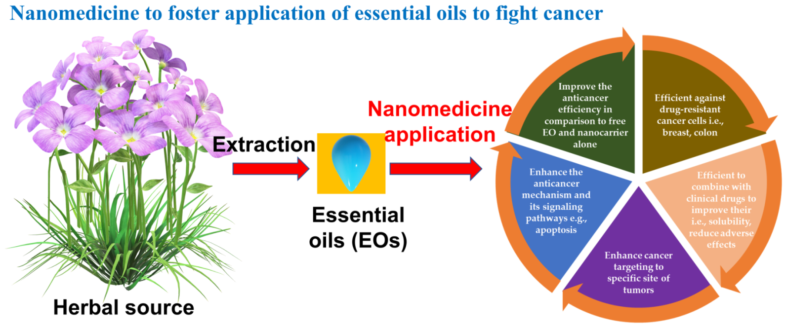
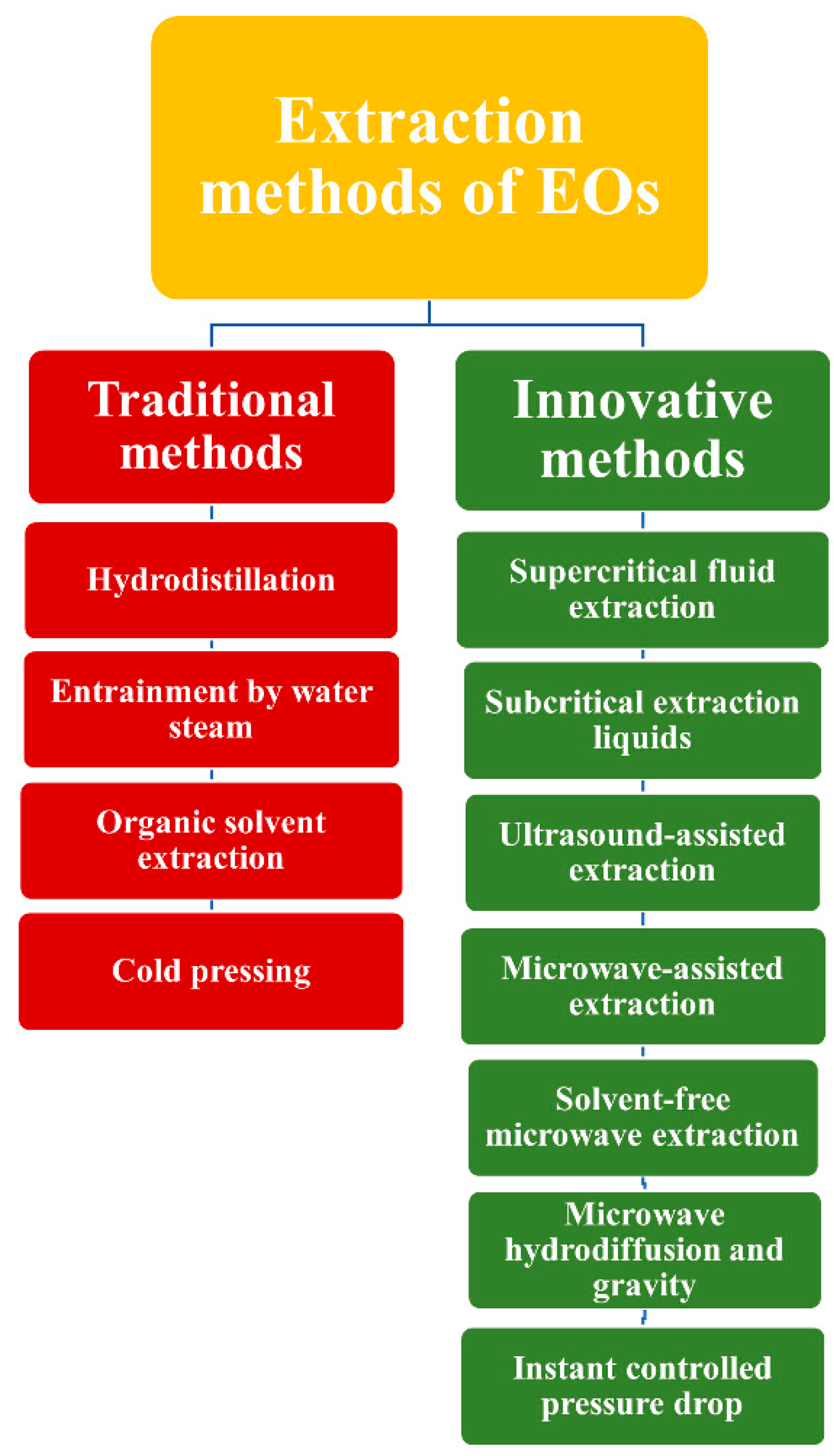
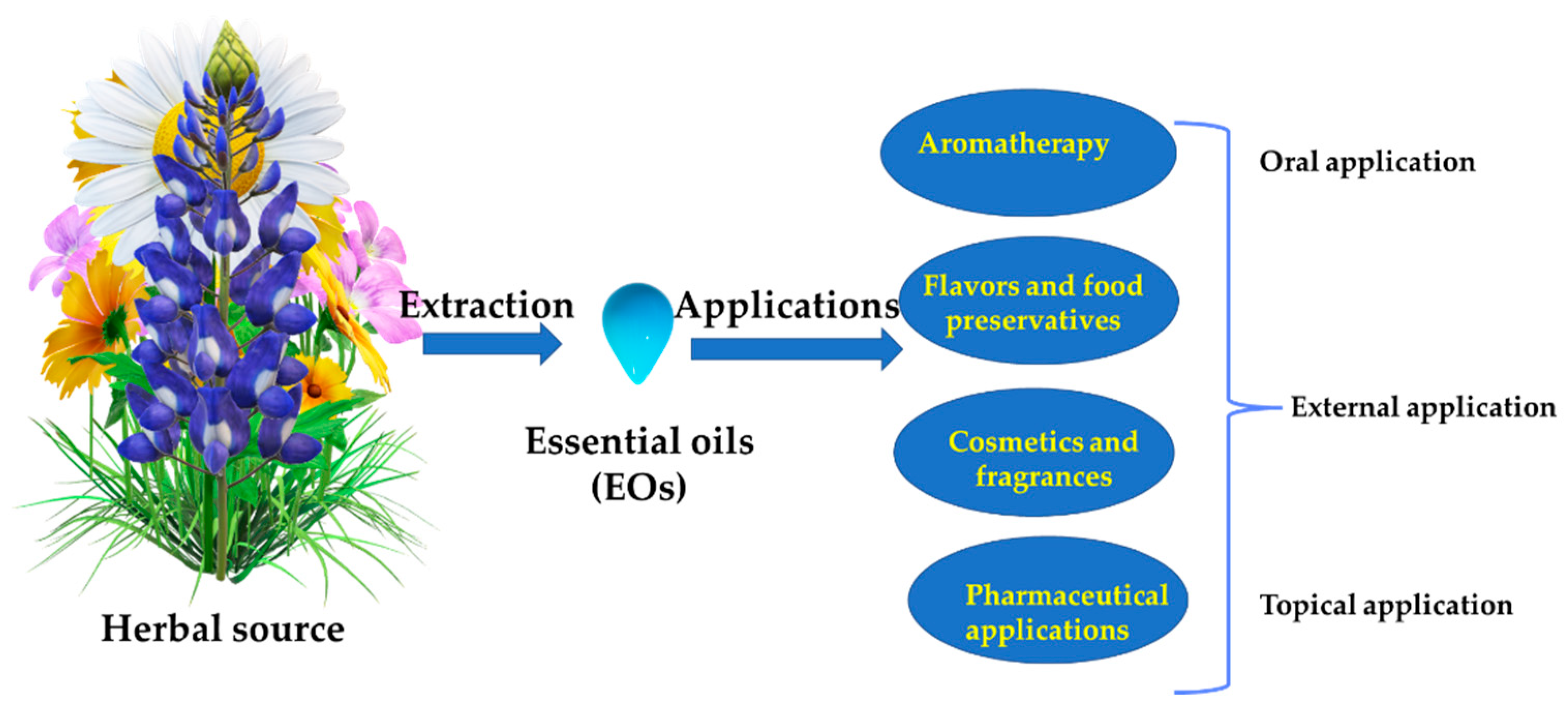
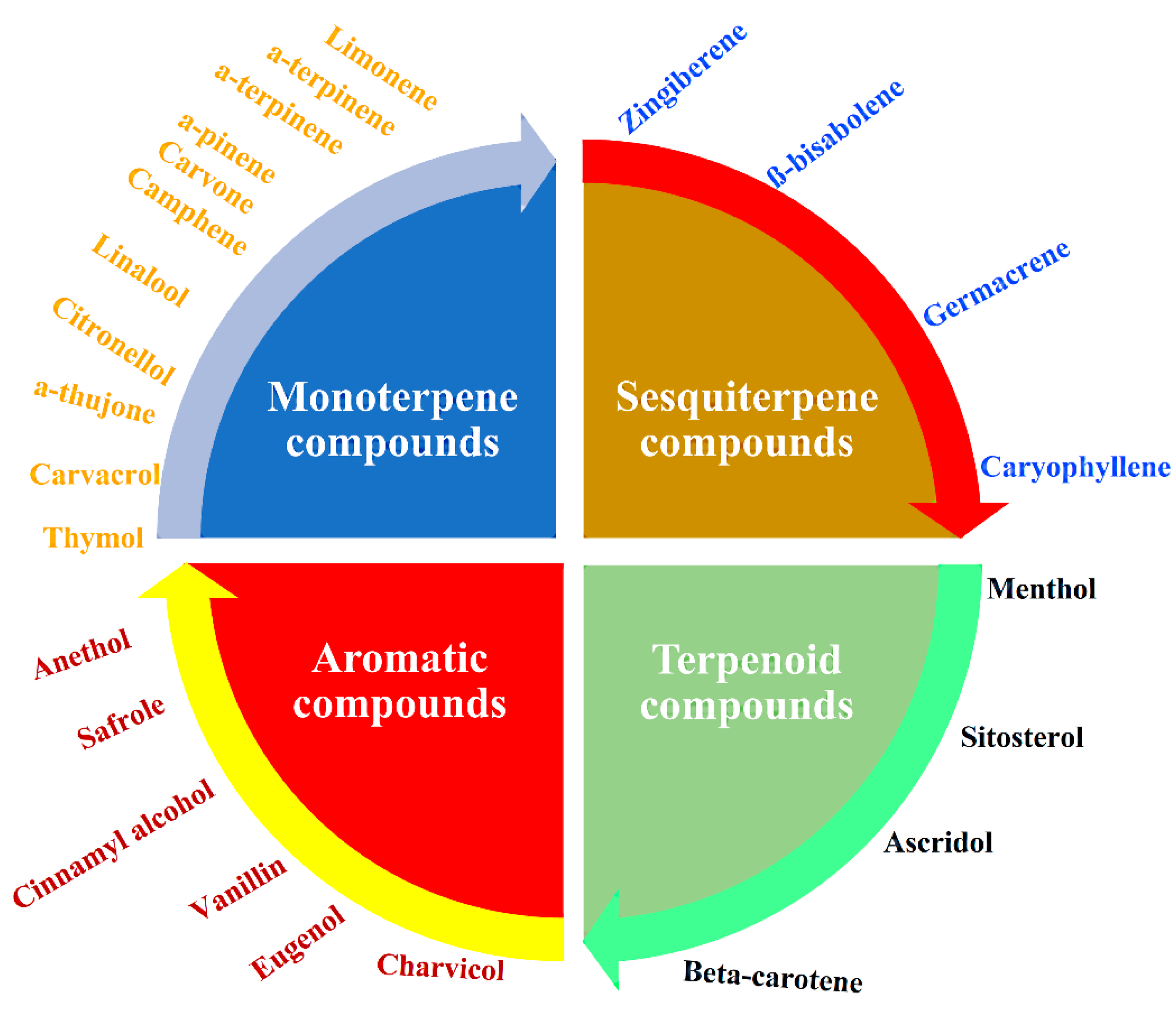
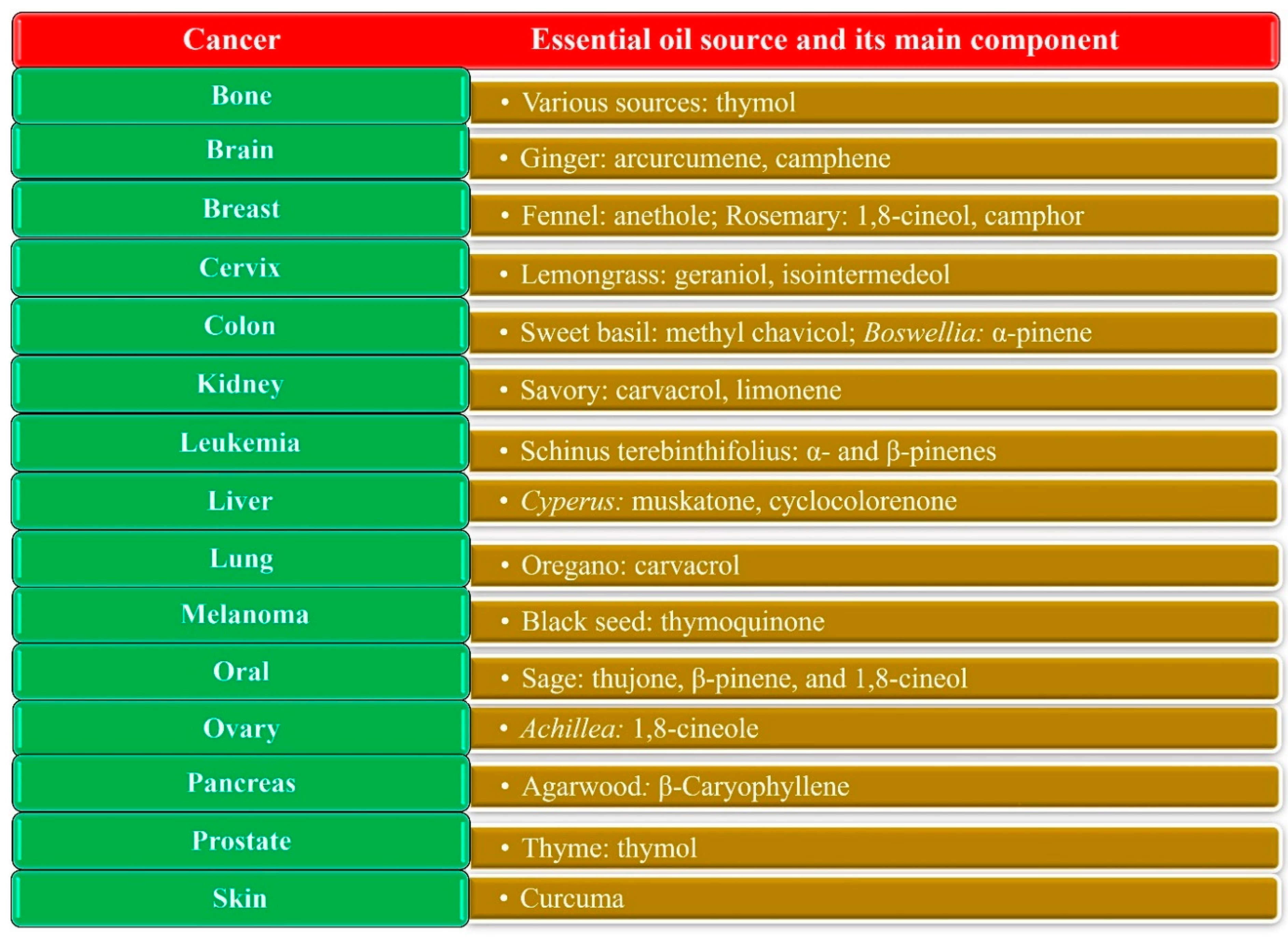
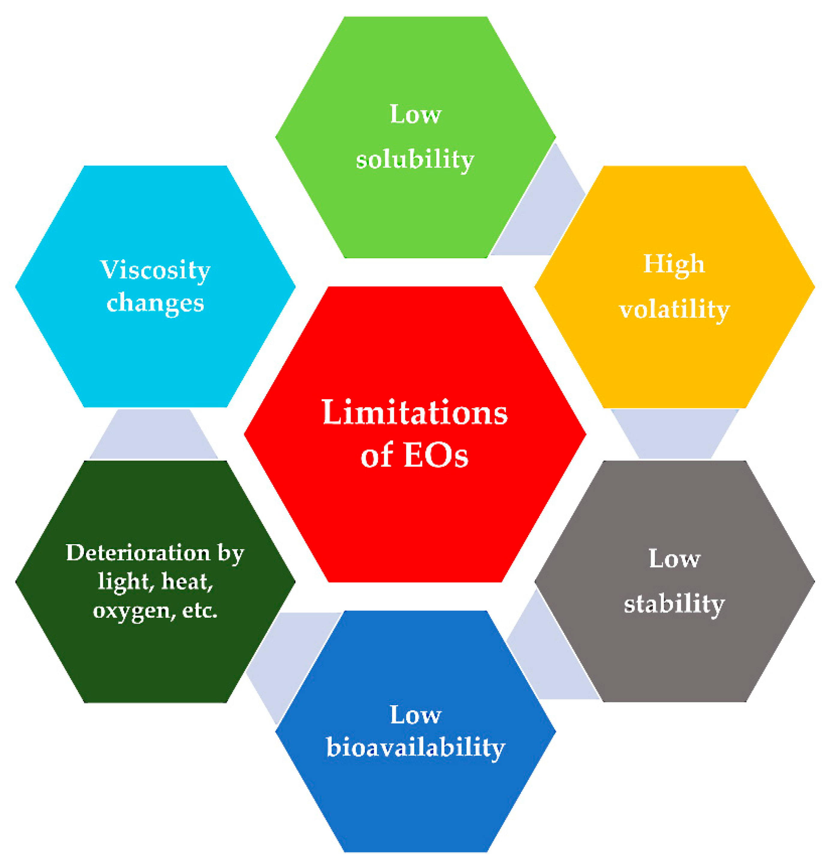
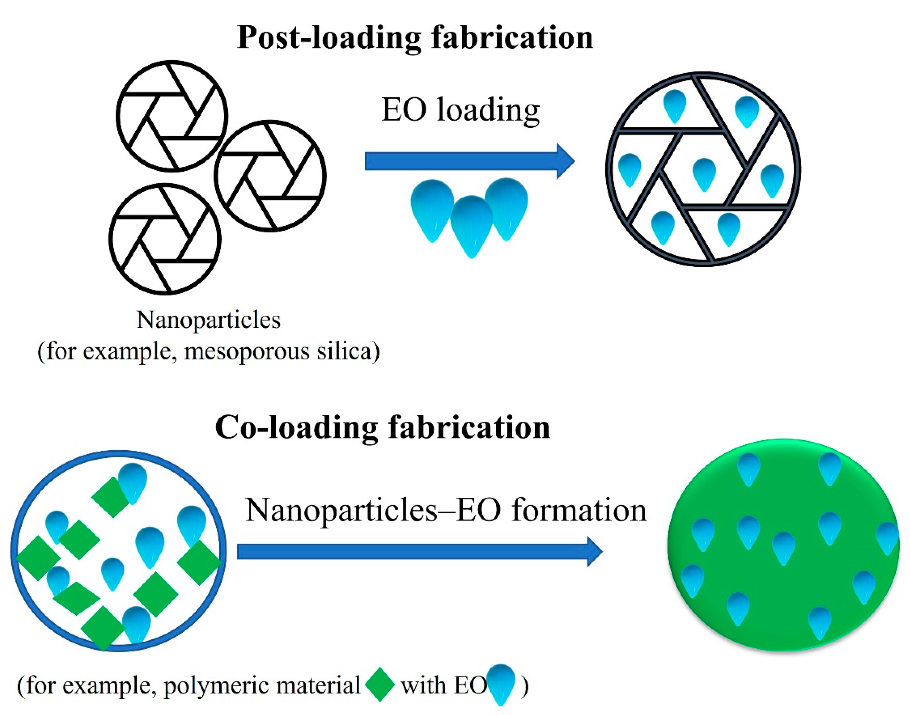
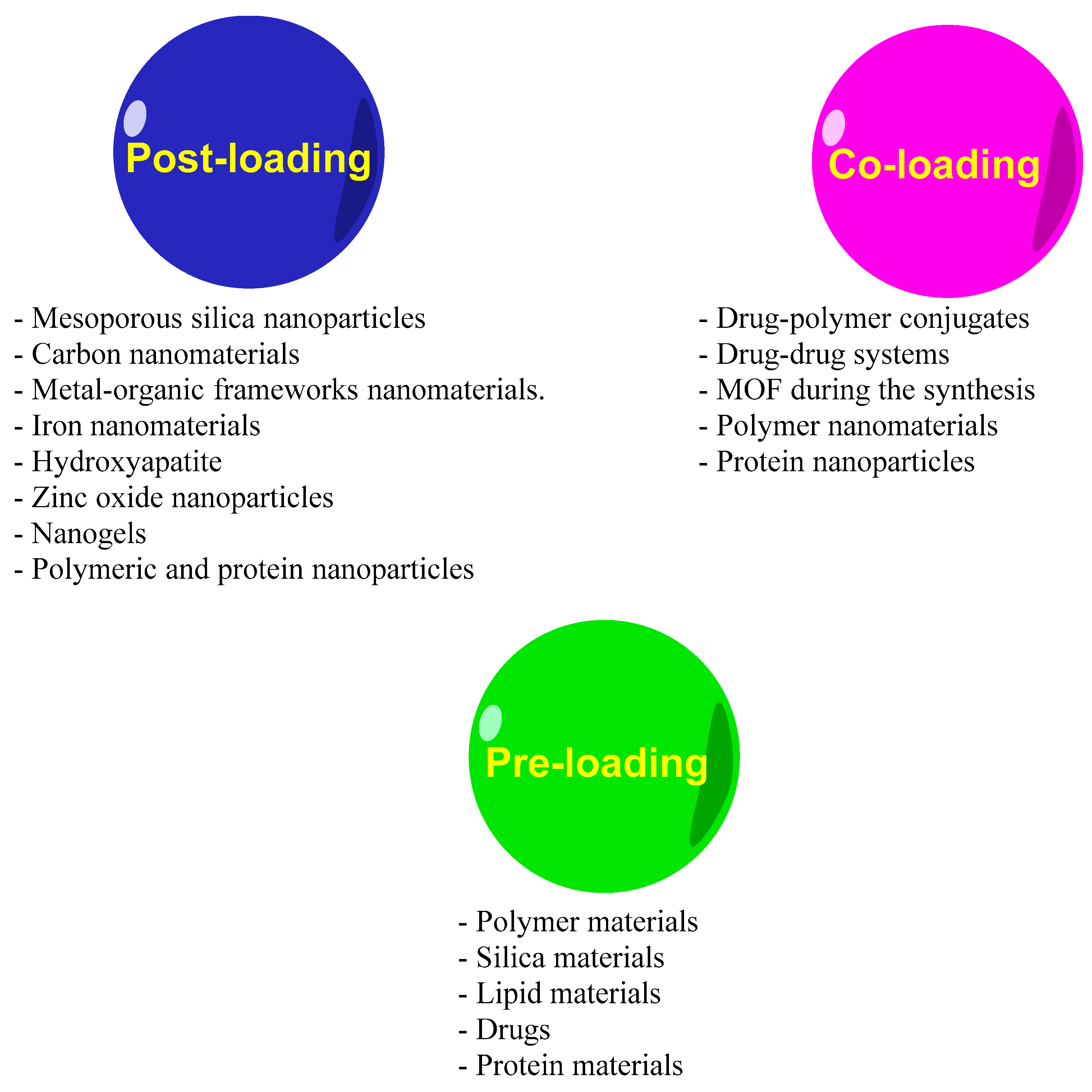
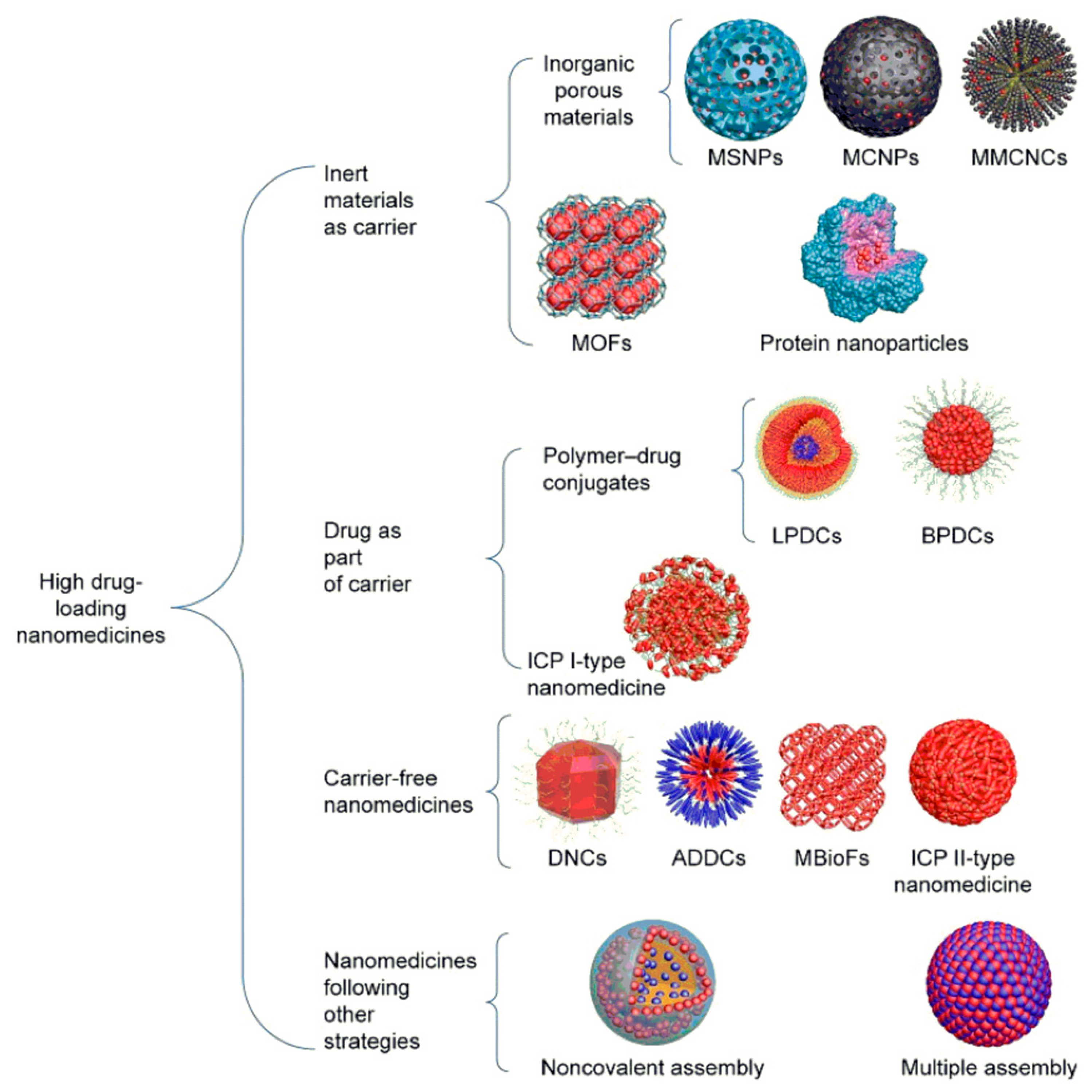
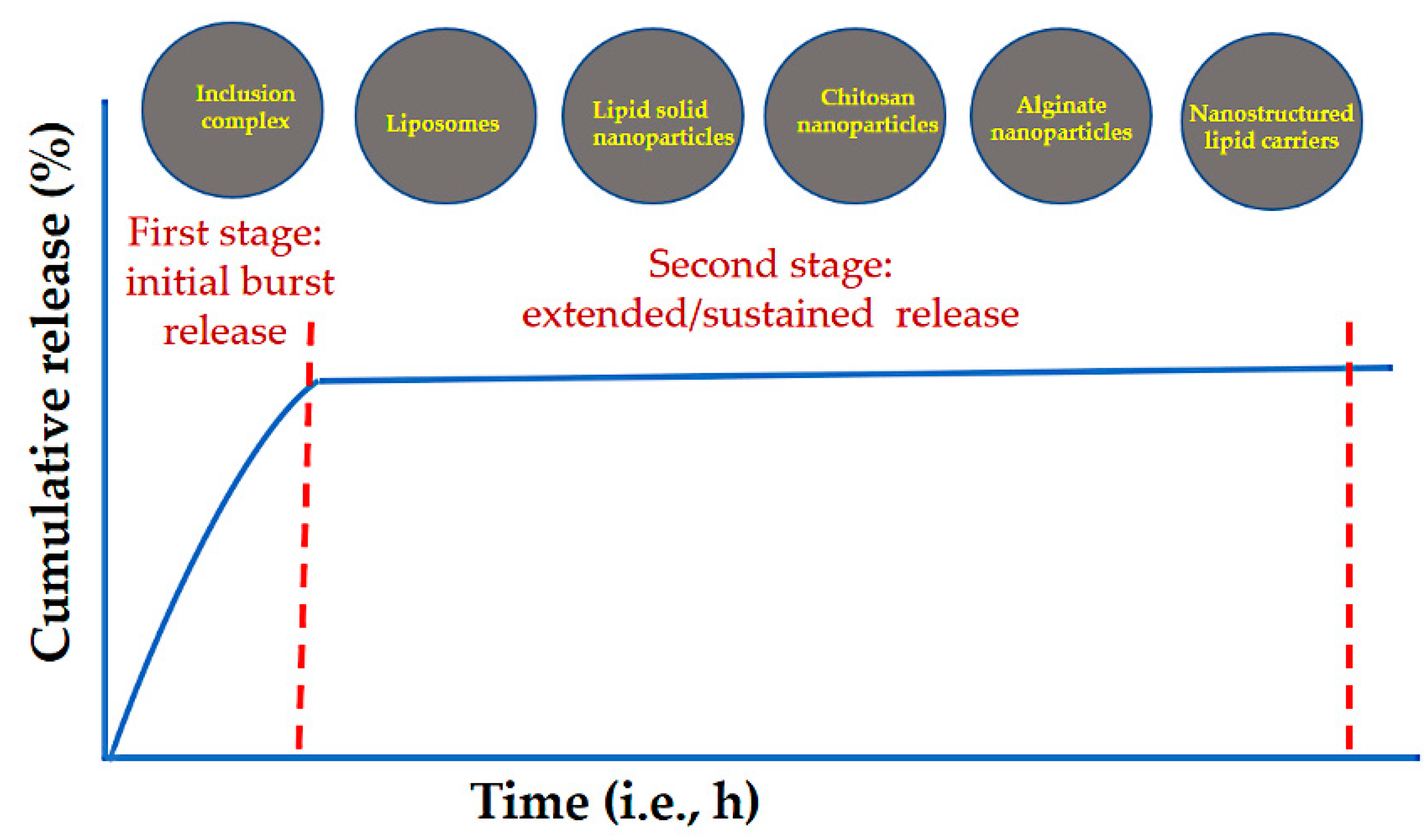
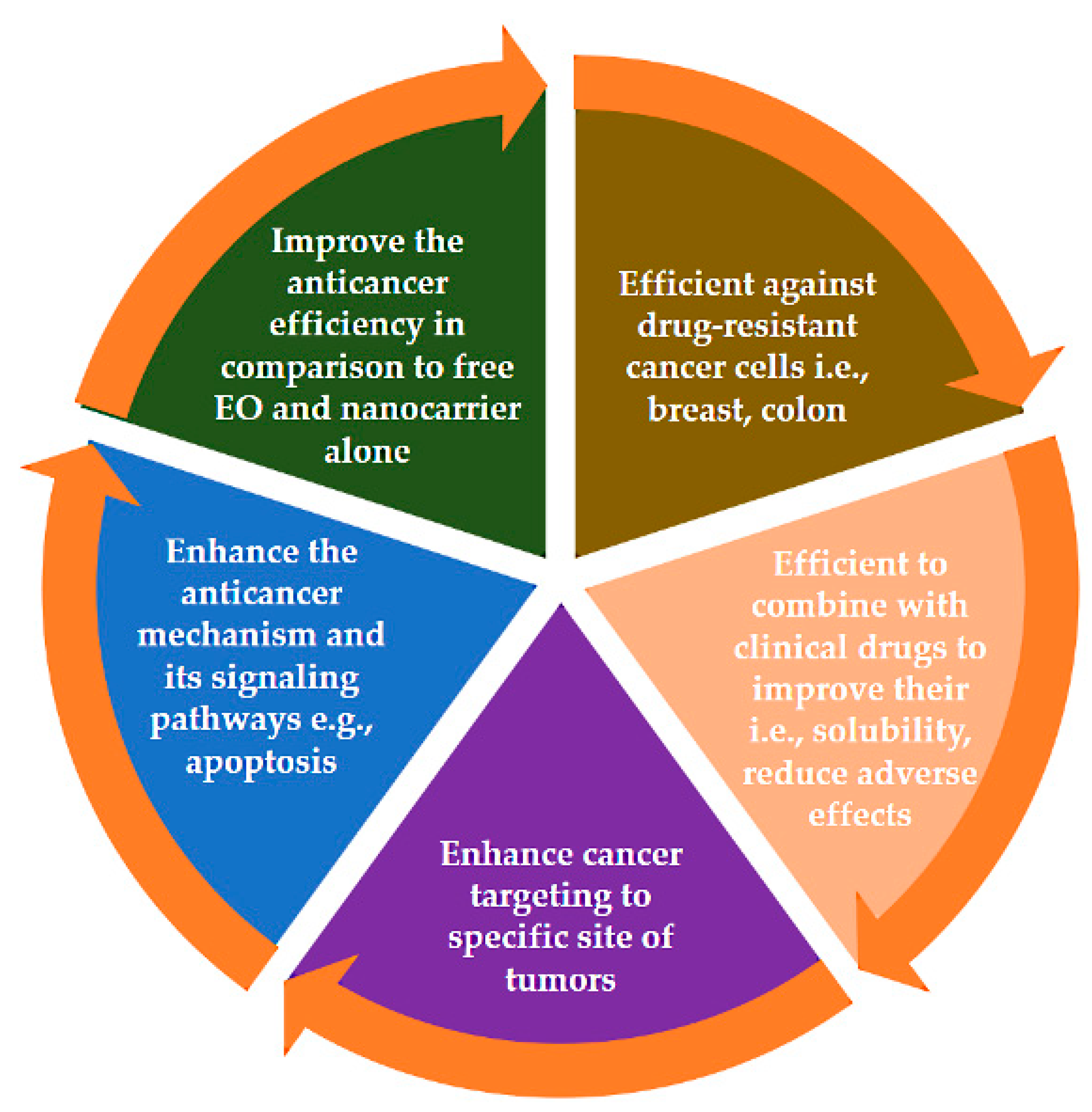
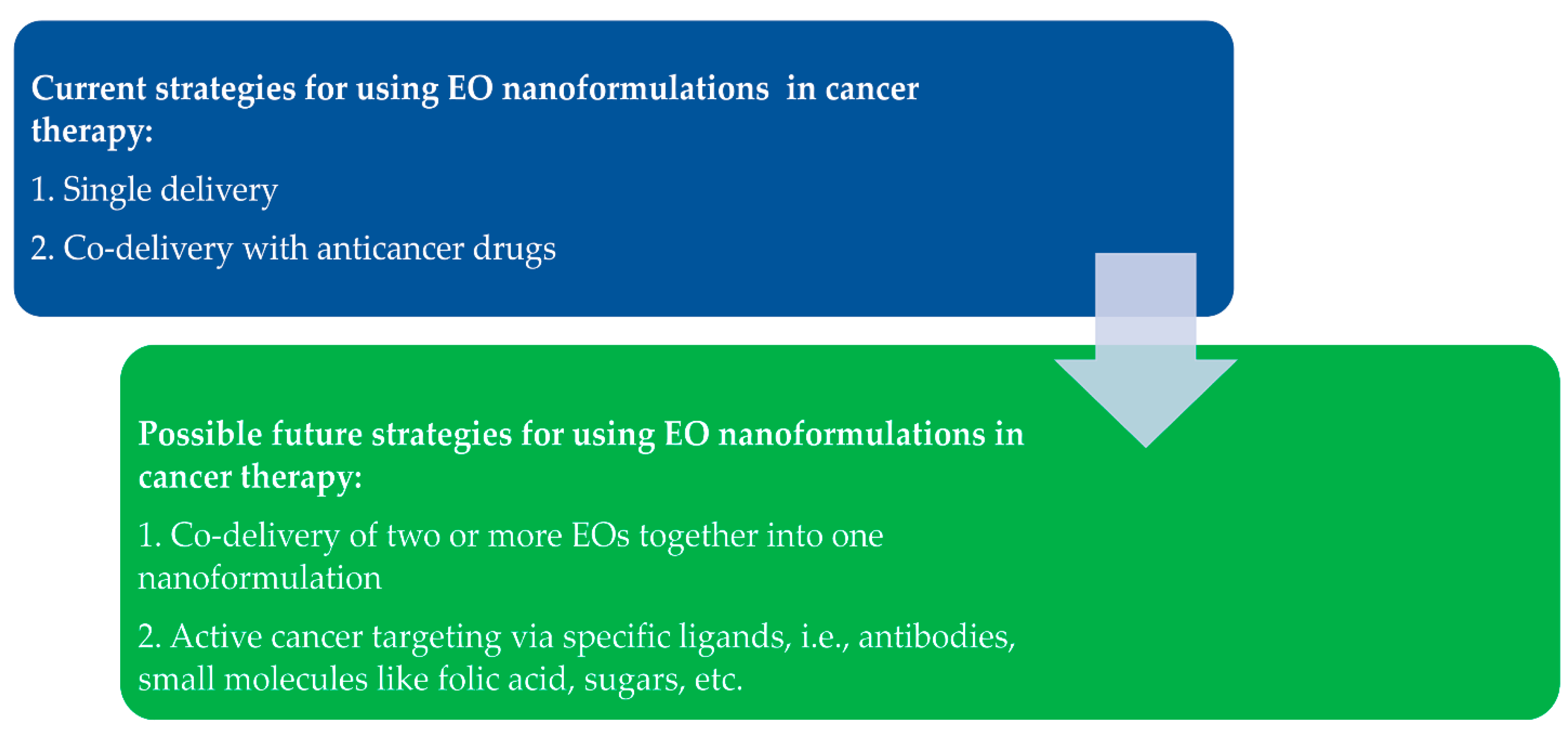
Table 1.
Loading capacity and method for various EOs with different nanocarriers.
| EO | Nanocarrier | Loading Capacity | Loading Method | References |
|---|---|---|---|---|
| Jasmine | Pectin/chitosan nanoparticles | 0.43–6.06% | Direct preparation method | [49] |
| Zedoary | Chitosan-grafted mesoporous silica nanoparticles | 41% | Gate-penetration by supercritical CO2 | [109] |
| Thymol compound | SBA-15 mesoporous silica nanoparticles | ~71 | Post-fabrication method | [110] |
| Carvacrol compound | Chitosan | Up to 21 | Direct preparation method | [111] |
| Carvacrol compound | Zein | Up to 13% | Direct preparation method | [112] |
| Peppermint and green tea | Chitosan nanoparticles | Up to 23% | Direct preparation method | [113] |
| Cuminum cyminum | Nano lipid carrier | Over 70% | Direct preparation method | [114] |
| Lippia origanoides | Nanostructured lipid carriers/cyclodextrin inclusion complexes | 2.6% | Direct preparation method | [115] |
Table 2.
Experimental in vitro release and analysis techniques of EOs from nanoformulations of different nanocarriers.
Table 2.
Experimental in vitro release and analysis techniques of EOs from nanoformulations of different nanocarriers.
| EO | Nanocarrier | In Vitro Experiment | Analysis Technique | References |
|---|---|---|---|---|
| Peppermint and green tea | Chitosan nanoparticles | Lyophilized encapsulated nanoparticles were placed in a dialysis bag containing release media: PBS or acetate buffer. This bag was incubated in an additional volume of the release medium. Left under gentle shaking at room temperature. Sampling: a predetermined volume of release media was taken at specific intervals, then an equal volume of fresh release media was added during the entire experiment. | UV–Vis spectrophotometer | [113] |
| Chamomile | Chitosan nanocapsules | Freeze-dried nanocapsules placed in PBS buffer release medium containing 0.5% Tween 80 adjusted to pH 5.5 or 7.4. The solution was orbitally stirred (150 rpm) at 37 °C. Sampling: at predetermined times, the solution was washed twice with hexane (using a separatory funnel), and the supernatant was used for analysis. | Gas chromatography (GC) | [121] |
| Turmeric and lemongrass | Chitosan-alginate nanocapsules | The loaded nanocapsules were dispersed in a small volume of PBS solution and put in a dialysis bag, then surrounded by PBS containing 20% ethanol adjusted to pH 1.5 and 7.4. Next, the solution was incubated at 37 °C under gentle agitation. Sampling: a specific volume was withdrawn and replaced with fresh medium. | UV–Vis spectrophotometer | [122] |
| Lavender | Hydrogel microparticles | Headspace GC | GC analysis | [123] |
| Pomelo citrus | Zein-alginate solid nanoparticles | The lyophilized encapsulated nanoparticles were placed in simulated fluid into dialysis bags, sealed, and suspended in the same simulated fluid. The solution was stirred (100 rpm) at 37 °C. Sampling: a known volume was taken from the solution and replaced with the same volume of fresh simulated fluid. For analysis, and the withdrawn sample was partitioned with an equal volume of ethyl acetate. | UV–Vis spectrophotometer | [124] |
| Lemon | Chitosan-modified starch nanocapsules | Freeze-dried nanocapsules were placed in a microtube containing 40% ethanol. This mixture solution was incubated at room temperature under stirring. Sampling: at specified time intervals, the samples were centrifuged. The supernatant was removed for analysis. | UV–Vis spectrophotometer | [125] |
| Cinnamon | Poly(e-caprolactone) electrospun nanofibers | The nanofibers were placed in a simulated dissolution medium at 37 °C, and the paddle was set at 100 r/min. Sampling: at fixed time intervals, aliquots were taken and replaced with an equal volume of fresh release medium. The samples were filtered and diluted to be used for analysis. | UV–vis spectrophotometer | [126] |
| Eucalyptus | Β-cyclodextrins, functionalized zeolite materials | Headspace extraction with GC | GC analysis | [127] |
| Cinnamon | Β-cyclodextrin/ chitosan nanoparticles | The loaded nanoparticles were dispersed in acetate and phosphate buffers and vortexed at room temperature. Sampling: at predetermined specific time intervals, the samples were centrifuged (for 5 min at 14,000 rpm); then, aliquots of the supernatant were withdrawn (for analysis) and replaced with the same volume of buffer, and the experiment continued. | UV–Vis spectrophotometer | [118] |
Publisher’s Note: MDPI stays neutral with regard to jurisdictional claims in published maps and institutional affiliations. |
© 2022 by the authors. Licensee MDPI, Basel, Switzerland. This article is an open access article distributed under the terms and conditions of the Creative Commons Attribution (CC BY) license (https://creativecommons.org/licenses/by/4.0/).
Share and Cite
MDPI and ACS Style
AbouAitah, K.; Lojkowski, W. Nanomedicine as an Emerging Technology to Foster Application of Essential Oils to Fight Cancer. Pharmaceuticals 2022, 15, 793. https://doi.org/10.3390/ph15070793
AMA Style
AbouAitah K, Lojkowski W. Nanomedicine as an Emerging Technology to Foster Application of Essential Oils to Fight Cancer. Pharmaceuticals. 2022; 15(7):793. https://doi.org/10.3390/ph15070793
Chicago/Turabian StyleAbouAitah, Khaled, and Witold Lojkowski. 2022. "Nanomedicine as an Emerging Technology to Foster Application of Essential Oils to Fight Cancer" Pharmaceuticals 15, no. 7: 793. https://doi.org/10.3390/ph15070793
Note that from the first issue of 2016, this journal uses article numbers instead of page numbers. See further details here.