The Evolving Interplay among Abundant Adipokines in Patients with Hepatitis C during Viral Clearance

Abstract
:1. Introduction
2. Materials and Methods
2.1. Patients
2.2. Study Design
2.3. Adipokine Enzyme-Linked Immunosorbent Assays (ELISAs)
2.4. Biochemistry
2.5. Statistical Analysis
2.6. Informed Consent
3. Results
3.1. Baseline Characteristics
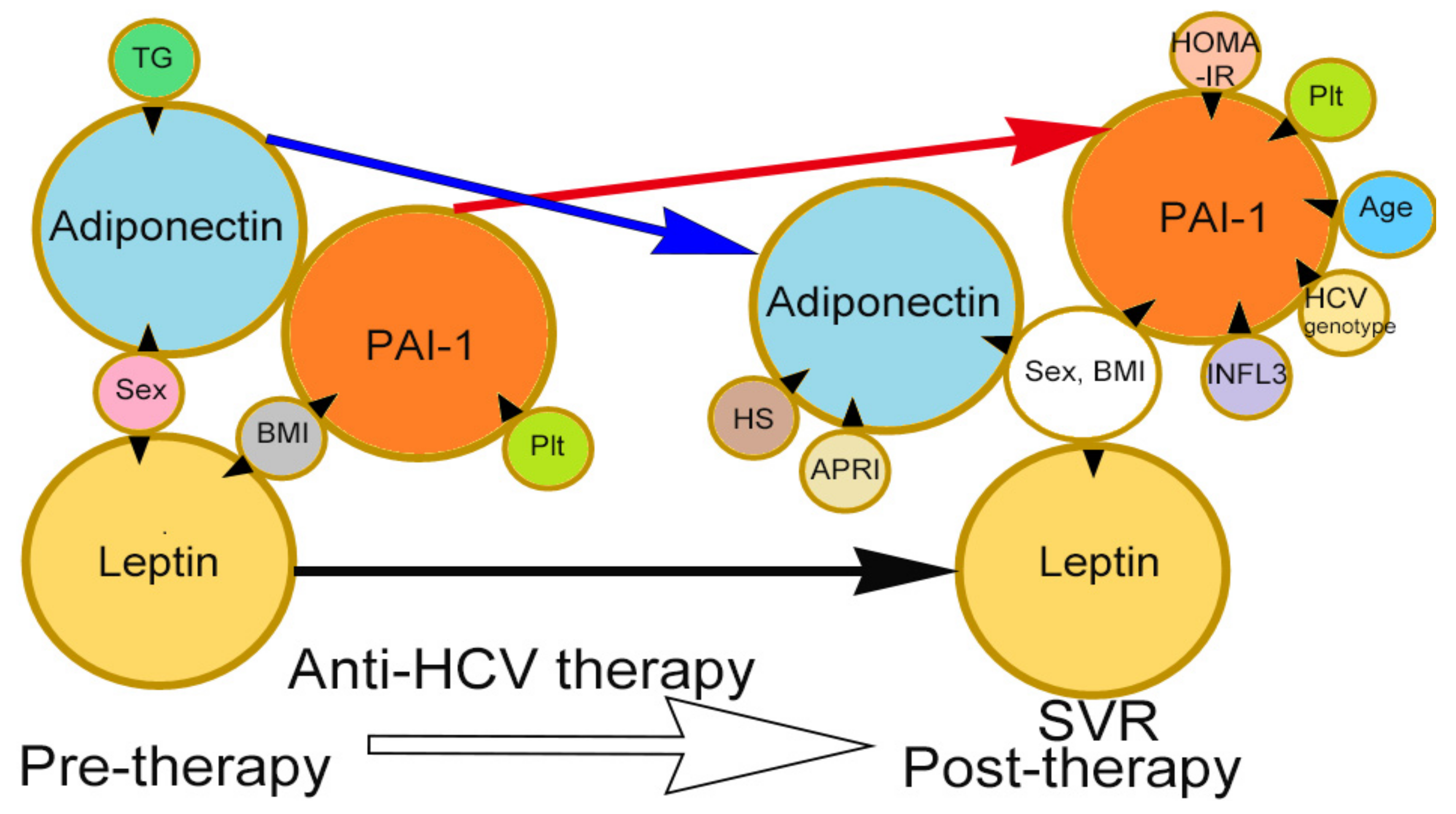
3.2. Independent Pre-Therapy Factors Associated with Pre-Therapy Leptin, Adiponectin, and PAI-1 Levels in CHC Patients
3.3. Independent Post-Therapy Factors Associated with 24-Week Post-Therapy Leptin, Adiponectin and PAI-1 Levels in CHC Patients Who Achieved SVR
3.4. Comparisons between the Pre- and Post-Therapy Levels of Each Variable in CHC Patients
4. Discussion and Conclusions
Acknowledgments
Author Contributions
Conflicts of Interest
References
- Smith, D.B.; Bukh, J.; Kuiken, C.; Muerhoff, A.S.; Rice, C.M.; Stapleton, J.T.; Simmonds, P. Expanded classification of hepatitis C virus into 7 genotypes and 67 subtypes: Updated criteria and genotype assignment web resource. Hepatology 2014, 59, 318–327. [Google Scholar] [CrossRef] [PubMed]
- Chang, M.L. Metabolic alterations and hepatitis C: From bench to bedside. World J. Gastroenterol. 2016, 22, 1461–1476. [Google Scholar] [CrossRef] [PubMed]
- Syed, G.H.; Amako, Y.; Siddiqui, A. Hepatitis C virus hijacks host lipid metabolism. Trends Endocrinol. Metab. 2010, 21, 33–40. [Google Scholar] [CrossRef] [PubMed]
- Scheja, L.; Heeren, J. Metabolic interplay between white, beige, brown adipocytes and the liver. J. Hepatol. 2016, 64, 1176–1186. [Google Scholar] [CrossRef] [PubMed]
- Chang, M.L.; Hsu, C.M.; Tseng, J.H.; Tsou, Y.K.; Chen, S.C.; Shiau, S.S.; Chiu, C.T. Plasminogen activator inhibitor-1 is independently associated with non-alcoholic fatty liver disease whereas leptin and adiponectin vary between genders. J. Gastroenterol. Hepatol. 2015, 30, 329–336. [Google Scholar] [CrossRef] [PubMed]
- Deng, Y.; Scherer, P.E. Adipokines as novel biomarkers and regulators of the metabolic syndrome. Ann. N. Y. Acad. Sci. 2010, 1212, E1–E19. [Google Scholar] [CrossRef] [PubMed]
- Jung, U.J.; Choi, M.S. Obesity and its metabolic complications: The role of adipokines and the relationship between obesity, inflammation, insulin resistance, dyslipidemia and nonalcoholic fatty liver disease. Int. J. Mol. Sci. 2014, 15, 6184–6223. [Google Scholar] [CrossRef] [PubMed]
- Podor, T.J.; Loskutoff, D.J. Immunoelectron microscopic localization of type 1 plasminogen activator inhibitor in the extracellular matrix of transforming growth factor-beta-activated endothelial cells. Ann. N. Y. Acad. Sci. 1992, 667, 46–49. [Google Scholar] [CrossRef] [PubMed]
- Ballantyne, G.H.; Gumbs, A.; Modlin, I.M. Changes in insulin resistance following bariatric surgery and the adipoinsular axis: role of the adipocytokines, leptin, adiponectin and resistin. Obes. Surg. 2005, 15, 692–699. [Google Scholar] [CrossRef] [PubMed]
- Heinrich, G.; Russo, L.; Castaneda, T.R.; Pfeiffer, V.; Ghadieh, H.E.; Ghanem, S.S.; Hill, J.W. Leptin resistance contributes to obesity in mice with null mutation of carcinoembryonic antigen cell adhesion molecule 1. J. Biol. Chem. 2016, 291, 11124–11132. [Google Scholar] [CrossRef] [PubMed]
- Ohashi, K.; Ouchi, N.; Matsuzawa, Y. Adiponectin and hypertension. Am. J. Hypertens. 2011, 24, 263–269. [Google Scholar] [CrossRef] [PubMed]
- Francés, D.E.; Motiño, O.; Agrá, N.; González-Rodríguez, Á.; Fernández-Álvarez, A.; Cucarella, C.; Carnovale, C.E. Hepatic cyclooxygenase-2 expression protects against diet-induced steatosis, obesity, and insulin resistance. Diabetes 2015, 64, 1522–1531. [Google Scholar] [CrossRef] [PubMed]
- Schäffler, A.; Schölmerich, J.; Büchler, C. Mechanisms of disease: adipocytokines and visceral adipose tissue--emerging role in nonalcoholic fatty liver disease. Nat. Clin. Pract. Gastroenterol. Hepatol. 2005, 2, 273–280. [Google Scholar] [CrossRef] [PubMed]
- Janeckova, R. The role of leptin in human physiology and pathophysiology. Physiol. Res. 2001, 50, 443–459. [Google Scholar] [PubMed]
- Pajvani, U.B.; Du, X.; Combs, T.P.; Berg, A.H.; Rajala, M.W.; Schulthess, T.; Scherer, P.E. Structure-function studies of the adipocyte-secreted hormone Acrp30/adiponectin. Implications FPR metabolic regulation and bioactivity. J. Biol. Chem. 2003, 278, 9073–9085. [Google Scholar] [CrossRef] [PubMed]
- Ruscica, M.; Baragetti, A.; Catapano, A.L.; Norata, G.D. Translating the biology of adipokines in atherosclerosis and cardiovascular diseases: Gaps and open questions. Nutr. Metab. Cardiovasc. Dis. 2017, 27, 379–395. [Google Scholar] [CrossRef] [PubMed]
- Tsaroucha, A.; Daniil, Z.; Malli, F.; Georgoulias, P.; Minas, M.; Kostikas, K.; Gourgoulianis, K.I. Leptin, adiponectin, and ghrelin levels in female patients with asthma during stable and exacerbation periods. J. Asthma 2013, 50, 188–197. [Google Scholar] [CrossRef] [PubMed]
- Kappelle, P.J.; Dullaart, R.P.; Van Beek, A.P.; Hillege, H.L.; Wolffenbuttel, B.H. The plasma leptin/adiponectin ratio predicts first cardiovascular event in men: a prospective nested case-control study. Eur. J. Intern. Med. 2012, 23, 755–759. [Google Scholar] [CrossRef] [PubMed]
- Lalić, K.; Jotić, A.; Rajković, N.; Singh, S.; Stošić, L.; Popović, L.; Stanarčić, J. Altered Daytime Fluctuation Pattern of Plasminogen Activator Inhibitor 1 in Type 2 Diabetes Patients with Coronary Artery Disease: A Strong Association with Persistently Elevated Plasma Insulin, Increased Insulin Resistance, and Abdominal Obesity. Int. J. Endocrinol. 2015, 2015, 390185. [Google Scholar] [CrossRef] [PubMed]
- Samad, F.; Ruf, W. Inflammation, obesity, and thrombosis. Blood 2013, 122, 3415–3422. [Google Scholar] [CrossRef] [PubMed]
- Kortlever, R.M.; Bernards, R. Senescence, wound healing and cancer: The PAI-1 connection. Cell Cycle 2006, 5, 2697–2703. [Google Scholar] [CrossRef] [PubMed]
- Eitzman, D.T.; Westrick, R.J.; Nabel, E.G.; Ginsburg, D. Plasminogen activator inhibitor-1 and vitronectin promote vascular thrombosis in mice. Blood 2000, 95, 577–580. [Google Scholar] [PubMed]
- Targher, G.; Marra, F.; Marchesini, G. Increased risk of cardiovascular disease in non-alcoholic fatty liver disease: Causal effect or epiphenomenon? Diabetologia 2008, 51, 1947–1953. [Google Scholar] [CrossRef] [PubMed]
- Chang, M.L.; Lin, Y.S.; Pao, L.H.; Huang, H.C.; Chiu, C.T. Link between plasminogen activator inhibitor-1 and cardiovascular risk in chronic hepatitis C after viral clearance. Sci. Rep. 2017, 7, 42503. [Google Scholar] [CrossRef] [PubMed]
- Chang, M.L.; Kuo, C.J.; Huang, H.C.; Chu, Y.Y.; Chiu, C.T. Association between Leptin and Complement in Hepatitis C Patients with Viral Clearance: Homeostasis of Metabolism and Immunity. PLoS ONE 2016, 11, e0166712. [Google Scholar] [CrossRef] [PubMed]
- Chang, M.L.; Kuo, C.J.; Pao, L.H.; Hsu, C.M.; Chiu, C.T. The evolving relationship between adiponectin and insulin sensitivity in hepatitis C patients during viral clearance. Virulence 2017. [Google Scholar] [CrossRef] [PubMed]
- Chang, M.L.; Tsou, Y.K.; Hu, T.H.; Lin, C.H.; Lin, W.R.; Sung, C.M.; Yeh, C.T. Distinct patterns of the lipid alterations between genotype 1 and 2 chronic hepatitis C patients after viral clearance. PLoS ONE 2014, 9, e104783. [Google Scholar] [CrossRef] [PubMed]
- Chang, M.L.; Cheng, M.L.; Chang, S.W.; Tang, H.Y.; Chiu, C.T.; Yeh, C.T.; Shiao, M.S. Recovery of pan-genotypic and genotype-specific amino acid alterations in chronic hepatitis C after viral clearance: Transition at the crossroad of metabolism and immunity. Amino Acids 2017, 49, 291–302. [Google Scholar] [CrossRef] [PubMed]
- Chang, M.L.; Liang, K.H.; Ku, C.L.; Lo, C.C.; Cheng, Y.T.; Hsu, C.M.; Chiu, C.T. Resistin reinforces interferon λ-3 to eliminate hepatitis C virus with fine-tuning from RETN single-nucleotide polymorphisms. Sci. Rep. 2016, 6, 30799. [Google Scholar] [CrossRef] [PubMed]
- Udompap, P.; Mannalithara, A.; Heo, N.Y.; Kim, D.; Kim, W.R. Increasing prevalence of cirrhosis among U.S. adults aware or unaware of their chronic hepatitis C virus infection. J. Hepatol. 2016, 64, 1027–1032. [Google Scholar] [CrossRef] [PubMed]
- Finucane, F.M.; Luan, J.; Wareham, N.J.; Sharp, S.J.; O’Rahilly, S.; Balkau, B.; Savage, D.B. Correlation of the leptin: Adiponectin ratio with measures of insulin resistance in non-diabetic individuals. Diabetologia 2009, 52, 2345–2349. [Google Scholar] [CrossRef] [PubMed]
- Dullaart, R.P.; Kappelle, P.J.; Dallinga-Thie, G.M. Carotid intima media thickness is associated with plasma adiponectin but not with the leptin:adiponectin ratio independently of metabolic syndrome. Atherosclerosis 2010, 211, 393–396. [Google Scholar] [CrossRef] [PubMed]
- Canavesi, E.; Porzio, M.; Ruscica, M.; Rametta, R.; Macchi, C.; Pelusi, S.; Valenti, L. Increased circulating adiponectin in males with chronic HCV hepatitis. Eur. J. Intern. Med. 2015, 26, 635–639. [Google Scholar] [CrossRef] [PubMed]
- Valenti, L.; Rametta, R.; Ruscica, M.; Dongiovanni, P.; Steffani, L.; Motta, B.M.; Magni, P. The I148M PNPLA3 polymorphism influences serum adiponectin in patients with fatty liver and healthy controls. BMC Gastroenterol. 2012, 12, 111. [Google Scholar] [CrossRef] [PubMed]
- Rametta, R.; Ruscica, M.; Dongiovanni, P.; Macchi, C.; Fracanzani, A.L.; Steffani, L.; Valenti, L. Hepatic steatosis and PNPLA3 I148M variant are associated with serum Fetuin-A independently of insulin resistance. Eur. J. Clin. Investig. 2014, 44, 627–633. [Google Scholar] [CrossRef] [PubMed]
- Valenti, L.; Rumi, M.; Galmozzi, E.; Aghemo, A.; Del Menico, B.; De Nicola, S.; Colombo, M. Patatin-like phospholipase domain-containing 3 I148M polymorphism, steatosis, and liver damage in chronic hepatitis C. Hepatology 2011, 53, 791–799. [Google Scholar] [CrossRef] [PubMed]


| Total (n = 450) | SVR (+) (n = 372) | SVR (−) (n = 78) | p values Obtained Using Student’s t-Test or the Chi-Squared Test | |
|---|---|---|---|---|
| Male, n (%) # | 256 (56.8) | 212 (56.9) | 44 (56.4) | 0.394 |
| Age (years) | 54.01 ± 11.51 | 53.68 ± 11.60 | 55.66 ± 10.92 | 0.160 |
| BMI | 24.99 ± 3.73 | 24.84 ± 3.63 | 25.72 ± 4.09 | 0.057 |
| HCV RNA (Log IU/mL) | 5.95 ± 1.14 | 5.85 ± 1.18 | 6.45 ± 0.74 | <0.001 * |
| HCV genotype, G1, n (%) | 234 (52) | 177 (47.5) | 57 (73) | 0.003 * |
| ALT (U/L) | 94.28 ± 84.95 | 97.18 ± 88.37 | 79.80 ± 63.85 | 0.116 |
| TC (mg/dL) | 171.76 ± 32.22 | 171.53 ± 32.75 | 172.91 ± 29.62 | 0.735 |
| TGs (mg/dL) | 103.25 ± 50.15 | 101.21 ± 46.21 | 113.57 ± 66.05 | 0.123 |
| Platelet count (103 cells/mm) | 176.89 ± 64.54 | 181.27 ± 57.86 | 156.89 ± 58.72 | 0.001 * |
| HOMA-IR | 3.14 ± 4.91 | 2.89 ± 4.70 | 4.42 ± 5.73 | 0.043 * |
| Hepatic steatosis, n (%) # | 220 (48.8) | 186 (50) | 34 (43.5) | 0.373 |
| Liver cirrhosis, n (%) # | 117 (26) | 83 (22.3) | 34 (43.5) | 0.001 * |
| APRI | 1.51 ± 1.66 | 1.47 ± 1.63 | 1.74 ± 1.83 | 0.210 |
| Leptin (ng/mL) | 9.82 ± 10.1 | 9.33 ± 9.05 | 12.2 ± 14.1 | 0.267 |
| Adiponectin (μg/mL) | 9.74 ± 7.15 | 10.1 ± 7.48 | 8.04 ± 5.22 | 0.097 |
| Leptin (ng/mL)/adiponectin (μg/mL) ratio | 2.14 ± 3.79 | 2.06 ± 4.10 | 2.45 ± 3.00 | 0.603 |
| PAI-1 (ng/mL) | 6.73 ± 3.11 | 6.76 ± 2.98 | 6.56 ± 3.69 | 0.658 |
| rs12979860 (CC) n (%) # | 379 (84.2) | 328 (88.1) | 88 (65.3) | 0.013 * |
| Pre-Therapy HCV RNA Level (Log IU/mL) | Pre-Therapy Leptin Level (ng/mL) | Pre-Therapy Adiponectin Level (μg/mL) | Pre-Therapy PAI-1 Level (ng/mL) | |||||||||
|---|---|---|---|---|---|---|---|---|---|---|---|---|
| Pre-Therapy Factors | Univariate | Multivariate | Univariate | Multivariate | Univariate | Multivariate | Univariate | Multivariate | ||||
| p Values | 95% CI of Estimated β (Estimated β) | p Values | p Values | 95% CI of Estimated β (Estimated β) | p Values | p Values | 95% CI of Estimated β (Estimated β) | p Values | p Values | 95% CI of Estimated β (Estimated β) | p Values | |
| Sex (Male) | 0.322 | <0.001 * | −12.7–−8.6 (−10.6) | <0.001 * | <0.001 * | −5.2–−1.5 (−3.4) | <0.001 * | 0.094 | −1.08–1.33 (0.123) | 0.84 | ||
| Age (years) | 0.415 | 0.874 | 0.012 * | −0.03–0.13 (0.05) | 0.22 | <0.001 * | −0.075–0.009 (−0.033) | 0.122 | ||||
| BMI | 0.795 | <0.001 * | 1.09–1.70 (1.39) | <0.001 * | <0.001 * | −0.48–0.038 (−0.222) | 0.094 | <0.001 * | 0.041–0.37 (0.205) | 0.015 * | ||
| HCV RNA (Log IU/mL) | NA | NA | 0.231 | 0.887 | 0.200 | |||||||
| HCV genotype | <0.001 * | −0.59–0.25 (−0.42) | <0.001 * | 0.356 | 0.074 | −0.1–3.5 (1.7) | 0.172 | 0.064 | −0.425–1.44 (0.509) | 0.284 | ||
| ALT (U/L) | 0.631 | 0.468 | 0.607 | 0.288 | ||||||||
| TC (mg/dL) | 0.027 * | 0.000–0.005 (0.003) | 0.036 * | 0.839 | 0.312 | 0.86 | ||||||
| TGs (mg/dL) | 0.375 | 0.341 | 0.028 * | −0.036–−0.003 (−0.017) | 0.045 * | 0.095 | −0.002–0.018 (0.008) | 0.113 | ||||
| Platelet count (103 cells/mm) | 0.419 | 0.659 | 0.008 * | −0.027–0.005 (−0.011) | 0.185 | <0.001 * | 0.005–0.026 (0.015) | 0.005 * | ||||
| HOMA-IR | 0.063 | 0.001–0.03 (0.015) | 0.04 * | 0.231 | 0.03 * | -0.254–0.046 (−0.104) | 0.171 | 0.909 | ||||
| Hepatic steatosis (Yes) | 0.837 | 0.001 * | −0.93–3.36 (1.22) | 0.265 | 0.01 * | −4.01–−0.014 (−0.302) | 0.052 | 0.009 * | −1.14–0.985 (−0.078) | 0.885 | ||
| Cirrhosis (Yes) | 0.213 | 0.577 | 0.76 | 0.005 * | −1.23–1.56 (0.167) | 0.813 | ||||||
| APRI | 0.496 | 0.489 | 0.213 | 0.012 * | −0.163–0.435 (0.136) | 0.371 | ||||||
| Leptin (ng/mL) | 0.231 | NA | 0.313 | 0.015 * | −0.048–0.181 (0.017) | 0.616 | ||||||
| Adiponectin (μg/mL) | 0.887 | 0.313 | NA | NA | NA | <0.001 * | −0.142–0.007 (−0.067) | 0.076 | ||||
| Leptin (ng/mL) | 0.207 | NA# | NA # | <0.001 * | NA # | NA # | ||||||
| adiponectin (μg/mL) ratio | ||||||||||||
| PAI-1 (ng/mL) | 0.2 | 0.015* | −0.19~0.45 (0.126) | 0.442 | <0.001 * | −0.603~0.000 (−0.302) | 0.05 | NA | NA | NA | ||
| rs12979860 (CC genotype) | 0.246 | 0.636 | 0.743 | 0.236 | ||||||||
| Post-therapy Leptin Level (ng/mL) | Post-therapy Adiponectin Level (μg/mL) | Post-therapy PAI-1 Level (ng/mL) | |||||||
|---|---|---|---|---|---|---|---|---|---|
| Univariate | Multivariate | Univariate | Multivariate | Univariate | Multivariate | ||||
| Post-Therapy Factors | p Values | 95% CI of Estimated β (Estimated β) | p Values | p Values | 95% CI of Estimated β (Estimated β) | p Values | p Values | 95% CI of Estimated β (Estimated β) | p Values |
| Sex (Male) | <0.001 * | −18.9–−10.3 (−15.2) | <0.001 * | <0.001 * | −5.7–−2.6 (−4.2) | 0.001 * | 0.008 * | 0.8–1.86 (0.97) | 0.033 * |
| Age | 0.317 | 0.223 | <0.001 * | −0.12–−0.04 (−0.08) | <0.001 * | ||||
| BMI | <0.001 * | 1.31–2.52 (1.88) | <0.001 * | <0.001 * | −0.53–−0.05 (−0.29) | 0.017 * | <0.001 * | 0.06–0.308 (0.186) | 0.003 * |
| HCV genotype | 0.193 | 0.545 | 0.034 * | −1.6–−0.005 (−0.81) | 0.049 * | ||||
| ALT (U/L) | 0.958 | 0.07 | −0.08–0.04 (−0.02) | 0.515 | 0.471 | ||||
| TC (mg/dL) | 0.079 | −0.003–0.09 (0.05) | 0.066 | 0.201 | 0.246 | ||||
| TGs (mg/dL) | 0.549 | <0.001 * | −0.02–0.008 (−0.006) | 0.382 | <0.001 * | −0.005–0.001 (0.002) | 0.551 | ||
| Platelet counts (103 cells/mm) | 0.523 | 0.448 | <0.001 * | 0.014–0.035 (0.025) | <0.001 * | ||||
| HOMA-IR | 0.089 | −0.32–0.69 (0.184) | 0.473 | 0.006 * | −0.46–−0.003 (−0.22) | 0.053 | 0.039 * | 0.05–0.30 (0.182) | 0.004 * |
| APRI | 0.564 | 0.015 * | 0.11–5.59 (2.85) | 0.042 * | <0.001 * | −0.77–2.4 (0.814) | 0.313 | ||
| Hepatic steatosis (Yes) | 0.069 | −2.9–4.6 (0.884) | 0.656 | <0.001 * | −3.8–−0.56 (−2.2) | 0.008 * | 0.008 * | −0.29–1.41 (0.56) | 0.794 |
| Cirrhosis (Yes) | 0.315 | 0.419 | <0.001 * | −2.2–0.19 (−0.98) | 0.100 | ||||
| Leptin (ng/mL) | NA | NA | NA | 0.29 | 0.781 | ||||
| Adiponectin(μg/mL) | 0.29 | NA | NA | NA | 0.008 * | −0.05–0.09 (0.02) | 0.592 | ||
| Leptin (ng/mL)/adiponectin (μg/mL) ratio | NA # | NA # | 0.044 * | NA # | NA # | ||||
| PAI-1 (ng/mL) | 0.781 | 0.008 * | −0.18–0.28 (0.053) | 0.65 | NA | NA | NA | ||
| rs12979860 (CC) | 0.853 | 0.915 | 0.076 | 0.46–2.4 (1.49) | 0.004 * | ||||
| SVR (+) (n = 372) | Paired t-Test p Values | SVR (−) (n = 78) | Paired t-Test p Values | |||
|---|---|---|---|---|---|---|
| Factors | Pre-TherapyValue | Post-TherapyValue | Pre-TherapyValue | Post-TherapyValue | ||
| BMI | 24.84 ± 3.63 | 24.35 ± 3.51 | <0.001 * | 25.72 ± 4.09 | 24.87 ± 3.62 | <0.001 * |
| ALT (U/L) | 97.18 ± 88.37 | 20.0 ± 10.5 | <0.001 * | 79.80 ± 63.85 | 63.7 ± 43.3 | 0.151 |
| TC (mg/dL) | 171.53 ± 32.75 | 184.28 ± 37.39 | <0.001 * | 172.91 ± 29.62 | 174.29 ± 36.12 | 0.7021 |
| TGs (mg/dL) | 101.21 ± 46.21 | 120.54 ± 74.75 | <0.001 * | 113.57 ± 66.05 | 102.77 ± 42.88 | 0.059 |
| Platelet count (103 cells/mm) | 181.27 ± 57.86 | 184.10 ± 55.68 | 0.243 | 156.89 ± 58.72 | 149.40 ± 54.55 | 0.179 |
| HOMA-IR | 2.89 ± 4.70 | 2.83 ± 3.96 | 0.5493 | 4.42 ± 5.73 | 5.40 ± 11.55 | 0.7332 |
| APRI | 1.47 ± 1.63 | 0.418 ± 0.297 | <0.001 * | 1.74 ± 1.83 | 1.28 ± 0.929 | 0.162 |
| Hepatic steatosis (Yes), n (%) | 186 (50) | 193 (51.8) | 0.499 | 34 (43.5) | 37 (47.4) | 0.443 |
| Cirrhosis (Yes), n (%) | 83 (22.3) | 86 (23) | 0.504 | 34 (43.5) | 39 (50) | 0.058 |
| Leptin (ng/mL) | 9.33 ± 9.05 | 9.61 ± 8.77 | 0.15 | 12.2 ± 14.1 | 10.1 ± 12.5 | 0.187 |
| Adiponectin (μg/mL) | 10.1 ± 7.48 | 8.14 ± 5.09 | 0.003* | 8.04 ± 5.16 | 8.77 ± 6.32 | 0.473 |
| Leptin (ng/mL)/adiponectin (μg/mL) ratio | 1.96 ± 4.07 | 2.26 ± 3.57 | 0.188 | 2.44 ± 2.96 | 3.02 ± 6.43 | 0.506 |
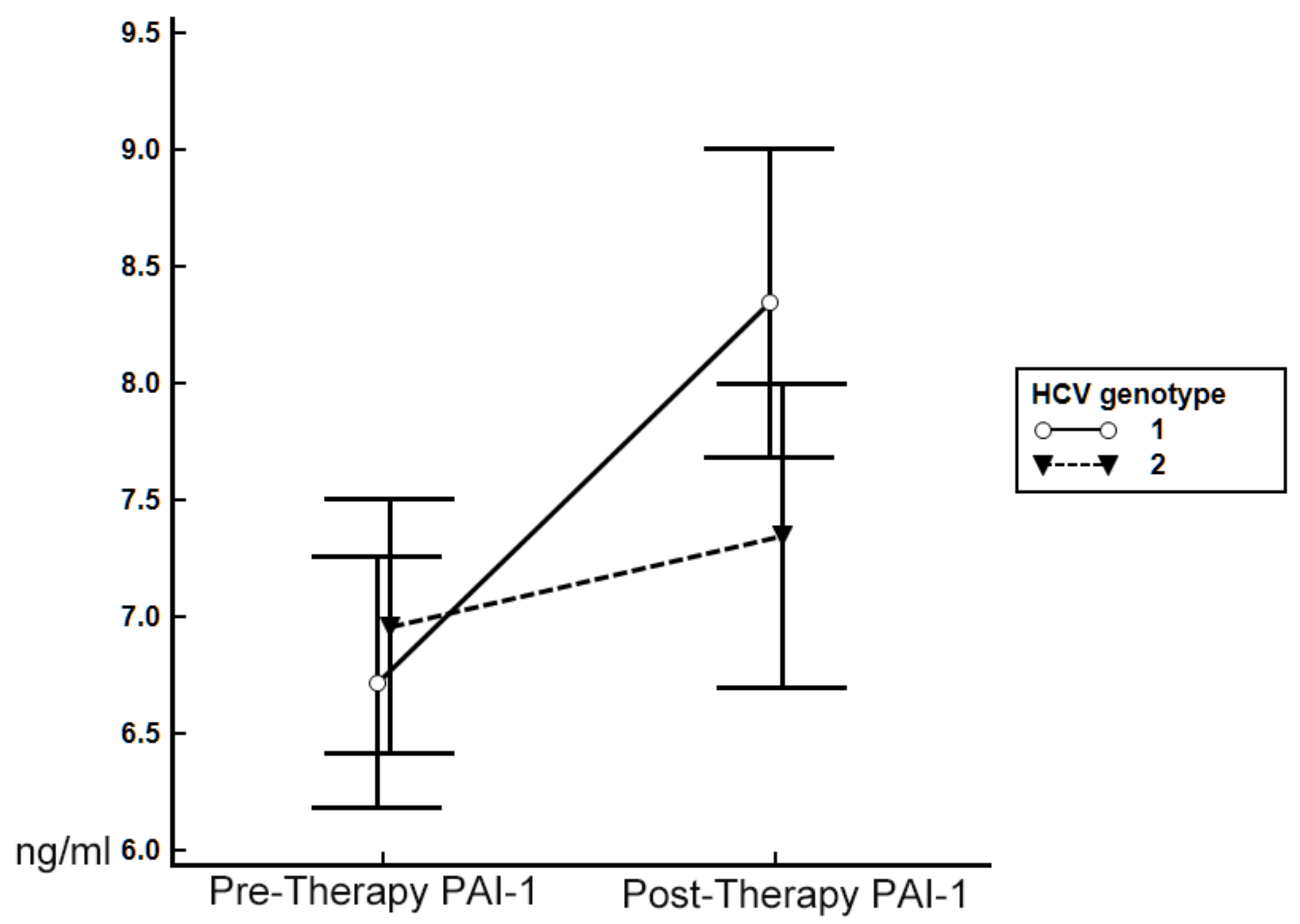
| PAI-1 (ng/mL) | 6.76 ± 2.98 | 9.08 ± 4.43 | 0.003* | 6.56 ± 3.69 | 6.45 ± 4.42 | 0.9355 |
© 2017 by the authors. Licensee MDPI, Basel, Switzerland. This article is an open access article distributed under the terms and conditions of the Creative Commons Attribution (CC BY) license (http://creativecommons.org/licenses/by/4.0/).
Share and Cite
Chang, M.-L.; Chen, T.-H.; Hsu, C.-M.; Lin, C.-H.; Lin, C.-Y.; Kuo, C.-J.; Huang, S.-W.; Chen, C.-W.; Cheng, H.-T.; Yeh, C.-T.; et al. The Evolving Interplay among Abundant Adipokines in Patients with Hepatitis C during Viral Clearance. Nutrients 2017, 9, 570. https://doi.org/10.3390/nu9060570
Chang M-L, Chen T-H, Hsu C-M, Lin C-H, Lin C-Y, Kuo C-J, Huang S-W, Chen C-W, Cheng H-T, Yeh C-T, et al. The Evolving Interplay among Abundant Adipokines in Patients with Hepatitis C during Viral Clearance. Nutrients. 2017; 9(6):570. https://doi.org/10.3390/nu9060570
Chicago/Turabian StyleChang, Ming-Ling, Tsung-Hsing Chen, Chen-Ming Hsu, Cheng-Hui Lin, Cheng-Yu Lin, Chia-Jung Kuo, Shu-Wei Huang, Chun-Wei Chen, Hao-Tsai Cheng, Chau-Ting Yeh, and et al. 2017. "The Evolving Interplay among Abundant Adipokines in Patients with Hepatitis C during Viral Clearance" Nutrients 9, no. 6: 570. https://doi.org/10.3390/nu9060570