
Anti-Inflammatory Effect of Korean Propolis on Helicobacter pylori-Infected Gastric Mucosal Injury Mice Model

College of Pharmacy and Institute of Pharmaceutical Sciences, CHA University, Seongnam 13488, Korea
*
Author to whom correspondence should be addressed.
Nutrients 2022, 14(21), 4644; https://doi.org/10.3390/nu14214644
Submission received: 10 October 2022
/
Revised: 28 October 2022
/
Accepted: 29 October 2022
/
Published: 3 November 2022
(This article belongs to the Section Nutrition and Metabolism)
Abstract
:Propolis, a natural resinous substance obtained from a variety of buds and plants, has been reported to possess various biological functions. Several recent studies have demonstrated the inhibitory effects of propolis on the growth of Helicobacter pylori (H. pylori) in vitro; however, current research efforts on Korean propolis (KP) remain insufficient especially in vivo. Our study aims to investigate the anti-inflammatory effect and molecular mechanism of KP on mouse gastric mucosa during H. pylori infection. We examined an in vivo H. pylori-induced gastric mucosal injury mice model. We found that KP inhibited the growth of H. pylori and attenuated the expression of H. pylori virulence factors such as cytotoxin-associated gene A, encoding urease A subunit, surface antigen gene and neutrophil-activating protein A. Moreover, KP reduced both gross lesions and pathological scores in H. pylori-challenged mice. In addition, KP markedly restrained the production of pro-inflammatory cytokines and nitric oxide levels compared with an untreated H. pylori-infected group. In particular, we found that KP repressed the phosphorylation of IκBα and NF-κB p65 subunit, and subsequently suppressed their downstream target genes. Taken together, these findings demonstrate the beneficial effects of KP on inflammation through the inhibition of NF-κB signaling as well as inhibition of H. pylori growth in a mouse model infected with H. pylori. This suggests the potential application of KP as a natural supplement for patient’s suffering from gastric mucosal injury caused by H. pylori infection.
1. Introduction
Helicobacter pylori (H. pylori) is a Gram-negative spiral-shaped bacillus that colonizes and spreads disease to the human stomach, affecting nearly half of the world’s population [1,2,3]. H. pylori infection leads to the pathogenesis of chronic gastritis, peptic ulcer and gastric adenocarcinoma [4,5]. Ever since the World Health Organization identified H. pylori as a high-risk carcinogen, considerable efforts have been devoted to therapeutic research aimed at combatting its harmful effects [6,7]. Notably, many studies have reported that the eradication and inhibition of H. pylori is particularly crucial for attenuating the pathogenesis of H. pylori-related diseases [8].
Nuclear factor-κB (NF-κB) is considered to be one of the major regulators of the inflammatory response, and can activate and amplify gastric disorders during H. pylori infection [9,10]. Interestingly, H. pylori mediates NF-κB activation through H. pylori virulence factors including cytotoxin-associated gene A (CagA) [11]. When infected with H. pylori, the inhibitors of NF-κB (IκBs) become phosphorylated [12]. In consequence, NF-κB is translocated from the cytosol to the nucleus, leading to the release of pro-inflammatory cytokines such as tumor necrosis factor-α (TNF-α), interleukin (IL)-1β and IL-8 [13,14]. This process ultimately results in strong exacerbation of gastric mucosal injury and inflammation [15,16,17]. Recently, several studies have provided evidence supporting the importance of natural compounds in the treatment of various diseases [18,19,20] and their effects on antibiotic resistance and inhibition of H. pylori growth [21,22,23].
Propolis is known as a natural remedy for H. pylori infection, and is produced and collected by honeybees [24,25]. Many studies have identified that propolis contains vital constituents of essential biologically active compounds such as flavonoids, caffeic acids, and phenolic esters [26]. Moreover, propolis has been shown to have anti-oxidant, anti-inflammation, anti-carcinogenic and anti-bacterial properties [27,28,29,30]. There are a variety of propolis-based mixtures for different pharmacological composites depending on the geographic region of extraction [28]. Therefore, the potency of propolis compounds also differs slightly depending on the country of origin. According to reports, Korean propolis (KP) contains a high total phenolic content and shows strong effects against various diseases [27,30,31]. In fact, our previous in vitro study demonstrated the anti-inflammation and anti-oxidant effects of KP against H. pylori-infected gastric damage [30]. Nevertheless, studies on the effects of KP against H. pylori-induced inflammation in animal models have not yet been published in the literature. In the present study, we demonstrate anti-inflammatory effects on gastric mucosal injury in experimental mice infected with H. pylori using KP samples obtained from 10 different regions in the Republic of Korea.
2. Materials and Methods
2.1. Propolis Solution
The KP used in the present study was extracted, purified and collected from Korea Beekeeping Association (Republic of Korea) [30]. Ethanol extracts of KP samples were diluted with 0.5% carboxymethyl cellulose sodium salt (CMC) and adjusted to the required concentration (weight to volume), followed by storage in no-light conditions at 4 °C and warmed to room temperature before use.
2.2. Bacterial Culture
H. pylori Sydney Strain 1 (SS1) strain (Korea Collection for Type Culture, Daejeon, Republic of Korea) was used for the in vivo experiment. Bacteria were cultured in Tryptic soy agar with 5% sheep blood and incubated for 3–5 days under 10% CO2, 37 °C micro-aerobic conditions. H. pylori SS1 strain was collected and resuspended in cold phosphate buffered saline (PBS) in triplicate, and then used for the experiments.
2.3. Ethics
The mice experiments were reviewed and approved by the Institutional Animal Care and Use Committee of CHA University Animal Center (reference number: IACUC200110 and IACUC210159). The animals were handled in an accredited animal facility according to the guidelines and regulations for the use and care of animals of CHA University Animal Center (Seongnam, Republic of Korea).
2.4. Animal Experiment
C57BL/6 male mice (4-weeks old) were purchased from Orient Bio (Seoul, Republic of Korea). H. pylori SS1 strain was used for inoculation. Mice were acclimatized for 1 week and then orally administered with 0.2 mL of 5% sodium bicarbonate (as inhibitor of gastric acid) for 3 days and fasted for 12 h before H. pylori infection. H. pylori cultured in all groups except the normal group was adjusted to the number of bacteria of 5.0 × 10⁹ Colony-Forming Unit (CFU)/mL H. pylori suspension, four times at 2-day intervals. Normal group was orally administered an equivalent volume of PBS. All mice were handled in an experimental setting under a 12 h light/dark cycle and specific pathogen-free conditions.
To confirm the maintenance of H. pylori infection, serum was isolated one-week later from venous blood extracted from the tails of all mice. H. pylori numbers were measured using the H. pylori IgG antibody ELISA kit (Cusabio Biotech Co., Houston, TX, USA), and mice with elevated H. pylori IgG levels were subsequently selected. Next, the mice were divided into three groups: Normal group; H. pylori group orally administered with 0.2 mL/kg PBS, 3-times weekly over a 4-week period, performed in triplicate; KP-treated group orally administered with 0.2 mL/kg of 200 mg/kg KP, 3-times weekly over a 4-week period.
2.5. H. pylori Antigen Test in Mouse Serum
The H. pylori antigen was calculated using the H. pylori IgG antibody ELISA kit (Cusabio Biotech Co., Houston, TX, USA), according to the manufacturer’s instructions. Samples were diluted with a solution and incubated at room temperature for 30 min and then 100 µL was added to the H. pylori antigen. The results were detected after 30 min. All samples were read on 450 nm absorbance.
2.6. Campylobacter-Like Organism (CLO) Test
The gastric mucosa tissue was determined using CLO test kit (Asan Pharmaceutical Co., Seoul, Republic of Korea). After incubation for 2 h at 37 °C, infection of H. pylori in mice produced a positive reaction, changing the color from yellow to purple. All samples were scored using the following rate: no color change: 0 points; slightly red color: 1 points; light purple: 2 points; dark purple: 3 points.
2.7. Histological Analysis
For histopathological assessment, stomach portions were fixed in 10% formalin, embedded in paraffin blocks, sectioned to 4 μm, stained with hematoxylin-eosin (H&E) and Periodic Acid Schiff (PAS), and then the histopathological scores were evaluated. Histopathological scores estimated overall gastritis in the entire corpus and antrum region of mice including inflammatory cell infiltration, submucosa edema, damage of the surface epithelium, and total pathologic score. The degree of gastritis was checked in the previous studies, and the samples were calculated in terms of scores [32]. A score of 0 points was given if no pathology was observed for each item, 0.5 points for mild, and 1 point for moderate. Subsequently, three different researchers measured all pathological index cases to increase objectivity.
2.8. Quantitative Reverse Transcriptase Polymerase Chain Reaction (qRT-PCR) Analysis
The qRT-PCR was performed on a ViiATM 7 Real-time PCR system (Applied Biosystems, Waltham, MA, USA) using Luna universal qPCR master mix (New England Biolabs, Beverly, MA, USA). The relative quantities of genes were calculated from triplicate samples after normalization to 18S ribosomal RNA (18S rRNA, as internal control). All oligonucleotide primers were purchased from Macrogen (Seoul, Republic of Korea) and are listed in Table 1 below.
2.9. Western Blot Analysis
The stomach tissues were homogenized with ice-cold cell lysis buffer (Cell Signaling, Danvers, MA, USA) plus phosphatase and protease inhibitors (Roche Applied Science, Mannheim, Germany), and then centrifuged. Western blot analysis was performed as previously described [33]. Proteins were visualized using an enhanced chemiluminescence system (Thermo Fisher Scientific, Waltham, MA, USA). The primary antibodies to detect pp65, p65, pp50, p50, p-IκBα, and IκBα used in this study were purchased from Cell Signaling Technology (Danvers, MA, USA). Antibodies for β-actin and CagA were purchased from Santa Cruz Biotechnology (Dallas, TX, USA). Antibodies for iNOS were purchased from BD Biosciences (Franklin Lakes, NJ, USA).
2.10. Cytokine Measurement via Enzyme-Linked Iimmunoassay (ELISA)
The serum levels of IL-8, IL-1β and TNF-α were measured with an ELISA assay kit (R&D Systems, Minneapolis, MN, USA) following the manufacturer’s instruction.
2.11. Measurement of Nitric Oxide (NO) Production
The mouse serum was quantified using a NO detection kit (iNtRON Biotechnology, Seongnam, Republic of Korea) following the manufacturer’s instruction.
2.12. Statistical Analysis
Results were expressed as the mean ± standard deviation (SD). Statistical analysis of the data was performed using GraphPad (GraphPad Software, San Diego, CA, USA). All other experiments were performed on individual mice. The statistical significance was analyzed by one-way analysis of variance (ANOVA). Statistical significance was accepted at p < 0.05.
3. Results
3.1. Gastric Mucosal Therapeutic Effect of KP in H. pylori-Infected Mice
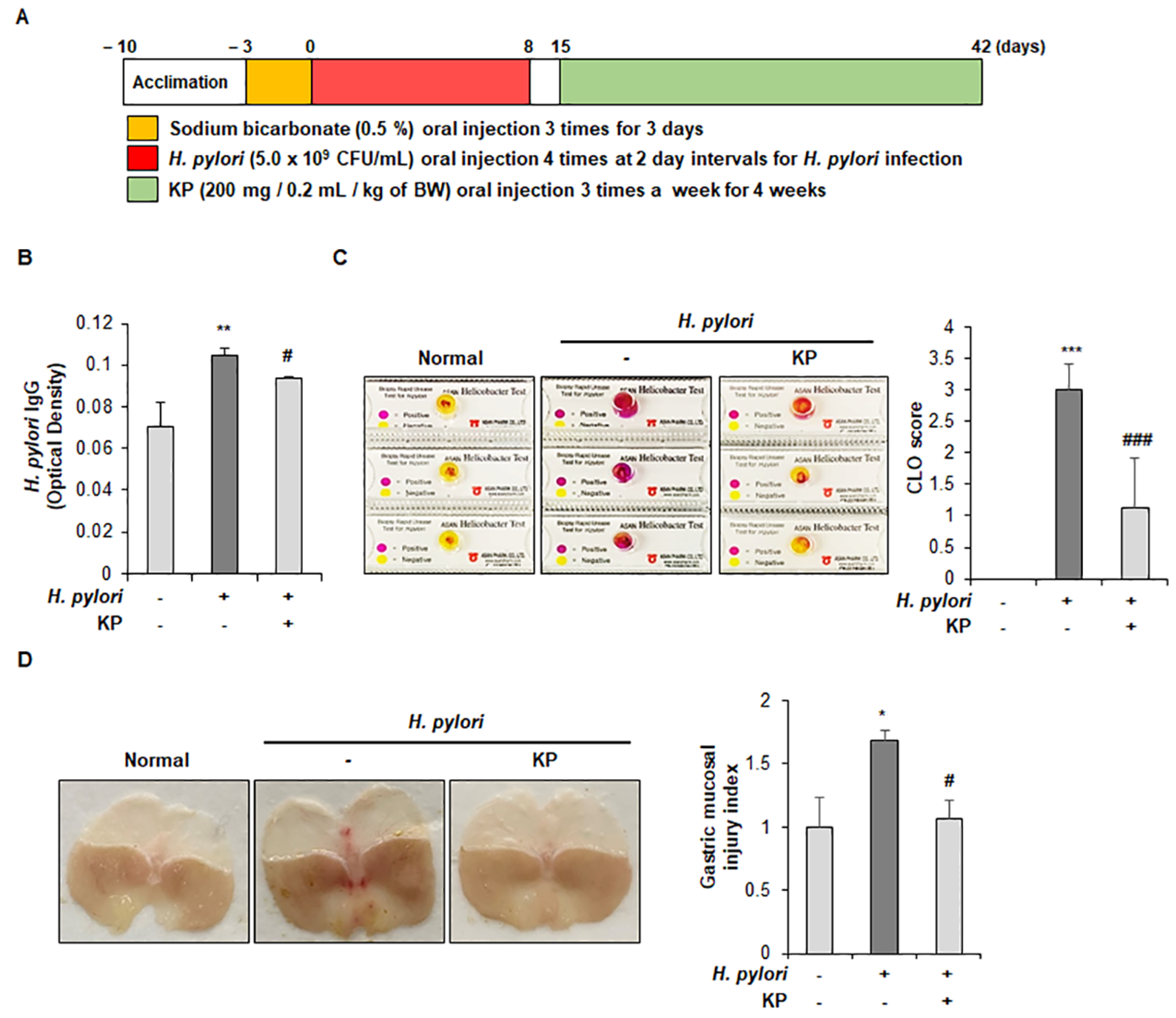
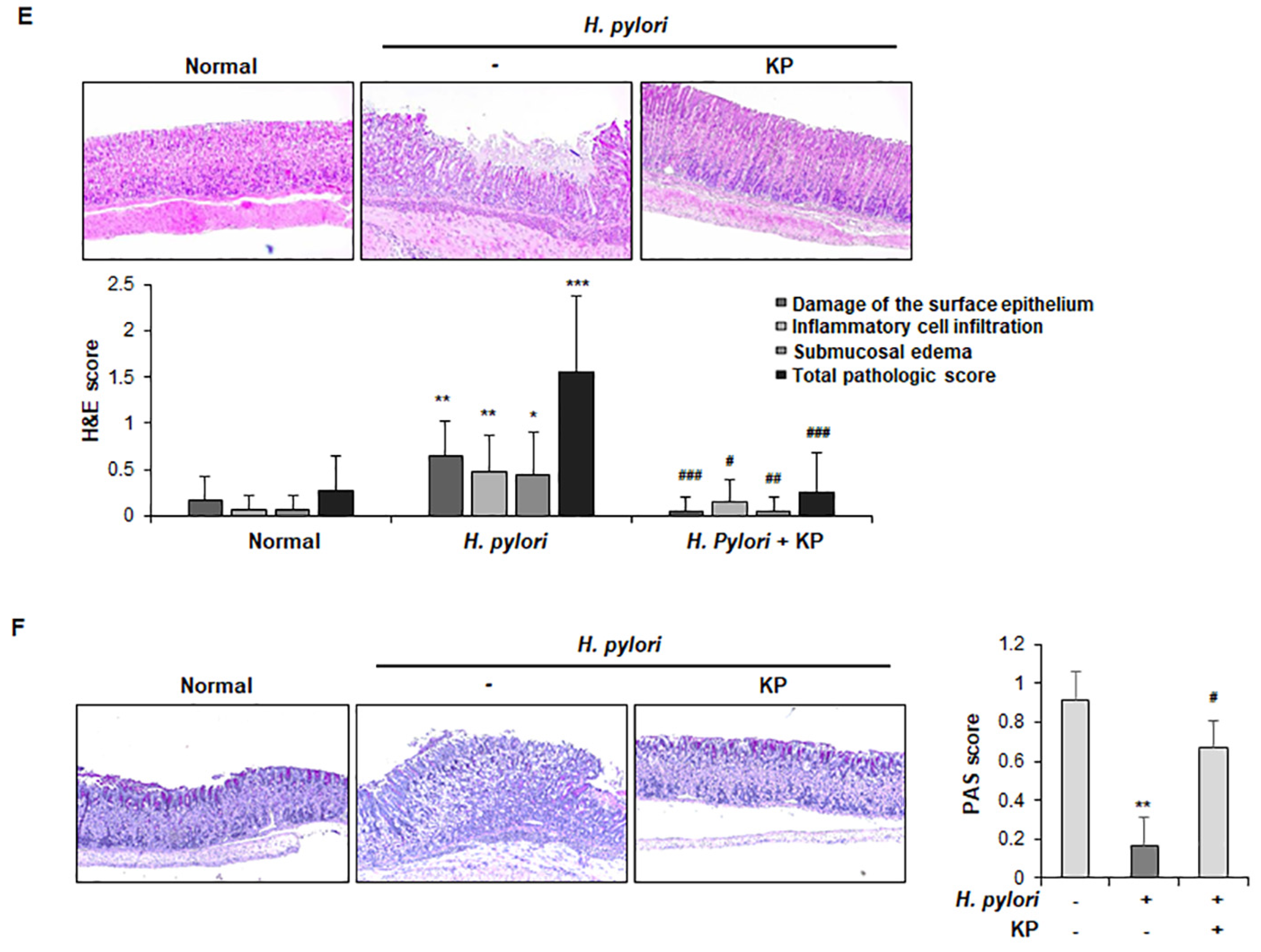
To investigate the anti-H. pylori effect of KP, we established H. pylori-infected gastric mucosal injury mice model. KP (200 mg/kg) was orally administered three times a week over a four-week period after inoculated with an H. pylori (5.0 × 10⁹ CFU/mL) suspension four times in two-day intervals (Figure 1A). After examination, first, we performed a test to check H. pylori IgG levels in mice. Upregulation of IgG levels is a classic indicator of H. pylori infection and was found to be significantly higher than in non-infection groups [34]. As expected, H. pylori-infected group showed a high IgG level, whereas it was significantly inhibited in the KP-treated group compared with the H. pylori-infected group (Figure 1B). We also checked for H. pylori infection using the CLO test, a sensitive and rapid method of detection for H. pylori [35]. The CLO test was conducted on the extracted gastric mucosal to examine the cure rate. As shown in Figure 1C, the H. pylori-infected group produced a positive reaction (purple color), but the KP-administrated group displayed a negative reaction (yellow color). These data indicate that the KP-treated group significantly inhibited the growth of H. pylori compared with the H. pylori-infected group. Next, we investigated gross legions present in the gastric mucosal layers in each group. The gastric mucosa of the H. pylori-infected group were thicker than the normal group, indicating infiltration of inflammatory cells in the sub-mucosal and mucosal layers [36]. As shown in Figure 1D, inflammatory cells were significantly observed in the gastric sub-mucosa and mucosa of H. pylori-infected mice compared with normal mice, whereas these inflammatory cell infiltrations were markedly decreased in the KP-administrated mice (p < 0.05). Overall, the gross legions showed that H. pylori-infected mice rapidly developed edema, slight bleeding, and considerable destruction of gastric mucosal layer, but the KP-treated mice were protected from gastric mucosal damage (Figure 1D). The severity of H. pylori-infected gastric mucosal injury was determined according to the degree of surface epithelium damage, total pathological score, inflammatory cell infiltration, and sub-mucosal edema (Figure 1E). The degree of inflammatory histopathological scores was measured using the previous strategy [32]. As demonstrated by the H&E staining technique, the H. pylori-infected mice group showed significant damage to the gastric mucosal surface epithelium, whereas the KP-treatment group showed dramatic preservation of the gastric mucosal layer (Figure 1E). Next, we validated whether KP protects the gastric mucosal layer against H. pylori infection via PAS staining in gastric tissues. Gastric surface mucous cells are mucous-producing cells that protect against the corrosive properties of stomach acid. In particular, H. pylori infection caused damage to the mucosal cells and accelerated gastric mucosal injury [37]. As shown in Figure 1F, the H. pylori group showed significant loss of mucous cells in gastric mucosal layer tissue. However, the KP-treatment group showed abundant presence and restoration of mucous within the cells, and gave a positive reaction of PAS staining results as indicated by purple coloring in the gastric mucosal layer (Figure 1F). These results provide supporting evidence for the protective effects of KP on mucous cells and mucosal layer from H. pylori-infection.
3.2. KP Attenuates H. pylori-Related Virulence Factors in H. pylori-Infected Mice
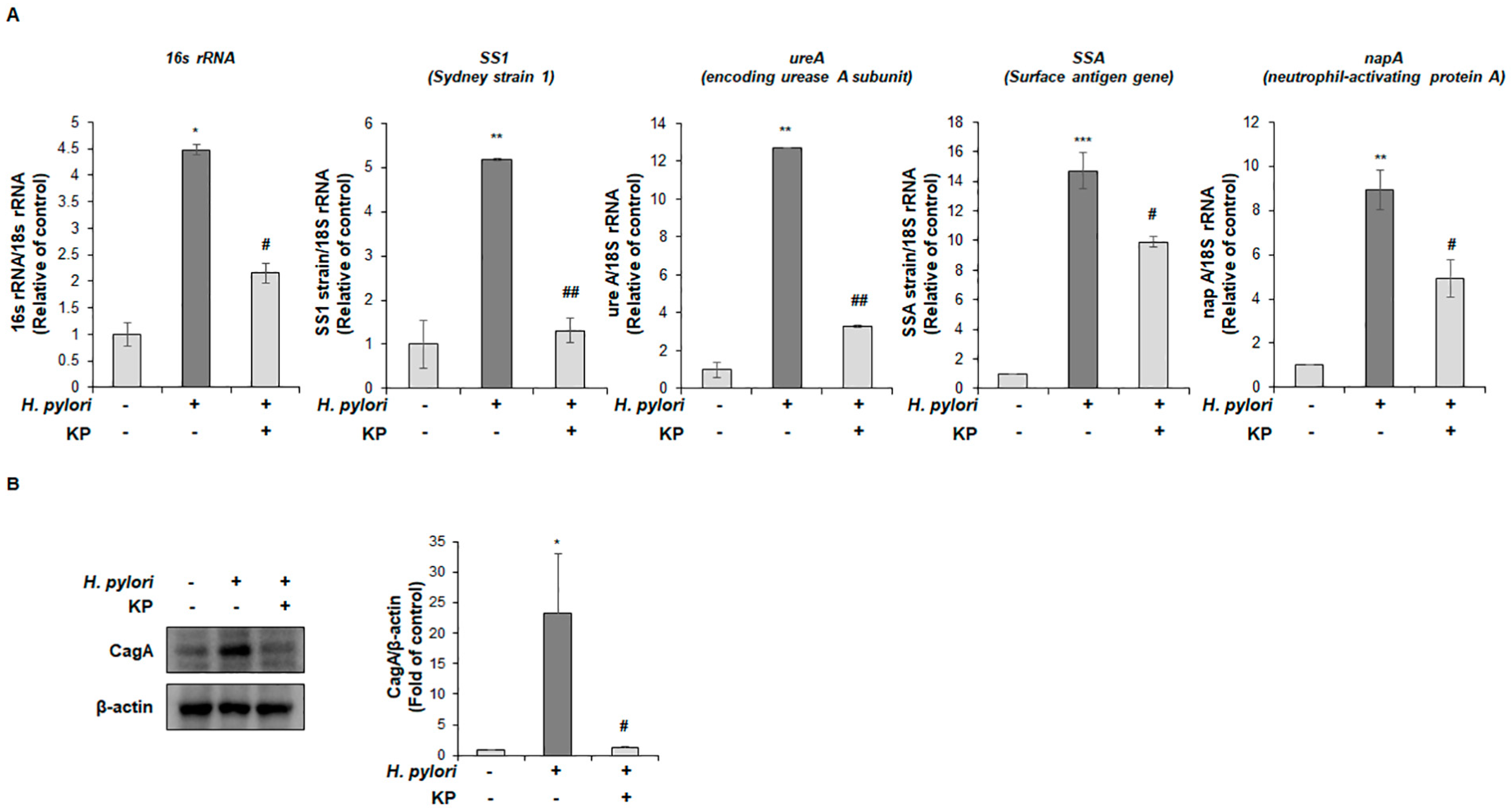
A previous study by Han et al. revealed the effect of KP against eradication of H. pylori using paper disc diffusion method [31]. Thus, we hypothesized the possibility of KP suppressing the expression of virulence factors in H. pylori. The virulence factors of H. pylori are associated with pathogenic responses, colony formation, immune system disruption and disease development [38]. To evaluate the mitigating effects of KP against the virulence factors of H. pylori, we conducted qPCR test for H. pylori-related genes and virulence factors. As shown in Figure 2A, the H. pylori-infected group demonstrated significantly increased expression levels of H. pylori virulence factors such as 16s rRNA, Sydney strain 1 (SS1), encoding urease A subunit (ureA), Surface antigen gene (SSA) and neutrophil-activating protein A (napA). In comparison, the KP-treatment group demonstrated extreme reduction and attenuation in mRNA expression levels of virulence factors. These results demonstrated that the KP-treatment group repressed gastric mucosal injury by inhibiting H. pylori growth and virulence factors of H. pylori. Next, we also confirmed the protein levels of CagA, pathogenicity factors of the bacterial pathogen H. pylori using gastric tissue extracts. As a result, the H. pylori-infected group showed increased CagA protein expression, whereas the KP-treatment group showed strongly decreased the protein levels (Figure 2B). Therefore, we considered that KP might regulate virulence factors concerning H. pylori infection in gastric tissue extracts.
3.3. KP Restrains the Pro-Inflammatory Response and NO Production in H. pylori-Infected Mice
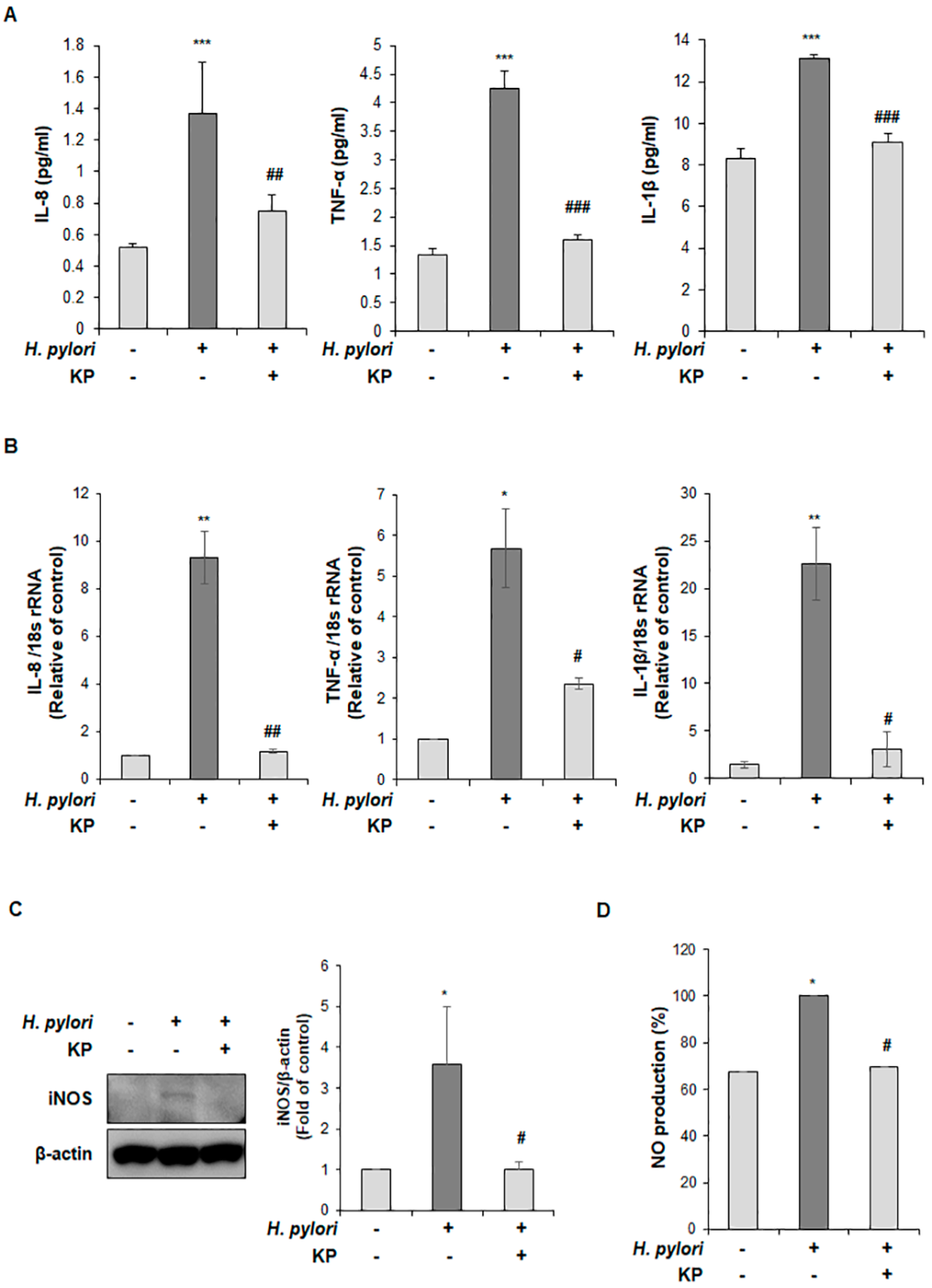
To demonstrate the effects of KP on pro-inflammatory responses in H. pylori-infected gastric mucosal injury mice, we examined the production levels of pro-inflammatory cytokines including IL-8, TNF-α, and IL-1β via ELISA and qPCR analysis. As shown in Figure 3A, the secretory serum levels of IL-8, TNF-α and IL-1β were significantly augmented after H. pylori infection, while the KP-treatment group showed an inhibitory effect on pro-inflammatory cytokines. Furthermore, the mRNA levels of IL-8, TNF-α and IL-1β were up-regulated in the H. pylori-infected group; however, mRNA expression was down-regulated in the KP-treatment group (Figure 3B). Next, we investigated whether the anti-inflammatory effect of KP on H. pylori-infected gastric mucosal injury is caused by inhibiting the production of NO. We performed the protein expression of inducible nitric oxide synthase (iNOS) via Western blot analysis and secretory serum levels of NO production. As a result, expression levels of iNOS and NO were markedly increased in the H. pylori-infected group, whereas the KP-treatment group showed significantly decreased levels of iNOS and NO production despite active infection with H. pylori (Figure 3C,D). These data suggest that the anti-inflammatory effect of KP occurs by suppressing IL-8, TNF-α, IL-1β and NO production in H. pylori-infected gastric mucosal injury mice model.
3.4. KP Regulates the NF-κB Signaling Pathway in H. pylori-Infected Mice
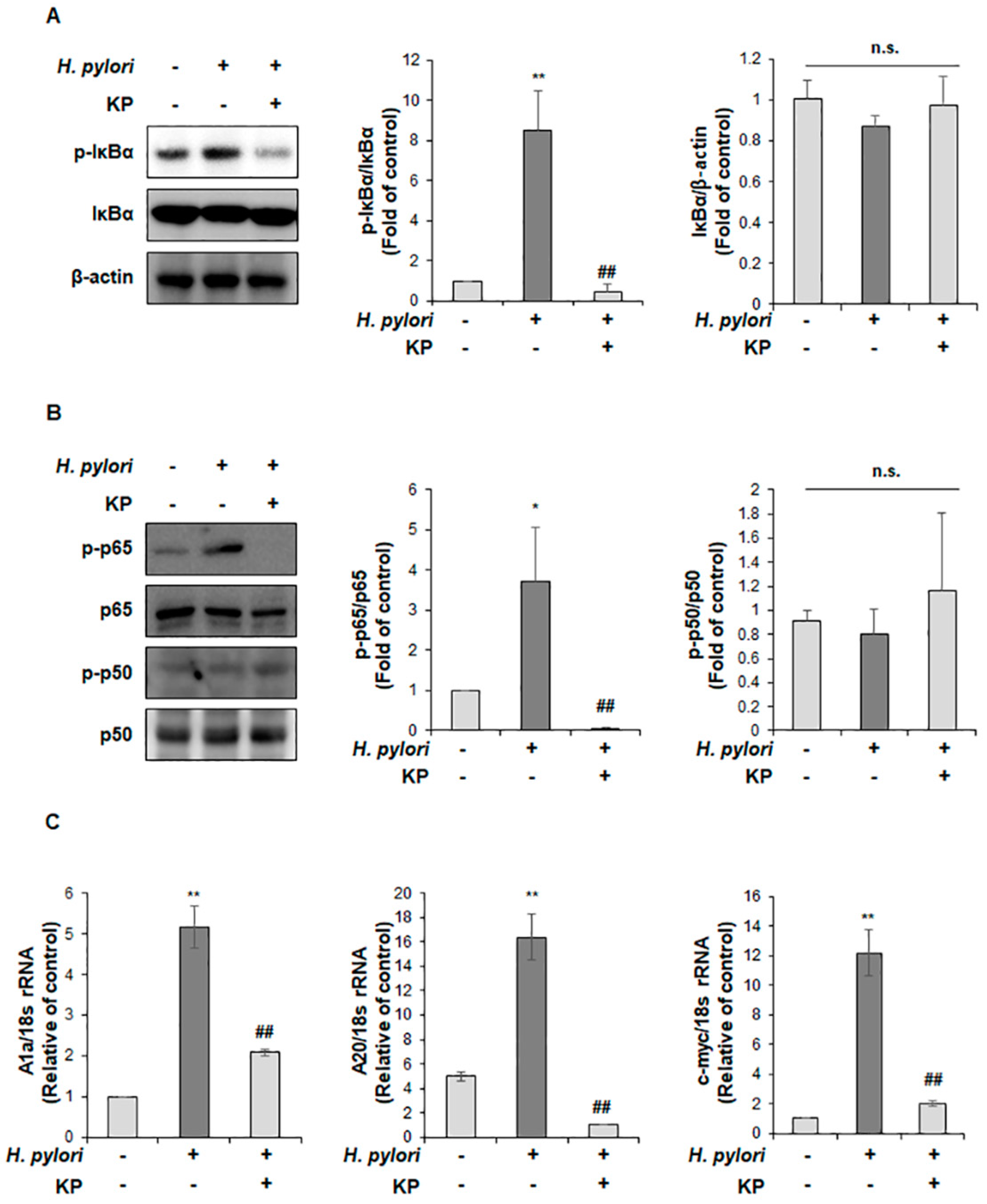
iNOS is mediated by NF-κB activation and correlated with pro-inflammatory responses [39,40,41]. Therefore, we hypothesized that the anti-inflammatory effect of KP in H. pylori-infected gastric mucosal injury may be regulated by NF-κB signaling. As shown in Figure 4A, the H. pylori-infected group showed increased protein expression of IκBα phosphorylation, but this level was significantly restrained in the KP-treatment group (Figure 4A). Moreover, phosphorylation of p65 was significantly promoted in the H. pylori-infected group, which was dramatically abrogated by treatment with KP (Figure 4B). In contrast, there was no change in the p50 phosphorylated by H. pylori administration or treatment with propolis (Figure 4B). Additionally, we validated the c-myc, A20 and A1a mRNA levels of NF-κB target genes, which are correlated with pro-inflammatory responses [42,43,44,45]. As expected, the H. pylori-infected group showed increased expression of these target genes; however, these were remarkably reduced in the KP-treatment group (Figure 4C).
4. Discussion
Our study results demonstrated the anti-inflammatory and anti-bacterial effects of KP in an H. pylori-infected mice model. Traditionally, H. pylori eradication therapy is commonly treated with proton-pump inhibitors and antibiotics [46,47]. However, antibiotic resistance presents a significant obstacle in the successful eradication of H. pylori [48,49]. Hence, in this study, we investigated an alternative therapeutic approach using a natural compound for the treatment and eradication of H. pylori infection.
Propolis has anti-inflammatory properties in the treatment of various diseases. Croatian propolis was reportedly used to treat obese patients with hyperlipidemic disorders, following prior success in mice studies [50]. In addition, Brazilian green propolis was suggested as a good candidate to suppress acute inflammation against hepatocellular necrosis in rats [51] and also indicated for use as a target in asthma immunotherapy [52]. In addition, Kai Wang demonstrated the protective effect of Chinese propolis against dextran sulfate sodium-induced acute colitis in rats [53]. Interestingly, several studies have shown that a variety of propolis such as Brazilian [54], Italian [55], Turkish [56], Bulgarian [57], Chilian [58] and Korean [31] inhibited the growth of H. pylori in vitro. Notably, our previous study has been clarified, in that KP attenuates inflammation against H. pylori-infected gastric epithelial cells via the NF-κB signaling pathway in vitro. However, the effect of KP on the reduction of inflammation against H. pylori on in vivo mice models has not been reported yet. Therefore, the main objective of our study was to elucidate the ability of KP to reduce H. pylori-induced inflammation through inhibition of NF-kB in vivo.
H. pylori infection is characterized by accelerated inflammation and damage to the gastric mucosa via the elaboration of virulence factors such as CagA, ureA, SSA and napA [59,60]. These virulence factors contribute to the flagella, chemotactic systems, ureases, and adhesions upon H. pylori, thereby affecting the inflammatory response and immune system through a variety of pathways [61]. For example, VacA is associated with immunosuppression of T cell activation and mediates NF-κB activity within targeted T cells [62,63]. Peptidoglycan enters host epithelial cells via T4SS and activates NF-κB signals for the acceleration of pro-inflammatory cytokines [64,65]. In particular, the H. pylori virulence factor CagA has been extensively studied for its inflammation responses. Several studies have noted that CagA conducts an important role in the acceleration of pro-inflammatory cytokines through the NF-κB pathway [15,66]. Indeed, pro-inflammatory cytokines such as TNF-α, IL-1β and IL-8 have been found to be triggered during H. pylori-infection and utilizing various mechanisms for the induction of inflammation [67].
The NO produced by L-arginine in response to H. pylori infection may accelerate the inflammatory processes in gastric mucosal damage [68,69]. NO is divided into a family of three distinct NOS isoforms including nNOS, eNOS and iNOS. It is well known that its two isoforms, nNOS and eNOS, are both calcium-dependent. nNOS is constitutively expressed in neurons of the nervous system and eNOS is mainly expressed in vascular endothelial cells [70]. The third calcium-independent isoform, iNOS, is expressed in response to inflammatory cytokines and bacterial virulence factors inducing the release of NO [40,71]. Over-production of NO triggers the pathogenesis of a variety of diseases including gastritis, and upregulation of iNOS levels has been observed in gastritis patients with H. pylori infection [72,73]. Interestingly, Nam et al. showed that iNOS levels were significantly diminished after inhibition and eradication of H. pylori [74]. Additionally, NO production activates NF-κB signaling, thereby accelerating the pathological conditions and inflammatory responses, implicated in gastric mucosal damage against H. pylori infection [30,41]. Here, we show that KP inhibited NF-κB signaling and significantly reduced the expression of iNOS and production of NO in H. pylori-infected mice, which may contribute to the treatment of gastric mucosal injury.
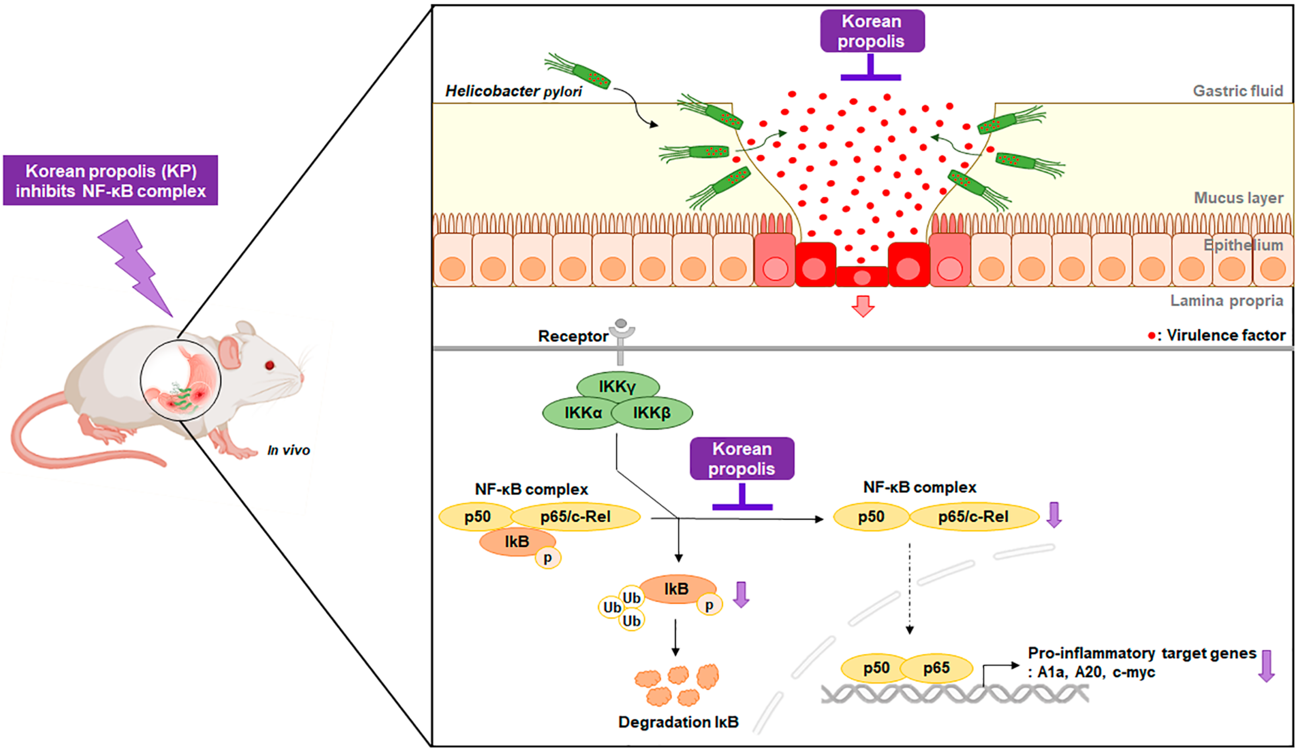
The transcription factor NF-κB is one of the major mediators of the inflammatory response against H. pylori infection [75]. H. pylori activates NF-κB through the canonical pathway, namely IKK complex phosphorylation/degradation of the IκB inhibitor, leading to the nuclear translocation of NF-κB, and thereby resulting in the acceleration of its target genes [15,68,76]. In this study, we showed that KP reduced the phosphorylation of IκBα and p65, thereby repressing the translocation of p65 from the cytosol to the nucleus in H. pylori-infected gastric mucosal injury mice. These findings suggest that KP attenuated the pro-inflammatory target genes through inhibition of NF-κB in H. pylori-infected mice. As a result, KP dramatically abrogated H. pylori virulence factors as well as host inflammatory responses during H. pylori infection (Figure 5).
Lastly, we investigated a human equivalent dose for the effect of KP using the recommended body surface area normalization method [77]. KP demonstrated a therapeutic effect against H. pylori-infected gastric mucosal injury at a dose of 200 mg/kg. Based on our studies, a daily KP dosage ranging from 900 to 1000 mg is required for a 60 kg subject. However, in order to clarify the exact concentration of human equivalent dose for the effect of KP on H. pylori infection, further studies on the minimum inhibitory concentration in vivo are needed.
5. Conclusions
In conclusion, we verified the anti-bacterial and anti-inflammatory effects of KP against H. pylori infection. KP attenuated H. pylori virulence factors and the production of pro-inflammatory cytokines in H. pylori-infected mice, thereby decreasing damage to the gastric mucosa. We provided a clarified molecular mechanism whereby NF-κB becomes inhibited following KP administration during active H. pylori infection. Therefore, our data suggest that KP may have potential therapeutic application against H. pylori infection in a gastric mucosal injury mice model.
Author Contributions
Conceptualization, E.-H.K. and M.-Y.S.; methodology, M.-Y.S. and D.-Y.L.; validation, M.-Y.S. and D.-Y.L.; formal analysis, M.-Y.S.; writing—original draft preparation, M.-Y.S.; writing—review and editing, Y.-M.H.; visualization, D.-Y.L. and Y.-M.H.; project administration, E.-H.K.; funding acquisition, E.-H.K.; supervision, E.-H.K. All authors have read and agreed to the published version of the manuscript.
Funding
This work was supported by the GRRC program of Gyeonggi province (GRRC-201900830002-CHA2019-B01, Production of physiologically active substances), Republic of Korea.
Institutional Review Board Statement
Not applicable.
Informed Consent Statement
Not applicable.
Data Availability Statement
The data presented in this study are available on request from the corresponding author.
Conflicts of Interest
The authors declare no conflict of interest.
References
- Hooi, J.K.; Lai, W.Y.; Ng, W.K.; Suen, M.M.; Underwood, F.E.; Tanyingoh, D.; Malfertheiner, P.; Graham, D.Y.; Wong, V.W.; Wu, J.C. Global prevalence of Helicobacter pylori infection: Systematic review and meta-analysis. Gastroenterology 2017, 153, 420–429. [Google Scholar] [CrossRef] [PubMed] [Green Version]
- Ranjbar, R.; Behzadi, P.; Farshad, S. Advances in diagnosis and treatment of Helicobacter pylori infection. Acta Microbiol. Immunol. Hung. 2017, 64, 273–292. [Google Scholar] [CrossRef] [PubMed] [Green Version]
- Zamani, M.; Ebrahimtabar, F.; Zamani, V.; Miller, W.; Alizadeh-Navaei, R.; Shokri-Shirvani, J.; Derakhshan, M. Systematic review with meta-analysis: The worldwide prevalence of Helicobacter pylori infection. Aliment. Pharmacol. Ther. 2018, 47, 868–876. [Google Scholar] [CrossRef] [PubMed] [Green Version]
- Cover, T.L.; Blaser, M.J. Helicobacter pylori in health and disease. Gastroenterology 2009, 136, 1863–1873. [Google Scholar] [CrossRef] [PubMed] [Green Version]
- Testerman, T.L.; Morris, J. Beyond the stomach: An updated view of Helicobacter pylori pathogenesis, diagnosis, and treatment. World J. Gastroenterol. 2014, 20, 12781. [Google Scholar] [CrossRef] [PubMed]
- Marcus, E.A.; Sachs, G.; Scott, D.R. Eradication of Helicobacter pylori infection. Curr. Gastroenterol. Rep. 2016, 18, 1–9. [Google Scholar] [CrossRef] [Green Version]
- Suerbaum, S.; Michetti, P. Helicobacter pylori infection. N. Engl. J. Med. 2002, 347, 1175–1186. [Google Scholar] [CrossRef] [Green Version]
- Roesler, B.M.; Costa, S.C.; Zeitune, J.M. Eradication treatment of Helicobacter pylori infection: Its importance and possible relationship in preventing the development of gastric cancer. Int. Sch. Res. Notices 2012, 2012, 935410. [Google Scholar] [CrossRef] [Green Version]
- Sepulveda, A.R. Helicobacter, inflammation, and gastric cancer. Curr. Pathobiol. Rep. 2013, 1, 9–18. [Google Scholar] [CrossRef] [Green Version]
- Sharma, S.A.; Tummuru, M.K.; Blaser, M.J.; Kerr, L.D. Activation of IL-8 gene expression by Helicobacter pylori is regulated by transcription factor nuclear factor-κB in gastric epithelial cells. J. Immunol. Res. 1998, 160, 2401–2407. [Google Scholar]
- Lamb, A.; Yang, X.D.; Tsang, Y.H.N.; Li, J.D.; Higashi, H.; Hatakeyama, M.; Peek, R.M.; Blanke, S.R.; Chen, L.F. Helicobacter pylori cagA activates NF-κB by targeting TAK1 for TRAF6-mediated Lys 63 ubiquitination. EMBO Rep. 2009, 10, 1242–1249. [Google Scholar] [CrossRef] [PubMed]
- Hayden, M.S.; Ghosh, S. Signaling to NF-κB. Genes Dev. 2004, 18, 2195–2224. [Google Scholar] [CrossRef] [PubMed] [Green Version]
- Aggarwal, B.B. Nuclear factor-κB: The enemy within. Cancer Cell 2004, 6, 203–208. [Google Scholar] [CrossRef] [Green Version]
- Crabtree, J.E.; Lindley, I. Mucosal interleukin-8 and Helicobacter pylori-associated gastroduodenal disease. Eur. J. Gastroenterol. Hepatol. 1994, 6, S33–S38. [Google Scholar] [PubMed]
- Brandt, S.; Kwok, T.; Hartig, R.; König, W.; Backert, S. NF-κB activation and potentiation of proinflammatory responses by the Helicobacter pylori CagA protein. Proc. Natl. Acad. Sci. USA 2005, 102, 9300–9305. [Google Scholar] [CrossRef] [Green Version]
- Veenendaal, R.A.; Götz, J.M.; Lamers, C.B. Mucosal inflammation and disease in Helicobacter pylori infection. Scand. J. Gastroenterol. 1996, 218, 86–91. [Google Scholar] [CrossRef]
- Wang, Y.-C. Medicinal plant activity on Helicobacter pylori related diseases. World J. Gastroenterol. 2014, 20, 10368. [Google Scholar] [CrossRef]
- Islam, M.; Kusumoto, Y.; Al-Mamun, M.A. Cytotoxicity and cancer (HeLa) cell killing efficacy of aqueous garlic (Allium sativum) extract. J. Sci. Res. 2011, 3, 375–382. [Google Scholar] [CrossRef] [Green Version]
- Kumar, D.; Kumar, M.; Saravanan, C.; Singh, S.K. Curcumin: A potential candidate for matrix metalloproteinase inhibitors. Expert Opin. Ther. Targets 2012, 16, 959–972. [Google Scholar] [CrossRef]
- Shapla, U.M.; Raihan, J.; Islam, A.; Alam, F.; Solayman, N.; Gan, S.H.; Hossen, S.; Khalil, I. Propolis: The future therapy against Helicobacter pylori-mediated gastrointestinal diseases. J. Appl. Biomed. 2018, 16, 81–99. [Google Scholar] [CrossRef] [Green Version]
- Cardos, I.A.; Zaha, D.C.; Sindhu, R.K.; Cavalu, S. Revisiting therapeutic strategies for H. pylori treatment in the context of antibiotic resistance: Focus on alternative and complementary therapies. Molecules 2021, 26, 6078. [Google Scholar] [CrossRef] [PubMed]
- Salatino, A.; Teixeira, É.W.; Negri, G. Origin and chemical variation of Brazilian propolis. Evid.-Based Complement. Altern. Med. 2005, 2, 33–38. [Google Scholar] [CrossRef] [PubMed] [Green Version]
- Sarkar, A.; De, R.; Mukhopadhyay, A.K. Curcumin as a potential therapeutic candidate for Helicobacter pylori associated diseases. World J. Gastroenterol. 2016, 22, 2736. [Google Scholar] [CrossRef] [PubMed]
- Osés, S.M.; Pascual-Maté, A.; Fernández-Muiño, M.A.; López-Díaz, T.M.; Sancho, M.T. Bioactive properties of honey with propolis. Food Chem. 2016, 196, 1215–1223. [Google Scholar] [CrossRef]
- Pascual, C.; Gonzalez, R.; Torricella, R. Scavenging action of propolis extract against oxygen radicals. J. Ethnopharmacol. 1994, 41, 9–13. [Google Scholar] [CrossRef]
- Huang, S.; Zhang, C.-P.; Wang, K.; Li, G.Q.; Hu, F.-L. Recent advances in the chemical composition of propolis. Molecules 2014, 19, 19610–19632. [Google Scholar] [CrossRef] [Green Version]
- Ahn, M.R.; Kumazawa, S.; Hamasaka, T.; Bang, K.S.; Nakayama, T. Antioxidant activity and constituents of propolis collected in various areas of Korea. J. Agric. Food Chem. 2004, 52, 7286–7292. [Google Scholar] [CrossRef]
- Kujumgiev, A.; Tsvetkova, I.; Serkedjieva, Y.; Bankova, V.; Christov, R.; Popov, S. Antibacterial, antifungal and antiviral activity of propolis of different geographic origin. J. Ethnopharmacol. 1999, 64, 235–240. [Google Scholar] [CrossRef]
- Lotfy, M. Biological activity of bee propolis in health and disease. Asian Pac. J. Cancer Prev. 2006, 7, 22–31. [Google Scholar]
- Song, M.Y.; Lee, D.Y.; Kim, E.H. Anti-inflammatory and anti-oxidative effect of Korean propolis on Helicobacter pylori-induced gastric damage in vitro. J. Microbiol. 2020, 58, 878–885. [Google Scholar] [CrossRef]
- Han, S.M.; Hong, I.P.; Woo, S.O.; Kim, S.G.; Jang, H.R.; Jang, J.S. Anti-Helicobacter pylori activity of Korean propolis. Korean J. Food Nutr. 2016, 29, 73–78. [Google Scholar] [CrossRef] [Green Version]
- Sipponen, P.; Price, A.B. The Sydney system for classification of gastritis 20 years ago. J. Gastroenterol. Hepatol. 2011, 26, 31–34. [Google Scholar] [CrossRef] [PubMed]
- Song, M.-Y.; Lee, D.-Y.; Yun, S.-M.; Kim, E.-H. GLUT3 promotes epithelial–mesenchymal transition via TGF-β/JNK/ATF2 signaling pathway in colorectal cancer cells. Biomedicines 2022, 10, 1837. [Google Scholar] [CrossRef] [PubMed]
- Li, S.; Lu, A.P.; Zhang, L.; Li, Y.D. Anti-Helicobacter pylori immunoglobulin G (IgG) and IgA antibody responses and the value of clinical presentations in diagnosis of H. pylori infection in patients with precancerous lesions. World J. Gastroenterol. 2003, 9, 755–758. [Google Scholar] [CrossRef]
- Unver, S.; Kubilay, U.; Sezen, O.S.; Coskuner, T. Investigation of Helicobacter pylori colonization in adenotonsillectomy specimens by means of the CLO Test. Laryngoscope 2001, 111, 2183–2186. [Google Scholar] [CrossRef] [PubMed]
- Allen, A.; Newton, J.; Oliver, L.; Jordan, N.; Strugala, V.; Pearson, J.P.; Dettmar, P.W. Mucus and H. pylori. J. Physiol. Pharmacol. 1997, 48, 297–305. [Google Scholar]
- Hidaka, E.; Ota, H.; Hidaka, H.; Hayama, M.; Matsuzawa, K.; Akamatsu, T.; Nakayama, J.; Katsuyama, T. Helicobacter pylori and two ultrastructurally distinct layers of gastric mucous cell mucins in the surface mucous gel layer. Gut 2001, 49, 474–480. [Google Scholar] [CrossRef] [Green Version]
- Chang, W.L.; Yeh, Y.C.; Sheu, B.S. The impacts of H. pylori virulence factors on the development of gastroduodenal diseases. J. Biomed. Sci. 2018, 25, 68. [Google Scholar] [CrossRef] [Green Version]
- Dincă, A.L.; Meliț, L.E.; Mărginean, C.O. Old and new aspects of H. pylori-associated Inflammation and gastric cancer. Children 2022, 9, 1083. [Google Scholar] [CrossRef]
- Sharma, J.; Al-Omran, A.; Parvathy, S. Role of nitric oxide in inflammatory diseases. Inflammopharmacology 2007, 15, 252–259. [Google Scholar] [CrossRef]
- Cho, K.; Lee, H.G.; Piao, J.Y.; Kim, S.J.; Na, H.K.; Surh, Y.J. Protective effects of Silibinin on Helicobacter pylori-induced gastritis: NF-κB and STAT3 as potential targets. J. Cancer Prev. 2021, 26, 118–127. [Google Scholar] [CrossRef] [PubMed]
- Santoro, M.G.; Rossi, A.; Amici, C. NF-κB and virus infection: Who controls whom. EMBO Rep. 2003, 22, 2552–2560. [Google Scholar] [CrossRef] [PubMed]
- Catrysse, L.; Vereecke, L.; Beyaert, R.; van Loo, G. A20 in inflammation and autoimmunity. Trends Immunol. 2014, 35, 22–31. [Google Scholar] [CrossRef]
- Moser, B.; Hochreiter, B.; Basílio, J.; Gleitsmann, V.; Panhuber, A.; Pardo-Garcia, A.; Hoesel, B.; Salzmann, M.; Resch, U.; Noreen, M.; et al. The inflammatory kinase IKKα phosphorylates and stabilizes c-Myc and enhances its activity. Mol. Cancer 2021, 20, 16. [Google Scholar] [CrossRef] [PubMed]
- Cho, J.M.; Yun, S.M.; Choi, Y.H.; Heo, J.; Kim, N.J.; Kim, S.H.; Kim, E.H. Xanthohumol prevents dextran sulfate sodium-induced colitis via inhibition of IKKβ/NF-κB signaling in mice. Oncotarget 2018, 9, 866–880. [Google Scholar] [CrossRef] [Green Version]
- Beales, I.L. Efficacy of Helicobacter pylori eradication therapies: A single centre observational study. BMC Gastroenterol. 2001, 1, 7. [Google Scholar] [CrossRef] [Green Version]
- Ford, A.C.; Yuan, Y.; Moayyedi, P. Helicobacter pylori eradication therapy to prevent gastric cancer: Systematic review and meta-analysis. Gut 2020, 69, 2113–2121. [Google Scholar] [CrossRef]
- Ghotaslou, R.; Leylabadlo, H.E.; Asl, Y.M. Prevalence of antibiotic resistance in Helicobacter pylori: A recent literature review. World J. Methodol. 2015, 5, 164. [Google Scholar] [CrossRef]
- Thung, I.; Aramin, H.; Vavinskaya, V.; Gupta, S.; Park, J.; Crowe, S.; Valasek, M. The global emergence of Helicobacter pylori antibiotic resistance. Aliment. Pharmacol. Ther. 2016, 43, 514–533. [Google Scholar] [CrossRef] [Green Version]
- Oršolić, N.; Landeka Jurčević, I.; Đikić, D.; Rogić, D.; Odeh, D.; Balta, V.; Perak Junaković, E.; Terzić, S.; Jutrić, D. Effect of Propolis on diet-Induced hyperlipidemia and atherogenic Indices in mice. Antioxidants 2019, 8, 156. [Google Scholar] [CrossRef] [Green Version]
- Tsuchiya, Y.; Sakai, H.; Hirata, A.; Yanai, T. Brazilian green propolis suppresses acetaminophen-induced hepatocellular necrosis by modulating inflammation-related factors in rats. J. Toxicol. Pathol. 2018, 31, 275–282. [Google Scholar] [CrossRef] [Green Version]
- Piñeros, A.R.; de Lima, M.H.F.; Rodrigues, T.; Gembre, A.F.; Bertolini, T.B.; Fonseca, M.D.; Berretta, A.A.; Ramalho, L.N.Z.; Cunha, F.Q.; Hori, J.I.; et al. Green propolis increases myeloid suppressor cells and CD4+Foxp3+ cells and reduces Th2 inflammation in the lungs after allergen exposure. J. Ethnopharmacol. 2020, 252, 112496. [Google Scholar] [CrossRef]
- Wang, K.; Jin, X.; Li, Q.; Sawaya, A.C.H.F.; Le Leu, R.K.; Conlon, M.A.; Wu, L.; Hu, F. Propolis from different geographic origins decreases intestinal inflammation and bacteroides spp. populations in a model of DSS-induced colitis. Mol. Nutr. Food Res. 2018, 62, 1800080. [Google Scholar] [CrossRef] [PubMed]
- Banskota, A.H.; Tezuka, Y.; Adnyana, I.K.; Ishii, E.; Midorikawa, K.; Matsushige, K.; Kadota, S. Hepatoprotective and anti-Helicobacter pylori activities of constituents from Brazilian propolis. Phytomedicine 2001, 8, 16–23. [Google Scholar] [CrossRef]
- Nostro, A.; Cellini, L.; Bartolomeo, S.D.; Cannatelli, M.A.; Campli, E.D.; Procopio, F.; Grande, R.; Marzio, L.; Alonzo, V. Effects of combining extracts (from propolis or Zingiber officinale) with clarithromycin on Helicobacter pylori. Phytother. Res. 2006, 20, 187–190. [Google Scholar] [CrossRef] [PubMed]
- Baltas, N.; Karaoglu, S.A.; Tarakci, C.; Kolayli, S. Effect of propolis in gastric disorders: Inhibition studies on the growth of Helicobacter pylori and production of its urease. J. Enzyme Inhib. Med. Chem. 2016, 31, 46–50. [Google Scholar] [CrossRef] [PubMed] [Green Version]
- Boyanova, L.; Derejian, S.; Koumanova, R.; Katsarov, N.; Gergova, G.; Mitov, I.; Nikolov, R.; Krastev, Z. Inhibition of Helicobacter pylori growth in vitro by Bulgarian propolis: Preliminary report. J. Med. Microbiol. 2003, 52, 417–419. [Google Scholar] [CrossRef]
- Romero, M.; Freire, J.; Pastene, E.; García, A.; Aranda, M.; González, C. Propolis polyphenolic compounds affect the viability and structure of Helicobacter pylori in vitro. Rev. Bras. Farmacogn. 2019, 29, 325–332. [Google Scholar] [CrossRef]
- Roesler, B.M.; Rabelo-Gonçalves, E.M.; Zeitune, J.M. Virulence Factors of Helicobacter pylori: A Review. Clin. Med. Insights Gastroenterol. 2014, 7, 9–17. [Google Scholar] [CrossRef]
- Šterbenc, A.; Jarc, E.; Poljak, M.; Homan, M. Helicobacter pylori virulence genes. World J. Gastroenterol. 2019, 25, 4870–4884. [Google Scholar] [CrossRef]
- Lamb, A.; Chen, L.F. Role of the Helicobacter pylori-induced inflammatory response in the development of gastric cancer. J. Cell. Biochem. 2013, 114, 491–497. [Google Scholar] [CrossRef] [PubMed] [Green Version]
- Nejati, S.; Karkhah, A.; Darvish, H.; Validi, M.; Ebrahimpour, S.; Nouri, H.R. Influence of Helicobacter pylori virulence factors CagA and VacA on pathogenesis of gastrointestinal disorders. Microb. Pathog. 2018, 117, 43–48. [Google Scholar] [CrossRef] [PubMed]
- Takeshima, E.; Tomimori, K.; Takamatsu, R.; Ishikawa, C.; Kinjo, F.; Hirayama, T.; Fujita, J.; Mori, N. Helicobacter pylori vacA activates NF-κB in T Cells via the classical but not alternative pathway. Helicobacter 2009, 14, 271–279. [Google Scholar] [CrossRef] [PubMed]
- Cover, T.L.; Lacy, D.B.; Ohi, M.D. The Helicobacter pylori cag type IV secretion system. Trends Microbiol. 2020, 28, 682–695. [Google Scholar] [CrossRef] [PubMed]
- Viala, J.; Chaput, C.; Boneca, I.G.; Cardona, A.; Girardin, S.E.; Moran, A.P.; Athman, R.; Mémet, S.; Huerre, M.R.; Coyle, A.J. Nod1 responds to peptidoglycan delivered by the Helicobacter pylori cag pathogenicity island. Nat. Immunol. 2004, 5, 1166–1174. [Google Scholar] [CrossRef] [PubMed]
- Waskito, L.A.; Salama, N.R.; Yamaoka, Y. Pathogenesis of Helicobacter pylori infection. Helicobacter 2018, 23, e12516. [Google Scholar] [CrossRef] [Green Version]
- McGee, D.J.; Mobley, H.L. Pathogenesis of Helicobacter pylori infection. Curr. Opin. Gastroenterol. 2000, 16, 24–31. [Google Scholar] [CrossRef]
- Pignatelli, B.; Bancel, B.; Esteve, J.; Malaveille, C.; Calmels, S.; Correa, P.; Patricot, L.; Laval, M.; Lyandrat, N.; Ohshima, H. Inducible nitric oxide synthase, anti-oxidant enzymes and Helicobacter pylori infection in gastritis and gastric precancerous lesions in humans. Eur. J. Cancer Prev. 1998, 7, 439–447. [Google Scholar] [CrossRef]
- Bothmer, C.v.; Edebo, A.; Lönroth, H.; Olbe, L.; Pettersson, A.; Fändriks, L. Helicobacter pylori infection inhibits antral mucosal nitric oxide production in humans. Scand. J. Gastroenterol. 2002, 37, 404–408. [Google Scholar] [CrossRef]
- Alderton, W.K.; Cooper, C.E.; Knowles, R.G. Nitric oxide synthases: Structure, function and inhibition. Biochem. J. 2001, 357, 593–615. [Google Scholar] [CrossRef]
- Cinelli, M.A.; Do, H.T.; Miley, G.P.; Silverman, R.B. Inducible nitric oxide synthase: Regulation, structure, and inhibition. Med. Res. Rev. 2020, 40, 158–189. [Google Scholar] [CrossRef]
- Fu, S.; Ramanujam, K.S.; Wong, A.; Fantry, G.T.; Drachenberg, C.B.; James, S.P.; Meltzer, S.J.; Wilson, K.T. Increased expression and cellular localization of inducible nitric oxide synthase and cyclooxygenase 2 in Helicobacter pylori gastritis. Gastroenterology 1999, 116, 1319–1329. [Google Scholar] [CrossRef]
- Saaed, H.K.; Chiggiato, L.; Webb, D.L.; Rehnberg, A.S.; Rubio, C.A.; Befrits, R.; Hellström, P.M. Elevated gaseous luminal nitric oxide and circulating IL-8 as features of Helicobacter pylori-induced gastric inflammation. UPS J. Med. Sci. 2021, 126, e8116. [Google Scholar] [CrossRef] [PubMed]
- Nam, K.-T.; Oh, S.-Y.; Ahn, B.-W.; Kim, Y.; Jang, D.; Yang, K.; Hahm, K.; Kim, D. Decreased Helicobacter pylori associated gastric carcinogenesis in mice lacking inducible nitric oxide synthase. Gut 2004, 53, 1250–1255. [Google Scholar] [CrossRef] [PubMed]
- Lamb, A.; Chen, L.-F. The many roads traveled by Helicobacter pylori to NF-κB activation. Gut Microbes 2010, 1, 109–113. [Google Scholar] [CrossRef] [Green Version]
- Saha, K.; Sarkar, D.; Khan, U.; Karmakar, B.C.; Paul, S.; Mukhopadhyay, A.K.; Dutta, S.; Bhattacharya, S. Capsaicin inhibits inflammation and gastric damage during H pylori infection by targeting NF-kB–miRNA axis. Pathogens 2022, 11, 641. [Google Scholar] [CrossRef]
- Nair, A.B.; Jacob, S. A simple practice guide for dose conversion between animals and human. J. Basic Clin. Pharm. 2016, 7, 27–31. [Google Scholar] [CrossRef]
Figure 1.
KP decreases the severity of H. pylori−infected gastric mucosal injury in mice. (A) Diagram shows the experimental design of H. pylori-infected gastric mucosal injury mice model. (B) Result of H. pylori IgG levels in mice using serum obtained from each group (n = 8). (C) Results of the CLO test in mice using gastric mucosal tissue from each group (n = 8, positive reaction shown by change in color from yellow to purple). (D) Representative images of the stomach and gastric mucosal injury index (n = 8). (E) Paraffin sections of stomach were stained with hematoxylin and eosin (H&E, magnification, ×100). Damage to the surface epithelium, inflammatory cell infiltration, submucosal edema and total pathological score in each group (n = 8) were quantified from H&E−stained sections. (F) Detection of mucous production was performed by PAS staining in each group (n = 3). Representative images of gastric sections were stained with PAS (magnification, ×100). Detection of mucous production were immunostained with PAS in mice gastric mucosal (positive result depicted by purple color), and one of three representative experiments is presented (×100). Graph shows the number of PAS-positive cells from at least 10 fields (bottom). Data are the mean ± standard deviation. Statistical significance was analyzed by analysis of variance. * p < 0.05, ** p < 0.01, and *** p < 0.001 vs. normal group; # p < 0.05, ## p < 0.01 and ### p < 0.001 vs. H. pylori group, significantly different compared with control.


Figure 2.
KP attenuates H. pylori virulence factors in H. pylori-infected gastric mucosal injury mice model. (A) mRNA expression levels in gastric mucosal tissues were examined via qRT-PCR. As an internal control, 18S rRNA was used for the expression of H. pylori-related genes. (B) The protein expression of CagA in gastric mucosal tissues was determined by Western blotting. β-actin was used as an internal control. Data are the mean ± standard deviation (n = 4). Statistical significance was analyzed by analysis of variance. * p < 0.05 and ** p < 0.01, and *** p < 0.001 vs. normal group; # p < 0.05 and ## p < 0.01 vs. H. pylori group, significantly different compared with control.

Figure 3.
KP inhibits pro-inflammatory response and NO production in H. pylori-infected gastric mucosal injury mice model. (A) IL-8, TNF-α, and IL-1β production levels of pro-inflammatory cytokines were measured in serum via ELISA. (B) The mRNA expressions of IL-8, TNF-α, and IL-1β in gastric mucosal tissues were examined via qRT-PCR. The 18S rRNA was used as an internal control. (C) The protein expression of iNOS in gastric mucosal tissues was determined by Western blotting. β-actin was used as an internal control. (D) The secretory serum levels were measured for NO production. Data are the mean ± standard deviation (n = 4). Statistical significance was analyzed by analysis of variance. * p < 0.05, ** p < 0.01, and *** p < 0.001 vs. normal group; # p < 0.05, ## p < 0.01 and ### p < 0.001 vs. H. pylori group, significantly different compared with control.

Figure 4.
KP inhibits NF-κB signaling in H. pylori-infected gastric mucosal injury mice model. (A) Expressions of p-IκBα and IκBα in gastric mucosal tissues were analyzed by Western blot analysis. β-actin was used as an internal control. (B) The expression of protein levels of pp65, p65, pp50 and p50 were conducted by Western blot analysis. (C) The mRNA levels of A1a, A20 and c-myc were determined by qRT-PCR. The 18S rRNA was used as an internal control for the mRNA levels. Data are the mean ± standard deviation (n = 4). Statistical significance was analyzed by analysis of variance. * p < 0.05 and ** p < 0.01 vs. normal group; ## p < 0.01 vs. H. pylori group, significantly different compared with control.

Figure 5.
Schematic representation depicting KP inhibition of NF-κB-mediated inflammation in H. pylori-infected gastric mucosal injury in mice. “Black color ↓” indicates induction; “Purple color↓” indicates reduction; “├” indicates inhibition.
Figure 5.
Schematic representation depicting KP inhibition of NF-κB-mediated inflammation in H. pylori-infected gastric mucosal injury in mice. “Black color ↓” indicates induction; “Purple color↓” indicates reduction; “├” indicates inhibition.


Table 1.
Mouse Primer sequences used for qRT-PCR.
| Identification | Gene | Primer Sequence (5′ to 3′) | |
|---|---|---|---|
| Specific for H. pylori | 16S rRNA | Forward | CTC ATT GCG AAG GCG ACC T |
| Reverse | TCT AAT CCT GTT TGC TCC CCA | ||
| SS1 | Forward | CTT AAC CAT AGA ACT GCA TTT GAA ACT AC | |
| Reverse | GGT CGC CTT CGC AAT GAG TA | ||
| ureA | Forward | AGG AAA CAT CGC TTC AAT ACC | |
| Reverse | AGG AAA CAT CGC TTC AAT ACC | ||
| SSA | Forward | TGG CGT GTC TAT TGA CAG CGA GC | |
| Reverse | CCT GCT GGG CAT ACT TCA CCA TG | ||
| napA | Forward | TCC TTT CAG CGA GAT CGT CA | |
| Reverse | GAA TGT GAA AGG CAC CGA TT | ||
| Specific for inflammation | IL-8 | Forward | TCC TTG TTC CAC TGT GCC TTG |
| Reverse | TGC TTC CAC ATG TCC TCA CAA | ||
| TNF-α | Forward | TCA GAG GGC CTG TAC CTC AT | |
| Reverse | GGA AGA CCC CTC CCA GAT AG | ||
| IL-1β | Forward | TTA AAG CCC GCC TGA CAG A | |
| Reverse | GCG AAT GAC AGA GGG TTT CTT | ||
| A1a | Forward | TCC ACA AGA GCA GAT TGC CCT G | |
| Reverse | GCC AGC CAG ATT TGG GTT CAA AC | ||
| A20 | Forward | AGC AAG TGC AGG AAA GCT GGC T | |
| Reverse | GCT TTC GCA GAG GCA GTA ACA G | ||
| c-myc | Forward | GCT GTT TGA AGG CTG GAT TTC | |
| Reverse | GAT GAA ATA GGG CTG TAC GGA G | ||
| Internal control | 18s rRNA | Forward | GCA ATT ATT CCC CAT GAA CG |
| Reverse | GGC CTC ACT AAA CCA TCC AA | ||
qRT-PCR, quantitative reverse transcription PCR; 16S rRNA, 16S ribosomal RNA; SS1, Sydney strain 1; ureA, encoding urease A subunit; SSA, Surface antigen gene; napA, neutrophil-activating protein A; IL-8, Interleukin-8; TNF-α, Tumor necrosis factor-α; IL-1β, Interleukin-1β; 18S rRNA, 18S ribosomal RNA.
Publisher’s Note: MDPI stays neutral with regard to jurisdictional claims in published maps and institutional affiliations. |
© 2022 by the authors. Licensee MDPI, Basel, Switzerland. This article is an open access article distributed under the terms and conditions of the Creative Commons Attribution (CC BY) license (https://creativecommons.org/licenses/by/4.0/).
Share and Cite
MDPI and ACS Style
Song, M.-Y.; Lee, D.-Y.; Han, Y.-M.; Kim, E.-H. Anti-Inflammatory Effect of Korean Propolis on Helicobacter pylori-Infected Gastric Mucosal Injury Mice Model. Nutrients 2022, 14, 4644. https://doi.org/10.3390/nu14214644
AMA Style
Song M-Y, Lee D-Y, Han Y-M, Kim E-H. Anti-Inflammatory Effect of Korean Propolis on Helicobacter pylori-Infected Gastric Mucosal Injury Mice Model. Nutrients. 2022; 14(21):4644. https://doi.org/10.3390/nu14214644
Chicago/Turabian StyleSong, Moon-Young, Da-Young Lee, Young-Min Han, and Eun-Hee Kim. 2022. "Anti-Inflammatory Effect of Korean Propolis on Helicobacter pylori-Infected Gastric Mucosal Injury Mice Model" Nutrients 14, no. 21: 4644. https://doi.org/10.3390/nu14214644
Note that from the first issue of 2016, this journal uses article numbers instead of page numbers. See further details here.