
Rapid Nontranscriptional Effects of Calcifediol and Calcitriol
by
 , and
, and
Simone Donati
1, 
Gaia Palmini
1,
Cinzia Aurilia
1,
Irene Falsetti
1,
Francesca Miglietta
1,
Teresa Iantomasi
1 and
Maria Luisa Brandi
2,* 1
Department of Experimental and Clinical Biomedical Sciences, University of Florence, 50139 Florence, Italy
2
Fondazione Italiana Ricerca sulle Malattie dell’Osso (F.I.R.M.O. Onlus) Italian Foundation for the Research on Bone Diseases, 50141 Florence, Italy
*
Author to whom correspondence should be addressed.
Nutrients 2022, 14(6), 1291; https://doi.org/10.3390/nu14061291
Submission received: 14 February 2022
/
Revised: 11 March 2022
/
Accepted: 14 March 2022
/
Published: 18 March 2022
(This article belongs to the Special Issue Vitamin D Endocrine System: Calcifediol for Treatment and Prevention of Infection and Disease)
Abstract
:Classically, a secosteroid hormone, vitamin D, has been implicated in calcium and phosphate homeostasis and has been associated with the pathogenesis of rickets and osteomalacia in patients with severe nutritional vitamin D deficiency. The spectrum of known vitamin D-mediated effects has been expanded in recent years. However, the mechanisms of how exactly this hormone elicits its biological function are still not fully understood. The interaction of this metabolite with the vitamin D receptor (VDR) and, subsequently, with the vitamin D-responsive element in the region of specific target genes leading to the transcription of genes whose protein products are involved in the traditional function of calcitriol (known as genomic actions). Moreover, in addition to these transcription-dependent mechanisms, it has been recognized that the biologically active form of vitamin D3, as well as its immediate precursor metabolite, calcifediol, initiate rapid, non-genomic actions through the membrane receptors that are bound as described for other steroid hormones. So far, among the best candidates responsible for mediating rapid membrane response to vitamin D metabolites are membrane-associated VDR (VDRm) and protein disulfide isomerase family A member 3 (Pdia3). The purpose of this paper is to provide an overview of the rapid, non-genomic effects of calcifediol and calcitriol, whose elucidation could improve the understanding of the vitamin D3 endocrine system. This will contribute to a better recognition of the physiological acute functions of vitamin D3, and it could lead to the identification of novel therapeutic targets able to modulate these actions.
1. Introduction
Calcitriol (1α,25-dihydroxyvitamin D3 or 1α,25(OH)2D3), the biologically active form of vitamin D3, is a sterol hormone that is involved in the regulation of phosphate and plasma-ionized calcium levels by modulating their renal excretion, intestinal absorption, and calcium bone mobilization [1].
When low serum calcium concentrations are detected through the calcium-sensing receptor (CaSR) on the parathyroid glands, a G-protein coupled receptor, parathyroid hormone (PTH) secretion is triggered, thus activating calcitriol synthesis [2].
In addition to repressing parathyroid gene expression and cell proliferation by interacting with the vitamin D receptor (VDR), 1α,25(OH)2D3 maintains ideal serum calcium levels through its direct actions on the gut, the kidney, and the bone tissues [3].
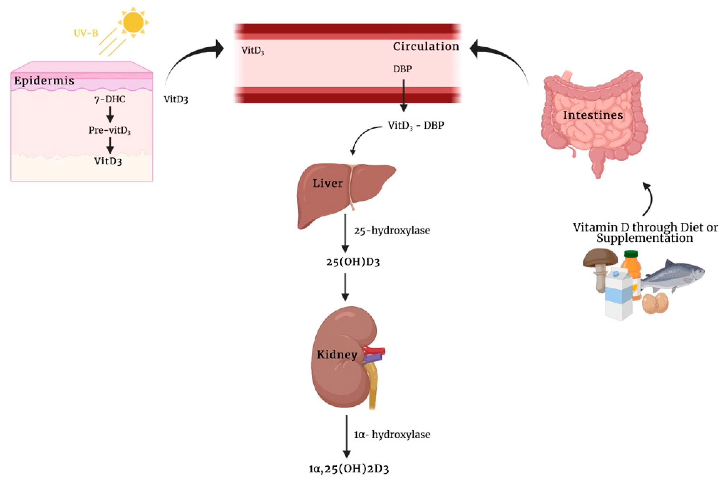
Vitamin D3 is typically produced in the skin, where provitamin D3 (7-dehydrocholesterol (7-DHC)) is transformed into previtamin D3 by the exposure to ultraviolet B (UVB) radiation, which then undergoes a thermal isomerization to produce vitamin D3 [4] (Figure 1). Subsequently, vitamin D3 enters the bloodstream and reaches the liver where it is promptly hydroxylated by the 25-hydroxylase, a member of cytochrome P450 enzyme subfamily, thus forming 25-hydroxyvitamin D3 or calcifediol (25(OH)D3) [5]. This compound shows an average plasma life of about three weeks, and therefore its serum levels are now currently used as indicators of the body vitamin D storage and status of patients. However, calcifediol is metabolically inactive and, once produced, it is secreted into blood bound up with vitamin D binding protein (DBP). In this case, the calcifediol needs a renal 1α-hydroxylation to obtain the biologically active form of vitamin D3, 1α,25(OH)2D3 [1] (Figure 1).
Vitamin D deficiency has been observed in different subpopulations, suggesting that it is an essential nutrient for people from certain ethnic groups, as well as people with specific lifestyle behaviors, such as elderly people, those who do not get enough sunlight exposure, and individuals with darker skin pigmentation [6]. Moreover, the relationship between the vitamin D status and the occurrence of several chronic illnesses (e.g., diabetes and cancer) proves that the physiological actions of this hormone go beyond the traditional effect on calcium homeostasis maintenance [7]. The presence of 1α-hydroxylases isoforms in extrarenal tissues implies that calcitriol could act as a paracrine or autocrine signal as well as its classical endocrine role [8].
Calcitriol acts through the binding with a VDR, which has been described in different species including humans, rats, and chickens [9]. It has a ligand-binding domain, named E-domain, a DNA-binding domain named C-domain, and an F-domain, which is one of the active domains. It acts by binding vitamin D-responsive elements (VDREs), repeated sequences located in the proximity of the start site of the target gene [10]. Upon the interaction between the ligand and the VDR, there is a conformational rearrangement of the receptor, preventing the bonding of the repressor. This results in the formation of a heterodimer between the VDR and the retinoid X receptor (RXR) at the VDREs, thus either initiating or suppressing the gene transcription whose protein products are involved in the controlling of calcium homeostasis (i.e., cytochrome P450 family 24 (CYP24), alkaline phosphatase (ALP), type I collagen (COL1A1), osteocalcin (bone gamma-carboxyglutamate protein, BGLAP), and transient receptor potential vanilloid type family member 6 (TRPV6)) [1,11,12]. Since VDR has been found in nearly all cell types, this could clarify the different vitamin D3 actions on several types of tissues [11].
In these circumstances, the increase or reduction of protein expression levels through gene transcription regulation from steroid hormones is the consequence of genomic steroid action [13]. These actions are not acute but delayed, because they require time for newly synthesized proteins and their processing [13]. In addition, molecules inhibiting of transcription and protein synthesis, such as cycloheximide or actinomycin D, may completely block these delayed effects if they do not take place at the time when steroid molecules are coupled to large proteins, thus preventing their cell entry [13].
Since the final active form was isolated and identified in 1971 [14], additional noncalcemic functions in the body have been described through further investigation of the importance of this hormone in the endocrine system.
In recent years, calcitriol, as well as its direct precursor, calcifediol, have been shown to exert rapid non-genomic steroid actions, indicating a more complex mechanism responsible for the wide range of actions of vitamin D [15,16].
The aim of this non-systematic is to provide an overview of the rapid, non-genomic effects of calcifediol and calcitriol, focusing on the mechanisms underlying these rapid responses that could lead to a better understanding of the vitamin D3 endocrine system, thus paving the way for the identification of potential novel therapeutic options for pathological conditions associated with vitamin D3 deficiency.
For this purpose, a rigorous search for literature on PubMed database has been conducted by employing different combinations of relevant keywords, including “vitamin D”, “1α,25(OH)2D3”, “25(OH)D3”, “non-genomic effects”, “VDR”, and “Pdia3”. All relevant studies released were selected and reviewed.
2. Rapid, Non-Genomic Steroid Actions
The first non-genomic action of steroid molecules was described in 1942 when Hans Selye observed the anesthetic effect of progesterone immediately after its injection into peritoneum of rats and mice differently from what was observed with respect to its main effect that took place only within hours after its administration [17]. Subsequently, Spach and Streeten demonstrated that Na+ ion variation occurred within few minutes after aldosterone administration in dog erythrocytes, offering new compelling evidence on non-genomic effects of this hormone because these cells lack nuclei, and therefore the observed in vitro effects might be attributable exclusively to its non-genomic mechanism [18]. However, these rapid hormone effects were not clarified until their recent recognition for several steroid hormones, including 1α,25(OH)2D3 [13].
Recently, 25(OH)D3, for a considerable time deemed only a metabolic precursor of 1α,25-(OH)2D3, has been proved to be an agonist ligand of VDR and capable of initiating rapid, non-genomic actions [16,19].
As opposed to their genomic counterpart, non-genomic rapid responses appear rapidly (within a range of seconds or minutes), are not susceptible to cycloheximide or actinomycin D, and also occur in response to steroids coupled to macromolecules which block their cell entering [20]. Therefore, a major difference for discerning between genomic and non-genomic actions is the time course and the sensitivity of transcription and protein synthesis inhibitors.
3. Mechanisms of Membrane-Associated Proteins for 1α,25(OH)2D3-Mediated Rapid, Non-Genomic Actions
Concerning the existence of non-genomic calcitriol actions, to our knowledge, the earliest observations for these actions were described by Nemere et al. in 1984 [21], who observed that this hormone was able to induce a rapid increase of intracellular calcium (Ca2+) concentrations both by promoting its release from intracellular stores and by stimulating its intestinal uptake in the vascularly perfused duodenum of normal, vitamin D-replete chicks. They observed that calcitriol significantly increased Ca2+ transport within 14 min compared with controls in a mechanism independent of genome activation and de novo protein synthesis. This rapid effect on transepithelial Ca2+ movement across the intestine has been termed transcaltachia.
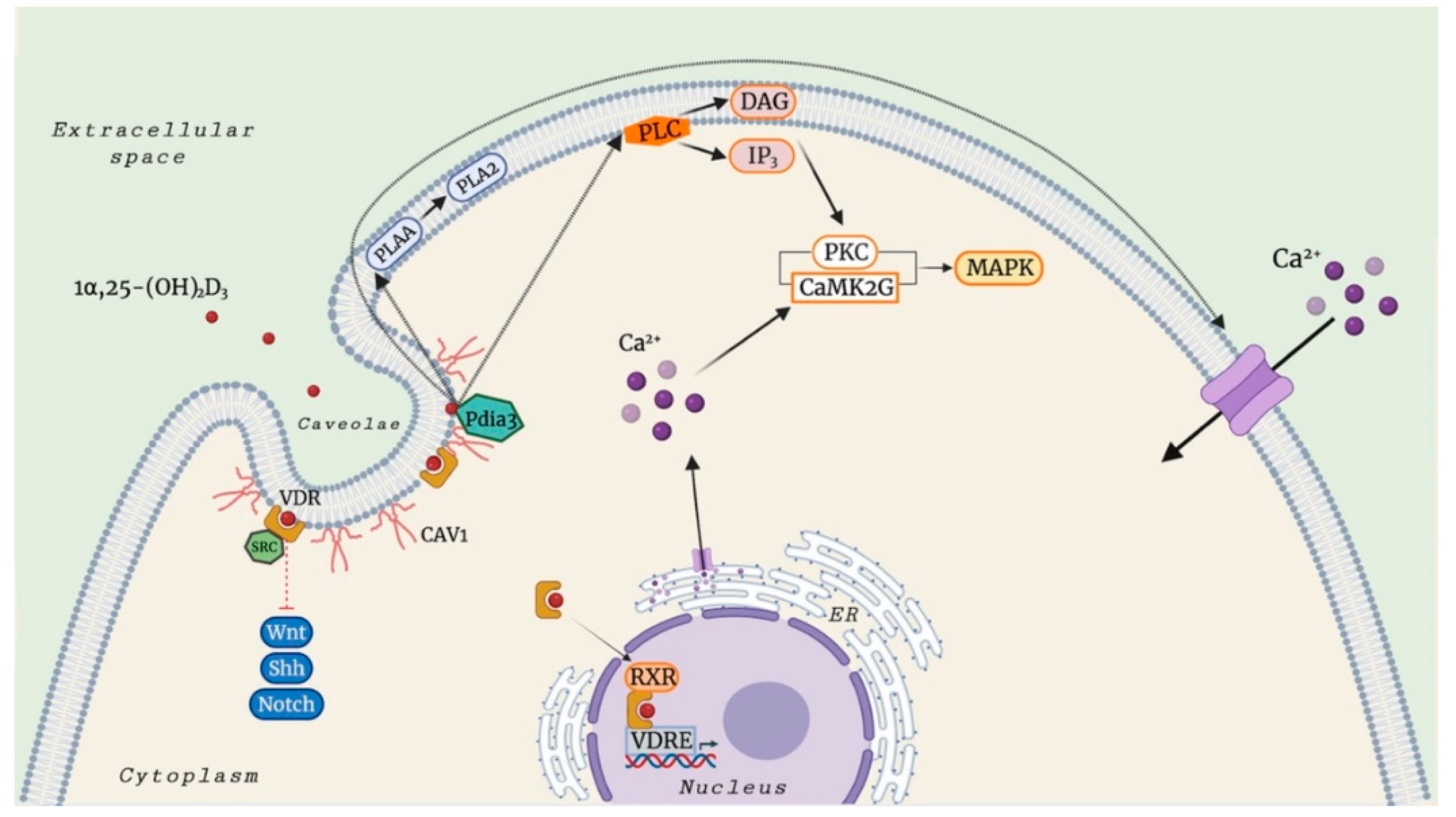
It is established that the biologically active vitamin D3 metabolite stimulates different signaling molecules, including phospholipase A2 (PLA2), phospholipase C (PLC), and phosphatidylinositol-3 kinase (PI3K), and promotes the generation of second messengers, such as Ca2+ ions, phosphatidylinositol (3,4,5)-trisphosphate (PIP3), and cyclic AMP (cAMP), culminating in the activation of several downstream protein kinases (protein kinase C (PKC), calcium/calmodulin-dependent protein kinase II gamma (CaMKIIG), Src, and mitogen-activated protein (MAP) kinases) [22,23,24,25,26]. In addition to the above-mentioned non-genomic actions of 1α,25(OH)2D3, this secosteroid hormone also mediates the opening of Ca2+, Cl−, and Pi channels.
Early studies performed by Norman and colleagues [27,28] suggested that a distinct membrane VDR could mediate non-genomic actions in response to 1α,25(OH)2D3. In their study, the authors reported that the biologically active form of vitamin D3 promotes the generation of non-genomic responses and, especially in the case of transcaltachia, in its 6-s-cis configuration. By contrast, binding of secosteroid in the 6-s-trans form could be responsible for genomic responses.
Further studies have shown that membrane-associated VDR (VDRm) could bind to proto-oncogene, non-receptor tyrosine kinase Src, and caveolin 1 (CAV1) in caveolae-enriched plasma membranes, thereby participating in the regulation of many signaling pathways, such as sonic hedgehog (Shh) [29,30,31,32,33,34], Wnt [35,36,37,38], and Notch [39,40,41].
In 1990, Civitelli et al. [42] observed an acute and transient rise in calcium mobilization in osteoblastic osteosarcoma cell line ROS 17/2.8 after a 1α,25(OH)2D3 stimulation in a mechanism independent of genomic activation, through both the influx of extracellular Ca2+ and release of Ca2+ from intracellular stores. The active form of vitamin D3 also produces a significant increase in the production of diacylglycerol (DAG) and inositol 1,4,5-trisphosphate (IP3), resulting in the activation of PLC.
This rapid effect was also observed in primary muscle cell cultures isolated from a chicken embryonic heart [43]. In particular, 1α,25(OH)2D3 induces a fast increase of both tissue Ca2+ uptake and cAMP levels within 10 min in primary myocytes. Moreover, authors found that this effect was inhibited by a specific protein kinase A (PKA) suppressor, suggesting that the regulation of Ca2+ ion channel activity by the biologically active form of vitamin D3 was mediated by the second messenger cAMP.
Baran et al. [44] demonstrated that the secosteroid 1α,25(OH)2D3 not only evokes a rapid opening of Ca2+ channels but also a rapid activation of phospholipase C (PLC) in ROS 24/1 osteoblastic cells lacking the VDR, implying that these effects occur independently of the VDR signaling mechanisms. This observation was also confirmed in vivo, where Boyan et al. [45] observed that 1α,25(OH)2D3 is involved in the regulation of protein kinase C (PKC) activity through rapid membrane-associated mechanisms in cultured costochondral chondrocytes derived from VDR−/− mice.
This could suggest that, in addition to membrane-associated VDR, the existence of other membrane receptors in conjunction with vitamin D might be essential for rapid, non-genomic effects of 1α,25(OH)2D3.
Given the highly liposoluble nature of vitamin D3, 1α,25(OH)2D3, can penetrate biological membranes, and when inside the cells, may interact with heat shock proteins (HSPs) so as to be transferred to the nucleus and the mitochondria [46]. Alternatively, vitamin D3 metabolites bound to a specific vitamin D3 binding protein (DBP) can undergo endocytosis through an LDL receptor-related protein 2 (LRP2)- and cubilin (CUBN)-mediated mechanism. Subsequently, 25(OH)D3 bound to DBP is transported into the proximal tubule epithelium of the kidneys via megalin-mediated endocytosis, where it undergoes a hydroxylation step to transform into 1α,25(OH)2D3 [47,48]. In this light, additional studies should be performed to clarify the physiological significance and the potential mechanistic aspect for vitamin D3 transporters.
One of best membrane-associated proteins able to bind vitamin D3 compounds is the protein disulfide isomerase family A member 3 (Pdia3), also known as 1α,25(OH)2D3-membrane-associated rapid response to steroid (MAARS), which has been described as a crucial protein in 1α,25(OH)2D3-initiated rapid membrane non-genomic signaling pathways [49].
This protein was first purified in the study of Nemere et al. [50], describing the existence of a putative plasmalemmal receptor for the biologically active form of vitamin D3 involved in the transcaltachia on the basal-lateral membranes (BLM) of chick intestinal epithelium. This conclusion was supported by the observation that an altered, but still present, specific binding for [3H] 1α,25(OH)2D3 was discovered in membrane fractions purified from vitamin D-deficient chicks with respect to the corresponding fractions obtained from normal animals. In addition, the BLM-VDR exhibited down-regulation of specific [3H] 1α,25(OH)2D3 binding upon exposure to nonradioactive 1α,25(OH)2D3.
Later studies showed that this candidate plasmalemmal receptor essential for the rapid responses by 1α,25(OH)2D3 is Pdia3 [51,52]. Its primary function is to catalyze the formation, reduction, and isomerization of disulfide bonds, interacting with lectin-like molecular chaperones, calnexin (CANX) and calreticulin (CALR), to ensure the correct folding of newly synthesized glycoproteins [53].
Pdia3 has been identified in the cell membrane, cytosol, mitochondria, and the nucleus and has demonstrated multiple distinct functions, such as the protection against oxidative stress and the prevention of diseases associated with the accumulation of unfolded/misfolded proteins [54].
One remarkable function of Pdia3 is its involvement in one of the most prominent rapid actions of 1α,25(OH)2D3, namely transcaltachia. Importantly, it has been observed that the rapid increase in intracellular Ca2+ levels in response to subnanomolar concentrations of 1α,25(OH)2D3 involves the interaction between Pdia3 and CAV1, the principal component of the caveolae plasma membranes and small plasma membrane invaginations [23,55,56].
Moreover, the interaction between the biologically active form of vitamin D3 and Pdia3 also has a significant implication for the cell protection against UV-caused thymine dimer formation [57], the activation of PKC signaling transduction pathway [58], and the repression of tumor necrosis factor receptor signaling produced by a rapid rise in intracellular Ca2+ concentration in aortic smooth muscle cells [59].
Interestingly, further studies have shown that Pdia3 protein binding of 1α,25(OH)2D3 is also involved in the activation of PLA2 via PLA2 activating protein (PLAA) [49], MAPK1, and MAPK2 via the regulation of CaMKIIG, PLC, PLA2, and PKC [23,55], and Wnt family member 5A (Wnt5A) [60].
Collectively, the above-mentioned studies investigating rapid, non-genomic actions in response to 1α,25(OH)2D3 support the hypothesis that Pdia3 is a crucial component of the machinery responsible for mediating these vitamin D3 activities.
Consistent with data derived from in vitro studies, it has been reported that Pdia3 is involved in vitamin D-mediated actions in vivo. Despite the observation of the fact that homozygous Pdia3 deletion causes embryonic lethality in mice, the animals with a deleted allele develop skeletal abnormalities, thus suggesting its function in maintaining calcium homeostasis [61,62]. In this regard, an analysis of enterocytes isolated from Pdia3 knockout mice showed a reduction in rapid, non-genomic effects following 1α,25(OH)2D3, such as the PKA signaling pathway and calcium uptake [63]. Moreover, the loss of Pdia3 in deficient mice resulted in significant attenuation of 1α,25(OH)2D3-related PKC activation and skeletal abnormalities [61]. Importantly, a study by Boyan et al. revealed that cultured chondrocytes isolated from VDR knockout mice retaining Pdia3 gene expression showed a rapid increase in PKC activity after 1α,25(OH)2D3 treatment [45].
Based on this evidence, Pdia3 could be involved in the activation of rapid non-genomic actions mediated by 1α,25(OH)2D3, which in turn might have a significant impact on musculoskeletal biology regulation, including calcium absorption through the intestinal epithelial and skeletal development.
However, crystallographic studies have not confirmed any binding site for the biologically active form of vitamin D3 in the partial structure of Pdia3 [64], although it has been assumed that its activity could be essential for the rapid, non-genomic actions of this secosteroid hormone. In this respect, not only was it postulated that 1α,25(OH)2D3 could interact with Pdia3 through its a’ domain, which has been shown to be essential for triggering non-genomic actions, but also that Pdia3 could function as a molecular chaperone for VDR, DPB, or other unknown membrane-associated proteins [64,65].
In Table 1, we summarized the 1α,25(OH)2D3-mediated rapid, non-genomic actions.
4. 25(OH)D3-Mediated Rapid, Non-Genomic Actions
In the study conducted by Lou et al. [19], it was assumed that calcifediol is an agonist ligand of VDR with anti-proliferative effects and gene regulatory functions, despite the fact it binds to VDR with a lower affinity compared to the biologically active metabolite of vitamin D3.
Based on this interesting observation, our previous research article [16] aimed to evaluate whether this metabolite could initiate rapid, non-genomic pathways, such as an increase in intracellular Ca2+ concentrations, in line with what was established for calcitriol. We observed for the first time that calcifediol produces a rapid increase in Ca2+ levels in mesenchymal stem cells derived from human adipose tissue (hADMSCs), although at a higher concentration compared to those present in normal physiological conditions. In this regard, calcifediol at subnanomolar concentrations has been revealed incapable of triggering an increase of intracellular Ca2+ concentration in human spermatozoa although an evident but delayed effect was found at higher dose [66,67].
Another study by Asano et al. showed an interesting non-genomic mechanism of 25(OH)D3, where this compound could regulate lipogenesis, thereby reducing the risk of metabolic disease-associated complications by altering sterol regulatory element-binding proteins (SREBPs) activation via the ubiquitin-mediated proteasomal degradation of SREBP cleavage-activating protein (SCAP) [68].
In Table 2, we summarized the 25(OH)D3-mediated rapid, non-genomic actions.
5. Discussion
In recent years, 1α,25(OH)2D3, the biologically active metabolite of vitamin D3, has attracted attention due to its involvement in several biological processes, including the regulation of the serum levels of calcium and phosphate, as well as its influence on bone and mineral metabolism.
There is now compelling evidence that 1α,25(OH)2D3 affects the target cells through genomic pathways and membrane receptor-mediated rapid, non-genomic responses. This latter mechanism has also been described for virtually all the steroid molecules, such as aldosterone, testosterone, estrogens, and cortisol [16,49,69].
Recently, even the direct metabolic precursor of 1α,25(OH)2D3, named calcifediol, it has been revealed able to activate rapid, nontranscriptional actions, such as an acute and sustained rise in intracellular Ca2+ levels, similarly to that observed with the biologically active form of vitamin D3 [16,70].
As described in this review, although growing evidence has led to a significant knowledge concerning calcifediol and calcitriol rapid, non-genomic activities, their impact on physiological processes needs to be clarified. In fact, it is difficult to identify vitamin D-deficiency-associated diseases which occur exclusively due to aberrations of its rapid actions.
The vitamin D3 endocrine system is primarily involved in different biological processes that maintain calcium and bone homeostasis, and presumably, it could not benefit from rapid membrane-mediated non-genomic responses. However, these actions could have important physiological implications for some processes, in particular on the protection of cells against DNA damage caused by solar UV radiation exposure and intestinal Ca2+ absorption.
In this regard, one of the most noticeable rapid actions of 1α,25(OH)2D3 is transcaltachia, the rapid stimulation of intestinal Ca2+ transport. However, further studies are needed to link this rapid physiological manifestation of 1α,25(OH)2D3 with meal feeding and therefore with calcium absorption physiology.
Accumulating evidence has proposed that the rapid, non-genomic actions could positively or negatively affect the genomic function mediated by 1α,25(OH)2D3 [15]. In fact, the secosteroid hormone could activate various signaling molecules involved in several rapid transduction pathways (i.e., PKC, PI3K, and PLA2) which could affect gene expression either through transcriptional regulatory elements present in the promoter or by using activated VDR as a substrate [15,71]. Moreover, this cross-talk could regulate both the efficacy and potency of genomic function [71].
Although the subject of vitamin D-mediated rapid non-genomic effects has been explored in recent years, important questions should be taken into account in the foreseeable future.
The current data come from a combination of results of studies performed on different species and multiple cells. Since these actions could be dependent on cell types, cell cycle stage and species, future studies should be carried out to elucidate the mechanisms underpinning these effects in a wide range of cells.
In addition, it should be considered that most of the in vitro studies about non-genomic effects of vitamin D3 compounds use higher concentrations than those subnanomolar existing under normal physiological circumstances [16,49,66]. This finding could be because secosteroid-related rapid, non-genomic actions could require higher concentrations than the genomic counterpart.
It has now been established that the VDR is merely the only protein that binds with the active form of vitamin D3 at a high level of affinity. Moreover, it is scattered in more than 38 tissues; this evidence could explain the diversity of actions on different tissues [1,71].
The vitamin D-endocrine system is complex, and there are several conversion and transport processes necessary to produce compounds in the skin by UV radiation until the synthesis of the active metabolite of vitamin D3 is reached.
During its production, besides undergoing the action of specific enzymes, vitamin D3 compounds interact with not only VDR but also other binding/transport proteins, such as DPB in the circulation and heat shock protein family A (HSPA) when inside the cells.
A growing body of evidence suggests that Pdia3, the best outlined membrane-associated protein capable of binding with vitamin D3 compounds, could play a key role in 1α,25(OH)2D3-mediated non-genomic responses [23,55,56]. Although studies regarding the crystal structure of Pdia3 did not reveal any binding pocket for the secosteroid hormone, its activity is critical for the rapid actions of 1α,25(OH)2D3. In this respect, even though it might not bind directly 1α,25(OH)2D3, this protein could serve as a molecular chaperone for DBP or VDR [49].
However, little is known about the presence of other nonclassical membrane receptors that stand at the beginning of membrane-based rapid responses to vitamin D3 and its metabolites as well as the existence of alternative cell signalling transduction pathways. Their comprehension could help in understanding of the non-genomic effects of vitamin D that have not been settled yet.
In summary, a significant progress has been made in this field in recent years, shifting the understanding of vitamin D3 beyond the originally reported role in calcium homeostasis and prevention of rickets in children and osteomalacia in adults. It has now been established not only that the vitamin D-related action mechanism is not based solely on its genomic activity, but also that either calcitriol or calcifediol can promote the generation of rapid, non-genomic effects, as well as described for other steroid hormones that can impact different physiological processes. There is convincing evidence that VDR has an essential role in these rapid responses. As described in this review, other proteins, such as Pdia3, may also bind to 1α,25(OH)2D3, although with a lower affinity compared to the VDR, thereby playing a crucial role in rapid membrane non-genomic response to vitamin D3 (Figure 2).
Further studies will need to be performed to better understand the mode of action sustaining these rapid responses to provide new avenues for the development of novel therapeutic approaches able to modulate the non-genomic actions of vitamin D, especially for people who are vitamin D3-deficient.
In fact, there are limited human data available for the non-genomic actions of vitamin D in vivo after a dietary supplementation and therefore additional investigations are required to elucidate whether vitamin D3 metabolites could elicit the same rapid nontranscriptional effects that have been reported under in vitro conditions.
Author Contributions
M.L.B., S.D. and G.P. conceived the idea; S.D., G.P. and M.L.B. wrote the manuscript in consultation with C.A., I.F. and F.M.; T.I. and M.L.B. revised the manuscript. All authors have read and agreed to the published version of the manuscript.
Funding
This research received no external funding.
Institutional Review Board Statement
Not applicable.
Informed Consent Statement
Not applicable.
Data Availability Statement
Not applicable.
Acknowledgments
All authors are indebted to FIRMO Foundation for secretarial support.
Conflicts of Interest
The authors declare no conflict of interest.
References
- Gil, Á.; Plaza-Diaz, J.; Mesa, M.D. Vitamin D: Classic and Novel Actions. Ann. Nutr. Metab. 2018, 72, 87–95. [Google Scholar] [CrossRef] [PubMed]
- Clinckspoor, I.; Verlinden, L.; Mathieu, C.; Bouillon, R.; Verstuyf, A.; Decallonne, B. Vitamin D in thyroid tumorigenesis and development. Prog. Histochem. Cytochem. 2013, 48, 65–98. [Google Scholar] [CrossRef] [PubMed]
- Naveh-Many, T.; Marx, R.; Keshet, E.; Pike, J.W.; Silver, J. Regulation of 1,25-dihydroxyvitamin D3 receptor gene expression by 1,25-dihydroxyvitamin D3 in the parathyroid in vivo. J. Clin. Investig. 1990, 86, 1968–1975. [Google Scholar] [CrossRef] [PubMed] [Green Version]
- Valero Zanuy, M.; Hawkins Carranza, F. Metabolismo, fuentes endógenas y exógenas de vitamina D. Rev. Esp. Enferm. Metab. Oseas 2007, 16, 63–70. [Google Scholar] [CrossRef]
- DeLuca, H.F. The Metabolism and Functions of Vitamin D. In Steroid Hormone Resistance: Mechanisms and Clinical Aspects; Chrousos, G.P., Loriaux, D.L., Lipsett, M.B., Eds.; Advances in Experimental Medicine and Biology; Springer: Boston, MA, USA, 1986; pp. 361–375. ISBN 978-1-4684-5101-6. [Google Scholar]
- Martin, C.E.; Veysey, M.; Yates, Z.R.; Lucock, M.D. Vitamin D: Genetics, Environment & Health. J. Food Nutr. Disord. 2014, 3, 1–19. [Google Scholar]
- Holick, M.F. Vitamin D: Importance in the prevention of cancers, type 1 diabetes, heart disease, and osteoporosis. Am. J. Clin. Nutr. 2004, 79, 362–371. [Google Scholar] [CrossRef] [Green Version]
- Zehnder, D.; Bland, R.; Williams, M.C.; McNinch, R.W.; Howie, A.J.; Stewart, P.M.; Hewison, M. Extrarenal expression of 25-hydroxyvitamin d(3)-1 alpha-hydroxylase. J. Clin. Endocrinol. Metab. 2001, 86, 888–894. [Google Scholar]
- DeLuca, H.F. Overview of general physiologic features and functions of vitamin D. Am. J. Clin. Nutr. 2004, 80, 1689S–1696S. [Google Scholar] [CrossRef] [Green Version]
- Feldman, D.; Malloy, P.J.; Gross, C. Chapter 9—Vitamin D: Biology, Action, and Clinical Implications. In Osteoporosis, 2nd ed.; Marcus, R., Feldman, D., Kelsey, J., Eds.; Academic Press: San Diego, LA, USA, 2001; pp. 257–303. ISBN 978-0-12-470862-4. [Google Scholar]
- Bouillon, R.; Carmeliet, G.; Verlinden, L.; van Etten, E.; Verstuyf, A.; Luderer, H.F.; Lieben, L.; Mathieu, C.; Demay, M. Vitamin D and human health: Lessons from vitamin D receptor null mice. Endocr. Rev. 2008, 29, 726–776. [Google Scholar] [CrossRef]
- DeLuca, H.F. Evolution of our understanding of vitamin D. Nutr. Rev. 2008, 66, S73–S87. [Google Scholar] [CrossRef]
- Schmidt, B.M.; Gerdes, D.; Feuring, M.; Falkenstein, E.; Christ, M.; Wehling, M. Rapid, nongenomic steroid actions: A new age? Front. Neuroendocrinol. 2000, 21, 57–94. [Google Scholar] [CrossRef] [PubMed]
- DeLuca, H.F.; Holick, M.F.; Schnoes, H.K.; Suda, T.; Cousins, R.J. Isolation and identification of 1,25-dihydroxycholecalciferol. A metabolite of vitamin D active in intestine. Biochemistry 1971, 10, 2799–2804. [Google Scholar] [CrossRef] [PubMed]
- Hii, C.S.; Ferrante, A. The Non-Genomic Actions of Vitamin D. Nutrients 2016, 8, 135. [Google Scholar] [CrossRef] [PubMed] [Green Version]
- Donati, S.; Palmini, G.; Romagnoli, C.; Aurilia, C.; Miglietta, F.; Falsetti, I.; Marini, F.; Zonefrati, R.; Galli, G.; Marcucci, G.; et al. In Vitro Non-Genomic Effects of Calcifediol on Human Preosteoblastic Cells. Nutrients 2021, 13, 4227. [Google Scholar] [CrossRef] [PubMed]
- Selye, H. Correlations between the chemical structure and the pharmacological actions of the steroids. Endocrinology 1942, 30, 437–453. [Google Scholar] [CrossRef]
- Spach, C.; Streeten, D.H.P. Retardation of Sodium Exchange in Dog Erythrocytes by Physiological Concentrations of Aldosterone, In Vitro. J. Clin. Investig. 1964, 43, 217–227. [Google Scholar] [CrossRef] [Green Version]
- Lou, Y.-R.; Molnár, F.; Peräkylä, M.; Qiao, S.; Kalueff, A.V.; St-Arnaud, R.; Carlberg, C.; Tuohimaa, P. 25-Hydroxyvitamin D(3) is an agonistic vitamin D receptor ligand. J. Steroid Biochem. Mol. Biol. 2010, 118, 162–170. [Google Scholar] [CrossRef]
- Gerdes, D.; Christ, M.; K. Haseroth, K.; Notzon, A.; Falkenstein, E.; Wehling, M. Nongenomic Actions of Steroids-From the Laboratory to Clinical Implications. J. Pediat. Endocrinol. Metab. 2000, 13, 853–878. [Google Scholar] [CrossRef]
- Nemere, I.; Yoshimoto, Y.; Norman, A.W. Calcium transport in perfused duodena from normal chicks: Enhancement within fourteen minutes of exposure to 1,25-dihydroxyvitamin D3. Endocrinology 1984, 115, 1476–1483. [Google Scholar] [CrossRef]
- Fleet, J.C. Rapid, Membrane-Initiated Actions of 1,25 Dihydroxyvitamin D: What Are They and What Do They Mean? J. Nutr. 2004, 134, 3215–3218. [Google Scholar] [CrossRef]
- Doroudi, M.; Schwartz, Z.; Boyan, B.D. Membrane-mediated actions of 1,25-dihydroxy vitamin D3: A review of the roles of phospholipase A2 activating protein and Ca (2+)/calmodulin-dependent protein kinase II. J. Steroid Biochem. Mol. Biol. 2015, 147, 81–84. [Google Scholar] [CrossRef] [PubMed] [Green Version]
- Dwivedi, P.P.; Hii, C.S.T.; Ferrante, A.; Tan, J.; Der, C.J.; Omdahl, J.L.; Morris, H.A.; May, B.K. Role of MAP kinases in the 1,25-dihydroxyvitamin D3-induced transactivation of the rat cytochrome P450C24 (CYP24) promoter. Specific functions for ERK1/ERK2 and ERK5. J. Biol. Chem. 2002, 277, 29643–29653. [Google Scholar] [CrossRef] [PubMed] [Green Version]
- Nutchey, B.K.; Kaplan, J.S.; Dwivedi, P.P.; Omdahl, J.L.; Ferrante, A.; May, B.K.; Hii, C.S.T. Molecular action of 1,25-dihydroxyvitamin D3 and phorbol ester on the activation of the rat cytochrome P450C24 (CYP24) promoter: Role of MAP kinase activities and identification of an important transcription factor binding site. Biochem. J. 2005, 389, 753–762. [Google Scholar] [CrossRef] [PubMed] [Green Version]
- Dwivedi, P.; Gao, X.; Tan, J.; Evdokiou, A.; Ferrante, A.; Morris, H.; May, B.; Hii, C. A role for the phosphatidylinositol 3-kinase--protein kinase C zeta-Sp1 pathway in the 1,25-dihydroxyvitamin D3 induction of the 25-hydroxyvitamin D3 24-hydroxylase gene in human kidney cells. Cell. Signal. 2010, 22, 543–552. [Google Scholar] [CrossRef] [PubMed]
- Norman, A.W. Vitamin D Receptor: New Assignments for an Already Busy Receptor. Endocrinology 2006, 147, 5542–5548. [Google Scholar] [CrossRef] [PubMed] [Green Version]
- Dormanen, M.C.; Bishop, J.E.; Hammond, M.W.; Okamura, W.H.; Nemere, I.; Norman, A.W. Nonnuclear effects of the steroid hormone 1 alpha,25(OH)2-vitamin D3: Analogs are able to functionally differentiate between nuclear and membrane receptors. Biochem. Biophys. Res. Commun. 1994, 201, 394–401. [Google Scholar] [CrossRef]
- Bikle, D.D.; Jiang, Y.; Nguyen, T.; Oda, Y.; Tu, C. Disruption of Vitamin D and Calcium Signaling in Keratinocytes Predisposes to Skin Cancer. Front. Physiol. 2016, 7, 296. [Google Scholar] [CrossRef] [Green Version]
- Bandera Merchan, B.; Morcillo, S.; Martin-Nuñez, G.; Tinahones, F.J.; Macías-González, M. The role of vitamin D and VDR in carcinogenesis: Through epidemiology and basic sciences. J. Steroid Biochem. Mol. Biol. 2017, 167, 203–218. [Google Scholar] [CrossRef]
- Hadden, M.K. Hedgehog and Vitamin D Signaling Pathways in Development and Disease. Vitam. Horm. 2016, 100, 231–253. [Google Scholar]
- Lisse, T.S.; Saini, V.; Zhao, H.; Luderer, H.F.; Gori, F.; Demay, M.B. The Vitamin D Receptor Is Required for Activation of cWnt and Hedgehog Signaling in Keratinocytes. Mol. Endocrinol. 2014, 28, 1698–1706. [Google Scholar] [CrossRef] [Green Version]
- Teichert, A.E.; Elalieh, H.; Elias, P.M.; Welsh, J.; Bikle, D.D. Overexpression of hedgehog signaling is associated with epidermal tumor formation in vitamin D receptor-null mice. J. Investig. Dermatol. 2011, 131, 2289–2297. [Google Scholar] [CrossRef] [PubMed] [Green Version]
- Teichert, A.; Elalieh, H.; Bikle, D. Disruption of the hedgehog signaling pathway contributes to the hair follicle cycling deficiency in Vdr knockout mice. J. Cell. Physiol. 2010, 225, 482–489. [Google Scholar] [CrossRef] [PubMed] [Green Version]
- Tapia, C.; Suares, A.; De Genaro, P.; González-Pardo, V. In vitro studies revealed a downregulation of Wnt/β-catenin cascade by active vitamin D and TX 527 analog in a Kaposi’s sarcoma cellular model. Toxicol. In Vitr. 2020, 63, 104748. [Google Scholar] [CrossRef] [PubMed]
- Muralidhar, S.; Filia, A.; Nsengimana, J.; Poźniak, J.; O’Shea, S.J.; Diaz, J.M.; Harland, M.; Randerson-Moor, J.A.; Reichrath, J.; Laye, J.P.; et al. Vitamin D–VDR Signaling Inhibits Wnt/β-Catenin–Mediated Melanoma Progression and Promotes Antitumor Immunity. Cancer Res. 2019, 79, 5986–5998. [Google Scholar] [CrossRef] [PubMed] [Green Version]
- Tang, L.; Fang, W.; Lin, J.; Li, J.; Wu, W.; Xu, J. Vitamin D protects human melanocytes against oxidative damage by activation of Wnt/β-catenin signaling. Lab. Investig. 2018, 98, 1527–1537. [Google Scholar] [CrossRef] [PubMed]
- Larriba, M.J.; González-Sancho, J.M.; Bonilla, F.; Muñoz, A. Interaction of vitamin D with membrane-based signaling pathways. Front. Physiol. 2014, 5, 60. [Google Scholar] [CrossRef] [PubMed] [Green Version]
- Wang, H.; Wang, X.; Xu, L.; Zhang, J.; Cao, H. A molecular sub-cluster of colon cancer cells with low VDR expression is sensitive to chemotherapy, BRAF inhibitors and PI3K-mTOR inhibitors treatment. Aging 2019, 11, 8587–8603. [Google Scholar] [CrossRef] [PubMed]
- Olsson, K.; Saini, A.; Strömberg, A.; Alam, S.; Lilja, M.; Rullman, E.; Gustafsson, T. Evidence for Vitamin D Receptor Expression and Direct Effects of 1α,25(OH)2D3 in Human Skeletal Muscle Precursor Cells. Endocrinology 2016, 157, 98–111. [Google Scholar] [CrossRef] [Green Version]
- Fuchs, E.; Raghavan, S. Getting under the skin of epidermal morphogenesis. Nat. Rev. Genet. 2002, 3, 199–209. [Google Scholar] [CrossRef]
- Civitelli, R.; Kim, Y.S.; Gunsten, S.L.; Fujimori, A.; Huskey, M.; Avioli, L.V.; Hruska, K.A. Nongenomic Activation of the Calcium Message System by Vitamin D Metabolites in Osteoblast-like Cells. Endocrinology 1990, 127, 2253–2262. [Google Scholar] [CrossRef]
- Selles, J.; Boland, R. Evidence on the participation of the 3′,5′-cyclic AMP pathway in the non-genomic action of 1,25-dihydroxy-vitamin D3 in cardiac muscle. Mol. Cell. Endocrinol. 1991, 82, 229–235. [Google Scholar] [CrossRef]
- Baran, D.T.; Ray, R.; Sorensen, A.M.; Honeyman, T.; Holick, M.F. Binding characteristics of a membrane receptor that recognizes 1 alpha,25-dihydroxyvitamin D3 and its epimer, 1 beta,25-dihydroxyvitamin D3. J. Cell. Biochem. 1994, 56, 510–517. [Google Scholar] [CrossRef] [PubMed]
- Boyan, B.D.; Sylvia, V.L.; McKinney, N.; Schwartz, Z. Membrane actions of vitamin D metabolites 1alpha,25(OH)2D3 and 24R,25(OH)2D3 are retained in growth plate cartilage cells from vitamin D receptor knockout mice. J. Cell. Biochem. 2003, 90, 1207–1223. [Google Scholar] [CrossRef] [PubMed]
- Wu, S.; Ren, S.; Chen, H.; Chun, R.F.; Gacad, M.A.; Adams, J.S. Intracellular vitamin D binding proteins: Novel facilitators of vitamin D-directed transactivation. Mol. Endocrinol. 2000, 14, 1387–1397. [Google Scholar] [CrossRef] [PubMed]
- Chapron, B.D.; Chapron, A.; Phillips, B.; Okoli, M.C.; Shen, D.D.; Kelly, E.J.; Himmelfarb, J.; Thummel, K.E. Reevaluating the role of megalin in renal vitamin D homeostasis using a human cell-derived microphysiological system. ALTEX 2018, 35, 504–515. [Google Scholar] [CrossRef] [PubMed] [Green Version]
- Nykjaer, A.; Fyfe, J.C.; Kozyraki, R.; Leheste, J.R.; Jacobsen, C.; Nielsen, M.S.; Verroust, P.J.; Aminoff, M.; de la Chapelle, A.; Moestrup, S.K.; et al. Cubilin dysfunction causes abnormal metabolism of the steroid hormone 25(OH) vitamin D (3). Proc. Natl. Acad. Sci. USA 2001, 98, 13895–13900. [Google Scholar] [CrossRef] [PubMed] [Green Version]
- Zmijewski, M.A.; Carlberg, C. Vitamin D receptor(s): In the nucleus but also at membranes? Exp. Dermatol. 2020, 29, 876–884. [Google Scholar] [CrossRef] [PubMed]
- Nemere, I.; Dormanen, M.C.; Hammond, M.W.; Okamura, W.H.; Norman, A.W. Identification of a specific binding protein for 1 alpha,25-dihydroxyvitamin D3 in basal-lateral membranes of chick intestinal epithelium and relationship to transcaltachia. J. Biol. Chem. 1994, 269, 23750–23756. [Google Scholar] [CrossRef]
- Nemere, I.; Farach-Carson, M.C.; Rohe, B.; Sterling, T.M.; Norman, A.W.; Boyan, B.D.; Safford, S.E. Ribozyme knockdown functionally links a 1,25(OH)2D3 membrane binding protein (1,25D3-MARRS) and phosphate uptake in intestinal cells. Proc. Natl. Acad. Sci. USA 2004, 101, 7392–7397. [Google Scholar] [CrossRef] [Green Version]
- Nemere, I.; Safford, S.E.; Rohe, B.; DeSouza, M.M.; Farach-Carson, M.C. Identification and characterization of 1,25D3-membrane-associated rapid response, steroid (1,25D3-MARRS) binding protein. J. Steroid Biochem. Mol. Biol. 2004, 89–90, 281–285. [Google Scholar] [CrossRef]
- Hettinghouse, A.; Liu, R.; Liu, C.-J. Multifunctional molecule ERp57: From cancer to neurodegenerative diseases. Pharmacol. Ther. 2018, 181, 34–48. [Google Scholar] [CrossRef] [PubMed]
- Mahmood, F.; Xu, R.; Awan, M.U.N.; Song, Y.; Han, Q.; Xia, X.; Zhang, J. PDIA3: Structure, functions and its potential role in viral infections. Biomed. Pharmacother. 2021, 143, 112110. [Google Scholar] [CrossRef] [PubMed]
- Doroudi, M.; Chen, J.; Boyan, B.D.; Schwartz, Z. New insights on membrane mediated effects of 1α,25-dihydroxy vitamin D3 signaling in the musculoskeletal system. Steroids 2014, 81, 81–87. [Google Scholar] [CrossRef] [PubMed]
- Doroudi, M.; Schwartz, Z.; Boyan, B.D. Phospholipase A2 activating protein is required for 1α,25-dihydroxyvitamin D3 dependent rapid activation of protein kinase C via Pdia3. J. Steroid Biochem. Mol. Biol. 2012, 132, 48–56. [Google Scholar] [CrossRef] [PubMed]
- Sequeira, V.B.; Rybchyn, M.S.; Tongkao-On, W.; Gordon-Thomson, C.; Malloy, P.J.; Nemere, I.; Norman, A.W.; Reeve, V.E.; Halliday, G.M.; Feldman, D.; et al. The role of the vitamin D receptor and ERp57 in photoprotection by 1α,25-dihydroxyvitamin D3. Mol. Endocrinol. 2012, 26, 574–582. [Google Scholar] [CrossRef] [Green Version]
- Khanal, R.; Nemere, I. Membrane receptors for vitamin D metabolites. Crit. Rev. Eukaryot. Gene Expr. 2007, 17, 31–47. [Google Scholar] [CrossRef]
- Yang, W.S.; Yu, H.; Kim, J.J.; Lee, M.J.; Park, S.-K. Vitamin D-induced ectodomain shedding of TNF receptor 1 as a nongenomic action: D3 vs D2 derivatives. J. Steroid Biochem. Mol. Biol. 2016, 155, 18–25. [Google Scholar] [CrossRef]
- Doroudi, M.; Olivares-Navarrete, R.; Boyan, B.D.; Schwartz, Z. A review of 1α,25(OH)2D3 dependent Pdia3 receptor complex components in Wnt5a non-canonical pathway signaling. J. Steroid Biochem. Mol. Biol. 2015, 152, 84–88. [Google Scholar] [CrossRef]
- Wang, Y.; Chen, J.; Lee, C.S.D.; Nizkorodov, A.; Riemenschneider, K.; Martin, D.; Hyzy, S.; Schwartz, Z.; Boyan, B.D. Disruption of Pdia3 gene results in bone abnormality and affects 1alpha,25-dihydroxy-vitamin D3-induced rapid activation of PKC. J. Steroid Biochem. Mol. Biol. 2010, 121, 257–260. [Google Scholar] [CrossRef]
- Garbi, N.; Tanaka, S.; Momburg, F.; Hämmerling, G.J. Impaired assembly of the major histocompatibility complex class I peptide-loading complex in mice deficient in the oxidoreductase ERp57. Nat. Immunol. 2006, 7, 93–102. [Google Scholar] [CrossRef]
- Nemere, I.; Garbi, N.; Hämmerling, G.J.; Khanal, R.C. Intestinal cell calcium uptake and the targeted knockout of the 1,25D3-MARRS (membrane-associated, rapid response steroid-binding) receptor/PDIA3/Erp57. J. Biol. Chem. 2010, 285, 31859–31866. [Google Scholar] [CrossRef] [Green Version]
- Chen, J.; Lobachev, K.S.; Grindel, B.J.; Farach-Carson, M.C.; Hyzy, S.L.; El-Baradie, K.B.; Olivares-Navarrete, R.; Doroudi, M.; Boyan, B.D.; Schwartz, Z. Chaperone properties of pdia3 participate in rapid membrane actions of 1α,25-dihydroxyvitamin d3. Mol. Endocrinol. 2013, 27, 1065–1077. [Google Scholar] [CrossRef] [Green Version]
- Gaucci, E.; Raimondo, D.; Grillo, C.; Cervoni, L.; Altieri, F.; Nittari, G.; Eufemi, M.; Chichiarelli, S. Analysis of the interaction of calcitriol with the disulfide isomerase ERp57. Sci. Rep. 2016, 6, 37957. [Google Scholar] [CrossRef] [Green Version]
- Blomberg Jensen, M.; Dissing, S. Non-genomic effects of vitamin D in human spermatozoa. Steroids 2012, 77, 903–909. [Google Scholar] [CrossRef]
- Blomberg Jensen, M.; Bjerrum, P.J.; Jessen, T.E.; Nielsen, J.E.; Joensen, U.N.; Olesen, I.A.; Petersen, J.H.; Juul, A.; Dissing, S.; Jørgensen, N. Vitamin D Is Positively Associated with Sperm Motility and Increases Intracellular Calcium in Human Spermatozoa. Hum. Reprod. 2011, 26, 1307–1317. [Google Scholar] [CrossRef] [PubMed] [Green Version]
- Asano, L.; Watanabe, M.; Ryoden, Y.; Usuda, K.; Yamaguchi, T.; Khambu, B.; Takashima, M.; Sato, S.-I.; Sakai, J.; Nagasawa, K.; et al. Vitamin D Metabolite, 25-Hydroxyvitamin D, Regulates Lipid Metabolism by Inducing Degradation of SREBP/SCAP. Cell Chem. Biol. 2017, 24, 207–217. [Google Scholar] [CrossRef] [PubMed] [Green Version]
- Lösel, R.; Wehling, M. Nongenomic actions of steroid hormones. Nat. Rev. Mol. Cell Biol. 2003, 4, 46–55. [Google Scholar] [CrossRef] [PubMed]
- Vazquez, G.; de Boland, A.R.; Boland, R. Stimulation of Ca2+ release-activated Ca2+ channels as a potential mechanism involved in non-genomic 1,25(OH)2-vitamin D3-induced Ca2+ entry in skeletal muscle cells. Biochem. Biophys. Res. Commun. 1997, 239, 562–565. [Google Scholar] [CrossRef]
- Haussler, M.R.; Jurutka, P.W.; Mizwicki, M.; Norman, A.W. Vitamin D receptor (VDR)-mediated actions of 1α,25(OH)₂vitamin D₃: Genomic and non-genomic mechanisms. Best Pract. Res. Clin. Endocrinol. Metab. 2011, 25, 543–559. [Google Scholar] [CrossRef] [PubMed]
Figure 1.
Schematic representation for the classical synthesis pathway of the biological active form of vitamin D3, 1α,25(OH)2D3. 7-DHC: 7-dehydrocholesterol; DBP: vitamin D binding protein. Image created by BioRender (https://app.biorender.com (accessed on 14 February 2022)).
Figure 1.
Schematic representation for the classical synthesis pathway of the biological active form of vitamin D3, 1α,25(OH)2D3. 7-DHC: 7-dehydrocholesterol; DBP: vitamin D binding protein. Image created by BioRender (https://app.biorender.com (accessed on 14 February 2022)).

Figure 2.
The proposed genomic and non-genomic mechanisms of the biological active form of vitamin D3, 1α,25(OH)2D3. Abbreviations: VDR: vitamin D3 receptor; RXR: retinoid X receptor; VDRE: vitamin D3 response elements; CAV1: caveolin 1; Shh: Sonic hedgehog; Pdia3: protein disulphide isomerase family A member 3; PLA2: phospholipase A2; PLAA: PLA2 activating protein; PLC: phospholipase C; DAG: diacylglycerol; IP3: inositol trisphosphate; PKC: protein kinase C; CaMK2G: calcium/calmodulin-dependent protein kinase II gamma; MAPK: mitogen-activated protein kinase; SRC: SRC proto-oncogene, non-receptor tyrosine kinase; Wnt: Wingless/Integrated. Image created by BioRender (https://app.biorender.com (accessed on 14 February 2022)).


Table 1.
Overview of the 1α,25(OH)2D3-mediated rapid, non-genomic actions.
| Study | 1α,25(OH)2D3-Mediated Rapid, Non-Genomic Effects | Putative Membrane-Associated Protein Responsible for 1α,25(OH)2D3-Related Rapid, Non-Genomic Effects |
|---|---|---|
| Nemere et al. [21] | Transcaltachia | / 1 |
| Dormanen et al. [27,28] | Transcaltachia | VDR |
| Lisse et al. [32] and Teichert et al. [33,34] | Regulation of the Hedeghog signalling pathway | VDR |
| Tapia et al. [35], Muralidhar et al. [36], and Tang et al. [37] | Regulation of the Wnt signalling pathway | VDR |
| Wang et al. [39] and Olsson et al. [40] | Regulation of the Notch signalling pathway | VDR |
| Civitelli et al. [42] | Increase in intracellular Ca2+ levels | / 1 |
| Selles et al. [43] | Involvement in cAMP signalling pathway | / 1 |
| Nemere et al. [50] | Transcaltachia | Pdia3 |
| Doroudi et al. [56] | Increase of intracellular Ca2+ levels | Pdia3 |
| Sequeira et al. [57] | Protection of UV-induced thymine dimer formation | Pdia3 |
| Khanal et. al. [58] | Regulation of PKC signalling pathway | Pdia3 |
| Yang et al. [59] | Regulation of TNF signalling pathway | Pdia3 |
| Zmijewski et al. [49] | Regulation of PLA2 activation | Pdia3 |
| Doroudi et al. [55] | Regulation of MAPK1 and MAPK2 activation | Pdia3 |
| Doroudi et al. [60] | Regulation of Wnt5A non-canonical signalling pathway | Pdia3 |
| Nemere et al. [63] | Regulation of PKA signalling pathway | Pdia3 |
| Wang et al. [61] and Boyan et al. [45] | Regulation of PKC activity | Pdia3 |
/ 1 Data not reported. VDR: vitamin D receptor; Pdia3: protein disulfide isomerase family A member 3; Wnt: Wingless/Integrated; cAMP: cyclic adenosine monophosphate; PKC: protein kinase C; TNF: tumor necrosis factor; PLA2: phospholipase A2; MAPK: mitogen-activated protein kinase; PKA: protein kinase A.
Table 2.
Overview of the 25(OH)D3-mediated rapid, non-genomic actions.
| Study | 25(OH)D3-Mediated Rapid, Non-Genomic Effects | Putative Membrane-Associated Protein Responsible for 25(OH)D3-Related Rapid, Non-Genomic Effects |
|---|---|---|
| Donati et al. [16] | Increase of intracellular Ca2+ levels | / 1 |
| Jensen et al. [67] | Increase of intracellular Ca2+ levels | VDR |
| Asano et al. [68] | Regulation of lipogenesis | SCAP |
/ 1 Data not reported. VDR: vitamin D receptor; SCAP: SREBP cleavage-activating protein.
Publisher’s Note: MDPI stays neutral with regard to jurisdictional claims in published maps and institutional affiliations. |
© 2022 by the authors. Licensee MDPI, Basel, Switzerland. This article is an open access article distributed under the terms and conditions of the Creative Commons Attribution (CC BY) license (https://creativecommons.org/licenses/by/4.0/).
Share and Cite
MDPI and ACS Style
Donati, S.; Palmini, G.; Aurilia, C.; Falsetti, I.; Miglietta, F.; Iantomasi, T.; Brandi, M.L. Rapid Nontranscriptional Effects of Calcifediol and Calcitriol. Nutrients 2022, 14, 1291. https://doi.org/10.3390/nu14061291
AMA Style
Donati S, Palmini G, Aurilia C, Falsetti I, Miglietta F, Iantomasi T, Brandi ML. Rapid Nontranscriptional Effects of Calcifediol and Calcitriol. Nutrients. 2022; 14(6):1291. https://doi.org/10.3390/nu14061291
Chicago/Turabian StyleDonati, Simone, Gaia Palmini, Cinzia Aurilia, Irene Falsetti, Francesca Miglietta, Teresa Iantomasi, and Maria Luisa Brandi. 2022. "Rapid Nontranscriptional Effects of Calcifediol and Calcitriol" Nutrients 14, no. 6: 1291. https://doi.org/10.3390/nu14061291
Note that from the first issue of 2016, this journal uses article numbers instead of page numbers. See further details here.