
Impact of Dietary Advanced Glycation End Products on Female Reproduction: Review of Potential Mechanistic Pathways


1
Rejuvenating Fertility Center, 315 W 57th Street, Suite 208, New York, NY 10019, USA
2
Division of Reproductive Endocrinology and Infertility, Department of Obstetrics and Gynecology, Maimonides Medical Center, Brooklyn, NY 11219, USA
3
Division of Reproductive Endocrinology and Infertility, Department of Obstetrics and Gynecology, SUNY Downstate Health Sciences University, Brooklyn, NY 11203, USA
*
Author to whom correspondence should be addressed.
Nutrients 2022, 14(5), 966; https://doi.org/10.3390/nu14050966
Submission received: 25 January 2022
/
Revised: 20 February 2022
/
Accepted: 22 February 2022
/
Published: 24 February 2022
(This article belongs to the Special Issue Advanced Glycation End Products (AGEs): Link between Modern Health and Disease)
Abstract
:Advanced glycation end products (AGEs), a heterogenous group of products formed by the reaction between protein and reducing sugars, can form endogenously due to non-enzymatic reactions or by exogenous sources such as diet where considerable increase in AGEs is observed due to the modification of food mainly by thermal processing. Recent studies have suggested that AGEs could impact, via inducing inflammation and oxidative stress, the reproductive health and fertility in both males and females. This review presents a summary of recently published data pertaining to the pathogenesis of dietary AGEs and their receptors as well as their potential impact on female reproductive health. More specifically, it will present data pertaining to dietary AGEs’ involvement in the mechanistic pathogenesis of polycystic ovary syndrome, ovarian dysfunction, as well as the AGEs’ effect perinatally on the female offspring reproduction. Understanding the mechanistic impact of dietary AGEs on female reproduction can help contribute to the development of targeted pharmacological therapies that will help curb rising female infertility.
1. Introduction
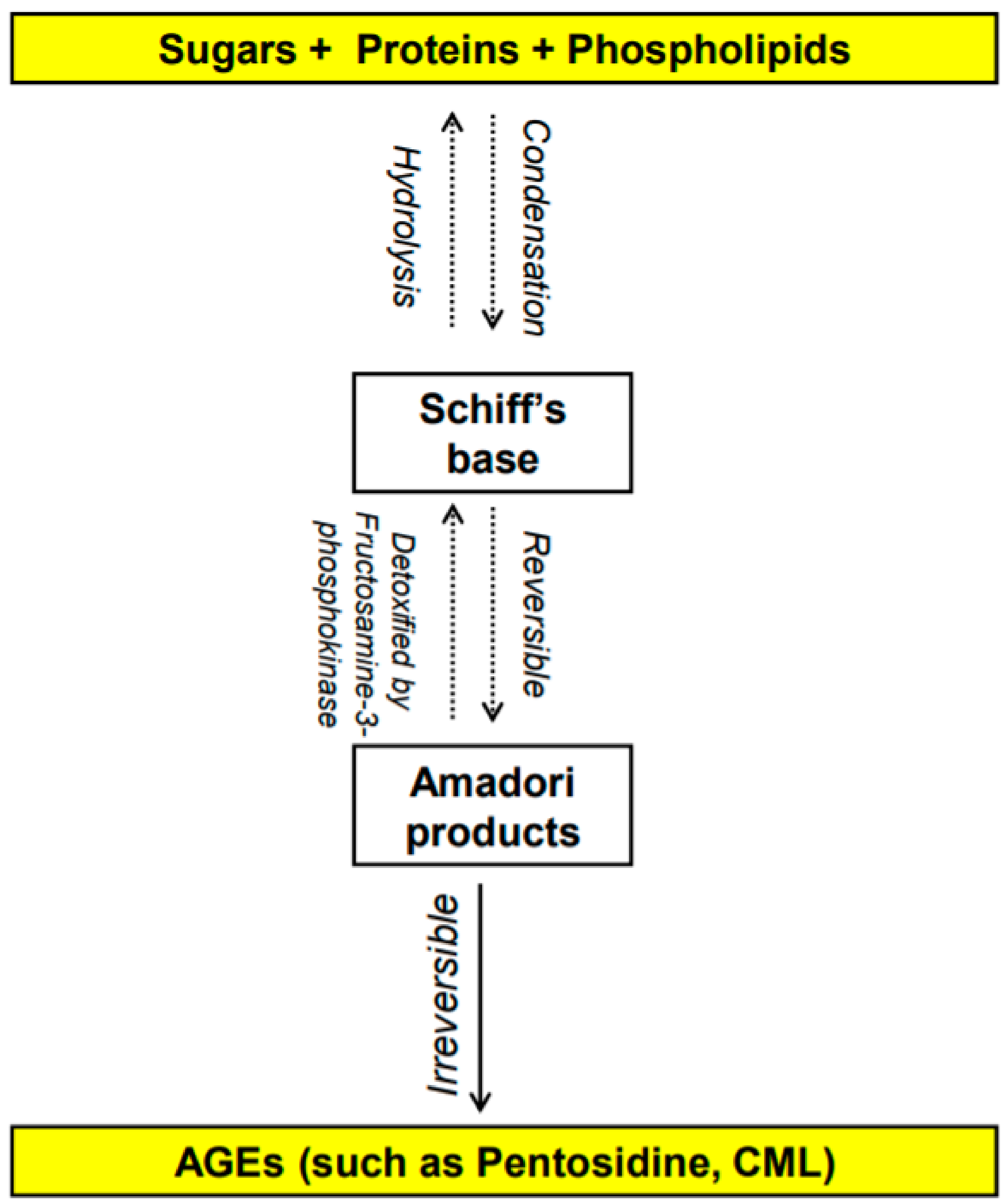
The Maillard reaction was first reported in 1912 by French scientist Louis Camille Maillard [1,2] and is defined as the chemical reaction in which the carbonyl group of carbohydrates reacts non-enzymatically with primary amino groups of proteins [3,4]. This reaction leads to the formation of advanced glycation end-products (AGEs). The early stages of the Maillard reaction lead to the formation of chemically reversible glycosylation products with proteins called Schiff bases and Amadori adducts [5]. The late stages of this glycation reaction forms complex glycation products which are the AGEs [6] (Figure 1). Since the 1980s, AGEs have been shown in several studies to be implicated in many health complications such as diabetes and aging [7], as well as many inflammatory diseases, obesity, cardiovascular diseases (CVD), metabolic syndrome and neurodegenerative disorders [8,9,10,11,12]. In the last decade, several studies have shown a potentially significant impact of AGEs on reproductive health in both males and females. This review will summarize the different types of AGEs and their receptors as well as the effect of dietary AGEs on female reproduction, in particular ovarian function, polycystic ovary syndrome (PCOS), and perinatally in utero on the female offspring reproduction. It also addresses the possible mechanistic pathways by which dietary AGEs alter female reproductive health.
1.1. What Are AGEs? How Do They Form?
AGEs are stable non-enzymatically catalyzed compounds which are formed by condensation of the amino groups of protein, lipid, amino acid and nucleic acid with the aldehyde group of reducing carbohydrate [13]. This nonenzymatic modification of proteins, lipids, and nucleic acids by glucose is one of the most important post-translational modifications in the formation of AGEs [14,15]. Once formed, the products of advanced glycation result in an irreversible cross-linking of proteins, loss of protein structure and function, followed by apoptosis and damage to cellular structures [14,15]. AGEs constitute a heterogeneous group of compounds of more than 20 members such as N-carboxymethyl-lysine (CML), pentosidine, 1,2-dicarbonyl precursor compounds glyoxal, and methylglyoxal. Pentosidine and CML are the most commonly studied AGEs [16,17] and have been used as markers of dietary AGE’s accumulation in various tissues [16,18,19,20,21,22]. Some of these compounds are fluorescent crosslinking (such as pentosidine [23]) products while others are non-fluorescent and/or non-crosslinking (such as CML [24,25]).
AGEs can be formed either endogenously by the body or from exogenous sources [1]. Endogenous AGEs are normally formed by glycosylation in different tissues of the body, occur slowly, increase progressively with aging and even faster with abnormal medical health conditions such as hyperglycemia and several chronic degenerative diseases [26]. Exogenous AGEs are obtained from food consumption and they are in very high levels in unhealthy food that is cooked at high temperature, such frying [27] and from smoking [28]. Contemporary methods of cooking (precooked fast-food meals), food high in protein and fat such as meat, cheese, and egg yolk dramatically increase serum AGEs’ concentration [27,29]. In addition to serum level, tissue AGEs can be influenced by diet as well [16,27]. Even though it is beyond the scope of the article, smoking has been identified as an exogenous source of AGEs [30]. Glycation products are present in tobacco and smoke in a form that can rapidly react with proteins to form AGEs [30].
1.2. How Do Dietary AGEs Act?
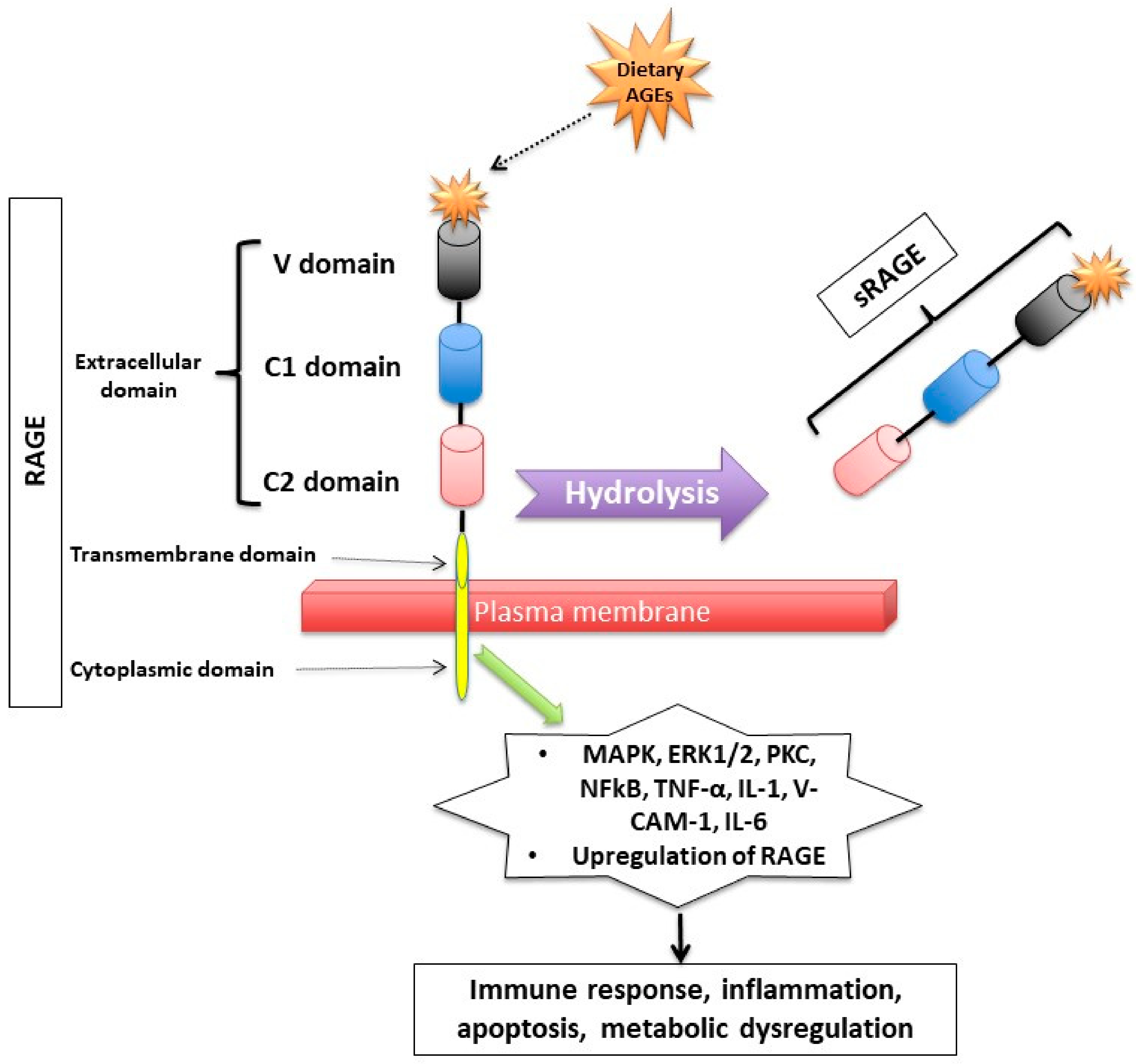
Dietary AGEs bind to several types of receptors (Figure 2). First, AGEs can act by binding to a receptor called RAGE (Receptor for Advanced Glycation End product) which is member of the immunoglobulin superfamily [31]. The expression of the RAGE protein is detected in human trophoblasts in chorionic villi early in fetal life (such as in endothelial cells of embryonic vessels and alveolar capillaries) and gradually increases after birth and in adulthood [32,33].
RAGE has a triple domain: transmembrane, a cytosolic and an extracellular [34], is expressed in cell membranes of several tissues such as heart, lung, skeletal muscle, the vessel wall and the reproductive system [10,34,35] and can be activated by many other ligands including amyloid β peptide, high-mobility group protein B1 (HMGB1) and the S100 group of protein [36].
After binding to RAGE, there is an activation of several intracellular inflammatory signaling pathways that include mitogen-activated protein kinase (MAPK), extracellular signal-regulated kinase1/2 (ERK1/2), protein kinase C (PKC) and nuclear factor kappa B (NF-κB) [37,38]. The activation of those pathways can lead to inflammatory state, cellular oxidative stress, and cellular damage [31] via upregulating markers of reactive oxygen species (ROS), and inflammatory molecules such as tumor necrosis factor (TNF-α), interleukin-1 (IL-1), vascular adhesion molecule-1 (VCAM-1), and interleukin-6 (IL-6) [39]. Interestingly, the binding of AGEs to RAGE upregulates RAGE expression itself, causing inflammation to get worse [31,40,41,42].
Second, RAGEs have been found to have multiple soluble forms detected mainly in body fluids and blood. The two most common forms are: sRAGE (soluble fragment of RAGE) and esRAGE (endogenous secretory RAGE). The sRAGE is produced by hydrolysis (MMPs and ADAM-10 induced proteolytic cleavage mechanisms [43,44]) of the RAGE receptor at the level of the cell surface and can be detected in the blood and bodily fluids [42,45]. Unlike RAGE, sRAGE contains only the extracellular domain of RAGE [42,45], and unlike RAGE, it has an “anti-inflammatory” action since it holds on to the circulating AGEs, thus inhibiting them from exercising their pro-inflammatory effect by binding to RAGE [46,47,48]. Unlike sRAGE, which is derived from the full-length form of RAGE [43], esRAGE is only derived from a part of the RAGE, specifically from pre-mRNA alternative splicing [49]. The esRAGE, also called variant RAGE-v1, usually comprises 20% of the total soluble RAGE receptors [49].
1.3. How Are Dietary AGEs Cleared from the Body?
Dietary AGEs are orally absorbed [50], with approximately 10% of them being absorbed in the GI tract and delivered to the liver and to other organs including but not limited to the reproductive system [51]. Dietary AGEs are mainly cleared by the urinary tract system (kidneys): nearly a third of dietary AGEs are excreted in the urine, with approximately 50% of the AGEs remaining quantified in the urine until approximately a few days following its consumption [52], and accumulating in the body leading to inflammation and oxidative stress [51]. The beginning of AGEs’ degradation occurs mainly intracellularly, therefore they first need to be inserted into the cell. Some of the AGE-receptors that are involved in the detoxification process are the AGE-R1/OST-48, AGE-R3/galectin-3 and scavenger-receptors [53]. These receptors compete with RAGE and try to bind the circulating dietary AGEs, thus they inhibit the toxic RAGE-mediated signaling pathways. The uptake of AGEs takes place through the activation of membrane receptors via phosphorylation or ubiquitinylation of the cytoplasmic side of the receptor, thus inducing its endocytosis [54].
2. Polycystic Ovary Syndrome (PCOS) and Dietary AGEs
PCOS is arguably the most common endocrinopathy in reproductive-aged women [55,56]. It is associated with significant metabolic changes and reproductive alterations, making it the most common cause (up to 70%) of anovulation [56]. Most women with PCOS display some type of metabolic dysfunction [57]. Studies have shown that women with PCOS have elevated circulating AGEs, which is exacerbated by exogenous absorption of AGEs from western heat processed diets [58]. AGEs contribute to the pathogenesis of PCOS as well as the consequential metabolic and reproductive system effects as proven by several in vitro experiments, animal models, and human studies [9,42,59,60,61,62].
When quantified at the ovarian tissue level by immunohistochemistry, RAGE and AGE-modified proteins are expressed in women with or without PCOS, though at much different concentrations [9]. There are alterations in the AGE system that have been shown to be related to reproductive impairment in women with PCOS [63]. It was first demonstrated in 2005 that overweight women with PCOS, compared to those without PCOS and independently of the hyperglycemia level (well known to be correlated to an increase in AGEs level), have increased AGEs’ levels and the upregulation of monocyte RAGE expression [42]. Then, in 2008, it was shown that lean women with PCOS without insulin resistance (another factor that is well known to be correlated with elevated body AGEs) also have elevated serum AGE levels compared to women with components of PCOS only (such as hyperandrogenemia with or without PCO-ovarian morphology) [59]. These findings suggest that these harmful molecules and the pro-inflammatory multi-ligand receptor RAGE have a pathological significance in reproductive abnormalities, in particular in ovarian dysfunction, in PCOS. Additionally, several studies in women who underwent IVF, assessed the relationship between sRAGE and PCOS and showed that compared to women without PCOS, those with PCOS had significantly lower sRAGE levels in the follicular fluid [64,65,66,67]. These findings suggest that there are alterations even in the anti-inflammatory sRAGE receptors in women with PCOS.
Other studies have demonstrated that women with PCOS given isocaloric diets high in AGEs for 2 months had significantly higher testosterone, free androgen index, and androstendione levels compared to women with PCOS on two-months low-AGE isocaloric diet [29]. Animal studies in animals confirmed the same findings, where rats put on a high-AGE diet for six months showed elevated AGE deposition in the reproductive system (theca cells), increased RAGE staining in granulosa cells, and higher plasma testosterone levels compared to low-AGE diet rats [16]. In another study, high-AGE diet showed increased plasma testosterone and decreased plasma estradiol and progesterone in female rats compared to their female rats counterparts on low-AGE diets [68]. This underscores an irrefutable correlation between dietary AGEs and hyperandrogenemia, solidifying the hypothesis that lowering dietary AGEs in PCOS could reduce some of the symptomatology of hyperandrogenemia.
At the cellular level, AGEs cause alterations in lysyl oxidase (LOX), which play a significant role in the regulation of ovarian follicular extracellular matrix organization and can explain some of the changes observed in PCOS [69]. In fact, AGE-mediated stimulation of LOX activity leads to the excessive deposition of collagen in the ovaries of women with PCOS [69]. In a rat model, dietary AGEs were able to reduce the activity of protective glyoxalase-I in the ovary of PCOS and that effect that was partially reversed by a diet low in AGEs [70]. These findings suggest that, at the cellular level, it is plausible that AGEs are significantly involved in the pathophysiology of PCOS, partially via LOX and glyoxalase-I [69]. Another study showed that AGEs may alter the enzymatic activity of specific enzymes such as cholesterol side-chain cleavage enzyme cytochrome P450, steroidogenic acute regulatory protein (StAR), 17α-hydroxylase, and 3β-hydroxysteroid dehydrogenase leading to the symptomatology of hyperandrogenism in PCOS [71]. AGEs also exert a direct effect in granulosa cells on the expression and signaling pathways of luteinizing hormone (LH) receptor and anti-Mullerian hormone receptor [71].
3. Ovarian Dysfunction
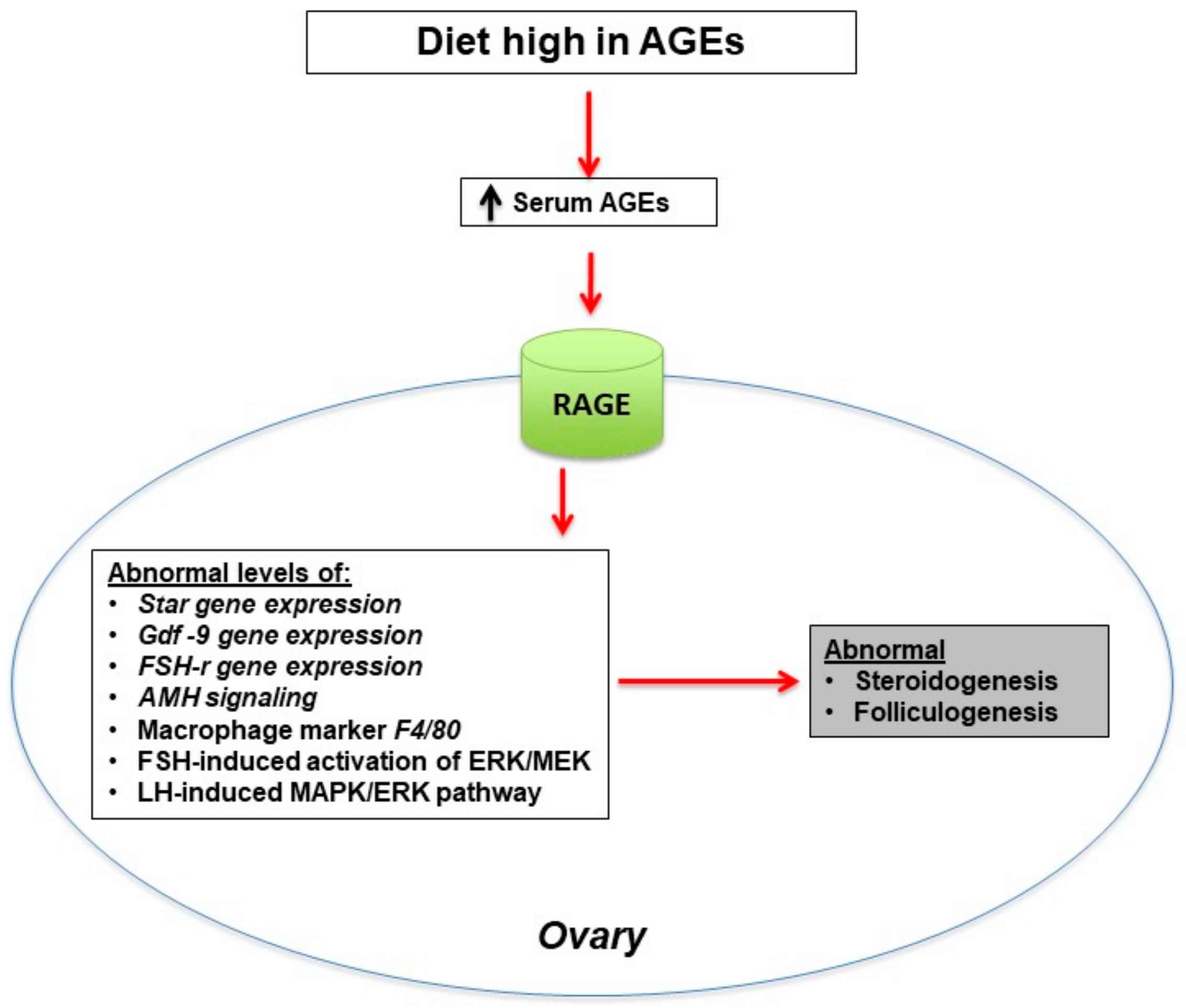
New data indicate that the high-AGE diet could disrupt ovarian function, particularly folliculogenesis and steroidogenesis [18,28,72]. Interestingly, the ingestion of dietary AGEs could also increase ovarian gene expression of inflammatory macrophage markers [72]. A mouse model study evaluated the effect of high-AGE diet on estrous cyclicity and ovarian function. In that study, six-week old C57BL/6J female mice were randomly subjected to either a diet low or high in AGEs for 13 weeks [72] during which daily assessment of estrous cyclicity was performed through vaginal smears, along with oocyte number assessment (following ovarian superovulation with gonadotropins), quantification of genes involved in folliculogenesis, steroidogenesis and ovarian macrophage markers (via whole ovarian tissue mRNA quantification by RT-PCR), and finally ovarian morphology for follicle count. Their results showed that, compared to mice on a diet low in AGEs, mice on a diet high in AGEs spent a significantly longer time in the diestrus phase, had significantly fewer corpora lutea, and showed significant alterations in genes involved in steroidogenesis, in particular an increase in StAR mRNA expression levels. They also showed significant alterations in genes involved in folliculogenesis, in particular an increase in growth differentiation factor 9 (Gdf-9) and follicular stimulating hormone (FSH) receptor (FSH-r) mRNA levels. Mouse macrophage marker F4/80 mRNA expression was upregulated in mice on a high-AGE diet. These results indicate that folliculogenesis and steroidogenesis could be disrupted by a high-AGE diet, leading to abnormal reproduction in female animals (Figure 3).
At the cellular level, the effect of MAPK/ERK is essential for normal follicle development (regulation of oocyte maturation) and proper ovulation [73]. One study evaluated the in vitro interference of exogenous AGEs with luteinizing hormone (LH)-induced MAPK/ERK signaling pathway in a cell line of KGN cell line granulosa cells [37]. The results of that study showed a direct abnormal effect of exogenous AGEs in this pathway, thus leading to a reduced activation of ERK1/2. Another pathway that was affected by AGEs was the FSH-induced phosphorylation of MEK1/2 and ERK1/2, leading to a reduced activation in the KGN granulosa cells [37]. These data, along with the fact that ERK1/2 activation is crucial for FSH-mediated granulosa cell mitogenesis [74], suggest that exogenous AGEs could potentially alter follicular development (Figure 3).
The effect of AGEs on pathways (such as Smad 1/5/8) and expression of genes (LH receptor [LHR], Anti-Mullerian hormone [AMH] and AMH receptor [AMHR-II]) involved in ovarian follicular development was studied in human luteinized granulosa cells of women who underwent oocyte collection for in vitro fertilization [75]. The AMH signals intracellularly via phosphorylating Smad 1/5/8 [76,77] and plays an important role in normal folliculogenesis by suppressing the differentiation of granulosa cells and follicular development thus protecting them from becoming atretic [78]. In that study, the human luteinized granulosa cells were treated with exogenous AGEs in vitro and showed significant increase in LHR and AMHR-II mRNA levels, but had no change in AMH mRNA expression levels. Compared to luteinized granulosa cells treated without exogenous AGEs in vitro, those treated with exogenous AGEs had significant increase in CYP11A1, 3β-HSD, StAR, and CYP17A1 mRNA expression levels as well as a significant secretion of estradiol [79]. These findings suggest that there is a relationship between exogenous AGEs and steroid expression. KGN granulosa cells treated with recombinant AMH (rAMH) showed a significant increase in the Smad 1/5/8 phosphorylation in the presence of exogenous AGEs in vitro, compared to the absence of AGEs in vitro [80]. Those findings suggest that exogenous AGEs could lead to ovulatory dysfunction partly via the elevated AMH-induced Smad 1/5/8 signaling pathway.
4. Perinatal Exposure to Elevated Dietary AGEs and Reproduction in Female Offspring
It is well known that maternal nutrition and the intrauterine environment are crucial in determining susceptibility to reproductive and metabolic disturbances later in life [81,82]. For instance, maternal obesity or consumption of a high fat diet during pregnancy and lactation have been shown to increase the risk of metabolic diseases in offspring [83]. The typical Western diet, commonly consumed by pregnant mothers, contains high amounts of the pro-inflammatory AGEs [27,84,85]. Data have shown that this dietary pattern results in reproductive disturbances [63], oxidative stress and inflammation in both humans and animals [27]. Maternal exposure to high dietary AGEs during pregnancy could predispose mice offspring to metabolic disturbances later in life; for example, perinatal exposure to high dietary AGEs have been shown to predispose the male offspring to weight gain and to metabolic alterations [86]. Additionally, reducing dietary AGEs throughout gestation, lactation, and early postnatal life have been shown to benefit the metabolism (for example pancreatic islet secretion) and the immune system in mice [87].
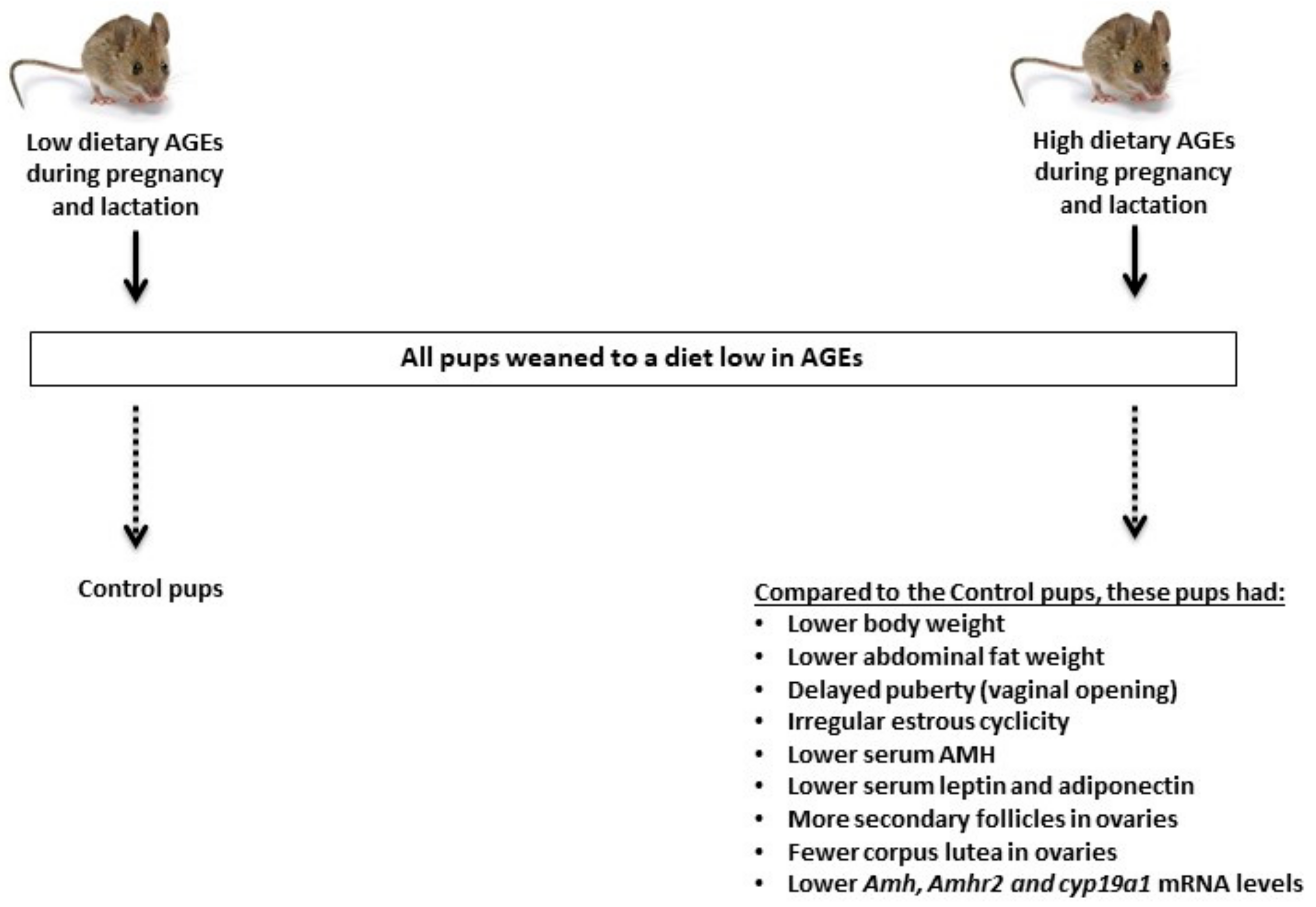
In a recent study [88], seven week old female mice were placed on either a diet low or high in AGEs before mating and then during pregnancy and lactation. All offspring were weaned onto a diet low in AGEs and studied until 16 weeks of age by counting them and weighing them at birth and then every week for a total of 11 weeks (Figure 4). The authors assessed the vaginal opening (an indicator of puberty), litter size, growth curve, ovarian follicular count, ovarian gene expression, liver and abdominal fat weights, serum levels of AMH (a marker of ovarian reserve), leptin and adiponectin (markers of adiposity) as well as insulin and glucose tolerance tests in both groups. Their results showed that compared to perinatal exposure to a diet low in AGEs, perinatal exposure to a diet high in AGEs caused lower body weight at birth, delayed growth in adult offspring, lower serum leptin and adiponectin levels, delayed vaginal opening, irregular estrous cyclicity, arrested follicular development, and significant alterations in the expression of genes involved in folliculogenesis such as Amh and Amhr2, and steroidogenesis such as Cyp19a1 (aromatase enzyme). These results indicate that perinatal exposure to a diet elevated in AGEs causes deficits in perinatal growth, pubertal onset, and reproductive organ development in female mice.
5. Conclusions
With the common ingestion of diets high in AGEs, reproductive-aged women might face more metabolic and reproductive complications. The reproductive function and body energy hemostasis are closely linked at the level of the brain, in particular the hypothalamus, and the ovaries; thus, future studies focusing on the neuroendocrine axis are necessary to further define the mechanisms by which dietary AGEs influence female reproduction. The ingestion of diets high in AGEs causes a systemic state of chronic inflammation, as shown by upregulation of ovarian macrophage marker F4/80 expression [72], which may directly affect ovarian function. Those dietary AGEs could potentially disrupt the ovarian microenvironment, compromising oocyte competence, formation of healthy embryos, and ultimately conception. Additionally, the elevation of AGEs in the serum and tissues of reproductive-aged women may exacerbate the reproductive dysfunction associated with PCOS.
This review underscores a critical need to unveil, in greater depth, the mechanistic pathways of AGEs, which are molecules present in high amounts in the daily Western diet. Cutting down on the ingestion of AGEs could be demanding and might not be maintainable, thus there is a need to promote therapies targeting AGEs and their intracellular signaling pathways in order to improve ovarian health not only now but in the future generations, since AGEs seem to have transgenerational effects on female reproduction.
Author Contributions
Conceptualization, M.M. and Z.M.; methodology, M.M. and Z.M.; resources, M.M. and Z.M.; data curation, M.M. and Z.M.; writing—review and editing, M.M. and Z.M.; visualization, Z.M.; supervision, Z.M. All authors have read and agreed to the published version of the manuscript.
Funding
This research received no external funding.
Institutional Review Board Statement
Not applicable.
Informed Consent Statement
Not applicable.
Conflicts of Interest
The authors declare no conflict of interest.
References
- Moschonas, D.P.; Piperi, C.; Korkolopoulou, P.; Levidou, G.; Kavantzas, N.; Trigka, E.-A.; Vlachos, I.; Arapostathi, C.; Perrea, D.; Mitropoulos, D.; et al. Impact of diet-induced obesity in male mouse reproductive system: The role of advanced glycation end product–receptor for advanced glycation end product axis. Exp. Biol. Med. 2014, 239, 937–947. [Google Scholar] [CrossRef] [PubMed]
- Maillard, L.C. Action des acides aminés sur les sucres; formation des méla-noidines par voie methodique. Comptes Rendus Acad. Sci. 1912, 154, 66–68. [Google Scholar]
- Unoki, H.; Yamagishi, S. Advanced glycation end products and insulin resistance. Curr. Pharm. Des. 2008, 14, 987–989. [Google Scholar] [CrossRef] [PubMed]
- Brownlee, M.; Cerami, A.; Vlassara, H. Advanced glycosylation end products in tissue and the biochemical basis of diabetic complications. N. Engl. J. Med. 1988, 318, 1315–1321. [Google Scholar] [CrossRef]
- Brownlee, M. Glycosylation products as toxic mediators of diabetic complications. Annu. Rev. Med. 1991, 42, 159–166. [Google Scholar] [CrossRef]
- Bucala, R.; Cerami, A. Advanced glycosylation: Chemistry, biology, and implications for diabetes and aging. Stud. Surf. Sci. Catal. 1992, 23, 1–34. [Google Scholar] [CrossRef]
- Monnier, V.M.; Stevens, V.J.; Cerami, A. Maillard reactions involving proteins and carbohydrates in vivo: Relevance to diabetes mellitus and aging. Prog. Food Nutr. Sci. 1981, 5, 315–327. [Google Scholar]
- Yamagishi, S.-I.; Nakamura, K.; Imaizumi, T. Advanced glycation end products (AGEs) and diabetic vascular complications. Curr. Diabetes Rev. 2005, 1, 93–106. [Google Scholar] [CrossRef]
- Diamanti-Kandarakis, E.; Piperi, C.; Patsouris, E.; Korkolopoulou, P.; Panidis, D.; Pawelczyk, L.; Papavassiliou, A.G.; Duleba, A.J. Immunohistochemical localization of advanced glycation end-products (AGEs) and their receptor (RAGE) in polycystic and normal ovaries. Histochem. Cell Biol. 2007, 127, 581–589. [Google Scholar] [CrossRef]
- Tatone, C.; Amicarelli, F. The aging ovary—the poor granulosa cells. Fertil. Steril. 2013, 99, 12–17. [Google Scholar] [CrossRef]
- Pertynska-Marczewska, M.; Merhi, Z. Relationship of advanced glycation end products with cardiovascular disease in menopausal women. Reprod. Sci. 2015, 22, 774–782. [Google Scholar] [CrossRef] [PubMed] [Green Version]
- Merhi, Z. Advanced glycation end-products: Pathway of potentially significant pathophysiological and therapeutic relevance for metabolic syndrome in menopausal women. J. Clin. Endocrinol. Metab. 2014, 99, 1146–1148. [Google Scholar] [CrossRef] [PubMed] [Green Version]
- Khalifah, R.G.; Baynes, J.W.; Hudson, B. Amadorins: Novel post-amadori inhibitors of advanced glycation reactions. Biochem. Biophys. Res. Commun. 1999, 257, 251–258. [Google Scholar] [CrossRef]
- Inagi, R. Inhibitors of advanced glycation and endoplasmic reticulum stress. Methods Enzymol. 2011, 491, 361–380. [Google Scholar] [CrossRef]
- Piperi, C.; Adamopoulos, C.; Dalagiorgou, G.; Diamanti-Kandarakis, E.; Papavassiliou, A.G. Crosstalk between advanced glycation and endoplasmic reticulum stress: Emerging therapeutic targeting for metabolic diseases. J. Clin. Endocrinol. Metab. 2012, 97, 2231–2242. [Google Scholar] [CrossRef] [PubMed] [Green Version]
- Diamanti-Kandarakis, E.; Piperi, C.; Korkolopoulou, P.; Kandaraki, E.; Levidou, G.; Papalois, A.; Patsouris, E.; Papavassiliou, A.G. Accumulation of dietary glycotoxins in the reproductive system of normal female rats. Klin. Wochenschr. 2007, 85, 1413–1420. [Google Scholar] [CrossRef] [Green Version]
- Takahashi, M.; Oikawa, M.; Nagano, A. Effect of age and menopause on serum concentrations of pentosidine, an advanced glycation end product. J. Gerontol. Ser. A 2000, 55, M137–M140. [Google Scholar] [CrossRef] [Green Version]
- Merhi, Z. Advanced glycation end products and their relevance in female reproduction. Hum. Reprod. 2014, 29, 135–145. [Google Scholar] [CrossRef] [Green Version]
- Desai, K.; Wu, L. Methylglyoxal and advanced glycation endproducts: New therapeutic horizons? Recent patents on cardiovascular drug discovery. Recent Pat. Cardiovasc. Drug Discov. (Discontin.) 2007, 2, 89–99. [Google Scholar] [CrossRef]
- Kerkeni, M.; Saïdi, A.; Bouzidi, H.; Ben Yahya, S.; Hammami, M. Elevated serum levels of AGEs, sRAGE, and pentosidine in Tunisian patients with severity of diabetic retinopathy. Microvasc. Res. 2012, 84, 378–383. [Google Scholar] [CrossRef]
- Saito, M.; Marumo, K. Bone quality. Nihon Rinsho 2015, 73, 1665–1672. [Google Scholar] [PubMed]
- Saeki, C.; Saito, M.; Kanai, T.; Nakano, M.; Oikawa, T.; Torisu, Y.; Saruta, M.; Tsubota, A. Plasma pentosidine levels are associated with prevalent fractures in patients with chronic liver disease. PLoS ONE 2021, 16, e0249728. [Google Scholar] [CrossRef] [PubMed]
- Ghanem, A.A.; Elewa, A.; Arafa, L.F. Pentosidine and N-Carboxymethyl-Lysine: Biomarkers for Type 2 diabetic retinopathy. Eur. J. Ophthalmol. 2011, 21, 48–54. [Google Scholar] [CrossRef] [PubMed]
- Brouwers, O.; Niessen, P.M.; Ferreira, I.; Miyata, T.; Scheffer, P.G.; Teerlink, T.; Schrauwen, P.; Brownlee, M.; Stehouwer, C.D.; Schalkwijk, C.G. Overexpression of glyoxalase-i reduces hyperglycemia-induced levels of advanced glycation end products and oxidative stress in diabetic rats. J. Biol. Chem. 2011, 286, 1374–1380. [Google Scholar] [CrossRef] [PubMed] [Green Version]
- Liu, X.; Zheng, L.; Zhang, R.; Liu, G.; Xiao, S.; Qiao, X.; Wu, Y.; Gong, Z. Toxicological evaluation of advanced glycation end product Nε-(carboxymethyl)lysine: Acute and subacute oral toxicity studies. Regul. Toxicol. Pharmacol. 2016, 77, 65–74. [Google Scholar] [CrossRef]
- Jud, P.; Sourij, H. Therapeutic options to reduce advanced glycation end products in patients with diabetes mellitus: A review. Diabetes Res. Clin. Pract. 2019, 148, 54–63. [Google Scholar] [CrossRef]
- Goldberg, T.; Cai, W.; Peppa, M.; Dardaine, V.; Baliga, B.S.; Uribarri, J.; Vlassara, H. Advanced glycoxidation end products in commonly consumed foods. J. Am. Diet. Assoc. 2004, 104, 1287–1291. [Google Scholar] [CrossRef]
- Gill, V.; Kumar, V.; Singh, K.; Kumar, A.; Kim, J.-J. Advanced glycation end products (AGEs) may be a striking link between modern diet and health. Biomolecules 2019, 9, 888. [Google Scholar] [CrossRef] [Green Version]
- Tantalaki, E.; Piperi, C.; Livadas, S.; Kollias, A.; Adamopoulos, C.; Koulouri, A.; Christakou, C.; Diamanti-Kandarakis, E. Impact of dietary modification of advanced glycation end products (AGEs) on the hormonal and metabolic profile of women with polycystic ovary syndrome (PCOS). Hormones 2014, 13, 65–73. [Google Scholar] [CrossRef]
- Cerami, C.; Founds, H.; Nicholl, I.; Mitsuhashi, T.; Giordano, D.; Vanpatten, S.; Lee, A.; Al-Abed, Y.; Vlassara, H.; Bucala, R.; et al. Tobacco smoke is a source of toxic reactive glycation products. Proc. Natl. Acad. Sci. USA 1997, 94, 13915–13920. [Google Scholar] [CrossRef] [Green Version]
- Kalea, A.Z.; Schmidt, A.M.; Hudson, B.I. RAGE: A novel biological and genetic marker for vascular disease. Clin. Sci. 2009, 116, 621–637. [Google Scholar] [CrossRef] [PubMed] [Green Version]
- Reynolds, P.R.; Kasteler, S.D.; Cosio, M.G.; Sturrock, A.; Huecksteadt, T.; Hoidal, J.R. RAGE: Developmental expression and positive feedback regulation by Egr-1 during cigarette smoke exposure in pulmonary epithelial cells. Am. J. Physiol. Cell. Mol. Physiol. 2008, 294, L1094–L1101. [Google Scholar] [CrossRef] [PubMed] [Green Version]
- Konishi, H.; Nakatsuka, M.; Chekir, C.; Noguchi, S.; Kamada, Y.; Sasaki, A.; Hiramatsu, Y. Advanced glycation end products induce secretion of chemokines and apoptosis in human first trimester trophoblasts. Hum. Reprod. 2004, 19, 2156–2162. [Google Scholar] [CrossRef] [PubMed] [Green Version]
- Basta, G. Receptor for advanced glycation endproducts and atherosclerosis: From basic mechanisms to clinical implications. Atherosclerosis 2008, 196, 9–21. [Google Scholar] [CrossRef] [PubMed]
- Fujii, E.Y.; Nakayama, M. The measurements of RAGE, VEGF, and AGEs in the plasma and follicular fluid of reproductive women: The influence of aging. Fertil. Steril. 2010, 94, 694–700. [Google Scholar] [CrossRef] [PubMed]
- Palanissami, G.; Paul, S.F.D. RAGE and Its Ligands: Molecular interplay between glycation, inflammation, and hallmarks of cancer—A review. Horm. Cancer 2018, 9, 295–325. [Google Scholar] [CrossRef] [PubMed]
- Kandaraki, E.A.; Chatzigeorgiou, A.; Papageorgiou, E.; Piperi, C.; Adamopoulos, C.; Papavassiliou, A.G.; Koutsilieris, M.; Diamanti-Kandarakis, E. Advanced glycation end products interfere in luteinizing hormone and follicle stimulating hormone signaling in human granulosa KGN cells. Exp. Biol. Med. 2017, 243, 29–33. [Google Scholar] [CrossRef] [PubMed]
- Verma, N.; Manna, S.K. Advanced Glycation End Products (AGE) potently induce autophagy through activation of RAF protein kinase and nuclear factor κB (NF-κB). J. Biol. Chem. 2016, 291, 1481–1491. [Google Scholar] [CrossRef] [Green Version]
- Guimarães, E.L.; Empsen, C.; Geerts, A.; van Grunsven, L.A. Advanced glycation end products induce production of reactive oxygen species via the activation of NADPH oxidase in murine hepatic stellate cells. J. Hepatol. 2010, 52, 389–397. [Google Scholar] [CrossRef]
- Dunaif, A.; Segal, K.R.; Futterweit, W.; Dobrjansky, A. Profound peripheral insulin resistance, independent of obesity, in polycystic ovary syndrome. Diabetes 1989, 38, 1165–1174. [Google Scholar] [CrossRef]
- Ramasamy, R.; Yan, S.F.; D’Agati, V.; Schmidt, A.M. Receptor for Advanced Glycation Endproducts (RAGE): A formidable force in the pathogenesis of the cardiovascular complications of diabetes & aging. Curr. Mol. Med. 2007, 7, 699–710. [Google Scholar] [CrossRef]
- Diamanti-Kandarakis, E.; Piperi, C.; Kalofoutis, A.; Creatsas, G. Increased levels of serum advanced glycation end-products in women with polycystic ovary syndrome. Clin. Endocrinol. 2005, 62, 37–43. [Google Scholar] [CrossRef] [PubMed]
- Raucci, A.; Cugusi, S.; Antonelli, A.; Barabino, S.M.; Monti, L.; Bierhaus, A.; Reiss, K.; Saftig, P.; Bianchi, M.E. A soluble form of the receptor for advanced glycation endproducts (RAGE) is produced by proteolytic cleavage of the membrane-bound form by the sheddase a disintegrin and metalloprotease 10 (ADAM10). FASEB J. 2008, 22, 3716–3727. [Google Scholar] [CrossRef] [PubMed]
- Zhang, L.; Bukulin, M.; Kojro, E.; Roth, A.; Metz, V.V.; Fahrenholz, F.; Nawroth, P.P.; Bierhaus, A.; Postina, R. Receptor for advanced glycation end products is subjected to protein ectodomain shedding by metalloproteinases. J. Biol. Chem. 2008, 283, 35507–35516. [Google Scholar] [CrossRef] [PubMed] [Green Version]
- Diamanti-Kandarakis, E. Insulin resistance in PCOS. Endocrine 2006, 30, 13–17. [Google Scholar] [CrossRef]
- Asadipooya, K.; Uy, E.M. Advanced Glycation End Products (AGEs), receptor for AGEs, diabetes, and bone: Review of the literature. J. Endocr. Soc. 2019, 3, 1799–1818. [Google Scholar] [CrossRef] [Green Version]
- Schmidt, A.M. Soluble RAGEs—Prospects for treating & tracking metabolic and inflammatory disease. Vasc. Pharmacol. 2015, 72, 1–8. [Google Scholar] [CrossRef] [Green Version]
- Selvin, E.; Halushka, M.K.; Rawlings, A.M.; Hoogeveen, R.C.; Ballantyne, C.M.; Coresh, J.; Astor, B.C. sRAGE and risk of diabetes, cardiovascular disease, and death. Diabetes 2013, 62, 2116–2121. [Google Scholar] [CrossRef] [Green Version]
- Yonekura, H.; Yamamoto, Y.; Sakurai, S.; Petrova, R.G.; Abedin, J.; Li, H.; Yasui, K.; Takeuchi, M.; Makita, Z.; Takasawa, S.; et al. Novel splice variants of the receptor for advanced glycation end-products expressed in human vascular endothelial cells and pericytes, and their putative roles in diabetes-induced vascular injury. Biochem. J. 2003, 370, 1097–1109. [Google Scholar] [CrossRef]
- He, C.; Sabol, J.; Mitsuhashi, T.; Vlassara, H. Dietary glycotoxins: Inhibition of reactive products by aminoguanidine facilitates renal clearance and reduces tissue sequestration. Diabetes 1999, 48, 1308–1315. [Google Scholar] [CrossRef]
- Semba, R.D.; Ang, A.; Talegawkar, S.A.; Crasto, C.; Dalal, M.; Jardack, P.; Traber, M.; Ferrucci, L.; Arab, L. Dietary intake associated with serum versus urinary carboxymethyl-lysine, a major advanced glycation end product, in adults: The Energetics Study. Eur. J. Clin. Nutr. 2011, 66, 3–9. [Google Scholar] [CrossRef] [PubMed] [Green Version]
- Förster, A.; Kühne, Y.; Henle, T. Studies on absorption and elimination of dietary maillard reaction products. Ann. N. Y. Acad. Sci. 2005, 1043, 474–481. [Google Scholar] [CrossRef] [PubMed]
- Vlassara, H.; Palace, M. Diabetes and advanced glycation endproducts. J. Intern. Med. 2002, 251, 87–101. [Google Scholar] [CrossRef] [PubMed]
- Araki, N.; Higashi, T.; Mori, T.; Shibayama, R.; Kawabe, Y.; Kodama, T.; Takahashi, K.; Shichiri, M.; Horiuchi, S. Macrophage scavenger receptor mediates the endocytic uptake and degradation of advanced glycation end products of the Maillard reaction. Eur. J. Biochem. 1995, 230, 408–415. [Google Scholar] [CrossRef]
- Diamanti-Kandarakis, E.; Kouli, C.R.; Bergiele, A.T.; Filandra, F.A.; Tsianateli, T.C.; Spina, G.G.; Zapanti, E.D.; Bartzis, M.I. A survey of the polycystic ovary syndrome in the Greek island of Lesbos: Hormonal and metabolic profile. J. Clin. Endocrinol. Metab. 1999, 84, 4006–4011. [Google Scholar] [CrossRef]
- Carmina, E.; Lobo, R.A. Polycystic ovary syndrome (PCOS): Arguably the most common endocrinopathy is associated with significant morbidity in women. J. Clin. Endocrinol. Metab. 1999, 84, 1897–1899. [Google Scholar] [CrossRef]
- Cussons, A.J.; Stuckey, B.G.; Watts, G.F. Cardiovascular disease in the polycystic ovary syndrome: New insights and perspectives. Atherosclerosis 2006, 185, 227–239. [Google Scholar] [CrossRef]
- Garg, D.; Merhi, Z. Advanced glycation end products: Link between diet and ovulatory dysfunction in PCOS? Nutrients 2015, 7, 10129–10144. [Google Scholar] [CrossRef] [Green Version]
- Diamanti-Kandarakis, E.; Katsikis, I.; Piperi, C.; Kandaraki, E.; Piouka, A.; Papavassiliou, A.G.; Panidis, D. Increased serum advanced glycation end-products is a distinct finding in lean women with polycystic ovary syndrome (PCOS). Clin. Endocrinol. 2008, 69, 634–641. [Google Scholar] [CrossRef]
- Rutkowska, A.Z.; Diamanti-Kandarakis, E. Do Advanced Glycation End Products (AGEs) contribute to the comorbidities of Polycystic Ovary Syndrome (PCOS)? Curr. Pharm. Des. 2016, 22, 5558–5571. [Google Scholar] [CrossRef]
- Pertynska-Marczewska, M.; Diamanti-Kandarakis, E.; Zhang, J.; Merhi, Z. Advanced glycation end products: A link between metabolic and endothelial dysfunction in polycystic ovary syndrome? Metabolism 2015, 64, 1564–1573. [Google Scholar] [CrossRef] [PubMed]
- Merhi, Z. Crosstalk between advanced glycation end products and vitamin D: A compelling paradigm for the treatment of ovarian dysfunction in PCOS. Mol. Cell. Endocrinol. 2019, 479, 20–26. [Google Scholar] [CrossRef] [PubMed]
- Merhi, Z.; Kandaraki, E.A.; Diamanti-Kandarakis, E. Implications and future perspectives of AGEs in PCOS pathophysiology. Trends Endocrinol. Metab. 2019, 30, 150–162. [Google Scholar] [CrossRef] [PubMed]
- Wang, B.; Li, J.; Yang, Q.; Zhang, F.; Hao, M.; Guo, Y. Decreased levels of sRAGE in follicular fluid from patients with PCOS. Reproduction 2017, 153, 285–292. [Google Scholar] [CrossRef]
- Garg, D.; Grazi, R.; Lambert-Messerlian, G.M.; Merhi, Z. Correlation between follicular fluid levels of sRAGE and vitamin D in women with PCOS. J. Assist. Reprod. Genet. 2017, 34, 1507–1513. [Google Scholar] [CrossRef]
- Irani, M.; Minkoff, H.; Seifer, D.; Merhi, Z. Vitamin D increases serum levels of the soluble receptor for advanced glycation end products in women with PCOS. J. Clin. Endocrinol. Metab. 2014, 99, E886–E890. [Google Scholar] [CrossRef] [Green Version]
- Merhi, Z.; Irani, M.; Doswell, A.D.; Ambroggio, J. Follicular fluid soluble receptor for advanced glycation end-products (sRAGE): A potential indicator of ovarian reserve. J. Clin. Endocrinol. Metab. 2014, 99, E226–E233. [Google Scholar] [CrossRef] [Green Version]
- Chatzigeorgiou, A.; Kandaraki, E.; Piperi, C.; Livadas, S.; Papavassiliou, A.G.; Koutsilieris, M.; Papalois, A.; Diamanti-Kandarakis, E. Dietary glycotoxins affect scavenger receptor expression and the hormonal profile of female rats. J. Endocrinol. 2013, 218, 331–337. [Google Scholar] [CrossRef] [Green Version]
- Papachroni, K.K.; Piperi, C.; Levidou, G.; Korkolopoulou, P.; Pawelczyk, L.; Diamanti-Kandarakis, E.; Papavassiliou, A.G. Lysyl oxidase interacts with AGE signalling to modulate collagen synthesis in polycystic ovarian tissue. J. Cell. Mol. Med. 2010, 14, 2460–2469. [Google Scholar] [CrossRef] [Green Version]
- Kandaraki, E.; Chatzigeorgiou, A.; Piperi, C.; Palioura, E.; Palimeri, S.; Korkolopoulou, P.; Koutsilieris, M.; Papavassiliou, A.G. Reduced ovarian glyoxalase-i activity by dietary glycotoxins and androgen excess: A causative link to polycystic ovarian syndrome. Mol. Med. 2012, 18, 1183–1189. [Google Scholar] [CrossRef]
- Garg, D.; Merhi, Z. Relationship between advanced glycation end products and steroidogenesis in PCOS. Reprod. Biol. Endocrinol. 2016, 14, 1–13. [Google Scholar] [CrossRef] [Green Version]
- Thornton, K.; Merhi, Z.; Jindal, S.; Goldsammler, M.; Charron, M.J.; Buyuk, E. Dietary Advanced Glycation End Products (AGEs) could alter ovarian function in mice. Mol. Cell. Endocrinol. 2020, 510, 110826. [Google Scholar] [CrossRef]
- Su, Y.Q.; Wigglesworth, K.; Pendola, F.L.; O’Brien, M.J.; Eppig, J.J. Mitogen-activated protein kinase activity in cumulus cells is essential for gonadotropin-induced oocyte meiotic resumption and cumulus expansion in the mouse. Endocrinology 2002, 143, 2221–2232. [Google Scholar] [CrossRef] [PubMed]
- Kayampilly, P.P.; Menon, K.M.J. Follicle-stimulating hormone inhibits adenosine 5′-Monophosphate-activated protein kinase activation and promotes cell proliferation of primary granulosa cells in culture through an akt-dependent pathway. Endocrinology 2009, 150, 929–935. [Google Scholar] [CrossRef] [PubMed] [Green Version]
- Merhi, Z.; Fadiel, A.; Buyuk, E.; Naftolin, F.; Cipolla, M. Vitamin D attenuates the adverse effect of advanced glycation end products on human granulosa cells: Implications for women with PCOS. Fertil. Steril. 2015, 104, e106. [Google Scholar] [CrossRef]
- Josso, N.; Clemente, N. Transduction pathway of anti-Mullerian hormone, a sex-specific member of the TGF-beta family. Trends Endocrinol. Metab. 2003, 14, 91–97. [Google Scholar] [CrossRef]
- Josso, N.; di Clemente, N.; Gouédard, L. Anti-müllerian hormone and its receptors. Mol. Cell. Endocrinol. 2001, 179, 25–32. [Google Scholar] [CrossRef]
- Durlinger, A.L.; Gruijters, M.J.; Kramer, P.; Karels, B.; Ingraham, H.A.; Nachtigal, M.W.; Uilenbroek, J.T.J.; Grootegoed, J.A.; Themmen, A.P. Anti-Mullerian hormone inhibits initiation of primordial follicle growth in the mouse ovary. Endocrinology 2002, 143, 1076–1084. [Google Scholar] [CrossRef]
- Merhi, Z.; Wang, S.; Cipolla, M. Special research presentation: Vitamin d reverses the adverse effects of advanced glycation end products on granulosa cells. Fertil. Steril. 2016, 106, e76. [Google Scholar] [CrossRef]
- Merhi, Z.; Büyük, E.; Cipolla, M.J. Advanced glycation end products alter steroidogenic gene expression by granulosa cells: An effect partially reversible by vitamin D. Mol. Hum. Reprod. 2018, 24, 318–326. [Google Scholar] [CrossRef]
- Barker, D.J. Maternal nutrition, fetal nutrition, and disease in later life. Nutrition 1997, 13, 807–813. [Google Scholar] [CrossRef]
- Dicken, C.L.; Israel, D.D.; Davis, J.B.; Sun, Y.; Shu, J.; Hardin, J.; Neal-Perry, G. Peripubertal vitamin D(3) deficiency delays puberty and disrupts the estrous cycle in adult female mice. Biol. Reprod. 2012, 87, 51. [Google Scholar] [CrossRef] [PubMed] [Green Version]
- Williams, L.; Seki, Y.; Vuguin, P.M.; Charron, M.J. Animal models of in utero exposure to a high fat diet: A review. Biochim. Biophys. Acta (BBA)—Mol. Basis Dis. 2014, 1842, 507–519. [Google Scholar] [CrossRef] [Green Version]
- Edelstein, D.; Brownlee, M. Mechanistic studies of advanced glycosylation end product inhibition by aminoguanidine. Diabetes 1992, 41, 26–29. [Google Scholar] [CrossRef]
- Horiuchi, S.; Araki, N.; Morino, Y. Immunochemical approach to characterize advanced glycation end products of the Maillard reaction. Evidence for the presence of a common structure. J. Biol. Chem. 1991, 266, 7329–7332. [Google Scholar] [CrossRef]
- Csongová, M.; Gurecká, R.; Koborová, I.; Celec, P.; Domonkos, E.; Uličná, O.; Somoza, V.; Šebeková, K. The effects of a maternal advanced glycation end product-rich diet on somatic features, reflex ontogeny and metabolic parameters of offspring mice. Food Funct. 2018, 9, 3432–3446. [Google Scholar] [CrossRef]
- Borg, D.J.; Yap, F.Y.T.; Keshvari, S.; Simmons, D.; Gallo, L.A.; Fotheringham, A.; Zhuang, A.; Slattery, R.M.; Hasnain, S.; Coughlan, M.; et al. Perinatal exposure to high dietary advanced glycation end products in transgenic NOD8.3 mice leads to pancreatic beta cell dysfunction. Islets 2017, 10, 10–24. [Google Scholar] [CrossRef] [PubMed]
- Merhi, Z.; Du, X.Q.; Charron, M.J. Perinatal exposure to high dietary advanced glycation end products affects the reproductive system in female offspring in mice. Mol. Hum. Reprod. 2020, 26, 615–623. [Google Scholar] [CrossRef]
Figure 1.
Overview of the formation of advanced glycation end products (AGEs) [1,2,3,4,5,6]. N-carboxymethyl-lysine (CML).

Figure 2.
Receptor for Advanced Glycation End (RAGE) products and its mechanism of action. RAGE consists of transmembrane, cytosolic and extracellular domains. The extracellular domain consists of V, C1, and C2 domains. The soluble fragment of RAGE (sRAGE) is produced by hydrolysis of the RAGE receptor and contains the RAGE’s extracellular domain only. The binding of AGEs to RAGE induces a series of inflammatory and apoptotic responses intracellularly and contributes to metabolic dysfunction [32,33,34,44,45,46,47,48,49]. Mitogen-activated protein kinase (MAPK), extracellular signal-regulated kinase1/2 (ERK1/2), protein kinase C (PKC) and nuclear factor kappa B (NF-κB), lysyl oxidase (LOX), tumor necrosis factor (TNF-α), interleukin-1 (IL-1), vascular adhesion molecule-1 (VCAM-1), and interleukin-6 (IL-6).

Figure 3.
Effect of dietary high levels of advanced glycation end products (AGEs) on ovary function. Diets with large amounts of AGEs increase serum level of AGEs which bind to RAGE in the ovary and cause abnormal expression of genes involved in steroidogenesis and folliculogenesis as well as increase macrophage infiltration in ovarian tissue, ultimately leading to ovarian dysfunction [28,37,72,73,74]. Growth differentiation factor 9 (GDF), follicular stimulating hormone receptor (FSH-r), anti-mullerian hormone (AMH), follicular stimulating hormone (FSH), extracellular-signal-regulated kinase (ERK), mitogen activated protein kinase (MEK), luteinizing hormone (LH), mitogen activated protein kinase (MAPK).

Figure 4.
Effect of high dietary AGEs during pregnancy and lactation on reproduction in offspring. Perinatal exposure to high dietary AGEs affects the growth of the offspring as well as the onset of puberty, estrous cyclicity, and ovarian follicular development, including ovulatory events. In addition, perinatal exposure to high dietary AGEs causes the lower expression of ovarian genes involved in folliculogenesis (Amh and its receptor Amhr2), and steroidogenesis (Cyp19a1, which is an aromatase enzyme responsible for conversion of testosterone to estradiol), as well as lower serum AMH levels, which are a marker of ovarian reserve [88]. Antimullerian hormone (AMH), anti-mullerian hormone receptor (anti-mullerian hormone receptor), messenger RNA (mRNA).

Publisher’s Note: MDPI stays neutral with regard to jurisdictional claims in published maps and institutional affiliations. |
© 2022 by the authors. Licensee MDPI, Basel, Switzerland. This article is an open access article distributed under the terms and conditions of the Creative Commons Attribution (CC BY) license (https://creativecommons.org/licenses/by/4.0/).
Share and Cite
MDPI and ACS Style
Mouanness, M.; Merhi, Z. Impact of Dietary Advanced Glycation End Products on Female Reproduction: Review of Potential Mechanistic Pathways. Nutrients 2022, 14, 966. https://doi.org/10.3390/nu14050966
AMA Style
Mouanness M, Merhi Z. Impact of Dietary Advanced Glycation End Products on Female Reproduction: Review of Potential Mechanistic Pathways. Nutrients. 2022; 14(5):966. https://doi.org/10.3390/nu14050966
Chicago/Turabian StyleMouanness, Marco, and Zaher Merhi. 2022. "Impact of Dietary Advanced Glycation End Products on Female Reproduction: Review of Potential Mechanistic Pathways" Nutrients 14, no. 5: 966. https://doi.org/10.3390/nu14050966
Note that from the first issue of 2016, this journal uses article numbers instead of page numbers. See further details here.