
Biomimetic Nanomaterials: Diversity, Technology, and Biomedical Applications

 , , ,
, , ,  ,
,
Abstract
:1. Introduction
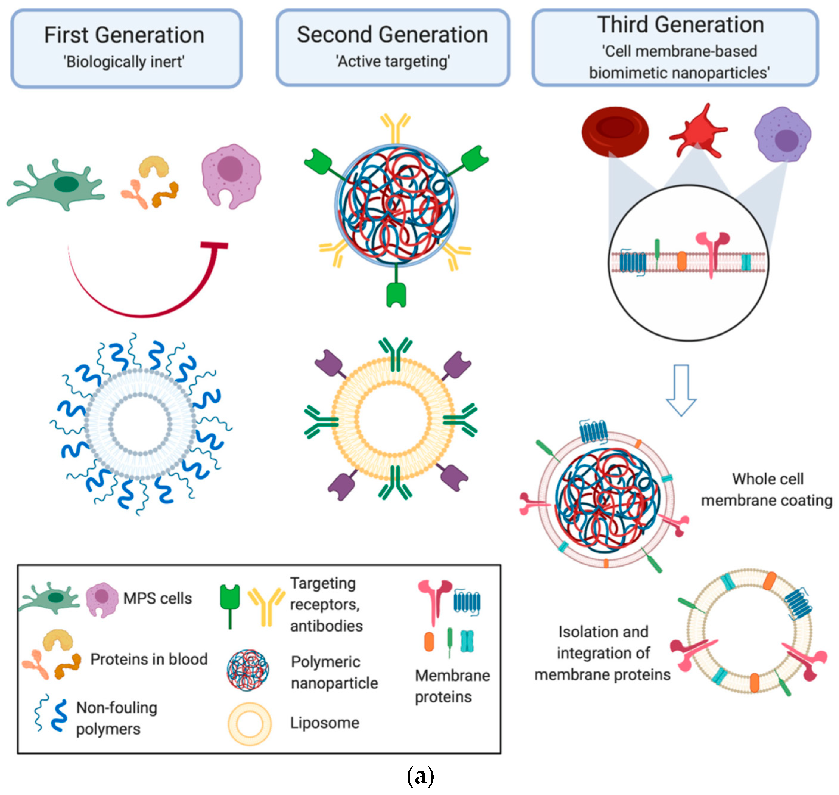
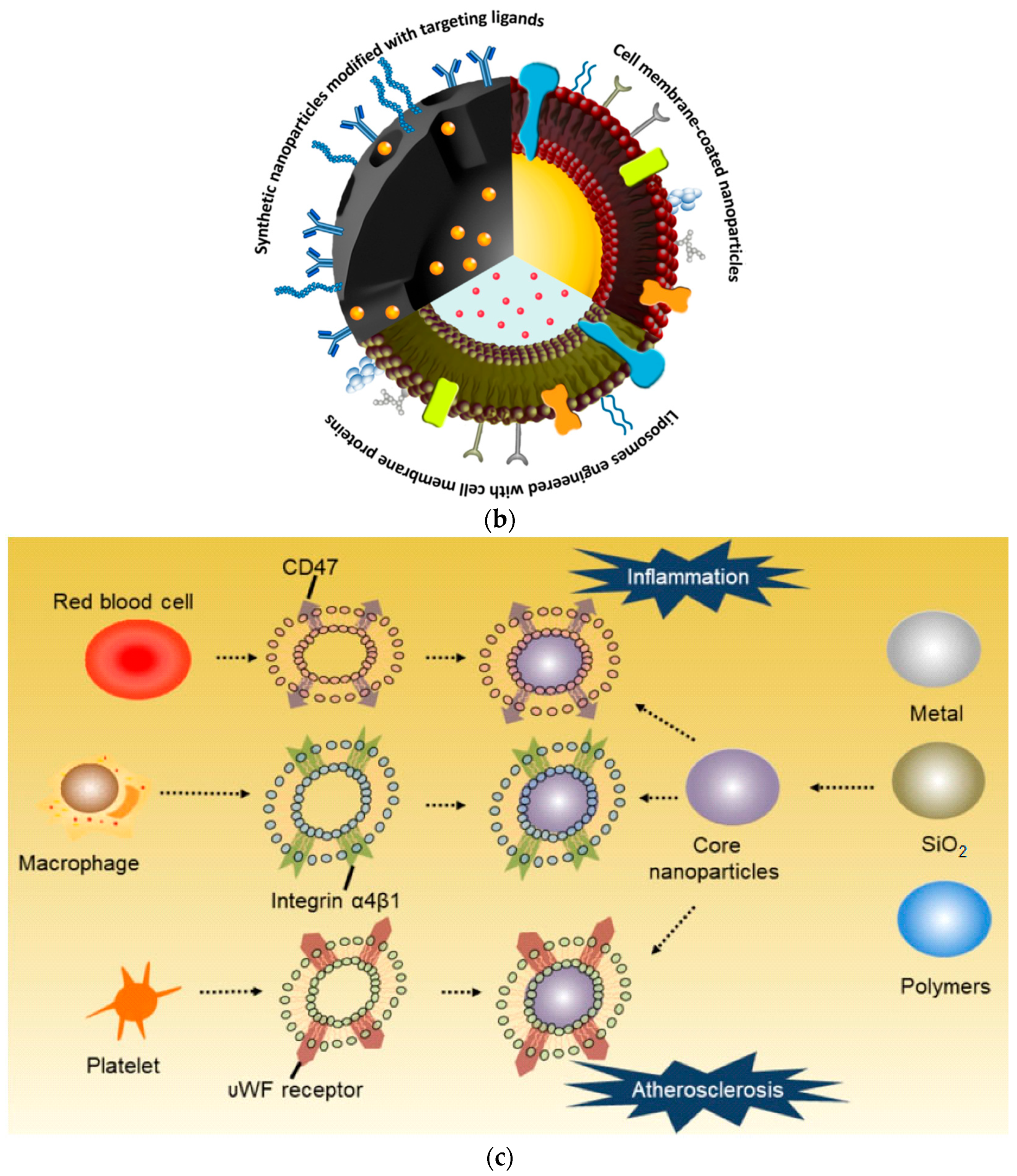
- (1)
- Synthetic NPs modified with targeting ligands that mimic cell surface proteins;
- (2)
- NPs covered with a native cell membrane;
- (3)
- Liposomes formed using cell membrane proteins (Figure 1b).
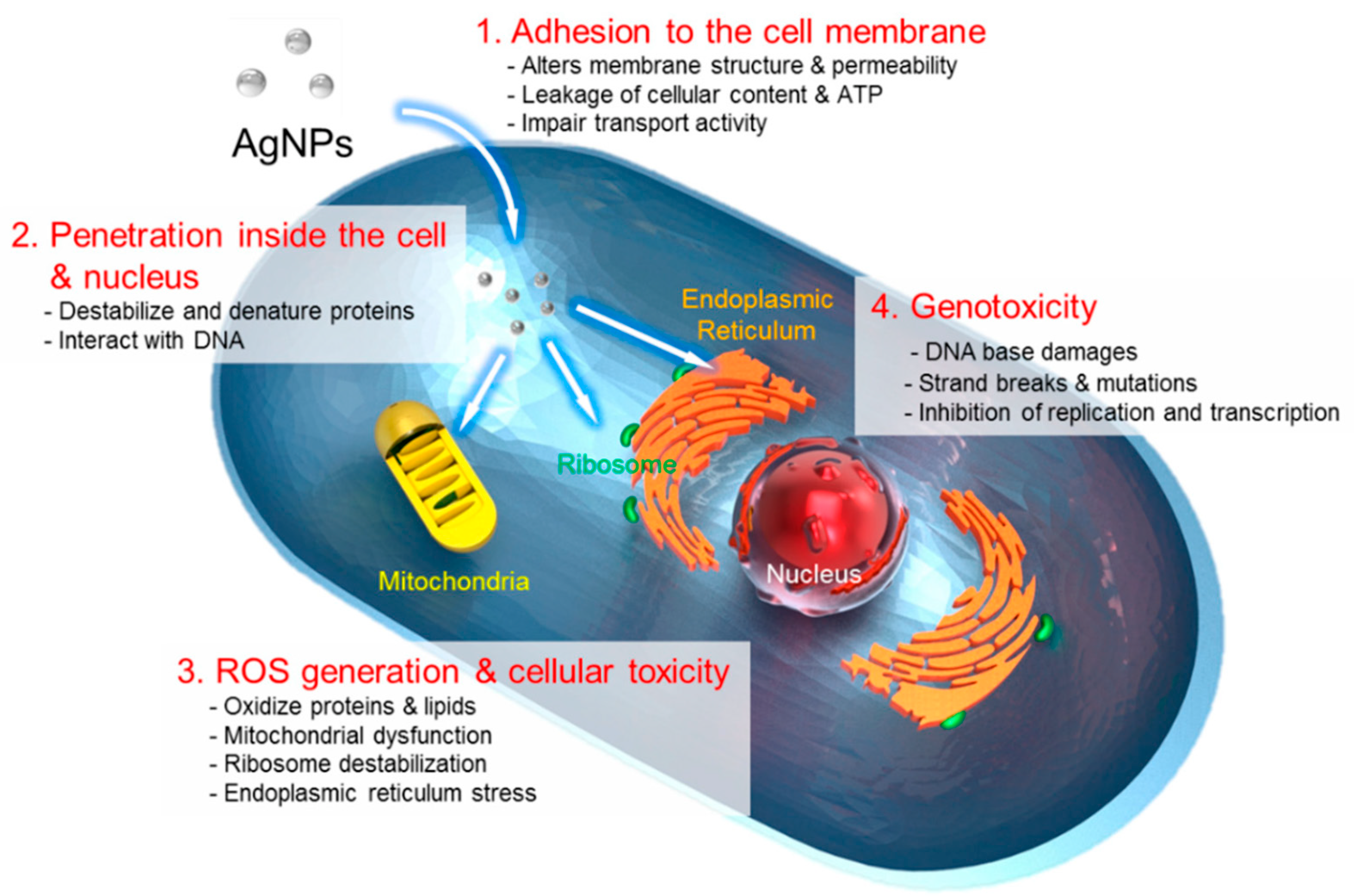
2. Interaction between Biomimetic Nanomaterials and Biological Tissue
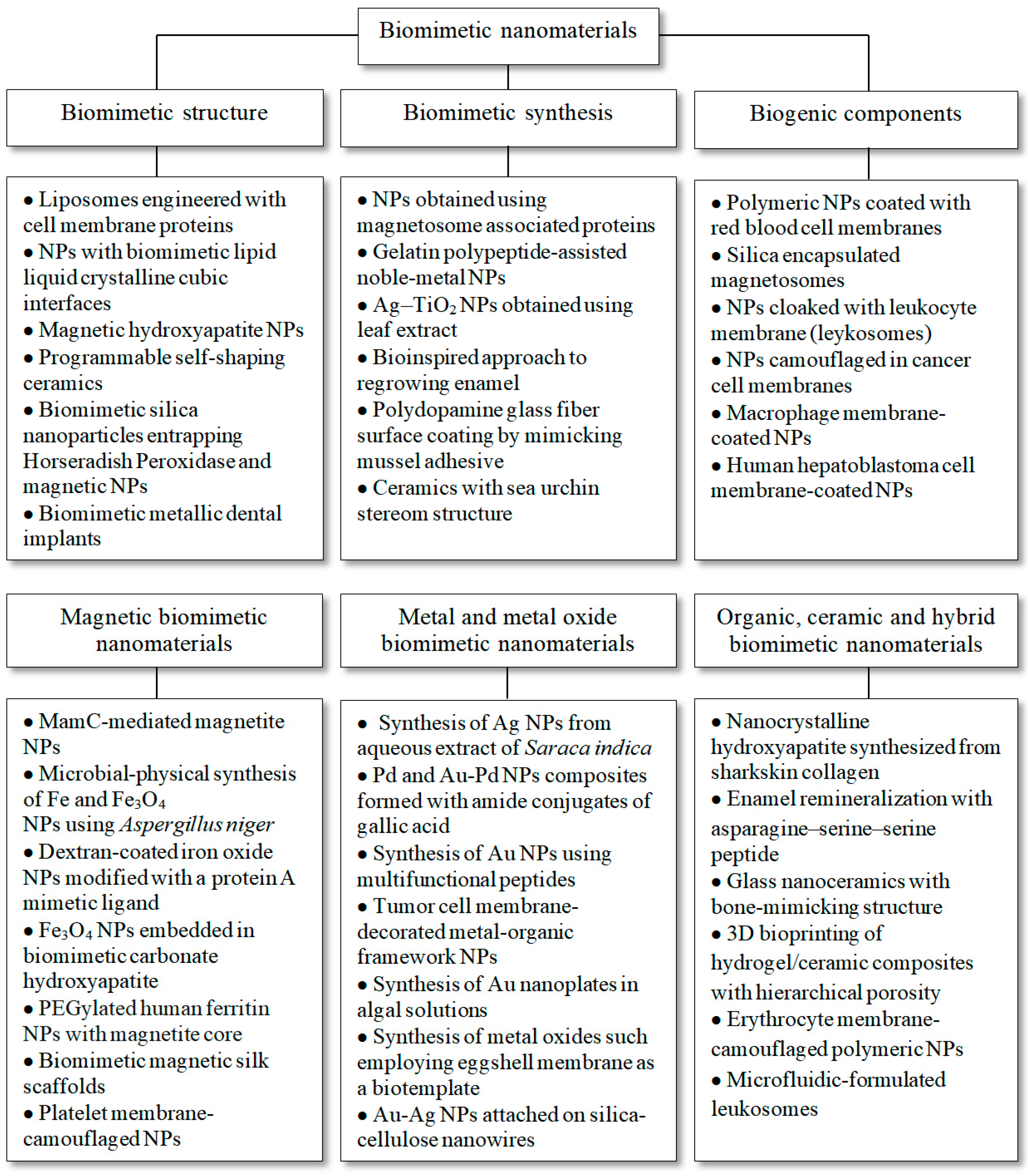
3. Magnetic Biomimetic Nanomaterials
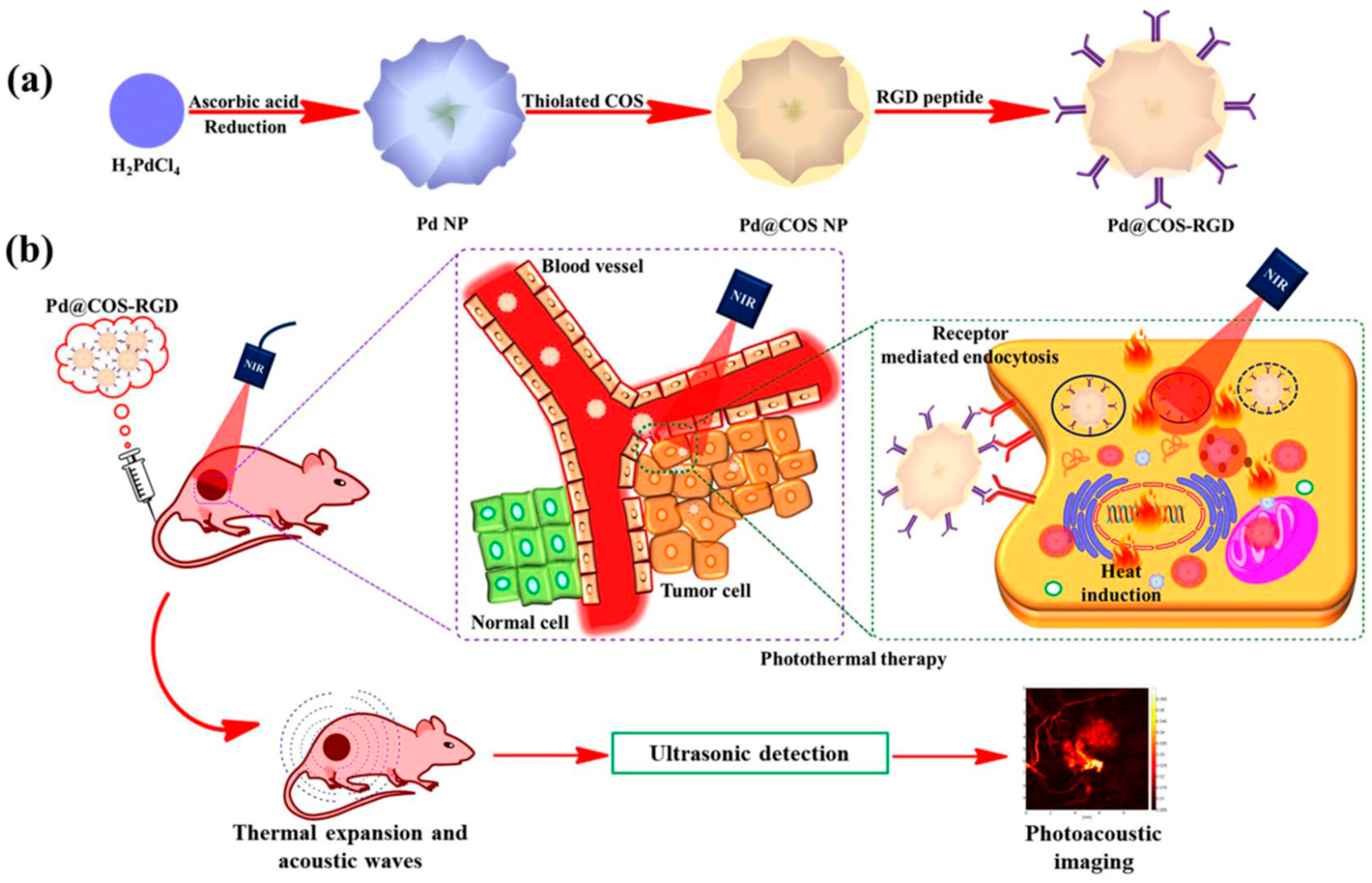
4. Metal and Metal Oxide Biomimetic Nanomaterials
5. Organic, Ceramic, and Hybrid Biomimetic Nanomaterials
6. Summary
Author Contributions
Funding
Institutional Review Board Statement
Informed Consent Statement
Data Availability Statement
Conflicts of Interest
References
- Zan, G.; Wu, Q. Biomimetic and Bioinspired Synthesis of Nanomaterials/Nanostructures. Adv. Mater. 2016, 28, 2099–2147. [Google Scholar] [CrossRef] [PubMed]
- Sheikhpour, M.; Barani, L.; Kasaeian, A. Biomimetics in drug delivery systems: A critical review. J. Control. Release 2017, 253, 97–109. [Google Scholar] [CrossRef] [PubMed]
- Das, D.; Noh, I. Biomimetic Medical Materials: From Nanotechnology to 3D Bioprinting; Springer: Berlin/Heidelberg, Germany, 2018; ISBN 9789811304446. [Google Scholar]
- Ruys, A.J. Biomimetic Biomaterials; Elsevier: Amsterdam, The Netherlands, 2013; ISBN 9780857092335. [Google Scholar]
- Poupon, E.; Nay, B. Biomimetic Organic Synthesis; Wiley: Hoboken, NJ, USA, 2011; Volume 1–2, ISBN 9783527325801. [Google Scholar]
- Moreira, F.T.C.; Guerreiro, J.R.L.; Brandão, L.; Sales, M.G.F. Synthesis of molecular biomimetics. In Biomimetic Technologies: Principles and Applications; Elsevier: Amsterdam, The Netherlands, 2015; pp. 3–31. ISBN 9780081002605. [Google Scholar]
- Takai, O. Biomimetic nanotechnology. Ann. N. Y. Acad. Sci. 2006, 1093, 84–97. [Google Scholar] [CrossRef] [PubMed]
- Garg, P.; Ghatmale, P.; Tarwadi, K.; Chavan, S. Influence of nanotechnology and the role of nanostructures in biomimetic studies and their potential applications. Biomimetics 2017, 2, 7. [Google Scholar] [CrossRef] [Green Version]
- Fatima, A.; Yasir, S.; Khan, M.S.; Manan, S.; Ullah, M.W.; Ul-Islam, M. Plant extract-loaded bacterial cellulose composite membrane for potential biomedical applications. J. Bioresour. Bioprod. 2021, 6, 26–32. [Google Scholar] [CrossRef]
- Saad, E.M.; Elshaarawy, R.F.; Mahmoud, S.A.; El-Moselhy, K.M. New Ulva lactuca Algae Based Chitosan Bio-composites for Bioremediation of Cd(II) Ions. J. Bioresour. Bioprod. 2021, 6, 223–242. [Google Scholar] [CrossRef]
- Chattopadhyay, S. Biomimetic Architectures by Plasma Processing: Fabrication and Applications; CRC Press: Boca Raton, FL, USA, 2014; ISBN 9789814463959. [Google Scholar]
- Ke, P.; Jiao, X.N.; Ge, X.H.; Xiao, W.M.; Yu, B. From macro to micro: Structural biomimetic materials by electrospinning. RSC Adv. 2014, 4, 39704–39724. [Google Scholar] [CrossRef]
- Vincent, J.F.V. Biomimetic materials. J. Mater. Res. 2008, 23, 3140–3147. [Google Scholar] [CrossRef]
- Gagliardi, M. Biomimetic and bioinspired nanoparticles for targeted drug delivery. Ther. Deliv. 2017, 8, 289–299. [Google Scholar] [CrossRef]
- Parodi, A.; Molinaro, R.; Sushnitha, M.; Evangelopoulos, M.; Martinez, J.O.; Arrighetti, N.; Corbo, C.; Tasciotti, E. Bio-inspired engineering of cell- and virus-like nanoparticles for drug delivery. Biomaterials 2017, 147, 155–168. [Google Scholar] [CrossRef]
- Jin, K.; Luo, Z.; Zhang, B.; Pang, Z. Biomimetic nanoparticles for inflammation targeting. Acta Pharm. Sin. B 2018, 8, 23–33. [Google Scholar] [CrossRef]
- Unnithan, A.R.; Sasikala, A.R.K.; Park, C.H.; Kim, C.S. Biomimetic Nanoengineered Materials for Advanced Drug Delivery; Elsevier: Amsterdam, The Netherlands, 2019; ISBN 9780128149447. [Google Scholar]
- Akagi, T.; Cabral, H.; Mi, P. Bio-inspired nanomaterials for biomedical innovation. Sci. Technol. Adv. Mater. 2020, 21, 420–421. [Google Scholar] [CrossRef]
- Wang, H.; Liu, Y.; He, R.; Xu, D.; Zang, J.; Weeranoppanant, N.; Dong, H.; Li, Y. Cell membrane biomimetic nanoparticles for inflammation and cancer targeting in drug delivery. Biomater. Sci. 2020, 8, 552–568. [Google Scholar] [CrossRef]
- Yang, L.; Zang, G.; Li, J.; Li, X.; Li, Y.; Zhao, Y. Cell-derived biomimetic nanoparticles as a novel drug delivery system for atherosclerosis: Predecessors and perspectives. Regen. Biomater. 2020, 7, 349–358. [Google Scholar] [CrossRef]
- Zhao, Y.; Li, A.; Jiang, L.; Gu, Y.; Liu, J. Hybrid Membrane-Coated Biomimetic Nanoparticles (HM@BNPs): A Multifunctional Nanomaterial for Biomedical Applications. Biomacromolecules 2021, 22, 3149–3167. [Google Scholar] [CrossRef]
- Feng, Z.; Fan, H.; Cheng, L.; Zhang, H.; Fan, H.; Liu, J. Advanced Biomimetic Nanomaterials for Non-invasive Disease Diagnosis. Front. Mater. 2021, 8, 664795. [Google Scholar] [CrossRef]
- Zhu, L.; Zhong, Y.; Wu, S.; Yan, M.; Cao, Y.; Mou, N.; Wang, G.; Sun, D.; Wu, W. Cell membrane camouflaged biomimetic nanoparticles: Focusing on tumor theranostics. Mater. Today Bio. 2022, 14, 100228. [Google Scholar] [CrossRef]
- Zhao, X.; Yan, C. Research Progress of Cell Membrane Biomimetic Nanoparticles for Tumor Therapy. Nanoscale Res. Lett. 2022, 17, 36. [Google Scholar] [CrossRef]
- Liu, R.; An, Y.; Jia, W.; Wang, Y.; Wu, Y.; Zhen, Y.; Cao, J.; Gao, H. Macrophage-mimic shape changeable nanomedicine retained in tumor for multimodal therapy of breast cancer. J. Control. Release 2020, 321, 589–601. [Google Scholar] [CrossRef]
- Yu, W.; He, X.; Yang, Z.; Yang, X.; Xiao, W.; Liu, R.; Xie, R.; Qin, L.; Gao, H. Sequentially responsive biomimetic nanoparticles with optimal size in combination with checkpoint blockade for cascade synergetic treatment of breast cancer and lung metastasis. Biomaterials 2019, 217, 119309. [Google Scholar] [CrossRef]
- Stratakis, E.; Ranella, A.; Fotakis, C. Biomimetic micro/nanostructured functional surfaces for microfluidic and tissue engineering applications. Biomicrofluidics 2011, 5, 013411. [Google Scholar] [CrossRef] [Green Version]
- Mano, J.F. Biomimetic Approaches for Biomaterials Development; Wiley: Hoboken, NJ, USA, 2012; ISBN 9783527329168. [Google Scholar]
- Pasha, M.; Muhammad, N.; Shahnawaz, S.; Najmi, Y.; Shahroz, N.; Liaqat, S. Ceramic Nanomaterials in Dental Applications. In Nanoengineering of Biomaterials; Wiley: Hoboken, NJ, USA, 2022; pp. 123–144. [Google Scholar] [CrossRef]
- Narayan, R.J.; Kumta, P.N.; Wagner, W.R. Advances in Biomedical and Biomimetic Materials; Wiley: Hoboken, NJ, USA, 2008; ISBN 9780470538357. [Google Scholar]
- Sushnitha, M.; Evangelopoulos, M.; Tasciotti, E.; Taraballi, F. Cell Membrane-Based Biomimetic Nanoparticles and the Immune System: Immunomodulatory Interactions to Therapeutic Applications. Front. Bioeng. Biotechnol. 2020, 8, 627. [Google Scholar] [CrossRef]
- Peigneux, A.; Jabalera, Y.; Vivas, M.A.F.; Casares, S.; Azuaga, A.I.; Jimenez-Lopez, C. Tuning properties of biomimetic magnetic nanoparticles by combining magnetosome associated proteins. Sci. Rep. 2019, 9, 8804. [Google Scholar] [CrossRef]
- Ben-Akiva, E.; Meyer, R.A.; Yu, H.; Smith, J.T.; Pardoll, D.M.; Green, J.J. Biomimetic anisotropic polymeric nanoparticles coated with red blood cell membranes for enhanced circulation and toxin removal. Sci. Adv. 2020, 6, eaay9035. [Google Scholar] [CrossRef]
- Cardellini, J.; Montis, C.; Barbero, F.; De Santis, I.; Caselli, L.; Berti, D. Interaction of Metallic Nanoparticles with Biomimetic Lipid Liquid Crystalline Cubic Interfaces. Front. Bioeng. Biotechnol. 2022, 10, 848687. [Google Scholar] [CrossRef]
- Mondal, S.; Manivasagan, P.; Bharathiraja, S.; Moorthy, M.S.; Kim, H.H.; Seo, H.; Lee, K.D.; Oh, J. Magnetic hydroxyapatite: A promising multifunctional platform for nanomedicine application. Int. J. Nanomed. 2017, 12, 8389–8410. [Google Scholar] [CrossRef] [Green Version]
- Bargardi, F.L.; Le Ferrand, H.; Libanori, R.; Studart, A.R. Bio-inspired self-shaping ceramics. Nat. Commun. 2016, 7, 13912. [Google Scholar] [CrossRef]
- Correa, S.; Puertas, S.; Gutiérrez, L.; Asín, L.; De La Fuente, J.M.; Grazú, V.; Betancor, L. Design of stable magnetic hybrid nanoparticles of Si-entrapped HRP. PLoS ONE 2019, 14, e0214004. [Google Scholar] [CrossRef] [PubMed]
- Al-Zubaidi, S.M.; Madfa, A.A.; Mufadhal, A.A.; Aldawla, M.A.; Hameed, O.S.; Yue, X.G. Improvements in Clinical Durability from Functional Biomimetic Metallic Dental Implants. Front. Mater. 2020, 7, 106. [Google Scholar] [CrossRef]
- Liu, Y.; Liu, X.; Wang, X. Biomimetic Synthesis of Gelatin Polypeptide-Assisted Noble-Metal Nanoparticles and Their Interaction Study. Nanoscale Res. Lett. 2011, 6, 22. [Google Scholar] [CrossRef] [PubMed] [Green Version]
- Bhardwaj, D.; Singh, R. Green biomimetic synthesis of Ag–TiO2 nanocomposite using Origanum majorana leaf extract under sonication and their biological activities. Bioresour. Bioprocess. 2021, 8, 1. [Google Scholar] [CrossRef]
- Mukherjee, K.; Ruan, Q.; Nutt, S.; Tao, J.; De Yoreo, J.J.; Moradian-Oldak, J. Peptide-Based Bioinspired Approach to Regrowing Multilayered Aprismatic Enamel. ACS Omega 2018, 3, 2546–2557. [Google Scholar] [CrossRef] [Green Version]
- Wen, X.; Xiao, Z.; Jiang, T.; Li, J.; Zhang, W.; Zhang, L.; Shao, H. Constructing novel fiber reinforced plastic (FRP) composites through a biomimetic approach: Connecting glass fiber with nanosized boron nitride by polydopamine coating. J. Nanomater. 2013, 2013, 470583. [Google Scholar] [CrossRef]
- Yu, H.; Li, J.; Li, J.; Chen, Y.; Hou, X.; Chen, S.; Yang, H. Biomimetic preparation of a ceramic combined with sea urchin stereom structure and nacre mineral bridge structure. Mater. Des. 2019, 178, 107844. [Google Scholar] [CrossRef]
- Mickoleit, F. Generation of nanomagnetic biocomposites by genetic engineering of bacterial magnetosomes. Bioinspired, Biomim. Nanobiomaterials 2019, 8, 86–98. [Google Scholar] [CrossRef] [Green Version]
- Jin, J.; Bhujwalla, Z.M. Biomimetic Nanoparticles Camouflaged in Cancer Cell Membranes and Their Applications in Cancer Theranostics. Front. Oncol. 2020, 9, 1560. [Google Scholar] [CrossRef] [Green Version]
- Tang, D.; Wang, Y.; Wijaya, A.; Liu, B.; Maruf, A.; Wang, J.; Xu, J.; Liao, X.; Wu, W.; Wang, G. ROS-responsive biomimetic nanoparticles for potential application in targeted anti-atherosclerosis. Regen. Biomater. 2021, 8, rbab033. [Google Scholar] [CrossRef]
- Wang, D.; Chen, L.; Li, C.; Long, Q.; Yang, Q.; Huang, A.; Tang, H. CRISPR/Cas9 delivery by NIR-responsive biomimetic nanoparticles for targeted HBV therapy. J. Nanobiotechnol. 2022, 20, 27. [Google Scholar] [CrossRef]
- Peigneux, A.; Glitscher, E.A.; Charbaji, R.; Weise, C.; Wedepohl, S.; Calderón, M.; Jimenez-Lopez, C.; Hedtrich, S. Protein corona formation and its influence on biomimetic magnetite nanoparticles. J. Mater. Chem. B 2020, 8, 4870–4882. [Google Scholar] [CrossRef]
- Abdeen, M.; Sabry, S.; Ghozlan, H.; El-Gendy, A.A.; Carpenter, E.E. Microbial-Physical Synthesis of Fe and Fe3O4 Magnetic Nanoparticles Using Aspergillus Niger YESM1 and Supercritical Condition of Ethanol. J. Nanomater. 2016, 2016, 9174891. [Google Scholar] [CrossRef] [Green Version]
- Santana, S.D.F.; Dhadge, V.L.; Roque, A.C.A. Dextran-coated magnetic supports modified with a biomimetic ligand for IgG purification. ACS Appl. Mater. Interfaces 2012, 4, 5907–5914. [Google Scholar] [CrossRef]
- Del Bianco, L.; Lesci, I.G.; Fracasso, G.; Barucca, G.; Spizzo, F.; Tamisari, M.; Scotti, R.; Ciocca, L. Synthesis of nanogranular Fe3O4/biomimetic hydroxyapatite for potential applications in nanomedicine: Structural and magnetic characterization. Mater. Res. Express 2015, 2, 65002. [Google Scholar] [CrossRef]
- Yang, C.; Cao, C.; Cai, Y.; Xu, H.; Zhang, T.; Pan, Y. Effects of PEGylation on biomimetic synthesis of magnetoferritin nanoparticles. J. Nanopart. Res. 2017, 19, 101. [Google Scholar] [CrossRef]
- Samal, S.K.; Dash, M.; Shelyakova, T.; Declercq, H.A.; Uhlarz, M.; Bañobre-López, M.; Dubruel, P.; Cornelissen, M.; Herrmannsdörfer, T.; Rivas, J.; et al. Biomimetic magnetic silk scaffolds. ACS Appl. Mater. Interfaces 2015, 7, 6282–6292. [Google Scholar] [CrossRef]
- Jiang, Q.; Wang, K.; Zhang, X.; Ouyang, B.; Liu, H.; Pang, Z.; Yang, W. Platelet Membrane-Camouflaged Magnetic Nanoparticles for Ferroptosis-Enhanced Cancer Immunotherapy. Small 2020, 16, e2001704. [Google Scholar] [CrossRef]
- Singh, M.; Renu; Kumar, V.; Upadhyay, S.K.; Singh, R.; Yadav, M.; Seema; Kumari, S.; Sharma, A.K.; Manikanadan, S. Biomimetic synthesis of silver nanoparticles from aqueous extract of saraca indica and its profound antibacterial activity. Biointerface Res. Appl. Chem. 2021, 11, 8110–8120. [Google Scholar] [CrossRef]
- Sarker, N.H.; Barnaby, S.N.; Dowdell, A.P.; Nakatsuka, N.; Banerjee, I.A. Biomimetic formation of Pd and Au-Pd nanocomposites and their catalytic applications. Soft Mater. 2013, 11, 403–413. [Google Scholar] [CrossRef]
- Slocik, J.M.; Stone, M.O.; Naik, R.R. Synthesis of gold nanoparticles using multifunctional peptides. Small 2005, 1, 1048–1052. [Google Scholar] [CrossRef] [PubMed]
- Min, H.; Wang, J.; Qi, Y.; Zhang, Y.; Han, X.; Xu, Y.; Xu, J.; Li, Y.; Chen, L.; Cheng, K.; et al. Biomimetic Metal–Organic Framework Nanoparticles for Cooperative Combination of Antiangiogenesis and Photodynamic Therapy for Enhanced Efficacy. Adv. Mater. 2019, 31, e1808200. [Google Scholar] [CrossRef] [PubMed]
- Xie, J.; Lee, J.Y.; Wang, D.I.C.; Ting, Y.P. Identification of active biomolecules in the high-yield synthesis of single-crystalline gold nanoplates in algal solutions. Small 2007, 3, 672–682. [Google Scholar] [CrossRef] [PubMed]
- Mallampati, R.; Valiyaveettil, S. Biomimetic metal oxides for the extraction of nanoparticles from water. Nanoscale 2013, 5, 3395–3399. [Google Scholar] [CrossRef]
- Gruber, S.; Zollfrank, C. Noble metal nanoparticles on biotemplated nanowires. Bioinspired Biomim. Nanobiomater. 2012, 1, 95–100. [Google Scholar] [CrossRef]
- Banerjee, P.; Das, J. Biomimetic synthesis of nanocrystalline hydroxyapatite from sharkskin collagen. Bioinspired Biomim. Nanobiomater. 2017, 7, 27–36. [Google Scholar] [CrossRef]
- Chung, H.Y.; Li, C.C. Microstructure and nanomechanical properties of enamel remineralized with asparagine-serine-serine peptide. Mater. Sci. Eng. C 2013, 33, 969–973. [Google Scholar] [CrossRef]
- Fu, C.; Ravindra, N.M. Magnetic iron oxide nanoparticles: Synthesis and applications. Bioinspired Biomim. Nanobiomater. 2012, 1, 229–244. [Google Scholar] [CrossRef]
- Mainardi, J.C.; Rezwan, K.; Maas, M. Genipin-crosslinked chitosan/alginate/alumina nanocomposite gels for 3D bioprinting. Bioprocess Biosyst. Eng. 2022, 45, 171–185. [Google Scholar] [CrossRef]
- Hu, C.M.J.; Zhang, L.; Aryal, S.; Cheung, C.; Fang, R.H.; Zhang, L. Erythrocyte membrane-camouflaged polymeric nanoparticles as a biomimetic delivery platform. Proc. Natl. Acad. Sci. USA 2011, 108, 10980–10985. [Google Scholar] [CrossRef] [Green Version]
- Molinaro, R.; Evangelopoulos, M.; Hoffman, J.R.; Corbo, C.; Taraballi, F.; Martinez, J.O.; Hartman, K.A.; Cosco, D.; Costa, G.; Romeo, I.; et al. Design and Development of Biomimetic Nanovesicles Using a Microfluidic Approach. Adv. Mater. 2018, 30, e1702749. [Google Scholar] [CrossRef]
- Singh, D.; Singh, D.; Zo, S.; Han, S.S. Nano-biomimetics for nano/micro tissue regeneration. J. Biomed. Nanotechnol. 2014, 10, 3141–3161. [Google Scholar] [CrossRef]
- Bharathiraja, S.; Bui, N.Q.; Manivasagan, P.; Moorthy, M.S.; Mondal, S.; Seo, H.; Phuoc, N.T.; Vy Phan, T.T.; Kim, H.; Lee, K.D.; et al. Multimodal tumor-homing chitosan oligosaccharide-coated biocompatible palladium nanoparticles for photo-based imaging and therapy. Sci. Rep. 2018, 8, 500. [Google Scholar] [CrossRef]
- Albert, K.; Huang, X.C.; Hsu, H.Y. Bio-templated silica composites for next-generation biomedical applications. Adv. Colloid Interface Sci. 2017, 249, 272–289. [Google Scholar] [CrossRef]
- Yang, Y.; Zhang, M.; Song, H.; Yu, C. Silica-Based Nanoparticles for Biomedical Applications: From Nanocarriers to Biomodulators. Acc. Chem. Res. 2020, 53, 1545–1556. [Google Scholar] [CrossRef]
- Ghanbari, H.; de Mel, A.; Seifalian, A.M. Cardiovascular application of polyhedral oligomeric silsesquioxane nanomaterials: A glimpse into prospective horizons. Int. J. Nanomed. 2011, 6, 775–786. [Google Scholar] [CrossRef] [Green Version]
- Hasanzadeh, M.; Babaie, P.; Jouyban-Gharamaleki, V.; Jouyban, A. The use of chitosan as a bioactive polysaccharide in non-invasive detection of malondialdehyde biomarker in human exhaled breath condensate: A new platform towards diagnosis of some lung disease. Int. J. Biol. Macromol. 2018, 120, 2482–2492. [Google Scholar] [CrossRef]
- Liang, K.; Chen, H. Protein-based nanoplatforms for tumor imaging and therapy. Wiley Interdiscip. Rev. Nanomed. Nanobiotechnology 2020, 12, e1616. [Google Scholar] [CrossRef]
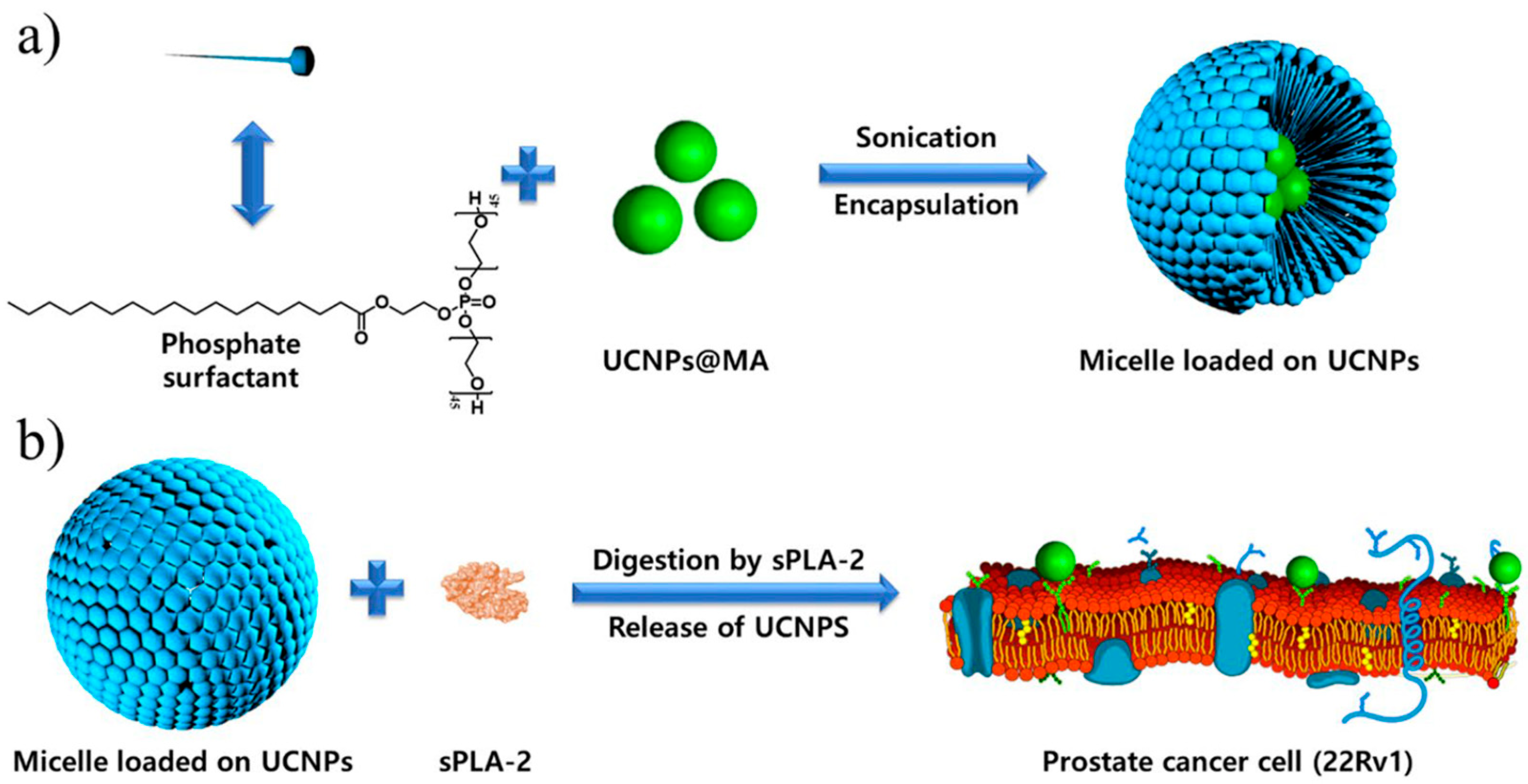
- Sharipov, M.; Tawfik, S.M.; Gerelkhuu, Z.; Huy, B.T.; Lee, Y.I. Phospholipase A2-Responsive Phosphate Micelle-Loaded UCNPs for Bioimaging of Prostate Cancer Cells. Sci. Rep. 2017, 7, 16073. [Google Scholar] [CrossRef] [Green Version]
- Renu, S.; Shivashangari, K.S.; Ravikumar, V. Incorporated plant extract fabricated silver/poly-D,L-lactide-co-glycolide nanocomposites for antimicrobial based wound healing. Spectrochim. Acta Part A Mol. Biomol. Spectrosc. 2020, 228, 117673. [Google Scholar] [CrossRef]
- Zhao, W.; Li, A.; Zhang, A.; Zheng, Y.; Liu, J. Recent Advances in Functional-Polymer-Decorated Transition-Metal Nanomaterials for Bioimaging and Cancer Therapy. ChemMedChem 2018, 13, 2134–2149. [Google Scholar] [CrossRef] [PubMed]
- Wu, K.; Su, D.; Liu, J.; Saha, R.; Wang, J.P. Magnetic nanoparticles in nanomedicine: A review of recent advances. Nanotechnology 2019, 30, 502003. [Google Scholar] [CrossRef] [PubMed] [Green Version]
- Knežević, N.; Gadjanski, I.; Durand, J.O. Magnetic nanoarchitectures for cancer sensing, imaging and therapy. J. Mater. Chem. B 2019, 7, 9–23. [Google Scholar] [CrossRef] [PubMed]
- Poon, C.; Gallo, J.; Joo, J.; Chang, T.; Bañobre-López, M.; Chung, E.J. Hybrid, metal oxide-peptide amphiphile micelles for molecular magnetic resonance imaging of atherosclerosis. J. Nanobiotechnol. 2018, 16, 92. [Google Scholar] [CrossRef] [Green Version]
- Lee, S.H.; Jun, B.H. Silver nanoparticles: Synthesis and application for nanomedicine. Int. J. Mol. Sci. 2019, 20, 865. [Google Scholar] [CrossRef] [Green Version]
- Klem, M.T.; Young, M.; Douglas, T. Biomimetic magnetic nanoparticles. Mater. Today 2005, 8, 28–37. [Google Scholar] [CrossRef]
- Altan, C.L. Biomimetic synthesis, Magnetic Properties and Applications of Magnetite Nanoparticles; Technische Universiteit Eindhoven: Eindhoven, The Netherlands, 2014; ISBN 978-94-6108-843-7. [Google Scholar]
- Kralj, S.; Marchesan, S. Bioinspired Magnetic Nanochains for Medicine. Pharmaceutics 2021, 13, 1262. [Google Scholar] [CrossRef]
- Vurro, F.; Jabalera, Y.; Mannucci, S.; Glorani, G.; Sola-Leyva, A.; Gerosa, M.; Romeo, A.; Romanelli, M.G.; Malatesta, M.; Calderan, L.; et al. Improving the cellular uptake of biomimetic magnetic nanoparticles. Nanomaterials 2021, 11, 766. [Google Scholar] [CrossRef]
- Jabalera, Y.; Sola-Leyva, A.; Carrasco-Jiménez, M.P.; Iglesias, G.R.; Jimenez-Lopez, C. Synergistic photothermal-chemotherapy based on the use of biomimetic magnetic nanoparticles. Pharmaceutics 2021, 13, 625. [Google Scholar] [CrossRef]
- Salem, K.; Jabalera, Y.; Puentes-Pardo, J.D.; Vilchez-Garcia, J.; Sayari, A.; Hmida-Sayari, A.; Jimenez-Lopez, C.; Perduca, M. Enzyme Storage and Recycling: Nanoassemblies of α-Amylase and Xylanase Immobilized on Biomimetic Magnetic Nanoparticles. ACS Sustain. Chem. Eng. 2021, 9, 4054–4063. [Google Scholar] [CrossRef]
- Gareev, K.G.; Grouzdev, D.S.; Kharitonskii, P.V.; Kosterov, A.; Koziaeva, V.V.; Sergienko, E.S.; Shevtsov, M.A. Magnetotactic bacteria and magnetosomes: Basic properties and applications. Magnetochemistry 2021, 7, 86. [Google Scholar] [CrossRef]
- Taher, Z.; Legge, C.; Winder, N.; Lysyganicz, P.; Rawlings, A.; Bryant, H.; Muthana, M.; Staniland, S. Magnetosomes and magnetosome mimics: Preparation, cancer cell uptake and functionalization for future cancer therapies. Pharmaceutics 2021, 13, 367. [Google Scholar] [CrossRef]
- Goyal, G.; Bhakta, S.; Mishra, P. Surface Molecularly Imprinted Biomimetic Magnetic Nanoparticles for Enantioseparation. ACS Appl. Nano Mater. 2019, 2, 6747–6756. [Google Scholar] [CrossRef]
- Lenders, J.J.M.; Bawazer, L.A.; Green, D.C.; Zope, H.R.; Bomans, P.H.H.; de With, G.; Kros, A.; Meldrum, F.C.; Sommerdijk, N.A.J.M. Combinatorial Evolution of Biomimetic Magnetite Nanoparticles. Adv. Funct. Mater. 2017, 27, 1604863. [Google Scholar] [CrossRef] [Green Version]
- Bu, Y.; Hu, Q.; Ke, R.; Sui, Y.; Xie, X.; Wang, S. Cell membrane camouflaged magnetic nanoparticles as a biomimetic drug discovery platform. Chem. Commun. 2018, 54, 13427–13430. [Google Scholar] [CrossRef]
- Ren, Y.; Rivera, J.G.; He, L.; Kulkarni, H.; Lee, D.K.; Messersmith, P.B. Facile, high efficiency immobilization of lipase enzyme on magnetic iron oxide nanoparticles via a biomimetic coating. BMC Biotechnol. 2011, 11, 63. [Google Scholar] [CrossRef] [PubMed] [Green Version]
- Tengood, J.E.; Alferiev, I.S.; Zhang, K.; Fishbein, I.; Levy, R.J.; Chorny, M. Real-time analysis of composite magnetic nanoparticle disassembly in vascular cells and biomimetic media. Proc. Natl. Acad. Sci. USA 2014, 111, 4245–4250. [Google Scholar] [CrossRef] [PubMed] [Green Version]
- Zhang, T.; Li, F.; Xu, Q.; Wang, Q.; Jiang, X.; Liang, Z.; Liao, H.; Kong, X.; Liu, J.; Wu, H.; et al. Ferrimagnetic Nanochains-Based Mesenchymal Stem Cell Engineering for Highly Efficient Post-Stroke Recovery. Adv. Funct. Mater. 2019, 29, 1900603. [Google Scholar] [CrossRef]
- Deravi, L.F.; Swartz, J.D.; Wright, D.W. The Biomimetic Synthesis of Metal Oxide Nanomaterials; Wiley: Hoboken, NJ, USA, 2010; Volume 2, ISBN 9783527321520. [Google Scholar]
- Prasad, R.; Kumar, M.; Kumar, V. Nanotechnology: An Agricultural Paradigm; Springer: Berlin/Heidelberg, Germany, 2017; ISBN 9789811045738. [Google Scholar]
- Singh, S.; Abbasi, B.H. Biomimetic synthesis of antimicrobial silver nanoparticles using in vitro-propagated plantlets of a medicinally important endangered species: Phlomis bracteosa. Int. J. Nanomed. 2016, 11, 1663–1675. [Google Scholar] [CrossRef] [Green Version]
- Bag, S.S.; Bora, A.; Golder, A.K. Biomimetic Synthesis of Silver Nanoparticles Using Bhimkol (Musa balbisiana) Peel Extract as Biological Waste: Its Antibacterial Activity and Role of Ripen Stage of the Peel. Curr. Nanomater. 2020, 5, 47–65. [Google Scholar] [CrossRef]
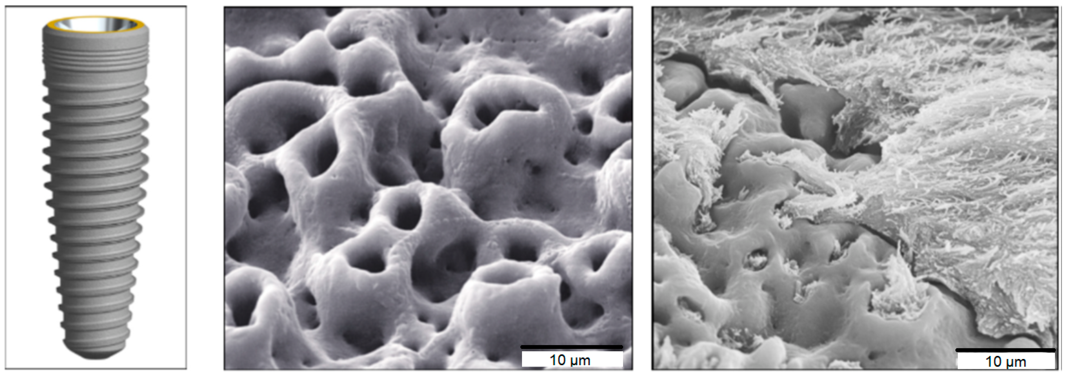
- Le Guéhennec, L.; Soueidan, A.; Layrolle, P.; Amouriq, Y. Surface treatments of titanium dental implants for rapid osseointegration. Dent. Mater. 2007, 23, 844–854. [Google Scholar] [CrossRef]
- Chen, H.; Qiu, Q.; Sharif, S.; Ying, S.; Wang, Y.; Ying, Y. Solution-Phase Synthesis of Platinum Nanoparticle-Decorated Metal-Organic Framework Hybrid Nanomaterials as Biomimetic Nanoenzymes for Biosensing Applications. ACS Appl. Mater. Interfaces 2018, 10, 24108–24115. [Google Scholar] [CrossRef]
- Azam, T.; Pervaiz, E.; Farrukh, S.; Noor, T. Biomimetic highly hydrophobic stearic acid functionalized MOF sponge for efficient oil/water separation. Mater. Res. Express 2021, 8, 015019. [Google Scholar] [CrossRef]
- Bim-Junior, O.; Alania, Y.; Tabatabaei, F.S.; Frem, R.; Bedran-Russo, A.K.; Lisboa-Filho, P.N. Biomimetic Growth of Metal–Organic Frameworks for the Stabilization of the Dentin Matrix and Control of Collagenolysis. Langmuir 2022, 38, 1600–1610. [Google Scholar] [CrossRef]
- Chen, K.; Wu, C. De Designed fabrication of biomimetic metal–organic frameworks for catalytic applications. Coord. Chem. Rev. 2019, 378, 445–465. [Google Scholar] [CrossRef]
- Cheng, X.; Zhang, S.; Liu, H.; Chen, H.; Zhou, J.; Chen, Z.; Zhou, X.; Xie, Z.; Kuang, Q.; Zheng, L. Biomimetic Metal-Organic Framework Composite-Mediated Cascade Catalysis for Synergistic Bacteria Killing. ACS Appl. Mater. Interfaces 2020, 12, 36996–37005. [Google Scholar] [CrossRef]
- Li, S.F.; Chen, Y.; Wang, Y.S.; Mo, H.L.; Zang, S.Q. Integration of enzyme immobilization and biomimetic catalysis in hierarchically porous metal-organic frameworks for multi-enzymatic cascade reactions. Sci. China Chem. 2022, 65, 1122–1128. [Google Scholar] [CrossRef]
- Gu, Z.Y.; Park, J.; Raiff, A.; Wei, Z.; Zhou, H.C. Metal-organic frameworks as biomimetic catalysts. ChemCatChem 2014, 6, 67–75. [Google Scholar] [CrossRef]
- Liang, K.; Ricco, R.; Doherty, C.M.; Styles, M.J.; Bell, S.; Kirby, N.; Mudie, S.; Haylock, D.; Hill, A.J.; Doonan, C.J.; et al. Biomimetic mineralization of metal-organic frameworks as protective coatings for biomacromolecules. Nat. Commun. 2015, 6, 4–11. [Google Scholar] [CrossRef] [Green Version]
- Singh, R.; White, J.F.; de Vries, M.; Beddome, G.; Dai, M.; Bean, A.G.; Mulet, X.; Layton, D.; Doherty, C.M. Biomimetic metal-organic frameworks as protective scaffolds for live-virus encapsulation and vaccine stabilization. Acta Biomater. 2022, 142, 320–331. [Google Scholar] [CrossRef]
- Wang, X.N.; Zhao, Y.; Li, J.L.; Pang, J.D.; Wang, Q.; Li, B.; Zhou, H.C. Biomimetic catalysts of iron-based metal-organic frameworks with high peroxidase-mimicking activity for colorimetric biosensing. Dalt. Trans. 2021, 50, 3854–3861. [Google Scholar] [CrossRef]
- Wright, A.M.; Wu, Z.; Zhang, G.; Mancuso, J.L.; Comito, R.J.; Day, R.W.; Hendon, C.H.; Miller, J.T.; Dincă, M. A Structural Mimic of Carbonic Anhydrase in a Metal-Organic Framework. Chem 2018, 4, 2894–2901. [Google Scholar] [CrossRef] [Green Version]
- Ma, X.; Zhao, S.; Tian, Z.; Duan, G.; Pan, H.; Yue, Y.; Li, S.; Jian, S.; Yang, W.; Liu, K.; et al. MOFs meet wood: Reusable magnetic hydrophilic composites toward efficient water treatment with super-high dye adsorption capacity at high dye concentration. Chem. Eng. J. 2022, 446, 136851. [Google Scholar] [CrossRef]
- Zou, D.; Yu, L.; Sun, Q.; Hui, Y.; Tengjisi; Liu, Y.; Yang, G.; Wibowo, D.; Zhao, C.X. A general approach for biomimetic mineralization of MOF particles using biomolecules. Colloids Surf. B Biointerfaces 2020, 193, 111108. [Google Scholar] [CrossRef]
- Wang, J.; Sun, Y.; Zhao, X.; Chen, L.; Peng, S.; Ma, C.; Duan, G.; Liu, Z.; Wang, H.; Yuan, Y.; et al. A poly(amidoxime)-modified MOF macroporous membrane for high-efficient uranium extraction from seawater. e-Polymers 2022, 22, 399–410. [Google Scholar] [CrossRef]
- Luo, J.; Wang, X.; Shi, Z.; Zeng, Y.; He, L.; Cao, J.; Sun, Y.; Zhang, T.; Huang, P. Enhancement of antitumor immunotherapy using mitochondria-targeted cancer cell membrane-biomimetic MOF-mediated sonodynamic therapy and checkpoint blockade immunotherapy. J. Nanobiotechnol. 2022, 20, 228. [Google Scholar] [CrossRef]
- Aghoutane, Y.; Bari, N.E.; Laghrari, Z.; Bouchikhi, B. Electrochemical Detection of Fenthion Insecticide in Olive Oils by a Sensitive Non-Enzymatic Biomimetic Sensor Enhanced with Metal Nanoparticles. Chem. Proc. 2021, 5, 64. [Google Scholar] [CrossRef]
- Yao, Y.; Lan, L.; Liu, X.; Ying, Y.; Ping, J. Spontaneous growth and regulation of noble metal nanoparticles on flexible biomimetic MXene paper for bioelectronics. Biosens. Bioelectron. 2020, 148, 111799. [Google Scholar] [CrossRef]
- Wu, K.; Yang, Y.; Zhang, Y.; Deng, J.; Lin, C. Antimicrobial activity and cytocompatibility of silver nanoparticles coated catheters via a biomimetic surface functionalization strategy. Int. J. Nanomed. 2015, 10, 7241–7252. [Google Scholar] [CrossRef] [Green Version]
- Tamerler, C.; Sarikaya, M. Molecular biomimetics: Nanotechnology and bionanotechnology using genetically engineered peptides. Philos. Trans. R. Soc. A Math. Phys. Eng. Sci. 2009, 367, 1705–1726. [Google Scholar] [CrossRef] [PubMed]
- Li, B.; Wang, F.; Gui, L.; He, Q.; Yao, Y.; Chen, H. The potential of biomimetic nanoparticles for tumor-targeted drug delivery. Nanomedicine 2018, 13, 2099–2118. [Google Scholar] [CrossRef] [PubMed]
- Liu, J.; Cai, B.; Cui, L.; Chen, C.L. Peptoid-based hierarchically-structured biomimetic nanomaterials: Synthesis, characterization and applications. Sci. China Mater. 2020, 63, 1099–1112. [Google Scholar] [CrossRef] [Green Version]
- Katsanevakis, E. Engineering a Biomimetic Structure for Human Long Bone Regeneration. Ph.D. Thesis, Clemson University, Clemson, SC, USA, 2013; 172p. [Google Scholar]
- Taale, M.; Schütt, F.; Carey, T.; Marx, J.; Mishra, Y.K.; Stock, N.; Fiedler, B.; Torrisi, F.; Adelung, R.; Selhuber-Unkel, C. Biomimetic Carbon Fiber Systems Engineering: A Modular Design Strategy to Generate Biofunctional Composites from Graphene and Carbon Nanofibers. ACS Appl. Mater. Interfaces 2019, 11, 5325–5335. [Google Scholar] [CrossRef] [PubMed] [Green Version]
- Diaz-Rodriguez, P.; Sánchez, M.; Landin, M. Drug-loaded biomimetic ceramics for tissue engineering. Pharmaceutics 2018, 10, 272. [Google Scholar] [CrossRef] [Green Version]
- Papagerakis, P. Odontogenesis; Humana Press Inc.: Totowa, NJ, USA, 2019; ISBN 9781493990115. [Google Scholar]
- Chan, C.M.; Ruys, A. Biomimetic Design of Haute-Temperature Lightweight Ceramics: A Review. J. Biomimetics, Biomater. Tissue Eng. 2009, 2, 73–93. [Google Scholar] [CrossRef]
- Luz, G.M.; Mano, J.F. Biomimetic design of materials and biomaterials inspired by the structure of nacre. Philos. Trans. R. Soc. A Math. Phys. Eng. Sci. 2009, 367, 1587–1605. [Google Scholar] [CrossRef]
- Rey, C.; Combes, C.; Drouet, C.; Cazalbou, S.; Grossin, D.; Brouillet, F.; Sarda, S. Surface properties of biomimetic nanocrystalline apatites; Applications in biomaterials. Prog. Cryst. Growth Charact. Mater. 2014, 60, 63–73. [Google Scholar] [CrossRef] [Green Version]
- Zhang, H.; Li, S.; Qu, A.; Hao, C.; Sun, M.; Xu, L.; Xu, C.; Kuang, H. Engineering of chiral nanomaterials for biomimetic catalysis. Chem. Sci. 2020, 11, 12937–12954. [Google Scholar] [CrossRef]
- Hussain, C.M.; Thomas, S. Handbook of Polymer and Ceramic Nanotechnology; Springer: Berlin/Heidelberg, Germany, 2021; ISBN 9783030405120. [Google Scholar]
- Chen, L.; Wei, X.; Sun, Y.; Xue, Y.; Wang, J.; Wu, Q.; Ma, C.; Yang, X.; Duan, G.; Wang, F.; et al. A bamboo/PNIPAM composite hydrogel assembly for both programmable and remotely-controlled light-responsive biomimetic actuations. Chem. Eng. J. 2022, 446, 137072. [Google Scholar] [CrossRef]
- Liu, L.; Song, W.; Jiang, S.; Duan, G.; Qin, X. Giving Penetrable Remote-Control Ability to Thermoresponsive Fibrous Composite Actuator with Fast Response Induced by Alternative Magnetic Field. Nanomaterials 2022, 12, 53. [Google Scholar] [CrossRef]
- Ruan, Q.; Zhang, Y.; Yang, X.; Nutt, S.; Moradian-Oldak, J. An amelogenin-chitosan matrix promotes assembly of an enamel-like layer with a dense interface. Acta Biomater. 2013, 9, 7289–7297. [Google Scholar] [CrossRef] [Green Version]
- Prajapati, S.; Ruan, Q.; Mukherjee, K.; Nutt, S.; Moradian-Oldak, J. The Presence of MMP-20 Reinforces Biomimetic Enamel Regrowth. J. Dent. Res. 2018, 97, 84–90. [Google Scholar] [CrossRef]
- Li, J.; Chen, L.; Liang, K.; Li, J.; Wu, D.; Zhou, X. Regeneration of biomimetic hydroxyapatite on etched human enamel by anionic PAMAM template in vitro. Arch. Oral Biol. 2013, 58, 975–980. [Google Scholar] [CrossRef]
- Liang, K.; Weir, M.D.; Xie, X.; Wang, L.; Reynolds, M.A.; Li, J.; Xu, H.H.K. Dentin remineralization in acid challenge environment via PAMAM and calcium phosphate composite. Dent. Mater. 2016, 32, 1429–1440. [Google Scholar] [CrossRef] [PubMed]
- Li, J.; Yang, J.J.; Li, J.; Chen, L.; Liang, K.; Wu, W.; Chen, X.; Li, J. Bioinspired intrafibrillar mineralization of human dentine by PAMAM dendrimer. Biomaterials 2013, 34, 6738–6747. [Google Scholar] [CrossRef] [PubMed]
- Liu, L.; Bai, X.; Martikainen, M.V.; Kårlund, A.; Roponen, M.; Xu, W.; Hu, G.; Tasciotti, E.; Lehto, V.P. Cell membrane coating integrity affects the internalization mechanism of biomimetic nanoparticles. Nat. Commun. 2021, 12, 5726. [Google Scholar] [CrossRef] [PubMed]
- Martinez, J.O.; Molinaro, R.; Hartman, K.A.; Boada, C.; Sukhovershin, R.; De Rosa, E.; Kirui, D.; Zhang, S.; Evangelopoulos, M.; Carter, A.M.; et al. Biomimetic nanoparticles with enhanced affinity towards activated endothelium as versatile tools for theranostic drug delivery. Theranostics 2018, 8, 1131–1145. [Google Scholar] [CrossRef]
- Engelmann, T.; Desante, G.; Labude, N.; Rütten, S.; Telle, R.; Neuss, S.; Schickle, K. Coatings based on organic/non-organic composites on bioinert ceramics by using biomimetic co-precipitation. Ceramics 2019, 2, 260–270. [Google Scholar] [CrossRef] [Green Version]
- Jin, P.; Wang, L.; Sha, R.; Liu, L.; Qian, J.; Ishimwe, N.; Zhang, W.; Qian, J.; Zhang, Y.; Wen, L. A blood circulation-prolonging peptide anchored biomimetic phage-platelet hybrid nanoparticle system for prolonged blood circulation and optimized anti-bacterial performance. Theranostics 2021, 11, 2278–2296. [Google Scholar] [CrossRef]
- Jud, C.; Ahmed, S.; Müller, L.; Kinnear, C.; Vanhecke, D.; Umehara, Y.; Frey, S.; Liley, M.; Angeloni, S.; Petri-Fink, A.; et al. Ultrathin Ceramic Membranes as Scaffolds for Functional Cell Coculture Models on a Biomimetic Scale. Biores. Open Access 2015, 4, 457–468. [Google Scholar] [CrossRef]
- Li, Z.; Zhang, X.; Liu, C.; Peng, Q.; Wu, Y.; Wen, Y.; Zheng, R.; Yan, Q.; Ma, J. Macrophage-Biomimetic Nanoparticles Ameliorate Ulcerative Colitis through Reducing Inflammatory Factors Expression. J. Innate Immun. 2021, 14, 380–392. [Google Scholar] [CrossRef]
- Evangelopoulos, M.; Parodi, A.; Martinez, J.O.; Yazdi, I.K.; Cevenini, A.; van de Ven, A.L.; Quattrocchi, N.; Boada, C.; Taghipour, N.; Corbo, C.; et al. Cell source determines the immunological impact of biomimetic nanoparticles. Biomaterials 2016, 82, 168–177. [Google Scholar] [CrossRef] [Green Version]
- Zhang, H.; Yang, J.; Liang, K.; Li, J.; He, L.; Yang, X.; Peng, S.; Chen, X.; Ding, C.; Li, J. Effective dentin restorative material based on phosphate-terminated dendrimer as artificial protein. Colloids Surfaces B Biointerfaces 2015, 128, 304–314. [Google Scholar] [CrossRef]
- Ma, Z.; Li, J.; Cao, F.; Yang, J.; Liu, R.; Zhao, D. Porous silicon carbide coated with tantalum as potential material for bone implants. Regen. Biomater. 2020, 7, 453–459. [Google Scholar] [CrossRef]
- Ruan, Q.; Liberman, D.; Zhang, Y.; Ren, D.; Zhang, Y.; Nutt, S.; Moradian-Oldak, J. Assembly of Layered Monetite-Chitosan Nanocomposite and Its Transition to Organized Hydroxyapatite. ACS Biomater. Sci. Eng. 2016, 2, 1049–1058. [Google Scholar] [CrossRef] [Green Version]
- Pullar, R.C.; Novais, R.M. Ecoceramics. Mater. Today 2017, 20, 45–46. [Google Scholar] [CrossRef]
- Pooyan, P.; Brewster, L.P.; Tannenbaum, R.; Garmestani, H. Biomimetic synthesis of two different types of renewable cellulosic nanomaterials for scaffolding in tissue engineering. Green Process. Synth. 2018, 7, 181–190. [Google Scholar] [CrossRef]
- Mainardi, J.C.; Demarchi, C.B.; Mirdrikvand, M.; Karim, M.N.; Dreher, W.; Rezwan, K.; Maas, M. 3D bioprinting of hydrogel/ceramic composites with hierarchical porosity. J. Mater. Sci. 2022, 57, 3662–3677. [Google Scholar] [CrossRef]
- Aksay, I.A.; Trau, M.; Manne, S.; Honma, I.; Yao, N.; Zhou, L.; Fenter, P.; Eisenberger, P.M.; Gruner, S.M. Biomimetic pathways for assembling inorganic thin films. Science 1996, 273, 892–898. [Google Scholar] [CrossRef]
- Ebrahimi, M.; Botelho, M.; Lu, W.; Monmaturapoj, N. Synthesis and characterization of biomimetic bioceramic nanoparticles with optimized physicochemical properties for bone tissue engineering. J. Biomed. Mater. Res. Part A 2019, 107, 1654–1666. [Google Scholar] [CrossRef]
- Fu, L.; Xie, L.; Fu, W.; Hu, S.; Zhang, Z.; Leifer, K.; Engqvist, H.; Xia, W. Ultrastrong Translucent Glass Ceramic with Nanocrystalline, Biomimetic Structure. Nano Lett. 2018, 18, 7146–7154. [Google Scholar] [CrossRef]
- Ling, S.; Jin, K.; Qin, Z.; Li, C.; Zheng, K.; Zhao, Y.; Wang, Q.; Kaplan, D.L.; Buehler, M.J. Combining In Silico Design and Biomimetic Assembly: A New Approach for Developing High-Performance Dynamic Responsive Bio-Nanomaterials. Adv. Mater. 2018, 30, 1802306. [Google Scholar] [CrossRef]


| Composition | Synthesis Technique | Declared Applications | Refs. |
|---|---|---|---|
| HAP 1-coated magnetite NPs | HAP precursors added into a solution containing iron oxide NPs | Magnetic hyperthermia, magnetic scaffold for bone tissue regeneration | [35,51] |
| Dextran-coated magnetite NPs modified with a protein and a mimetic ligand | Ligand directly synthesized on dextran-coated particles | Magnetic separation of biomolecules | [50] |
| MamC-mediated biomimetic Fe3O4 NPs with or without polymer coating | Biosynthesis from oxygen-free solutions containing recombinant MamC in anaerobic conditions for 30 days | Photothermia, chemotherapy, magnetic hyperthermia, immobilization of enzymes | [32,48,85,86,87] |
| Fe and Fe3O4 NPs | Microbial preparation of FeS and Fe2O3 using Aspergillus niger YESM 1, followed by physical process at supercritical conditions | Magnetic resonance imaging, magnetic hyperthermia | [49] |
| PEGylated magnetoferritin NPs with magnetite core | Magnetite biomineralization using PEGylated human ferritin NPs | Magnetic resonance imaging | [52] |
| Acrylamide-based biomimetic magnetic NPs | Acrylamide and ethylene glycol dimethacrylate copolymerized in the presence of S-naproxen on silica-coated Fe3O4 NPs | Separation of chiral drugs | [90] |
| Biomimetic silica entrapping Fe3O4 NPs and horseradish peroxidase | Tetramethyl orthosilicate hydrolysis in the presence of Fe3O4 NP suspension and horseradish peroxidase solution | Biocatalyst for direct enzyme prodrug therapy | [37] |
| Fe3O4 NPs | Copolypeptide-promoted Fe3O4 NP biomimetic mineralization | Separation technology, magnetic resonance imaging | [91] |
| Cell-membrane-camouflaged Fe3O4 NPs | Cell membrane adsorption onto silica-coated Fe3O4 or onto drug-loaded mesoporous Fe3O4 NPs | Drug targeting, cancer immunotherapy | [54,92] |
| Biomimetic magnetic silk scaffolds | Magnetic NP diffusion into silk fibroin protein via dip-coating | Tissue engineering, magnetic hyperthermia | [53] |
| Engineered bacterial magnetosomes | Silica encapsulation or biotinylation of isolated bacterial magnetosomes | Magnetic particle imaging, magnetic resonance imaging, magnetic hyperthermia | [44,89] |
| Polydopamine-coated Fe3O4 NPs | Preliminarily prepared Fe3O4 NP incubation in an alkaline dopamine solution | Immobilization of enzymes | [93] |
| Biodegradable polylactide-based Fe3O4 NPs | Modified emulsification–solvent evaporation method | Degradation pattern study of NP formulations | [94] |
| Magnetosome-like ferrimagnetic iron oxide nanochains | Self-assembly of Fe3O4 NPs coated with hydrophilic polymer into 1D nanochains in water | Post-stroke recovery | [95] |
| Composition | Synthesis Technique | Declared Applications | Refs. |
|---|---|---|---|
| Au, Ag, and Ag–Au bimetallic NPs | Biomimetic synthesis in aqueous gelatin solution with consequent addition of AgNO3 and HAuCl4 | Biosensing, nanotoxicology | [39] |
| Au and Ag NPs attached to model lipid cubic phase membranes | Incubation of preliminarily obtained Au or Ag NPs and cubosomes in aqueous dispersions | Cubosome-based targeted drug delivery | [34] |
| Electrochemical sensor based on Au-NP-imprinted polymer | Surface modification of metal electrode with 2-aminothiophenol and preliminarily obtained Au NPs, followed by electropolymerization | Organic pollutant detection | [116] |
| Ag NPs | Biological reduction of AgNO3 in aqueous solution with Musa balbisiana or Phlomis bracteosa plantlets or Saraca indica leaf extracts as reducing agents | Multidrug-resistant bacteria treatment | [55,98,99] |
| Au–Ag NPs attached on silica nanowire support | Silica nanowire formation using cellulose nanocrystals as biotemplates, followed by Au–Ag NP attachment via wet chemical process | Network substrate in surface-enhanced Raman scattering | [61] |
| Au, Pd, and Pt NPs on biomimetic MXene paper | Spontaneous growth of metal NPs from aqueous precursor solution on Ti3C2Tx paper obtained using vacuum filtration | Flexible bioelectronics | [117] |
| Au–Pd NPs in amide conjugate structure | Formation of Au–Pd NPs from HAuCl4 to PdCl2 self-assembled gallic acid amid conjugates | Catalytic degradation of organic pollutants | [56] |
| Porphyrinic Zr–MOF 1 NPs cloaked with cell membrane | Wet chemistry synthesis of Zr–MOFs, followed by MnO2-coating in KMnO4 solution and cell-membrane-cloaking | Antiangiogenesis and photodynamic therapy | [58] |
| Peptide-coated Au NPs | Reduction of HAuCl4 in aqueous solution of multifunctional peptides | Biosensing, targeting NPs into cells and organelles | [57] |
| Au nanoplates | Reduction of HAuCl4 in aqueous solution using Chlorella vulgaris extract | Near-infrared range hyperthermia | [59] |
| ZnO, NiO, CuO, Co3O4, and CeO2 | Eggshell membrane immersion in metal salt solutions, followed by drying at room temperature and calcination at 750 °C | Removal of NPs from an aqueous environment | [60] |
| Polydopamine-Ag NP membrane | Treatment of catheter surface with a dopamine solution, followed by AgNO3 solution immersion and vacuum-drying | Central venous catheter coating | [118] |
| Pt-NP-decorated metal–organic framework | Synthesis of Pt NPs templated with MOFs obtained using Fe(III) tetra(4-carboxyphenyl)porphine chloride | Biosensing | [101] |
| Nanostructured calcium-phosphate-coated Ti | HAP 2 ceramic particle injection into a plasma torch and projection on the surface of titanium | Dental implants | [38,100] |
| Composition | Synthesis Technique | Declared Applications | Refs. |
|---|---|---|---|
| Microstructured Al2O3 self-shaped bilayers | Al2O3 NP repeated coagulation with ferrofluid under magnetic field following sintering | Biomimetic complex-shaped ceramics | [36] |
| Cancer-cell-membrane-coated polymeric NPs | Drug-encapsulating PLGA prepared via nanoprecipitation consequently coated with cancer cell membrane | Bioimaging, phototheranostics, nanovaccines | [45] |
| ZrO2 coated with HAP 1–bovine serum albumin composite | ZrO2 substrate soaked in albumin and simulated body fluid solution, followed by calcium phosphate nanocrystal precipitation | Orthopedic and dentistry | [140] |
| Phage–platelet hybrid NPs | Binding of a blood-circulation-prolonging, peptide-modified bacteriophage to platelet membrane NPs derived via a repeated freeze–thaw procedure | Blood-retention-time-prolonging, antibacterial phage therapy | [141] |
| Ultrathin silicon nitride microporous membranes | Nonstoichiometric silicon nitride (SixNy) deposition on both sides of a silicon wafer by low-pressure chemical vapor deposition | Scaffolds for epithelial tissue cell models | [142] |
| PLGA 2 NPs wrapped with MMs 3 | Mixing of drug-containing PLGA NPs with purified macrophage membranes and following extrusion using a 200 nm polycarbonate membrane | Ulcerative colitis treatment | [143] |
| Fe3O4, ZIF 4, Au, PLGA, and porous Si coated with cell membrane | Sonication or extrusion coating of various NPs with HeLa, macrophages, platelets, and RBC 5 cell membranes | Cancer nanomedicine | [138] |
| Leukocyte-based biomimetic NPs | Combination of phospholipids and membrane proteins from leukocytes, followed by incubation with specific antibodies in batch or microfluidic processes | Anti-inflammatory therapy | [67,139] |
| Aprismatic, enamel-like, nanostructured HAP layers | HAP mineralization from CaCl2·2H2O and KH2PO4 in the presence of synthetic peptide solution | Development of enamel-like biomaterials | [41] |
| MM-camouflaged ROS 6-responsive biomimetic NPs | Camouflaging of ROS-responsive polymer NPs with MMs extruded through a 400 nm polycarbonate porous membrane | Atherosclerosis therapy | [46] |
| Lanthanide NPs-Cas9 7 complex coated with hepatoblastoma cell membrane | Synthesis of NaYF4:Yb/Tm/Ca@NaYF4:Yb/Nd core–shell NPs from LnCl3 aqueous solution, followed by Cas9 binding and coating with hepatoblastoma cell membranes | HBV 8-targeted therapy | [47] |
| NPs functionalized with leukocyte cellular membrane | Biodegradable NPs conjugated with (3-aminopropyl)triethoxysilane, followed by incubation with proteolipid solution | Development of drug delivery carriers | [144] |
| Anisotropic polymeric NPs coated with RBC membranes | Stretching of spherical PLGA NPs immobilized on a PVA 9-glycerol film, followed by sonication-assisted coating with ultrasound-derived RBC membranes | Detoxification of systemically administered bacterial toxin | [33] |
| BN NP-polydopamine-coated glass fiber-epoxy resin nanocomposite | Facile, water-assisted dopamine coating of glass fiber, followed by addition of BN NPs and epoxy resin components | Development of fiber-reinforced plastic composites | [42] |
| Al2TiO5–Al2O3 ceramics with sea urchin and nacre structure elements | Ball-milling of Al2O3, SiO2, MgO, and TiO2, followed by vacuum-drying and pressureless-sintering in air atmosphere | Composite ceramics, catalyst carriers, and sound absorbers | [43] |
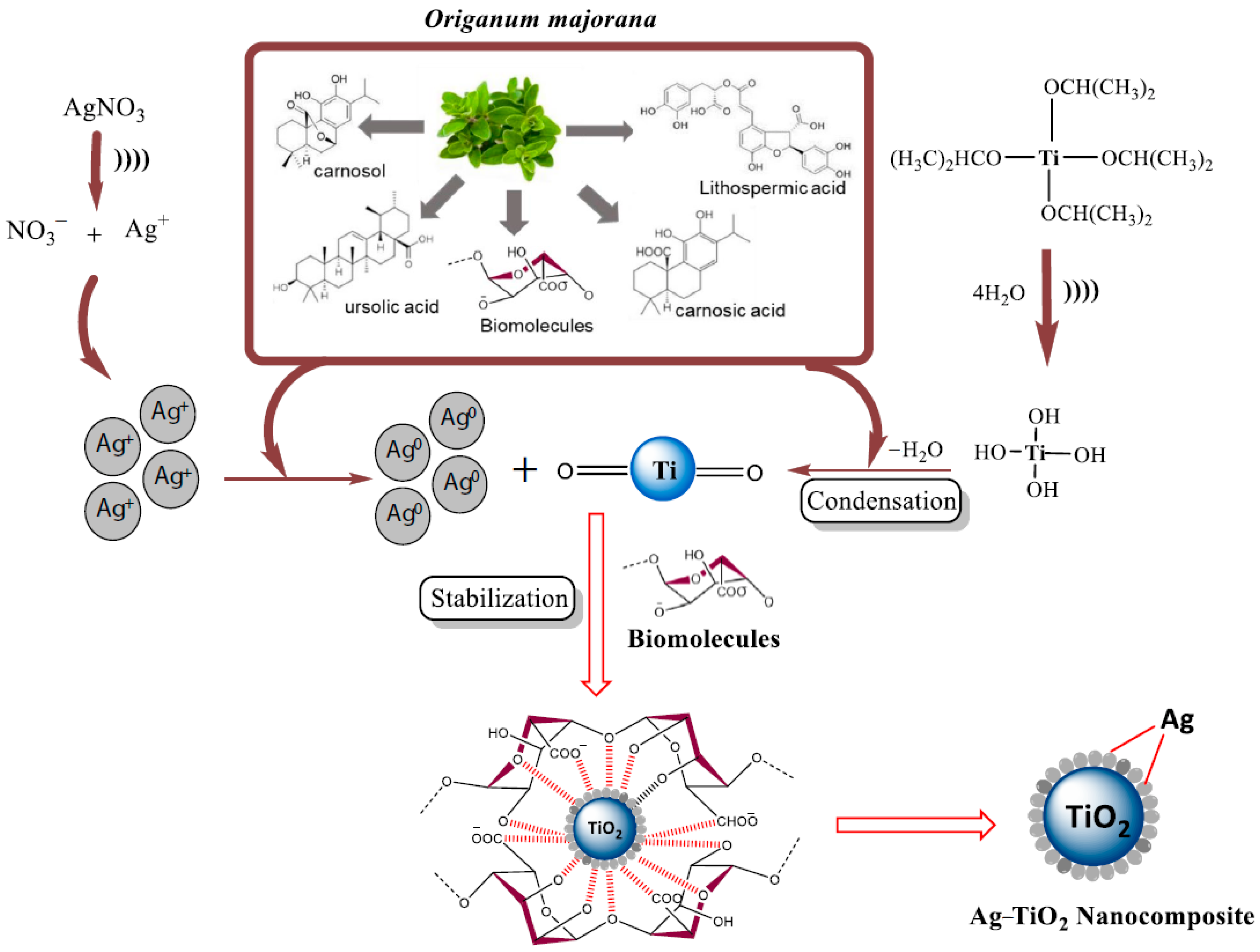
| Ag–TiO2 NPs | Sonochemical synthesis of NPs using leaf extract of Origanum majorana as a bioreductant and a stabilizing agent | Antibacterial and antioxidant therapy | [40] |
| Phosphate-terminated polyamidoamine dendrimer | G4 PAMAM 10 modification with dimethylhydrogenophosphonate, followed by treatment with bromotrimethylsilane | Bone and teeth restoration | [145] |
| Porous SiC coated with Ta | Bioactive metal (Ta) chemical vapor deposition on porous SiC scaffolds | Potential material for bone substitutes | [146] |
| HAP with multi-scale, hierarchically ordered structure | Self-assembly of layered chitosan–maleic acid matrix, followed by monetite mineralization and transformation to HAP | Developing bone substitute materials | [147] |
| Amelogenin-containing chitosan hydrogel (modified with enamel proteinase) | Mixing of chitosan solution, CaCl2, and recombinant full-length porcine amelogenin, followed by stirring overnight (and addition of enamel proteinase) | Enamel repair | [133,134] |
| Ceramic biomimetic 3-DOM 11 foam | Cork powder pyrolysis to carbon, followed by infiltration with precursor salt solution and calcination to form the oxide ceramic | Environmental and energy applications | [148] |
| Cellulose nanowhiskers in biopolymer matrices | Microcrystalline cellulose sulfuric acid hydrolysis and centrifugation | Scaffolding in tissue engineering | [149] |
| Genipin-crosslinked chitosan, alginate, and alumina nanocomposite gels | Alumina powder added to chitosan solution, followed by alginate dissolution and genipin (cross-linking agent) addition | 3D bioprinting | [65,150] |
| Ceramic–organic nanocomposite films | Templated supramolecular surfactant self-assembly on a mica surface | Low-temperature thin-film processing | [151] |
| Nanometer-sized HAP–collagen composite | Incubation of Tris-buffered CaCl2 with sharkskin collagen suspension | Orthopedic implants | [62] |
| PAMAM 12-dendrimer-templated HAP crystallization | Enamel immersion in a solution of CaCl2, KH2PO4, and PAMAM dendrimers modified with carboxylic acid groups | Enamel repair | [135,136,137] |
| HAP–tricalcium phosphate biphasic NPs | Wet-milling of CaHPO4 and CaCO3 powders, followed by double-sieving and high-temperature calcination | Bone tissue engineering | [152] |
| HAP NPs obtained using asparagine–serine–serine peptide | Enamel exposure to triplet repeats of asparagine–serine–serine solution, followed by soaking in artificial saliva | Enamel repair | [63] |
| Erythrocyte-membrane-camouflaged polymeric NPs | RBC hypotonic treatment and extrusion, followed by mixing with PLGA NPs via extrusion through a porous membrane | Targeted drug delivery | [66] |
| Monocrystalline ZrO2 NPs embedded in an amorphous SiO2 matrix | Spark-plasma-sintering of ZrO2 NPs and amorphous SiO2 powder with a molar ratio of 65% ZrO2/35% SiO2 at 1200 °C | High-strength translucent glass ceramic materials | [153] |
| Nacre-like composite of silk nanofibrils, HAP, and chitin nanofibrils | Self-assembly of silk nanofibrils, followed by HAP biomineralization, mixing with chitin nanofibril solution, and nacre-like membrane vacuum-assisted deposition | “Grab-and-release” actuators | [154] |
Publisher’s Note: MDPI stays neutral with regard to jurisdictional claims in published maps and institutional affiliations. |
© 2022 by the authors. Licensee MDPI, Basel, Switzerland. This article is an open access article distributed under the terms and conditions of the Creative Commons Attribution (CC BY) license (https://creativecommons.org/licenses/by/4.0/).
Share and Cite
Gareev, K.G.; Grouzdev, D.S.; Koziaeva, V.V.; Sitkov, N.O.; Gao, H.; Zimina, T.M.; Shevtsov, M. Biomimetic Nanomaterials: Diversity, Technology, and Biomedical Applications. Nanomaterials 2022, 12, 2485. https://doi.org/10.3390/nano12142485
Gareev KG, Grouzdev DS, Koziaeva VV, Sitkov NO, Gao H, Zimina TM, Shevtsov M. Biomimetic Nanomaterials: Diversity, Technology, and Biomedical Applications. Nanomaterials. 2022; 12(14):2485. https://doi.org/10.3390/nano12142485
Chicago/Turabian StyleGareev, Kamil G., Denis S. Grouzdev, Veronika V. Koziaeva, Nikita O. Sitkov, Huile Gao, Tatiana M. Zimina, and Maxim Shevtsov. 2022. "Biomimetic Nanomaterials: Diversity, Technology, and Biomedical Applications" Nanomaterials 12, no. 14: 2485. https://doi.org/10.3390/nano12142485