
Direct Synthesis of MoS2 Nanosheets in Reduced Graphene Oxide Nanoscroll for Enhanced Photodetection


Key Laboratory of Flexible Electronics (KLOFE) & Institute of Advanced Materials (IAM), Nanjing Tech University (Nanjing Tech), 30 South Puzhu Road, Nanjing 211816, China
*
Authors to whom correspondence should be addressed.
Nanomaterials 2022, 12(9), 1581; https://doi.org/10.3390/nano12091581
Submission received: 15 April 2022
/
Revised: 2 May 2022
/
Accepted: 4 May 2022
/
Published: 6 May 2022
(This article belongs to the Special Issue Advances in Nanotechnology of Perovskite and Silicon Solar Cells)
Abstract
:Due to their unique tubular and spiral structure, graphene and graphene oxide nanoscrolls (GONS) have shown extensive applications in various fields. However, it is still a challenge to improve the optoelectronic application of graphene and GONS because of the zero bandgap of graphene. Herein, ammonium tetrathiomolybdate ((NH4)2MoS4) was firstly wrapped into the ((NH4)2MoS4@GONS) by molecular combing the mixture of (NH4)2MoS4 and GO solution on hydrophobic substrate. After thermal annealing, the (NH4)2MoS4 and GO were converted to MoS2 nanosheets and reduced GO (RGO) simultaneously, and, thus, the MoS2@RGONS was obtained. Raman spectroscopy and high-resolution transmission electron microscopy were used to confirm the formation of MoS2 nanosheets among the RGONS. The amount of MoS2 wrapped in RGONS increased with the increasing height of GONS, which is confirmed by the atomic force microscopy and Raman spectroscopy. The as-prepared MoS2@RGONS showed much better photoresponse than the RGONS under visible light. The photocurrent-to-dark current ratios of photodetectors based on MoS2@RGONS are ~570, 360 and 140 under blue, red and green lasers, respectively, which are 81, 144 and 35 times of the photodetectors based on RGONS. Moreover, the MoS2@RGONS-based photodetector exhibited good power-dependent photoresponse. Our work indicates that the MoS2@RGONS is expected to be a promising material in the fields of optoelectronic devices and flexible electronics.
1. Introduction
By scrolling two-dimensional graphene nanosheet into a one-dimensional structure, a graphene nanoscroll (GNS) is formed with a tubular, spiral structure and open ends [1]. Due to the excellent properties resulting from its unique structure, graphene nanoscroll has attracted great attention in the fields of energy storage, sensors and flexible electronics [2,3,4,5]. In recent years, graphene oxide nanoscrolls (GONS) have been widely investigated instead of GNS because of the facile mass production of graphene oxide [2,6,7,8,9,10,11,12,13,14,15]. In order to further improve the performance of GONS, various functional nanomaterials were wrapped into GONS to extend their applications in supercapacitors, batteries and photocatalysts [16,17,18,19,20]. By encapsulating sulfur into GONS during the freeze-casting process, the as-prepared S/GONS was used as a good cathode material for a lithium-sulfur battery [21,22]. After Fe2O3 nanoparticles were wrapped into GONS as electrode materials by ultrasonication, a high volumetric energy density supercapacitor with quite good cycling stability was obtained [23]. Fe1−xS/Fe3O4 nanoparticles were also confined into GONSs and GO nanosheets by cold quenching and freeze drying, and the as-obtained composites were used as promising electrodes for a flexible lithium-ion battery [3].
It is well known that the application of graphene in high-performance photodetectors has been seriously hindered due to its low optical absorption ability [24]. To improve the photoresponse of graphene-based devices, a large number of functional nanomaterials have been integrated with graphene, including quantum dots [24], transition metal dichalcogenides [25,26,27], metallic nanostructures with plasmonic effects [28,29] and so on. Although the GONS has shown promising applications in the fields of energy storage, sensing and photocatalysis, it is still a challenge to apply the GONS as a high-performance photodetector. Previously, we have embedded chemical vapor deposition (CVD)-grown MoS2 nanoflakes into RGO nanoscrolls to improve the photodetection performance [25]. The photosensitivity of MoS2@RGO nanoscrolls increased by 20 times compared to the RGO nanosheet. However, it is difficult to further increase the amount of MoS2 grown on an RGO nanosheet by using the CVD method. Therefore, it is highly desirable to develop an alternative method to wrap a large amount of MoS2 into GONS and thus further enhance its photodetection performance.
In this work, a large amount of a precursor, ammonium tetrathiomolybdate ((NH4)2MoS4), was successfully wrapped into GONSs with length up to hundreds of micrometers by using the molecular combing method. After high temperature annealing, the (NH4)2MoS4 was in situ decomposed to MoS2 nanosheets, which were well encapsulated into the GONS. Meanwhile, the GONSs were simultaneously converted to reduced GONSs (RGONS). By optimizing precursor concentration and annealing temperature, high-quality MoS2@RGONS was facilely obtained with mass production. The optical microscopy (OM), atomic force microscopy (AFM), high-resolution transmission electron microscopy (HRTEM) and Raman spectroscopy were employed to demonstrate the uniform distribution of MoS2 nanosheets in RGONSs. The photodetectors based on MoS2@RGONSs showed photosensitivity two orders of magnitude higher than that of the RGONS under visible light. The MoS2@RGONSs-based photodetectors also exhibited good power dependent behavior. The improved performance could be attributed to the formation of multiple graphene/MoS2 interfaces in the scrolled structure of MoS2@RGONS, which increases the light absorption efficiency and enables the ultrafast charge transfer, resulting in a much higher photocurrent and photosensitivity.
2. Experimental Section
2.1. Preparation of (NH4)2MoS4 Solution and Hydrophobic Substrate
Graphene oxide (GO) was synthesized by the modified Hummers method. A total of 0.038 g of (NH4)2MoS4 was ground to fine powder and dissolved in water to form aqueous solutions with concentration of 5, 10, 20, 30 and 50 mM. Thereafter, the (NH4)2MoS4 solution was treated by ultrasonication to ensure complete dissolution. Finally, the (NH4)2MoS4@GO solution was prepared by mixing 0.2 mg/mL GO and (NH4)2MoS4 solution with a volume ratio of 1:1.
The preparation of hydrophobic substrate is a key step to form GO nanoscrolls by the molecular combing method [6,7,10,11]. Firstly, the cleaned 300 nm SiO2/Si substrate was immersed into a glass bottle containing a mixture of 8 mL toluene and 200 µL OTS (Octadecyltrimethoxysilane, purchased from Aladdin). Then, the glass bottle was sealed and heated at 60 °C for 24 h. After that, the SiO2/Si substrate was washed with ethanol and DI water three times. Therefore, the hydrophobic OTS-modified SiO2/Si substrate (OTS-SiO2/Si) was obtained.
2.2. Preparation of (NH4)2MoS4@GONS and MoS2@RGONS
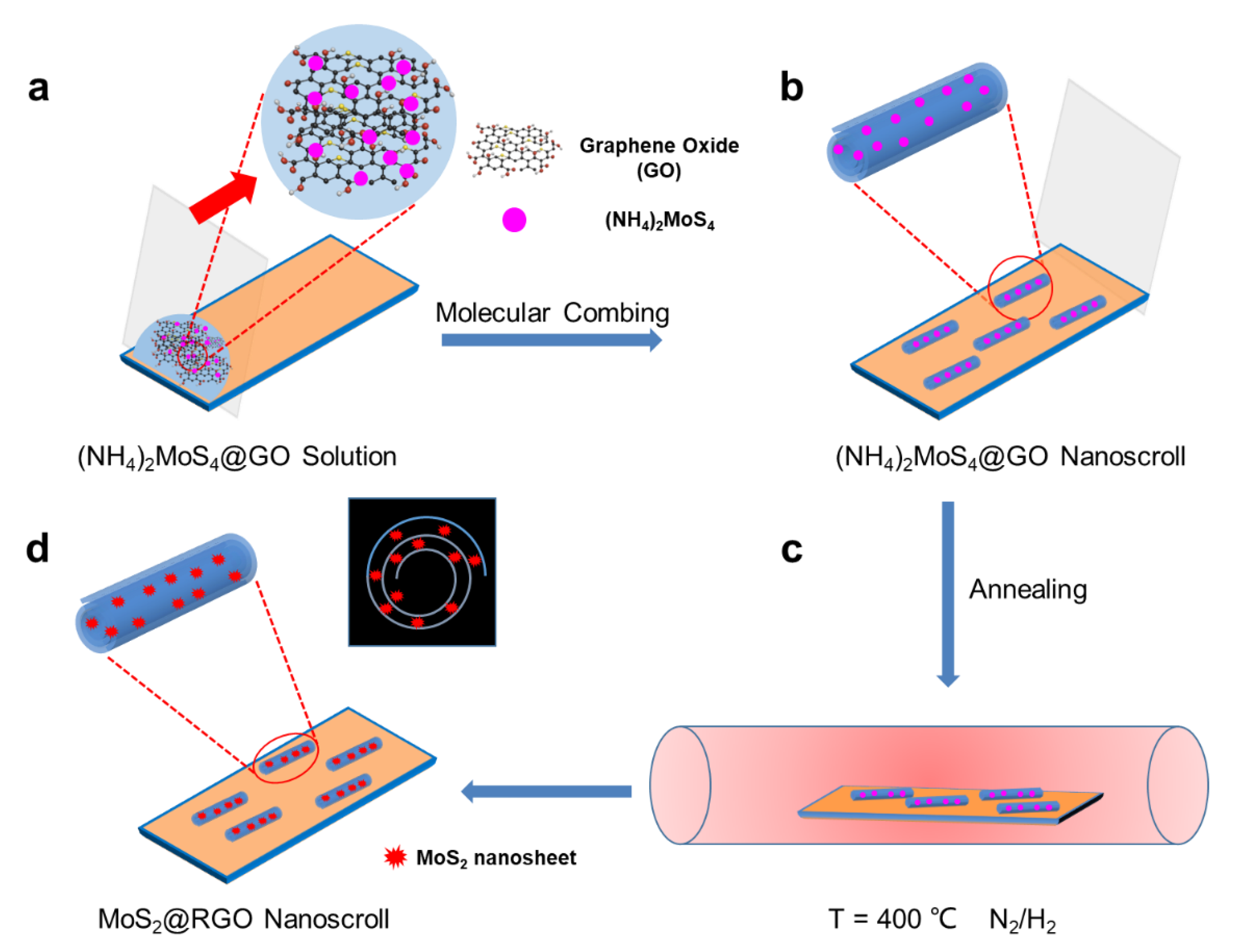
The (NH4)2MoS4@GO nanoscrolls ((NH4)2MoS4@GONSs) were prepared by the molecular combing method [6], as shown in Scheme 1a,b. Firstly, 30 μL of the (NH4)2MoS4@GO solution was dropped onto the hydrophobic OTS-SiO2/Si substrate. A glass coverslip was then used to slowly drag the droplet from one end to the other end of the substrate. In this way, the (NH4)2MoS4@GONSs were formed on the OTS-SiO2/Si substrate. The MoS2@RGO nanoscrolls (MoS2@RGONSs) were prepared as shown in Scheme 1c,d. After the (NH4)2MoS4@GONSs were put into a tube furnace, a mixture gas of N2/H2 (80/40 sccm) was introduced as a protective gas. Then, the temperature of the furnace increased to 400 °C gradually, with a speed of 10 °C/min, and was kept for 60 min. During this period, (NH4)2MoS4 was decomposed to MoS2, while GO was reduced to RGO at the same time. Therefore, the MoS2@RGONSs were successfully obtained.
2.3. Characterizations
An optical microscope (ECLIPSE LV100ND, Nikon, Tokyo, Japan), AFM (Dimension ICON with Nanoscope V controller, Bruker, Billerica, MA, USA) and TEM (JEM-2100F JEOL, Tokyo, Japan) were used to characterize the as-prepared (NH4)2MoS4@GONS and MoS2@RGONS. In addition, Raman spectroscopy and Raman mapping of the as-prepared (NH4)2MoS4@GONS and MoS2@RGONS were tested on a LabRAM HR Evolution Raman spectrometer (Horiba Jobin Yvon, Palaiseau, France) with a 532 nm laser focused through a 100× objective lens.
2.4. Device Fabrication and Photodetection
First, 30 nm thick gold and 5 nm thick chromium films were deposited on RGO and MoS2@RGO nanoscrolls, respectively, by thermal evaporation with a TEM grid (200 mesh) as a mask.
A probe station (model TTPX, Lake Shore Inc., Rhinelander, WI, USA) and Keithley 4200 semiconductor characterization system (Advanced Test Equipment Corp., San Diego, CA, USA) were used to monitor the real-time current change of the as-prepared devices. The photocurrent was collected from individual ROG and MoS2@RGO nanoscrolls with Au pads as the source and drain electrodes, respectively, as shown in Figure S1. The photo-detection test was recorded under blue (405 nm), green (532 nm) and red (633 nm) lasers. The power of the laser was measured with a laser power meter (Laser power meter LP1, SanWa, Okayama, Japan).
3. Result and Discussion
Scheme 1 shows the preparation of MoS2@RGONSs by molecular combing and thermal annealing. After the (NH4)2MoS4 was wrapped into GO nanoscrolls by molecular combing, the (NH4)2MoS4 was decomposed to MoS2 at a high temperature under the atmosphere of N2/H2 [30]. It was found that the (NH4)2MoS4 was decomposed to MoS3 as the temperature increased from 120 to 260 °C under an inert atmosphere. The MoS3 was further reduced to MoS2 as the temperature was higher than 230 °C under N2/H2 [30]. The process can be described as the following chemical reactions,
(NH4)2MoS4 → 2NH3 + H2S + MoS3 (120–260 °C)
MoS3 + H2 → MoS2 + H2S (230–450 °C)
MoS3 + H2 → MoS2 + H2S (230–450 °C)
We heated the (NH4)2MoS4@GONSs in the temperature range of 300 to 500 °C. The Raman spectroscopy was used to characterize the amount of MoS2 in GONSs by measuring the peak intensity of A1g. As shown in Figure S2a, the peak intensity of A1g increased as the temperature increased from 300 to 400 °C, while it decreased as the temperature further increased to 500 °C. The flow ratio of N2 to H2 also affects the formation of MoS2 during the thermal annealing process. As shown in Figure S2b, the A1g peak of the (NH4)2MoS4@GONS annealed with a gas stream of 80 sccm N2 and 40 sccm H2 showed the highest intensity. Therefore, a mixture gas of N2/H2 (80/40 sccm) was introduced as a protective gas during the experiment. The concentration of the (NH4)2MoS4 solution also has an important effect on the formation of (NH4)2MoS4@GONSs and MoS2@RGONSs. The peak intensity of A1g increased as the concentration of (NH4)2MoS4 solution increased from 5 mM to 30 mM, while it decreased when 50 mM (NH4)2MoS4 solution was used (Figure S2c). In addition, the diameter of the (NH4)2MoS4@GONSs increased as the concentration of (NH4)2MoS4 increased from 5 mM to 50 mM (Figure S3). Meanwhile, the long and straight nanoscrolls were changed to irregular and thick aggregations. In order to wrap more MoS2 and maintain the good scroll structure, (NH4)2MoS4 solution with a concentration of 30 mM was used as the optimal concentration and annealed at 400 °C.
Figure 1a,b show the OM images of an (NH4)2MoS4@GO nanoscroll before and after thermal annealing, respectively. It can be seen that the color of the nanoscroll changed from cyan to gray blue after thermal annealing. In addition, the height of the MoS2@RGO nanoscroll is quite smaller than that of the (NH4)2MoS4@GO nanoscrolls. As shown in Figure S4, the height of the (NH4)2MoS4@GO is 201.4 nm, while it decreases largely to 112.5 nm after high temperature annealing, which could be attributed to the evaporation of water molecules trapped in the nanoscroll and the decomposition of (NH4)2MoS4. In order to confirm the formation of the MoS2@RGO nanoscroll, Raman spectroscopy was conducted at the same position of the nanoscroll before and after annealing. As shown in Figure 1c, there are only two Raman peaks located at 1359 and 1587 cm−1 for the (NH4)2MoS4@GO nanoscroll, which are assigned to the D and G peaks of GO. After thermal annealing, there are two more peaks located at 384.4 and 404.2 cm−1 besides the D and G peaks, which are characteristics of MoS2 nanosheets [31,32]. In addition, the intensity ratio of the D to G peak decreased from 1.24 to 0.72 for the (NH4)2MoS4@GO nanoscroll after thermal annealing, indicating the formation of reduced GO (RGO). The D band originates from the lattice destruction of sp2-hybridized carbon, and the G band arises from the first-order scattering of the mode. The intensity ratio (ID/IG) reflects the disorder of the carbon structure, and the higher intensity ratio of ID/IG means more defective graphitic structures. Therefore, the Raman characterization confirms the successful conversion of the (NH4)2MoS4@GO nanoscroll to the MoS2@RGO nanoscroll after thermal annealing at 400 °C for 60 min. In order to investigate whether the MoS2 was uniformly wrapped into the RGO nanoscroll, the as-obtained MoS2@RGO nanoscroll was characterized by Raman mapping. As shown in Figure 1d,e, the G peak of GO nanoscroll was unchanged after thermal annealing. Meanwhile, the Raman mapping of the A1g peak of MoS2 showed a homogeneous signal (Figure 1f), indicating that the MoS2 was uniformly distributed in the RGO nanoscroll.
We found that the Raman peak intensity of MoS2 in the MoS2@RGO nanoscrolls varied with the height of the nanoscrolls. To investigate the relationship between the height of the MoS2@RGO nanoscroll and the amount of trapped MoS2 in it, the MoS2@RGO nanoscrolls with various heights were characterized using AFM and Raman spectroscopy, respectively. Figure 2a shows the OM image of the MoS2@RGO nanoscroll, where the boxes marked by d, e and f are three nanoscrolls with different heights. Figure 2d–f show the corresponding AFM images, and the measured heights of the three nanoscrolls are 137.4 nm, 83.5 nm and 35.4 nm, respectively. As shown in Figure 2b, the nanoscroll with a height of 137.4 nm presents a stronger Raman signal (box d), while the nanoscroll with a height of 35.4 nm has a weaker Raman signal. To further reveal the influence of the height of the MoS2@RGO nanoscrolls on the Raman peak intensity of MoS2, a lot of nanoscrolls were measured to plot the Raman peak intensity as a function of the height of the nanoscrolls. As shown in Figure 2c, with the increasing height of the MoS2@RGO nanoscrolls, the Raman peak intensity of MoS2 gradually increases, indicating that more MoS2 was trapped in a higher nanoscroll.
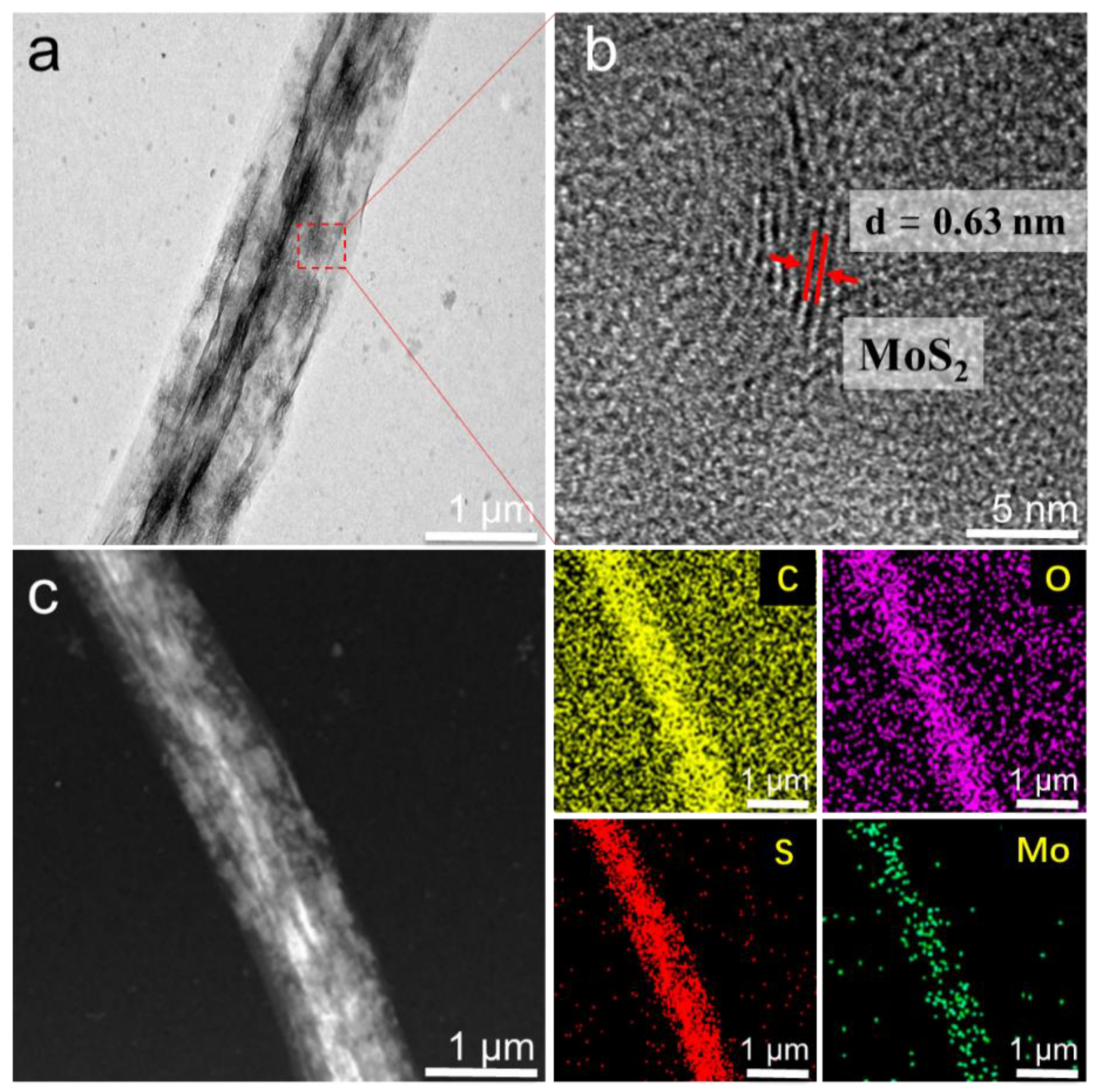
In order to clearly observe the detailed structure and confirm the formation of MoS2 in the as-prepared MoS2@RGO nanoscroll, high-resolution transmission electron microscopy (HRTEM) was used for characterization. Figure 3a shows the low-resolution TEM image of the MoS2@RGO nanoscroll. We can see that the MoS2@RGO nanoscroll exhibits a multilayer scrolled structure with a dark, dense inner layer. Figure 3b shows the HRTEM characterization result of the red dashed box shown in Figure 3a. The well resolved lattice stripes with spacing of 0.63 nm are clearly presented, which is consistent with the interlayer spacing of layered MoS2 [33]. Moreover, the energy dispersive X-ray (EDX) elemental mapping analysis of the MoS2@RGO nanoscroll shown in Figure 3c provides strong evidence for the homogeneous distribution of C, O, Mo and S elements in the MoS2@RGO nanoscroll. The HRTEM and EDX results confirm the uniform existence of the MoS2 nanosheet in the RGO nanoscroll.
Graphene and its derivatives are severely limited in optoelectronic applications due to their zero band gap and poor absorption of visible light. In order to improve the optoelectronic performance of graphene, MoS2, as a typical transition metal dichalcogenides (TMDCs) material, has been widely used to combine graphene for photodetection [34]. By wrapping MoS2 into the RGO nanoscrolls, we found that the MoS2@RGO nanoscroll also showed promising photodetection performance. Photodetectors based on RGO nanoscrolls and MoS2@RGO nanoscrolls were fabricated to investigate the effect of wrapped MoS2. It is well known that photosensitivity is an important parameter to evaluate the performance of photodetectors [35,36,37], which is usually defined by the ratio of photocurrent to dark current (PDR), as follows:
PDR = Iphoto/Idark (Iphoto: photocurrent; Idark: dark current)
The photocurrent and dark current of the RGO nanoscrolls and MoS2@RGO nanoscrolls-based photodetectors were firstly measured under the dark and the illumination of blue, red and green lasers with different laser power densities, respectively. Because of the low light absorption of RGO, we found that the RGO-based photodetectors exhibited photosensitivity of ~7, ~2.4 and ~4 under blue, red and green lasers (Figure S5). Figure 4a–c show the PDRs of photodetectors based on RGO nanoscrolls and MoS2@RGO nanoscrolls under blue (405 nm), red (633 nm) and green (532 nm) lasers. The PDRs of photodetectors based on the MoS2@RGO nanoscrolls were 570, 360 and 140 under blue, red and green lasers, which are almost 81, 144 and 35 times those of the photodetectors based on the RGO nanoscrolls measured under the same conditions. In addition, the photocurrent of the MoS2@RGO nanoscroll is highly dependent on the power density of the incident light. As shown in Figure 4d–f, the PDRs increased as the incident laser power density increased. The different photoresponse of MoS2@RGONS to blue, green and red light could be explained as follows. In our experiment, the power intensity of green light is the lowest. However, the PDR of MoS2@RGONS is around 140 at a power density of 0.56 mW/mm2 (Figure 4f), while the PDRs of MoS2@RGONS are around 100 and 70 for blue and red lasers at power densities of 1.05 mW/mm2 and 1.41 mW/mm2 (Figure 4d,e), respectively. The MoS2 nanosheets synthesized in GONS could be multilayer, which can be confirmed by the HRTEM images shown in Figure 3b. Therefore, the multilayer MoS2 trapped into RGONS should be more suitable for detecting green lasers than blue and red lasers given that they are at the same power intensity. A similar phenomenon has also been reported in a multilayer MoS2@glassy-graphene heterostructure [38]. In addition, the MoS2@RGONS shows higher PDR under the blue laser than under the red laser at similar power intensities. This could be attributed the higher photon energy of the blue laser compared to the red laser, which can generate more photoinduced carriers at the same power [39].
The excellent photodetection performance of the MoS2@RGO nanoscroll could be attributed to the formation of multiple heterojunction interfaces between the RGO and MoS2 nanosheets. It is known that the ultrafast separation and transfer of photogenerated carriers can be achieved at the heterojunction interface of graphene and MoS2, resulting in a substantial increase in photocurrent and photoresponse. Due to the roll-up structure of the MoS2@RGO nanoscroll, the MoS2 nanosheets are wrapped between adjacent RGO layers spirally, forming multiple heterojunction interfaces. When light was shined on the MoS2@RGO nanoscroll, the MoS2 nanosheets in each heterojunction interface could absorb light, and charge carriers were generated simultaneously. Meanwhile, the photogenerated charge carriers can be separated and transferred in an ultrafast way. Therefore, the photocurrent of the MoS2@RGO nanoscroll can be greatly enhanced due to the synergetic enhancement of photocurrent at each heterojunction interface. As a consequence, the photosensitivity of the MoS2@RGO nanoscroll is much higher than that of the RGO nanoscroll.
4. Conclusions
In summary, (NH4)2MoS4 was encapsulated into the GO nanoscrolls by the molecular combing method on hydrophobic substrate. By optimizing the precursor concentration and annealing temperature, the (NH4)2MoS4 and GO nanoscrolls were successfully converted to MoS2 and RGO nanoscrolls, forming the MoS2@RGO nanoscroll. The OM and AFM characterization results showed that the high-density MoS2@RGO nanoscrolls were successfully prepared. The uniform distribution of the MoS2 nanosheets in the RGO nanoscrolls was confirmed by the Raman spectroscopy and HRTEM characterization. Compared to the RGO nanoscroll, the MoS2@RGO nanoscroll showed much better photodetection performance. The PDRs of photodetectors based on the MoS2@RGO nanoscrolls were about two orders of magnitude higher than those of photodetectors based on the RGO nanoscrolls under blue, red and green lasers. The formation of multiple graphene/MoS2 heterojunction interfaces in a scrolled structure can not only enhance the light absorption of MoS2 but also accelerate the electron-hole separation. Our work indicates that the MoS2@RGO nanoscrolls could be promising materials for high-performance graphene-based photodetectors.
Supplementary Materials
The following supporting information can be downloaded at https://www.mdpi.com/article/10.3390/nano12091581/s1: Figure S1. The optical images of photodetectors based on individual (a) RGO nanoscroll and (b) MoS2@RGO nanoscroll with Au pads as source and drain electrodes. Figure S2. Plots of Raman peak intensity of A1g as function of (a) annealing temperature, (b) the flow ratio of N2 to H2, and (c) concentration of (NH4)2MoS4. Figure S3. OM images of (NH4)2MoS4@GONS prepared by molecular combing (NH4)2MoS4 solution with concentration of (a,e) 0.005 M, (b,f) 0.01 M, (c,g) 0.03 M, and (d,h) 0.05 M before and after thermal annealing. Figure S4. AFM height images of the same (NH4)2MoS4@GONS (a) before and (b) after thermal annealing. Figure S5. PDR plots of RGO nanoscrolls measured under (a) blue, (b) red, and (c) green lasers.
Author Contributions
Conceptualization, H.L.; supervision and project administration, X.H. and H.L.; methodology, H.L. and X.L.; formal analysis, Z.W., F.L., X.L. and Y.Y.; investigation, Z.W. and X.L.; writing—original draft preparation, H.L., Z.W. and X.L.; revision of the manuscript, H.L. and Z.W. All authors have read and agreed to the published version of the manuscript.
Funding
This work was supported by the National Natural Science Foundation of China (Grant No. 51832001, 21571101 and 51322202), the Natural Science Foundation of Jiangsu Province in China (Grant No. BK20161543), and the Natural Science Foundation of the Jiangsu Higher Education Institutions of China (Grant No. 15KJB430016).
Institutional Review Board Statement
Not applicable.
Informed Consent Statement
Not applicable.
Data Availability Statement
Data can be available upon request from the authors.
Conflicts of Interest
The authors declare no conflict of interest.
References
- Berman, D.; Deshmukh, S.A.; Sankaranarayanan, S.K.R.S.; Erdemir, A.; Sumant, A.V. Macroscale Superlubricity Enabled by Graphene Nanoscroll Formation. Science 2015, 348, 1118–1122. [Google Scholar] [CrossRef] [PubMed] [Green Version]
- Chen, Z.; Wang, J.R.; Pan, D.X.; Wang, Y.; Noetzel, R.; Li, H.; Xie, P.; Pei, W.L.; Umar, A.; Jiang, L.; et al. Mimicking a Dog’s Nose: Scrolling Graphene Nanosheets. ACS Nano 2018, 12, 2521–2530. [Google Scholar] [CrossRef] [PubMed]
- Zhao, Y.; Wang, J.J.; Ma, C.L.; Cao, L.J.; Shao, Z.P. A Self-Adhesive Graphene Nanoscroll/Nanosheet Paper with Confined Fe1−xS/Fe3O4 Hetero-Nanoparticles for High-Performance Anode Material of Flexible Li-Ion Batteries. Chem. Eng. J. 2019, 370, 536–546. [Google Scholar] [CrossRef]
- Liu, P.W.; Jin, Z.; Katsukis, G.; Drahushuk, L.W.; Shimizu, S.; Shih, C.J.; Wetzel, E.D.; Taggart-Scarff, J.K.; Qing, B.; Van Vliet, K.J.; et al. Layered and Scrolled Nanocomposites with Aligned Semi-Infinite Graphene Inclusions at the Platelet Limit. Science 2016, 353, 364–367. [Google Scholar] [CrossRef] [PubMed] [Green Version]
- Lai, Z.C.; Chen, Y.; Tan, C.L.; Zhang, X.; Zhang, H. Self-Assembly of Two-Dimensional Nanosheets into One-Dimensional Nanostructures. Chem 2016, 1, 59–77. [Google Scholar] [CrossRef] [Green Version]
- Li, H.; Wu, J.; Qi, X.Y.; He, Q.Y.; Liusman, C.; Lu, G.; Zhou, X.Z.; Zhang, H. Graphene Oxide Scrolls on Hydrophobic Substrates Fabricated by Molecular Combing and Their Application in Gas Sensing. Small 2013, 9, 382–386. [Google Scholar] [CrossRef]
- Wu, J.M.T.; Li, H.; Qi, X.Y.; He, Q.Y.; Xu, B.X.; Zhang, H. Graphene Oxide Architectures Prepared by Molecular Combing on Hydrophilic-Hydrophobic Micropatterns. Small 2014, 10, 2239–2244. [Google Scholar] [CrossRef]
- Wu, J.; Yang, J.; Huang, Y.; Li, H.; Fan, Z.X.; Liu, J.Q.; Cao, X.H.; Huang, X.; Huang, W.; Zhang, H. Graphene Oxide Scroll Meshes Prepared by Molecular Combing for Transparent and Flexible Electrodes. Adv. Mater. Technol. 2017, 2, 1600231. [Google Scholar] [CrossRef]
- Wang, L.; Yang, P.; Liu, Y.; Fang, X.R.; Shi, X.T.; Wu, S.Y.; Huang, L.; Li, H.; Huang, X.; Huang, W. Scrolling up Graphene Oxide Nanosheets Assisted by Self-Assembled Monolayers of Alkanethiols. Nanoscale 2017, 9, 9997–10001. [Google Scholar] [CrossRef]
- Liu, Y.; Wang, L.; Zhang, H.; Ran, F.R.; Yang, P.; Li, H. Graphene Oxide Scroll Meshes Encapsulated Ag Nanoparticles for Humidity Sensing. RSC Adv. 2017, 7, 40119–40123. [Google Scholar] [CrossRef] [Green Version]
- Zhao, W.H.; Wang, L.; Pei, C.J.; Wei, C.; You, H.; Zhang, J.D.; Li, H. Impact of pH on Regulating Ion Encapsulation of Graphene Oxide Nanoscroll for Pressure Sensing. Nanomaterials 2019, 9, 548. [Google Scholar] [CrossRef] [PubMed] [Green Version]
- Tang, B.; Gao, E.L.; Xiong, Z.Y.; Dang, B.; Xu, Z.P.; Wang, X.G. Transition of Graphene Oxide from Nanomembrane to Nanoscroll Mediated by Organic Solvent in Dispersion. Chem. Mater. 2018, 30, 5951–5960. [Google Scholar] [CrossRef]
- Fang, Q.L.; Zhou, X.F.; Deng, W.; Liu, Y.W.; Zheng, Z.; Liu, Z.P. Nitrogen-Doped Graphene Nanoscroll Foam with High Diffusion Rate and Binding Affinity for Removal of Organic Pollutants. Small 2017, 13, 201603779. [Google Scholar] [CrossRef] [PubMed]
- Zheng, B.N.; Gao, C. Preparation of Graphene Nanoscroll/Polyaniline Composites and Their Use in High Performance Supercapacitors. New Carbon Mater. 2016, 31, 315–320. [Google Scholar] [CrossRef]
- Rani, J.R.; Thangavel, R.; Oh, S.I.; Lee, Y.S.; Jang, J.H. An Ultra-High-Energy Density Supercapacitor: Fabrication Based on Thiol-functionalized Graphene Oxide Scrolls. Nanomaterials 2019, 9, 148. [Google Scholar] [CrossRef] [PubMed] [Green Version]
- Li, X.J.; Natsuki, J.; Natsuki, T. A Recyclable Silver Nanoparticles/Graphene Oxide Nanoscroll Composite Photocatalyst. Environ. Technol. Inno. 2021, 21, 101210. [Google Scholar] [CrossRef]
- Zhang, Y.F.; Zhao, C.Y.; Zeng, Z.H.; Ang, J.M.; Che, B.Y.; Wang, Z.; Lu, X.H. Graphene Nanoscroll/Nanosheet Aerogels with Confined SnS2 Nanosheets: Simultaneous Wrapping and Bridging for High-Performance Lithium-Ion Battery Anodes. Electrochim. Acta 2018, 278, 156–164. [Google Scholar] [CrossRef]
- Yang, B.J.; Chen, J.T.; Liu, B.; Ding, Y.X.; Tang, Y.; Yan, X.B. One Dimensional Graphene Nanoscroll-Wrapped MnO Nanoparticles for High-Performance Lithium Ion Hybrid Capacitors. J. Mater. Chem. A 2021, 9, 6352–6360. [Google Scholar] [CrossRef]
- Lin, Y.T.; Zhou, F.S.; Chen, M.; Zhang, S.; Deng, C. Building Defect-Rich Oxide Nanowires@Graphene Coaxial Scrolls to Boost High-Rate Capability, Cycling Durability and Energy Density for Flexible Zn-Ion Batteries. Chem. Eng. J. 2020, 396, 125259. [Google Scholar] [CrossRef]
- Cho, S.H.; Kim, J.H.; Kim, I.G.; Park, J.H.; Jung, J.W.; Kim, H.S.; Kim, I.D. Reduced Graphene-Oxide-Encapsulated MoS2/Carbon Nanofiber Composite Electrode for High-Performance Na-Ion Batteries. Nanomaterials 2021, 11, 2691. [Google Scholar] [CrossRef]
- Yoo, S.; Lee, J.; Kim, J.M.; Seong, C.Y.; Seong, K.D.; Piao, Y. Well-Dispersed Sulfur Wrapped in Reduced Graphene Oxide Nanoscroll as Cathode Material for Lithium-Sulfur Battery. J. Electroanal. Chem. 2016, 780, 19–25. [Google Scholar] [CrossRef]
- Guo, Y.; Zhao, G.; Wu, N.T.; Zhang, Y.; Xiang, M.W.; Wang, B.; Liu, H.; Wu, H. Efficient Synthesis of Graphene Nanoscrolls for Fabricating Sulfur-Loaded Cathode and Flexible Hybrid Interlayer toward High-Performance Li-S Batteries. ACS Appl. Mater. Inter. 2016, 8, 34185–34193. [Google Scholar] [CrossRef] [PubMed]
- Rani, J.R.; Thangavel, R.; Oh, S.I.; Woo, J.M.; Das, N.C.; Kim, S.Y.; Lee, Y.S.; Jang, J.H. High Volumetric Energy Density Hybrid Supercapacitors Based on Reduced Graphene Oxide Scrolls. ACS Appl. Mater. Inter. 2017, 9, 22398–22407. [Google Scholar] [CrossRef] [PubMed]
- Liu, C.H.; Chang, Y.C.; Norris, T.B.; Zhong, Z.H. Graphene Photodetectors with Ultra-Broadband and High Responsivity at Room Temperature. Nat. Nanotechnol. 2014, 9, 273–278. [Google Scholar] [CrossRef] [PubMed]
- Fan, H.C.; Wang, J.; Li, X.Y.; You, H.; Li, X.Z.; Pei, C.J.; Huang, X.; Li, H. Direct CVD Growth of MoS2 on Chemically and Thermally Reduced Graphene Oxide Nanosheets for Improved Photoresponse. APL Mater. 2021, 9, 051105. [Google Scholar] [CrossRef]
- Zhang, W.J.; Chuu, C.P.; Huang, J.K.; Chen, C.H.; Tsai, M.L.; Chang, Y.H.; Liang, C.T.; Chen, Y.Z.; Chueh, Y.L.; He, J.H.; et al. Ultrahigh-Gain Photodetectors Based on Atomically Thin Graphene-MoS2 Heterostructures. Sci. Rep. 2014, 4, 3826. [Google Scholar] [CrossRef] [Green Version]
- Gao, S.; Wang, Z.Q.; Wang, H.D.; Meng, F.X.; Wang, P.F.; Chen, S.; Zeng, Y.H.; Zhao, J.L.; Hu, H.G.; Cao, R.; et al. Graphene/MoS2/Graphene Vertical Heterostructure-Based Broadband Photodetector with High Performance. Adv. Mater. Interfaces 2021, 8, 2001730. [Google Scholar] [CrossRef]
- Rohizat, N.S.; Ripain, A.H.A.; Lim, C.S.; Tan, C.L.; Zakaria, R. Plasmon-Enhanced Reduced Graphene Oxide Photodetector with Monometallic of Au and Ag Nanoparticles at VIS-NIR Region. Sci. Rep. 2021, 11, 19688. [Google Scholar] [CrossRef]
- Liu, Y.; Cheng, R.; Liao, L.; Zhou, H.L.; Bai, J.W.; Liu, G.; Liu, L.X.; Huang, Y.; Duan, X.F. Plasmon Resonance Enhanced Multicolour Photodetection by Graphene. Nat. Commun. 2011, 2, 579. [Google Scholar] [CrossRef] [Green Version]
- Brito, J.L.; Ilija, M.; Hernandez, P. Thermal and Reductive Decomposition of Ammonium Thiomolybdates. Thermochim. Acta 1995, 256, 325–338. [Google Scholar] [CrossRef]
- Mao, Y.; Dong, N.N.; Wang, L.; Chen, X.; Wang, H.Q.; Wang, Z.X.; Kislyakov, I.M.; Wang, J. Machine Learning Analysis of Raman Spectra of MoS2. Nanomaterials 2020, 10, 2223. [Google Scholar] [CrossRef]
- Lai, Y.Y.; Yeh, Y.W.; Tzou, A.J.; Chen, Y.Y.; Wu, Y.S.; Cheng, Y.J.; Kuo, H.C. Dependence of Photoresponsivity and On/Off Ratio on Quantum Dot Density in Quantum Dot Sensitized MoS2 Photodetector. Nanomaterials 2020, 10, 1828. [Google Scholar] [CrossRef]
- Fang, X.R.; Wei, P.; Wang, L.; Wang, X.S.; Chen, B.; He, Q.Y.; Yue, Q.Y.; Zhang, J.D.; Zhao, W.; Wang, J.L.; et al. Transforming Monolayer Transition-Metal Dichalcogenide Nanosheets into One-Dimensional Nanoscrolls with High Photosensitivity. ACS Appl. Mater. Inter. 2018, 10, 13011–13018. [Google Scholar] [CrossRef]
- Seo, D.B.; Trung, T.N.; Bae, S.S.; Kim, E.T. Improved Photoelectrochemical Performance of MoS2 through Morphology-Controlled Chemical Vapor Deposition Growth on Graphene. Nanomaterials 2021, 11, 1585. [Google Scholar] [CrossRef]
- Wang, L.; Yue, Q.Y.; Pei, C.J.; Fan, H.C.; Dai, J.; Huang, X.; Li, H.; Huang, W. Scrolling Bilayer WS2/MoS2 Heterostructures for High-Performance Photo-Detection. Nano Res. 2020, 13, 959–966. [Google Scholar] [CrossRef]
- Yue, Q.Y.; Wang, L.; Fan, H.C.; Zhao, Y.; Wei, C.; Pei, C.J.; Song, Q.S.; Huang, X.; Li, H. Wrapping Plasmonic Silver Nanoparticles inside One-Dimensional Nanoscrolls of Transition-Metal Dichalcogenides for Enhanced Photoresponse. Inorg. Chem. 2021, 60, 4226–4235. [Google Scholar] [CrossRef]
- Zhao, Y.; You, H.; Li, X.Z.; Pei, C.J.; Huang, X.; Li, H. Solvent-Free Preparation of Closely Packed MoS2 Nanoscrolls for Improved Photosensitivity. ACS Appl. Mater. Inter. 2022, 14, 9515–9524. [Google Scholar] [CrossRef]
- Xu, H.; Han, X.Y.; Dai, X.; Liu, W.; Wu, J.; Zhu, J.T.; Kim, D.Y.; Zou, G.F.; Sablon, K.A.; Sergeev, A.; et al. High Detectivity and Transparent Few-Layer MoS2/Glassy-Graphene Heterostructure Photodetectors. Adv. Mater. 2018, 30, 1706561. [Google Scholar] [CrossRef]
- Naqi, M.; Kaniselvan, M.; Choo, S.; Han, G.; Kang, S.; Kim, E.; Yoon, Y.; Kim, S. Ultrasensitive Multilayer MoS2-Based Photodetector with Permanently Grounded Gate Effect. Adv. Electron. Mater. 2020, 6, 1901256. [Google Scholar] [CrossRef]
Scheme 1.
Schematic diagram of the preparation of (NH4)2MoS4@GO and MoS2@RGO nanoscrolls. (a) A drop of (NH4)2MoS4 and GO solution is dragged by the cover slip on the hydrophobic substrate. (b) The (NH4)2MoS4@GO nanoscroll is formed by molecular combing. (c) After the as-prepared (NH4)2MoS4@GO nanoscroll is treated at 400 °C in the N2/H2 environment, (NH4)2MoS4 is decomposed to MoS2 nanosheets and GO is reduced to RGO. (d) The as-obtained MoS2@RGO nanoscroll after high temperature annealing. The inset in the top right shows the cross-section structure of the MoS2@RGO nanoscroll.

Figure 1.
The OM images of the (NH4)2MoS4@GO nanoscroll (a) before and (b) after thermal annealing. (c) Raman spectra of the (NH4)2MoS4@GO and MoS2@RGO nanoscrolls. The Raman mapping images of (d) the (NH4)2MoS4@GO nanoscroll and (e) the RGO at peak G (1587 cm−1), and the MoS2@RGO nanoscroll at peak (f) A1g (404.2 cm−1) of MoS2.

Figure 2.
(a) The OM image of the MoS2@RGO nanoscrolls. (b) The Raman spectra of MoS2@RGO marked by dashed boxes of d, e and f shown in (a). (c) The plot of the Raman intensity of A1g peak as a function of the height of the MoS2@RGO nanoscrolls. (d–f) The corresponding AFM images of the MoS2@RGO nanoscroll marked by dashed boxes of d, e and f shown in (a).

Figure 3.
(a) The TEM image of the MoS2@RGO nanoscroll. (b) HRTEM image of the MoS2 nanosheets marked by the dashed box shown in (a). (c) The STEM image and EDX elemental mapping of the MoS2@RGO nanoscroll.
Figure 3.
(a) The TEM image of the MoS2@RGO nanoscroll. (b) HRTEM image of the MoS2 nanosheets marked by the dashed box shown in (a). (c) The STEM image and EDX elemental mapping of the MoS2@RGO nanoscroll.

Figure 4.
(a–c) The PDRs of photodetectors based on RGO and MoS2@RGO nanoscrolls under (a) blue, (b) red and (c) green lasers. (d–f) Plots of PDR values of photodetectors based on MoS2@RGO nanoscrolls under (d) blue, (e) red and (f) green lasers as a function of power density.
Figure 4.
(a–c) The PDRs of photodetectors based on RGO and MoS2@RGO nanoscrolls under (a) blue, (b) red and (c) green lasers. (d–f) Plots of PDR values of photodetectors based on MoS2@RGO nanoscrolls under (d) blue, (e) red and (f) green lasers as a function of power density.

Publisher’s Note: MDPI stays neutral with regard to jurisdictional claims in published maps and institutional affiliations. |
© 2022 by the authors. Licensee MDPI, Basel, Switzerland. This article is an open access article distributed under the terms and conditions of the Creative Commons Attribution (CC BY) license (https://creativecommons.org/licenses/by/4.0/).
Share and Cite
MDPI and ACS Style
Wu, Z.; Li, F.; Li, X.; Yang, Y.; Huang, X.; Li, H. Direct Synthesis of MoS2 Nanosheets in Reduced Graphene Oxide Nanoscroll for Enhanced Photodetection. Nanomaterials 2022, 12, 1581. https://doi.org/10.3390/nano12091581
AMA Style
Wu Z, Li F, Li X, Yang Y, Huang X, Li H. Direct Synthesis of MoS2 Nanosheets in Reduced Graphene Oxide Nanoscroll for Enhanced Photodetection. Nanomaterials. 2022; 12(9):1581. https://doi.org/10.3390/nano12091581
Chicago/Turabian StyleWu, Zhikang, Feifei Li, Xiya Li, Yang Yang, Xiao Huang, and Hai Li. 2022. "Direct Synthesis of MoS2 Nanosheets in Reduced Graphene Oxide Nanoscroll for Enhanced Photodetection" Nanomaterials 12, no. 9: 1581. https://doi.org/10.3390/nano12091581
Note that from the first issue of 2016, this journal uses article numbers instead of page numbers. See further details here.