1. Introduction
In recent decades, silver nanoparticles (AgNPs) have attracted considerable attention because of their tunable properties, allowing them to be used in various application fields such as sterilization [
1,
2], catalysis [
3,
4], electronics [
5,
6,
7], and optics [
8,
9]. Indeed, AgNP surface plasmon resonance (SPR) properties strongly depend on nanoparticle size and shape (e.g., spheres, rods, cubes, bipyramids, prisms, triangles, and hexagons) [
10,
11,
12], making them suitable for biolabeling [
13] or surface-enhanced Raman spectroscopy (SERS) [
14,
15]. In this context, two-dimensional plate-like nanostructures (also referred to as silver nanoprisms (AgNPrisms) [
12]) emerged because of their typical anisotropy (i.e., a lateral dimension larger than the thickness), which allows the tuning of their localized surface plasmon resonance (LSPR) linked to the aspect ratio [
12,
16,
17]. Several approaches have made it possible to synthesize AgNPrisms. Lithographic techniques [
17,
18] are often used to obtain surface-confined AgNP and AgNPrism arrays with a good size-shape control, but they are not adapted to solution-based applications. Light-mediated methods are based on the use of visible light to orient pre-formed NPs (i.e., photo-induced aggregation [
19]). As proposed by Jin et al. [
20], monodispersed AgNPrisms with an edge length of 30–120 nm can be obtained via a dual-beam illumination. In addition, photo-induced growth treatment can also be performed on Ag seeds in the presence of Ag
+ ions that are reduced at the seed surface [
21]. Callegari et al. [
22] indicate that irradiance conditions can transform AgNPs in suspension into larger NPs with different shapes, demonstrating that the photochemical growth of metallic NPs can be controlled by selecting the light color. Other methods to synthesize AgNPs and AgNPrisms are based on the chemical reduction of silver ions [
23,
24,
25]. Several studies [
10,
23,
25,
26,
27] propose the use of a reducing agent to produce the Ag seeds (e.g., sodium borohydride, NaBH
4) and the addition of a capping agent to promote AgNP stabilization (e.g., trisodium citrate (TSC); polyvinylpirrolidine (PVP); and dextran). For example, Sun et al. [
28] indicated that Ag triangular nanoplates can be produced from spherical 3.5-nm AgNPs previously synthesized by reducing Ag
+ ions in the presence of NaBH
4, PVP, and sodium citrate. The authors underline that PVP provided stable Ag seeds smaller than the critical size necessary for morphological transformation, whereas citrate effectively induced nanoplate formation. However, the extensive use of stabilizers could prevent AgNP and AgNPrism functionalization, preventing them from being used for sensing applications [
29]. In addition to capping agents, other authors [
10,
11,
30] propose the introduction of hydrogen peroxide (H
2O
2) to promote the formation of AgNPrisms. Indeed, the addition of H
2O
2 to the suspension where AgNPs are already synthesized induces the oxidative dissolution of unstable Ag seeds while preserving those with twin defects or stacking defaults [
11,
30,
31]. Moreover, under a neutral environment, H
2O
2 is able to reduce Ag
+ ions into Ag atoms, which then aggregate into seeds necessary for the formation of AgNPrisms. In their pioneering work, Métraux et al. [
23] proposed a simple procedure to prepare AgNPrisms in aqueous solution using AgNO
3/NaBH
4/PVPV/TSC/H
2O
2, controlling the AgNPrisms thickness. In 2011, an approach similar to the one proposed by Métraux et al. was adopted by Zhang et al. [
31] to identify the role of each agent involved in AgNPrism formation. They pointed out the critical role of H
2O
2 instead of citrate in the well-known chemical reduction route to AgNPrisms. Parnklang et al. [
32] developed a novel approach for AgNPrism synthesis with a tunable localized surface plasmon reference, focusing on the chemical shape transformation of AgNPs by the addition of H
2O
2 and proving that the H
2O
2 injection rate and mixing efficiency are the key parameters to control the LSPR wavelengths.
Finally, biopolymeric templates such as cellulose nanocrystals (CNCs) and nanofibers (CNFs) can also be used as stabilizers in the synthesis of AgNPs and AgNPrisms, allowing the production of new hybrid nanomaterials. These studies [
33,
34,
35,
36] used modified CNC and CNF surface chemistry. In this context, 2,2,6,6-tetramethylpyperidine-1-oxyl (TEMPO)-oxidized CNFs appear to be an excellent substrate for the stabilization of AgNPs synthesized by NaBH
4 reduction since most of the surface primary C6 hydroxyls can be converted to carboxylates by TEMPO oxidation [
37]. The introduction of H
2O
2 as redox post-treatment allows the conversion from AgNPs to AgNPrisms in this hybrid system as well. To our knowledge, all the post-treatments reported on AgNPs nucleated on CNCs required preliminary surface treatment.
In this study, for the first time, nanocellulose with unmodified surface is used to investigate the impact of a H2O2 redox post-treatment on the morphology, physicochemical properties, and structural organization of grafted AgNPs. We followed a two-step process: First, AgNP nucleation is initiated by chemical reduction on native CNCs to form a CNC/AgNP hybrid without any additional capping agent and second, a H2O2 redox post-treatment convert AgNPs into AgNPrisms. We elucidate the role of the initial H2O2/AgNP mass ratio (α), proposing a detailed characterization of the AgNP-AgNPrism conversion. In particular, we correlate the size-shape variations of AgNPs to their oxidation state evaluated by X-ray absorption near-edge structure (XANES) and to their crystallographic structure using X-ray powder diffraction (XRD) and extended X-ray absorption fine structure (EXAFS). Our approach allows a complete control of the properties of AgNPs in hybrid nanomaterials, opening the way to new application fields.
2. Experimental Section
Chemicals. Cellulose nanocrystals were purchased from CelluForce (Windsor, Canada, product number 2015-009). They were obtained from bleached Kraft pulp by acid hydrolysis and then neutralized to sodium form and spray-dried (length = 183 ± 88 nm; cross-section = 6 ± 2 nm; aspect ratio = 31) [
38]. Silver nitrate (AgNO
3 ≥ 99%), sodium borohydride (NaBH
4 ≥ 96%), and hydrogen peroxide (H
2O
2) were purchased from Sigma-Aldrich (France) and used without further purification. All of the aqueous suspensions and solutions were prepared using ultra-pure water.
Synthesis of CNC/AgNP hybrid suspensions and H2O2 post-reaction. To produce well-dispersed CNC/AgNP hybrid suspensions, CNC aqueous suspension (2 g/L) was dialyzed against water for 3 days (dialysis bath volume to sample volume = 10:1). Then, 10 mL were mixed at room temperature for 1 min with various amounts of AgNO
3 aqueous solution (50 mM, from 15 to 700 μL). A quantity of 500 μL of freshly-prepared NaBH
4 aqueous solution (100 mM) was then added to reduce Ag
+ ions, obtaining AgNPs. NaBH
4 aqueous solution was placed in ice to minimize its decomposition. AgNP formation induced a variation of color suspension (i.e., from translucent to yellow). The final suspension was covered with aluminum foil to prevent AgNP oxidation by light, mixed at room temperature for 24 h and then dialyzed against water for 24 h. For the H
2O
2 redox post-treatment, various amounts of H
2O
2 (0, 40, 80, 120, 160, and 250 μL) were added under stirring to the hybrid suspension immediately after the introduction of NaBH
4. An exothermic reaction took place, leading to the formation of gas bubbles resulting from the H
2O
2 decomposition [
10]. Finally, the suspension was dialyzed against water for 24 h to remove unreacted reagents (dialysis bath volume to sample volume = 10:1).
Characterization. A Mettler-Toledo UV7 spectrophotometer (Columbus, OH, USA) equipped with a 10-mm quartz cell was used to record the light-visible absorbance of hybrid suspensions in the 300–900 nm range. All the samples were diluted (1:10) and ultra-pure water was used as a blank reference.
The AgNP content in CNC/AgNP hybrid suspensions was determined by digesting 1 mL of sample with 40 mL water/aqua regia mixture (i.e., 30% v aqua regia; HCl/HNO3: 3/1) and then analyzing it by atomic absorption spectroscopy (AAS) (ICE 3300 AAS, Thermo Fisher, Waltham, MA, USA). A calibration curve was obtained using a silver standard solution (1000 μg/mL, Chem-Lab NV, Zedelgem, Belgium) at different concentrations, from 0.25 to 10 ppm. Two independent measurements were repeated for each sample. The final AgNP content was expressed in mg of AgNP per g of sample (wt%).
To obtain scanning transmission electron microscopy (STEM) acquisitions, hybrid suspensions were diluted with water at 0.5 g/L in CNC content. Then, 10 μL were deposited on glow-discharged carbon coated grids (200 meshes, Delta Microscopies, Mauressac, France) for two minutes and the excess was removed using Whatman filter paper. The grids were dried overnight in air and then coated with a 0.5-nm platinum layer by an ion-sputter coater (LEICA AM ACE600, Wetzlar, Germany). Images were recorded with a Quattro S field emission gun scanning microscope (Thermo Fisher Scientific, Waltham, MA, USA) at 10 kV using a STEM detector. The acquired STEM images were analyzed by ImageJ software to determine the mean AgNP Feret diameter (i.e., the largest distance between two tangents to the contour of the measured particle), averaged over the largest possible number of particles (from 20 to 100, depending on the sample).
XANES and EXAFS measurements were performed to study the AgNP oxidation state and bulk atomic structure (e.g., bond length, interatomic distance), respectively. XANES-EXAFS spectra were simultaneously recorded in transmission mode at the Ag K-edge from 25,250 to 27,750 eV on a SAMBA beamline at the SOLEIL synchrotron (Saint Aubin, France). The Si (220) monochromator was calibrated to 25,515.6 eV at the first inflection point of the Ag foil XANES spectrum. The freeze-dried hybrid samples were pressed to obtain circular pellets with a diameter of 6 mm with a controlled amount of AgNPs to reach an absorption edge jump close to 1. The pellets were placed on a sample rod and immersed in a liquid nitrogen bath before being introduced into the He cryostat (T = 20 K). Silver foil (Agfoil) and AgNO
3 aqueous solution with 1 wt% glycerol (AgH
2O) were used as standards. For each sample, one scan was collected in transmission and in continuous scan mode along the 25,250 to 27,750 eV energy range with 5 eV/s monochromator velocity and 0.08 s/point integration time. Scans were normalized and background-subtracted using the Athena software package [
39]. XANES data were analyzed by a linear combination fitting (LCF) procedure using the fit range [E
0 − 20 eV, E
0 + 50 eV] with E
0 set to 25,514 eV, and using Agfoil and AgH
2O standards as components. All component weights were forced to be positive, and the relative proportions of the components were forced to add up to 100%. The EXAFS oscillations were background-subtracted using an autobk algorithm (Rbkb = 1, k-weight = 3) and the Fourier transform of the k
3-weighted EXAFS spectra was calculated over a k range of 2.5–18 Å
−1 using a Hanning apodization window (width of the transition region window parameter = 1). k
3 EXAFS fitting was performed in the 2.35–7.7 Å distance range (R) with the Artemis [
39] interface to IFEFFIT library, using least-squares refinements. Paths used for fitting standards and samples were obtained from a metallic silver crystallographic model [
40] using the FEFF6 algorithm included in the Artemis interface. Only paths with a rank higher than 7% were considered. E
0 was fixed to 25520 eV. For sample fitting, the amplitude reduction factor S
0² was fixed to 0.978 after being determined by fitting the first coordination sphere of the Agfoil spectrum over a range of 2.30–2.83 Å. Degeneracy of the paths, energy shift ΔE
0, R shift ΔR, and thermal and static disorder σ² were fitted for each of the selected paths for a total of 52 independent points and 19 variables. All R-factors were lower than 0.05.
A Bruker D8 Discover diffractometer was used to record XRD diffractograms. Cu-Kα1 radiation (Cu Kα1,1.5405 Å) was produced in a sealed tube at 40 kV and 40 mA, parallelized using a Gobël mirror parallel optic system and then collimated to produce a 500-mm beam diameter. The data were collected in a 2
θ angle range from 3° to 70° (10 min of acquisition). The AgNP crystallite size (CS) was determined using Scherrer’s equation [
41]:
where K is the shape factor (0.9),
λ is the X-ray wavelength (1.54 Å),
β is the full-width at half-maximum (FWHM), and
θ is the angle of the diffraction peak of the crystalline phase (Bragg’s angle). The FWHM was determined considering the AgNP characteristic peak at 2
θ = 38°.
3. Results and Discussion
The H
2O
2 redox reaction is known to lead to the conversion of spherical AgNPs into AgNPrisms. To shed light on the role of the H
2O
2/AgNP mass ratio (α) on the AgNP-AgNPrism transition, we proposed a two-step approach. We first synthesized CNC/AgNP hybrid aqueous suspensions where hydrophilic CNCs are used as substrate to easily disperse and stabilize AgNPs of about 10 nm in water. Indeed, the good CNC dispersion is ensured by the negative surface charges (SO
3− groups), and the hydroxyl groups on the CNC surface act as nucleation points that allow the growth of well-dispersed AgNPs on the CNC surface [
42]. In a second step, the H
2O
2 redox post-treatment is performed adding various H
2O
2 volumes to the primary hybrid suspension. The following notations will be used from now on:
- (i)
AgNPs: the primary 10-nm spherical NPs nucleated on the CNC surface in initial CNC/AgNP hybrid suspensions;
- (ii)
AgNPs_H2O2: AgNPs after the addition of H2O2;
- (iii)
AgNPrism: AgNPs_H
2O
2 for which the H
2O
2 post-treatment leads to the formation of triangular shaped NPs [
12,
34].
Such an experimental approach made it possible to investigate the AgNP size-shape transition as a function of α.
Firstly, we focused on a CNC/AgNP hybrid aqueous suspension at 8.7 wt% AgNP treated with different volumes of H
2O
2 (0, 40, 80, 120, 160, and 250 µL), thus varying the α parameter from 0 up to 0.42 (
Figure 1a). After the addition of H
2O
2, the samples were dialyzed and the average amount of AgNP (AgNPs_H
2O
2) was estimated at 6.9 ± 1.2 wt% for the six samples, proving the efficiency of the H
2O
2 reduction. In
Figure 1a, the reference hybrid suspension (i.e., no H
2O
2) displays the typical yellow color of aqueous suspensions with well-dispersed and stabilized AgNPs. The increasing addition of H
2O
2 enshrines a variation of the color of the suspension from yellow to blue. In the UV-Vis spectra (
Figure 1b), the λ
max value of the in-plane dipole surface peak was shifted from 400 nm for the reference sample, to 450 nm and 495 nm for samples at α equal to 0.07 and 0.13, respectively. This shift is associated with an increase in the average diameter of AgNPs_H
2O
2 from 10 nm for the reference, to 34 nm and 52 nm for α of 0.07 and 0.13, respectively, as measured on STEM images (
Figure 1c). Furthermore, the presence of two shoulders around 340 and 380 nm in the UV-Vis spectra reveal the beginning of a morphological modification of the primary AgNPs, even if the AgNPs_H
2O
2 still displayed a quite well-defined spherical shape. At α = 0.20, a sharp low intensity peak is present at λ
max = 335 nm (red line in
Figure 1b), which is generally referred to as the out-of-plane quadrupole resonance peak, representing a good indicator of the AgNP architectural modification related to the aspect ratio [
19]. Indeed, the AgNPs_H
2O
2 lost their spherical shape, assuming an irregular triangular shape with an increase in their average size (i.e., shift of the in-plane peak to a higher wavelength). The low-intensity peak at 335 nm was also identified in the UV-Vis spectra of hybrids at α equal to 0.27 and 0.42, for which the in-plane dipole plasmon peak was outside of the measurement window. At these high α values, the AgNPs_H
2O
2 presented a well-defined triangular shape with an average diameter of up to 324 nm. Such a size-shape transition clearly identified the passage from 10-nm spherical NPs to 300-nm AgNPrisms. We emphasize here that the synthesis of primary AgNPs and the subsequent production of AgNPrisms by H
2O
2 redox treatment can be achieved using unmodified native CNCs as stabilizers, without the introduction of additional capping agents, since hydroxyl groups on the CNC surface act as perfect nucleation points [
42].
Since silver can exist in various forms (e.g., metallic Ag
0, ionic Ag
+, and Ag
2O oxide), the oxidation state of AgNPs and AgNPs_H
2O
2 in hybrid samples were characterized by XANES. An example of fitting of spectra in the XANES region by the LCF procedure was reported in
Figure S1. The analysis of the XANES spectra of CNC/AgNP hybrid suspensions at 8.7 wt% AgNP where the α parameter varied from 0 up to 0.42 (
Figure 2a) made it possible to reveal the increase in the variation of the Ag
0 content from 65% to 94% with the increase in the α value. The R-factor and the Chi-square values of the fits of the XANES spectra are reported in
Table S1.
As for the reference hybrid, the XRD diffractograms of all the CNC/AgNP_H
2O
2 (
Figure 2b) were characterized by peaks at 38.1°, 44.0°, and 64.2°, corresponding to (111), (200), and (220) planes, respectively. This clearly defined a face-centered cubic (fcc) silver structure, characterized by the isotropic nature of the crystals [
34] (JCPDS Card No. 89-3722). In addition to XANES, the EXAFS spectra of the same samples were recorded and analyzed. All Fourier transform spectra of the CNC/AgNP hybrid containing 8.7 wt% AgNP (
Figure S2a,b) were fitted with the same crystallographic structure of metallic silver (Ag
0), with an R-factor systematically lower than 0.016. The overall variation in interatomic distance (R) values obtained from the fits with increasing α from 0 to 0.47 (
Table S2) were systematically negligible as the error bars associated with the R values always partly overlapped with each other. This indicated that the interatomic distances in the hybrids did not significantly change in comparison to the metallic silver distances and that the space group of the AgNPs still corresponded to the fcc silver structure, as suggested by XRD. It therefore follows that the initial and final crystal structural organizations were not affected by the H
2O
2 redox post-reaction. These experimental results seemed to suggest that the size-shape transition from AgNPs to AgNPrisms could be achieved only above a critical α value of 0.20, and that the H
2O
2 post-reduction did not affect the crystalline structure while partially modifying the oxidation state of the AgNPs_H
2O
2.
To better shed light on the impact of the α parameter on the efficiency of the H
2O
2 redox post-treatment and on the morphological and physicochemical properties of AgNPs_H
2O
2, hybrid suspensions at a fixed concentration but containing 12.5, 18.6, or 24.7 wt% AgNP were also mixed with various H
2O
2 volumes (from 40 to 250 μL). All the α values considered in the study are summarized in
Table 1. After introducing H
2O
2, the Ag contents were found to be equal to 9.3, 17.1, and 24.1 wt%, respectively, confirming once again that this redox post-treatment occurs at a very high yield, close to 100%. In
Figure 3a, we propose STEM images of hybrids at various initial AgNP contents mixed with 160 µL of H
2O
2, thus varying α from 0.09 to 0.27. It appeared that the formation of AgNPrisms with a size of 150–300 nm was obtained only at α equal to 0.20 and 0.27. In contrast, AgNPs_H
2O
2 produced at α equal to 0.09 and 0.12 maintained a spherical shape with an average diameter of 15–20 nm. AgNP_H
2O
2 size distributions are reported in
Figure S3a,b, and the NP average diameters are summarized in
Table S3. These last results agreed with those at α equal to 0.12 (18.6 wt% AgNP) and 0.09 (24.7 wt% AgNP), we found the same spherical morphology as that obtained for hybrids at α equal to 0.13 and 0.07 (8.7 wt% and 12.5 wt% AgNP, respectively). On the other hand, AgNPrisms were formed for higher α, as shown at 0.20 and 0.27. Furthermore, it could be observed that smaller satellite AgNPs (between 10 and 35 nm) were formed near AgNPrisms, which were not observed before the H
2O
2 reduction step (
Figure 1c and
Figure 3a). Even if the average diameter of these smaller AgNPs was reported in
Figure S3a, they were not considered to determine the average size of the AgNPrisms.
The UV-Vis spectra (
Figure 3b) showed the shift of the main peak outside of the measurement window and the appearance of the low-intensity peak at λ
max = 335 nm only for hybrids at α of 0.20 and 0.27, indicating a size-shape transition of 10-nm spherical AgNPs to 150–350 nm AgNPrisms. For other hybrids, the in-plane peak remained at around λ
max = 445 nm, with a wide low-intensity shoulder between 345 nm and 375 nm. In this case, AgNP_H
2O
2 maintained a spherical shape grafted on CNC with a slight increase in the average diameter from 10 nm to 15–20 nm.
On the basis of these results, we propose a H
2O
2 redox mechanism where H
2O
2 first induced the oxidative dissolution of primary AgNPs, generating Ag
+ ions. After that, two scenarios are occurring; at α equal to or greater than 0.20 (i.e., hybrids at lower initial AgNP contents), the H
2O
2 oxidation affected quickly most of the AgNPs, except the contact location where AgNPs are effectively grafted onto the CNC surface. Indeed, H
2O
2 etching usually works on the most unstable NPs, while maintaining parts with high stability intact [
30,
31]. These residual sites on the CNC surface could actually work as nucleation sites for the formation of newly formed AgNPrisms. We assumed that the appearance of the bigger AgNPrisms was promoted by the limited number of Ag residual sites on the CNC surface. On the other hand, for α below 0.20 (i.e., hybrids at higher initial AgNP contents), even the introduction of the largest H
2O
2 volume probably induced only a partial etching of most of the primary AgNPs, due to the higher number of NPs with respect to the amount of H
2O
2 introduced. Thus, it appeared that the reduction step did not allow the synthesis of AgNPrisms, as in the previous case. The higher number of residual sites available on the CNC surface from the oxidative dissolution could prevent the formation of 300-nm AgNPrisms. A schematic representation of this H
2O
2 oxidation-reduction process is proposed in
Figure 3c. It could thus be concluded that the mechanism that controlled the size-shape transition of primary AgNPs in a CNC/AgNP hybrid suspension was mainly governed by the H
2O
2/AgNP ratio. As for the primary AgNPs, the AgNPrism stabilization was provided by CNCs without any additional stabilizer since, otherwise, AgNP and AgNPrism aggregation and sedimentation occurred.
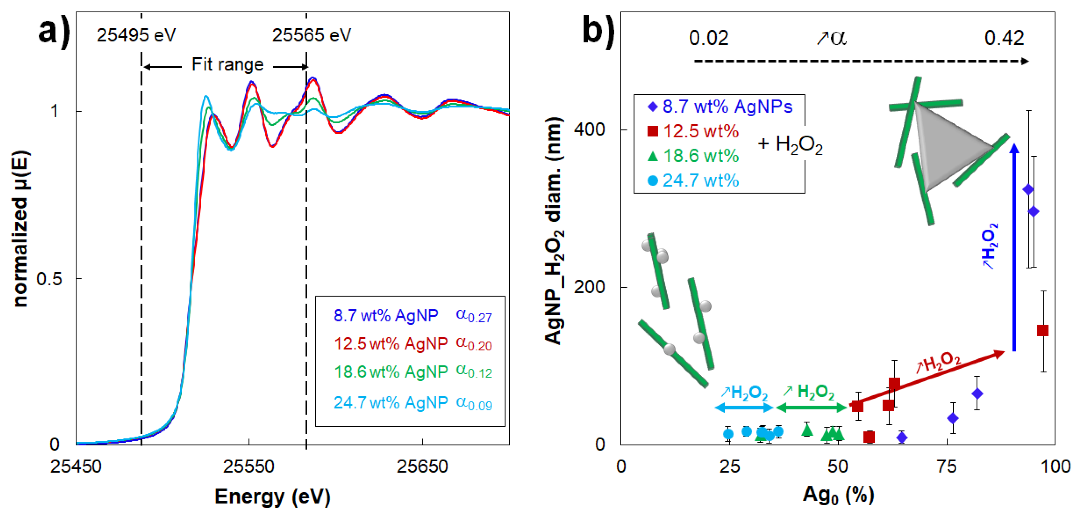
As an example, the XANES spectra for hybrids treated with 160 µL of H
2O
2 at different initial AgNP contents (α from 0.09 to 0.27) are shown in
Figure 4a and the R-factor and Chi-square values of the fits are reported in
Table S4. Extending this analysis,
Table 1 reports the evolution of the average diameter of the AgNPs_H
2O
2 as a function of their Ag
0 content for all the studied hybrids (
Figure 4b). It is shown that the Ag
0 content of AgNPs_H
2O
2 varied with their morphological modification. Notably, hybrids at 18.6 and 24.7 wt% AgNP_H
2O
2 always displayed a α lower than 0.20, maintaining 15-nm AgNPs_H
2O
2 with a spherical shape and a rather low Ag
0 content that remained between 35% and 50%. On the other hand, when the AgNP–AgNPrism transition occurred (i.e., α ≥ 0.20), the Ag
0 content increased up to 100%. Such a trend confirmed that the size-shape transition from 10-nm AgNPs to 300-nm AgNPrisms was always associated with a modification of the oxidation state. In other words, it could be concluded that it was not possible to obtain AgNPrisms by a H
2O
2 redox post-treatment without affecting their oxidation state. Thus,
Figure 4b could be considered as a guide map to tune the AgNP morphological characteristics with respect to their oxidation state. Moreover, our experimental results proved that AgNPs and AgNPrisms were undoubtedly composed of different fractions of both Ag
0 and Ag
+.
Concerning the structure, the XRD diffractograms showed the persistence of a fcc Ag crystalline structure for all the samples (
Figure S4), independently of the α value. Hybrids formulated with the introduction of H
2O
2 still showed a well-defined (111) peak, indicating that even bigger AgNPrisms with a triangular shape had a fcc structure and that they were preferentially oriented parallel to the substrate during the acquisition. However, the crystallite size increased from 3.2 nm up to 7 nm with the progressive addition of H
2O
2, which confirmed that oxidation/reduction steps occurred and maintained the fcc structure intact, with a slight variation of the CS.
The EXAFS Fourier transform spectra (
Figure S5a,b) of the CNC/AgNP hybrid were fitted with the crystallographic structure of metallic silver, with an R-factor systematically lower than 0.048 (
Table S5). The overall variation in interatomic distance values, R, obtained from the fits were systematically negligible as the error bars associated to the R values always partly overlapped with each other.
This result showed that the interatomic distances in the CNC/AgNP_H2O2 hybrids do not significantly change in comparison to the metallic silver distances and that the space group of the samples still corresponds to the fcc silver structure, as shown by XRD. As previously shown, the final crystal structural organization was neither affected by the H2O2 redox post-reaction, nor by the initial Ag content in CNC/AgNP hybrids.
Finally, we checked the influence of the NaBH
4/AgNO
3 initial molar ratio on the H
2O
2 redox post-treatment. CNC/AgNP suspensions at the highest silver content (i.e., the lowest NaBH
4/AgNO
3 ratio equal to 1.5) were prepared to obtain the NaBH
4/AgNO
3 molar ratio of the sample at the lowest AgNP content (i.e., NaBH
4/AgNO
3 ratio = 30) and then mixed with 160 μL of H
2O
2 (α = 0.09). Even in these conditions, the UV-Vis spectra of such suspensions at an NaBH
4/AgNO
3 ratio of 30 overlapped the one of the same sample prepared at a NaBH
4/AgNO
3 ratio of 1.5 (
Figure S6), thus proving that this parameter did not affect the H
2O
2 post-reduction in our experimental conditions.
4. Conclusions
In this study, we investigated the impact of the H2O2 redox post-treatment on the morphology, physicochemical properties, and structural organization of CNC/AgNP aqueous hybrid suspensions formulated using unmodified CNCs as bio-based support to stabilize AgNPs without the addition of any other capping agent. Hybrids at various AgNP contents (i.e., 8.7, 12.5, 18.6, and 24.7 wt%) were mixed with different H2O2 volumes (i.e., 0, 40, 80, 120, 160, and 250 µL) to obtain various H2O2/AgNP mass ratios (α) up to 0.42.
We demonstrated that a critical α value of 0.20 had to be overcome to achieve a size-shape transition from 10-nm spherical NPs (AgNPs) to 300-nm triangular or prismatic NPs (AgNPrisms). Furthermore, we proposed an H2O2 redox mechanism that considered the CNCs as stabilizers for AgNP_H2O2 and AgNPrisms. We speculated that at large amounts of H2O2, for α values higher than 0.20, the H2O2 oxidative action concerned most of the NP except its part effectively grafted onto the CNC surface, which acted as a nucleation seed for AgNPrism formation. On the other hand, at α lower than 0.20, primary AgNPs were only partially oxidized and spherical AgNPs-H2O2 of about 15–20 nm were formed again.
We proved that the transition from 10-nm spherical AgNPs to 300-nm triangular AgNPrisms (i.e., α ≥ 0.20) was associated with an increase in the Ag0 content up to 100%. However, the oxidation state was slightly affected when the H2O2 post-reaction did not modify the size and shape of AgNPs_H2O2 (i.e., α < 0.20). Finally, the AgNP_H2O2 structure was not affected by the H2O2 redox reaction since a fcc model structure was maintained, regardless of the α value.
The present results make it possible to create a guide map to fully control AgNP properties in hybrid NPs where CNCs serve as substrate, making these hybrid NPs suitable for new applications fields.


