
Crystallographic and Spectroscopic Investigations on Oxidative Coordination in the Heteroleptic Mononuclear Complex of Cerium and Benzoxazine Dimer

 , and
, and
Abstract
:1. Introduction
2. Materials and Methods
2.1. Chemicals
2.2. Preparation of the Ligand (MMD)
2.3. Preparation and Characterization of the Cerium-MMD Complex
3. Results and Discussion
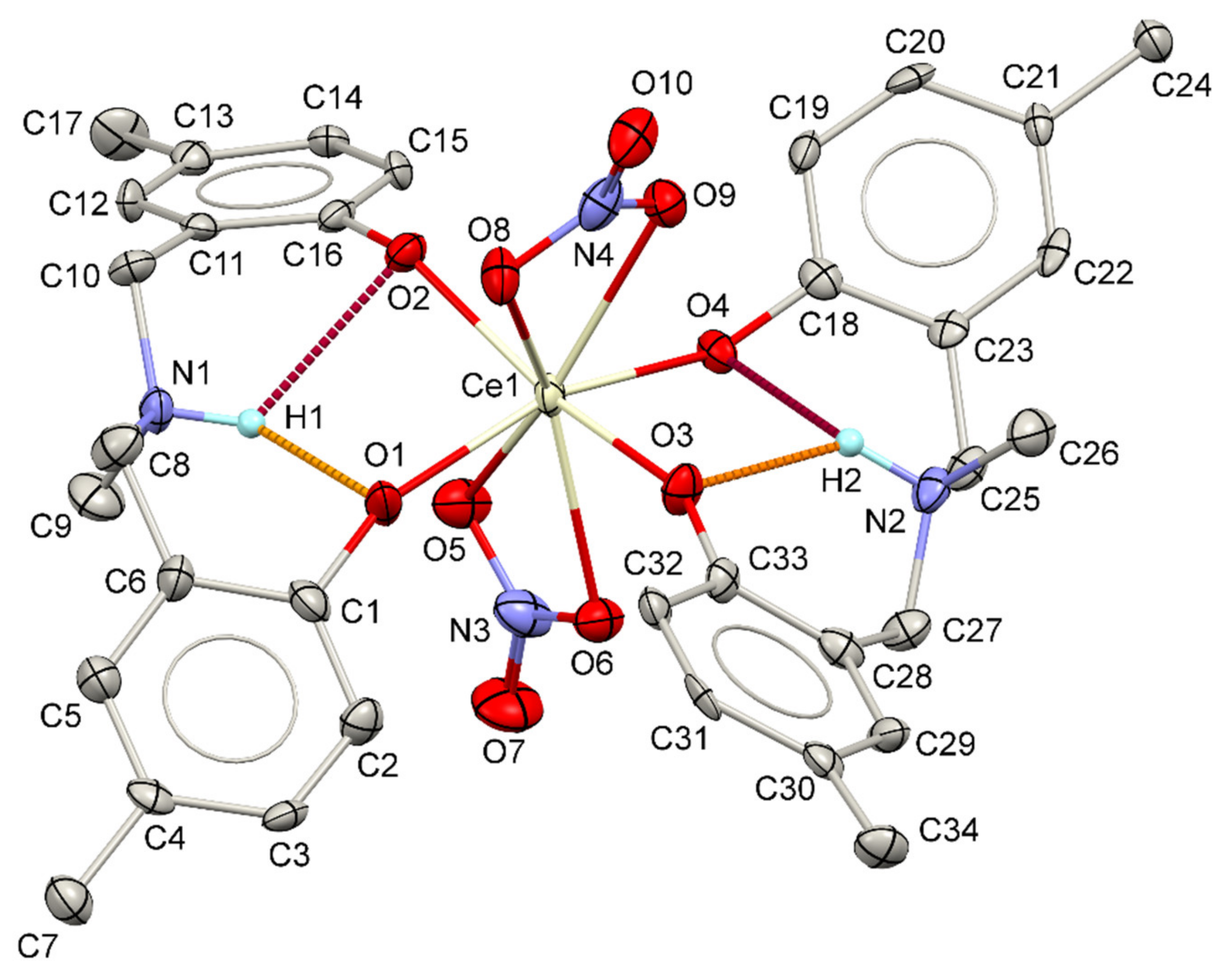
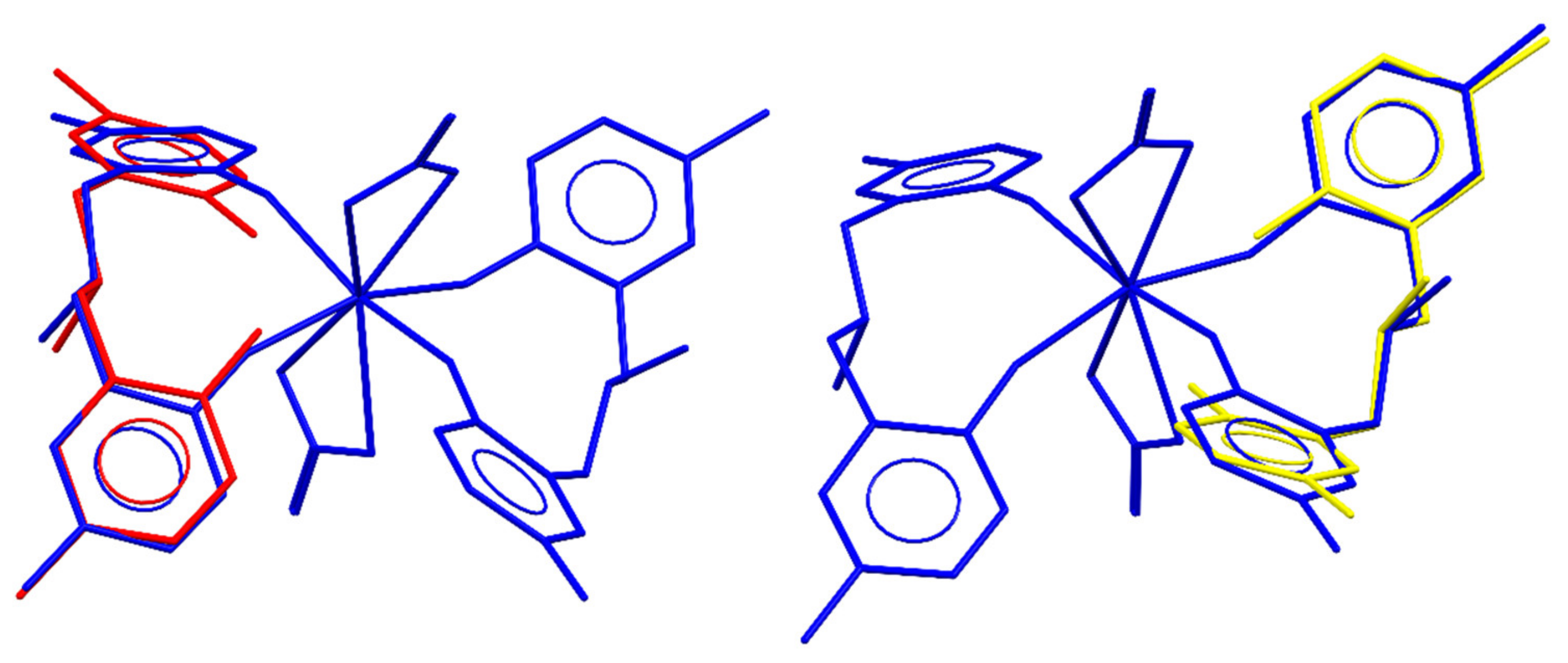
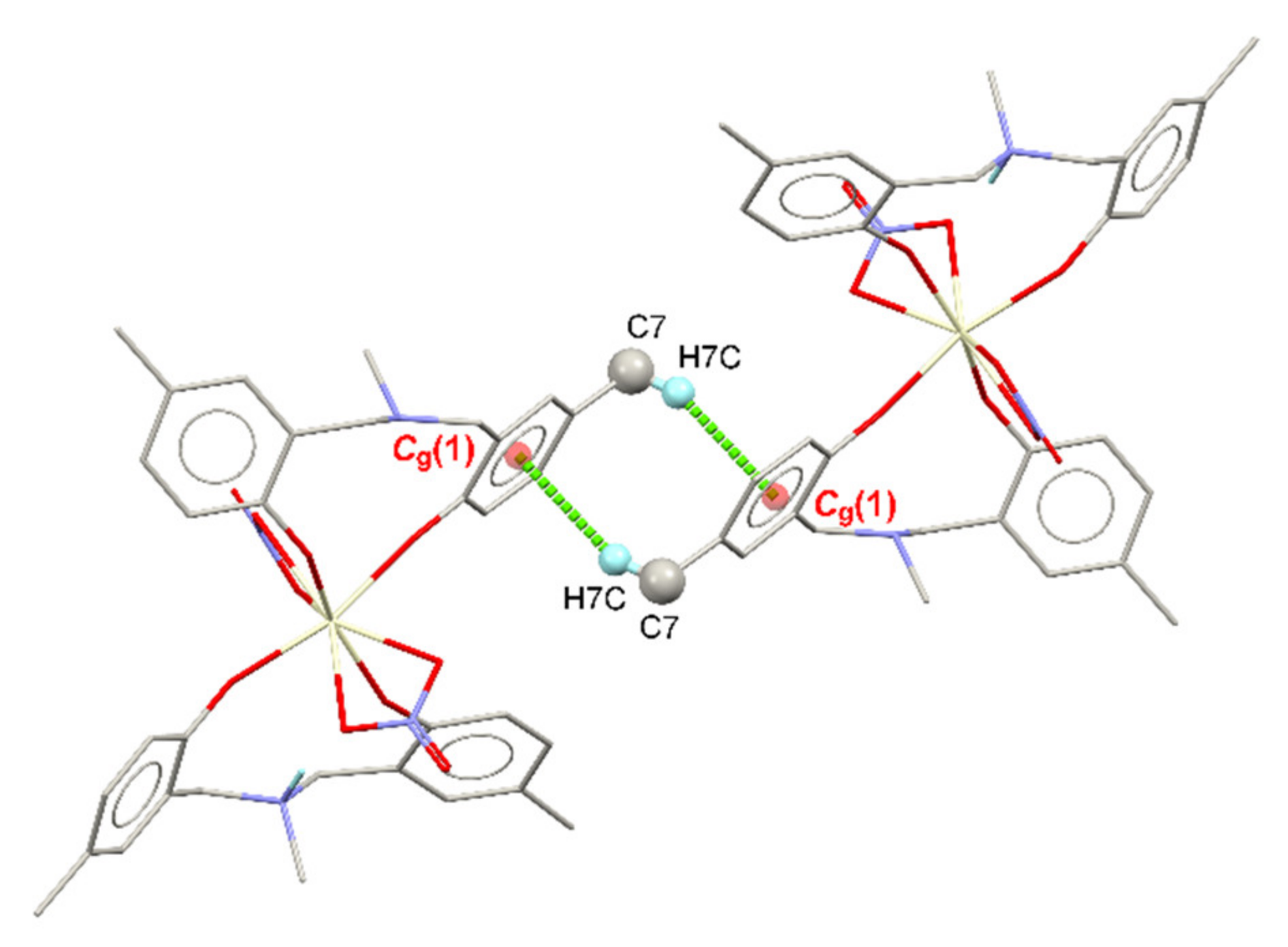
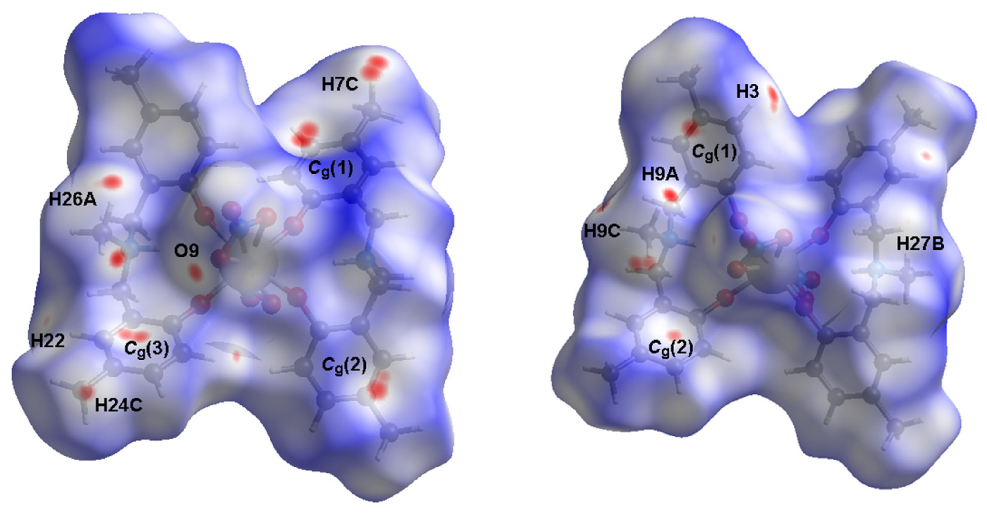
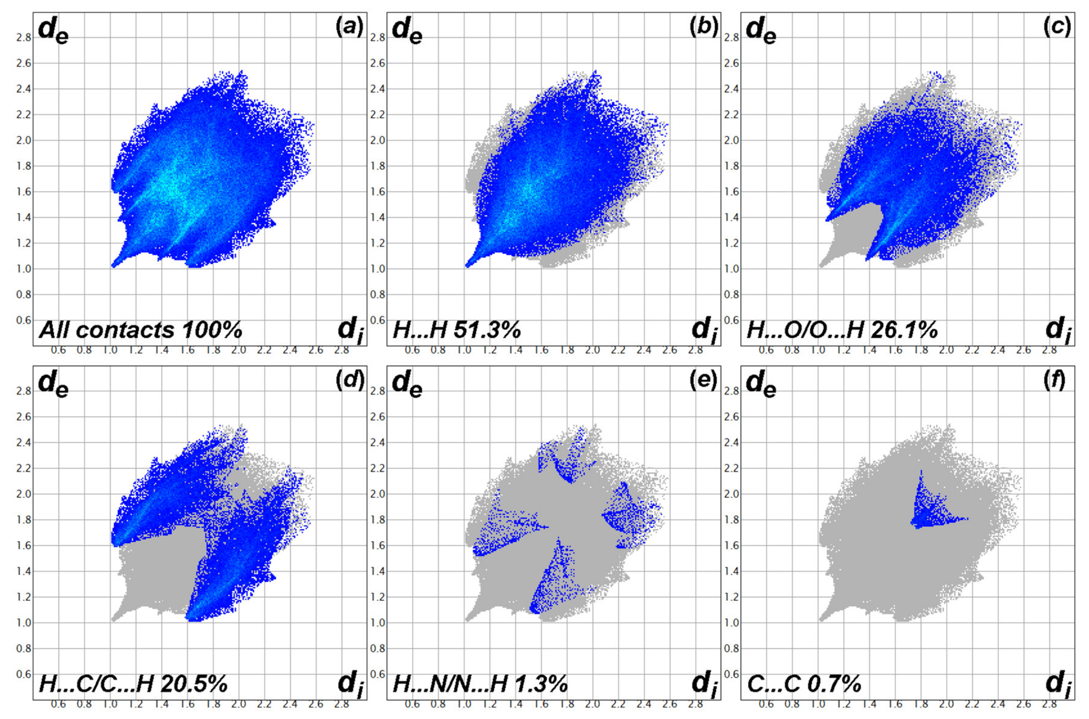
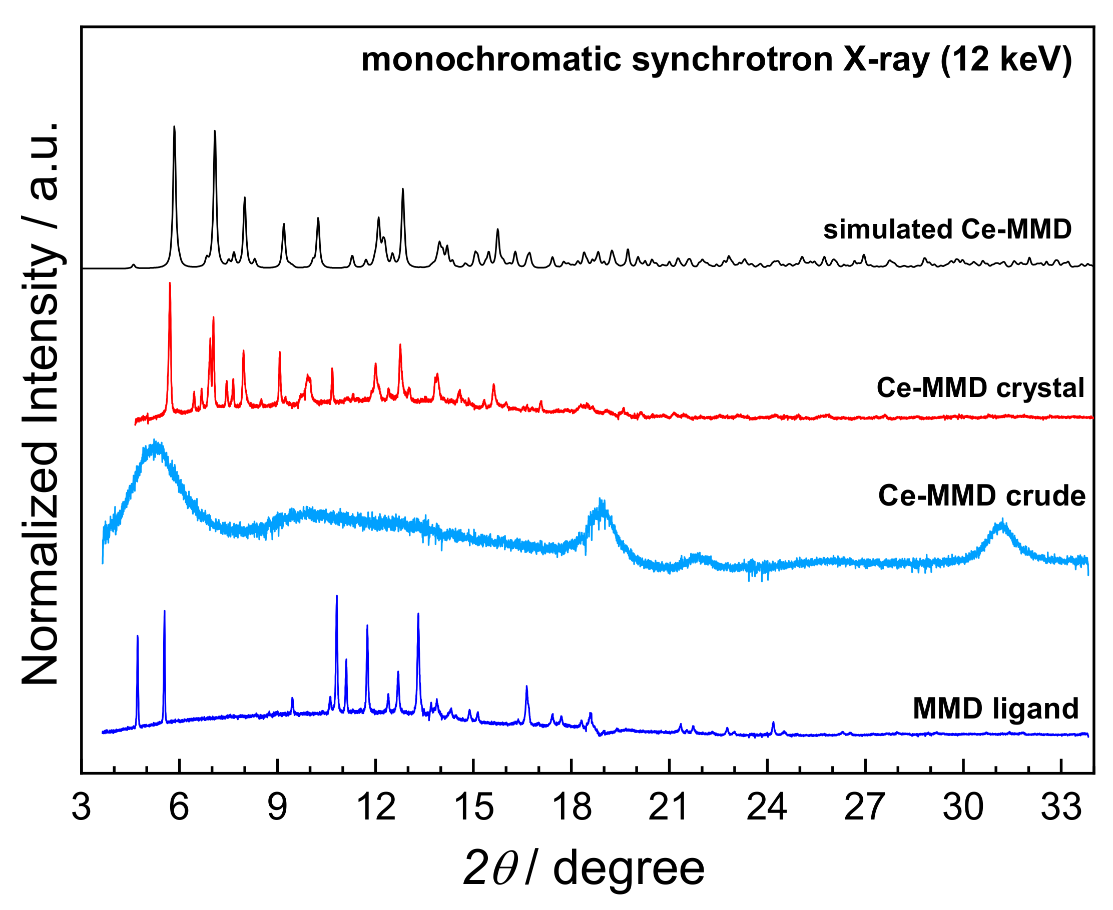
3.1. Crystal Structure and Hirshfeld Surface Analysis of the Cerium-MMD Complex
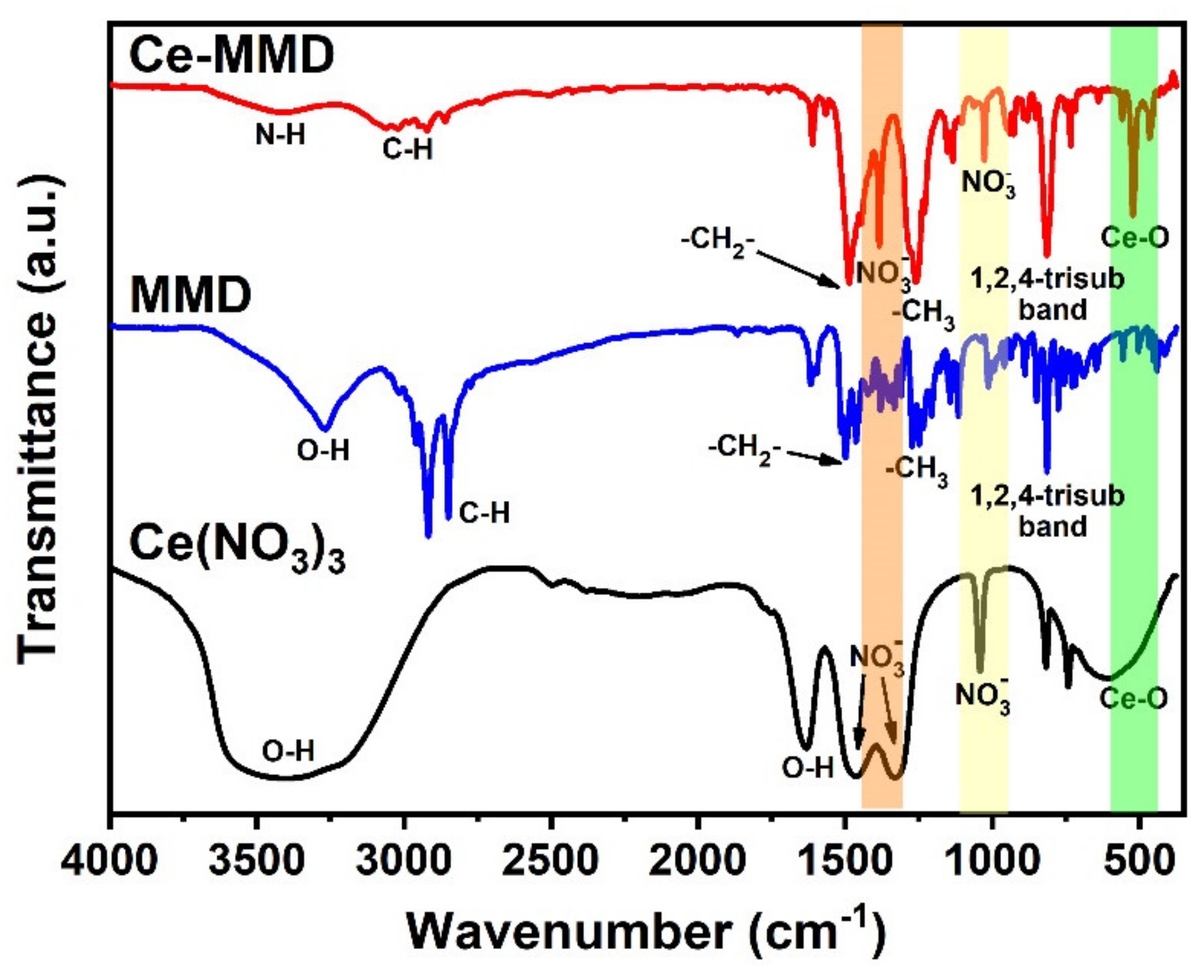
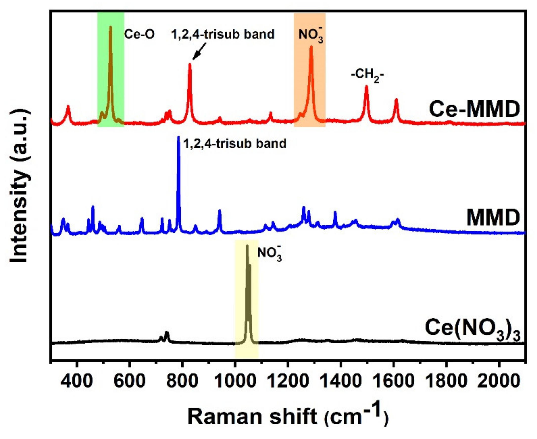
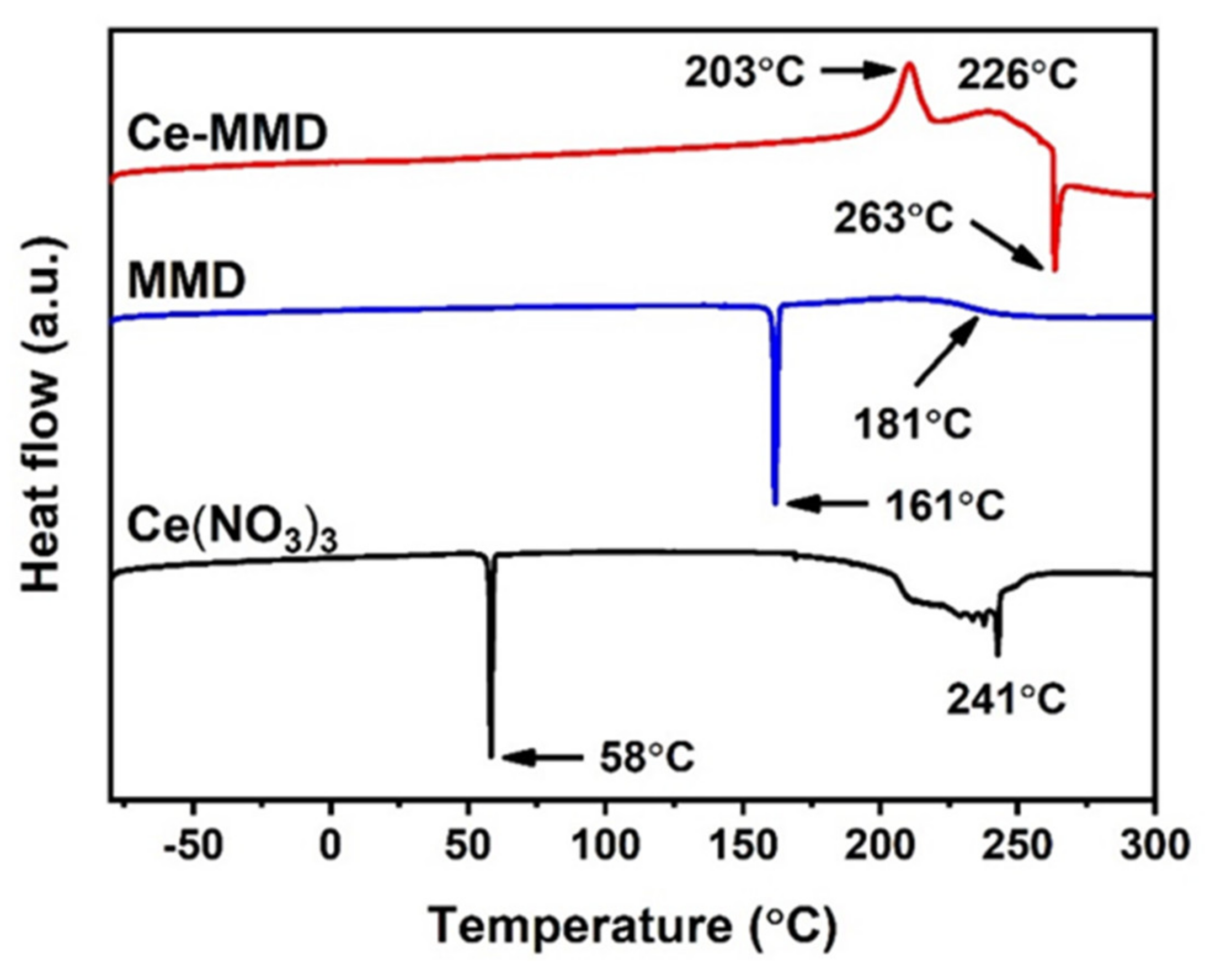
3.2. Spectroscopic Studies and Thermal Properties of the Ce-MMD Complex
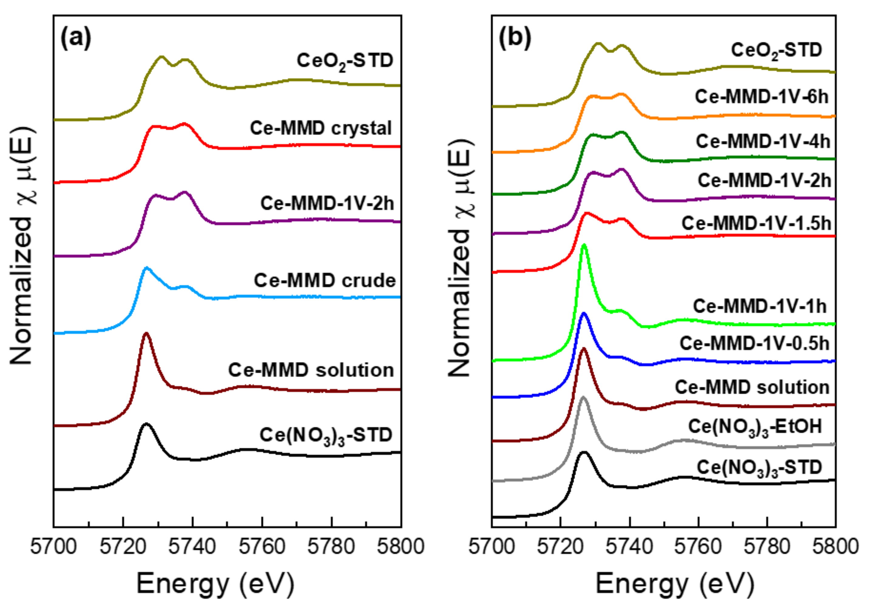
3.3. Monitoring the Oxidative Complexation of the Cerium-MMD Complex
4. Conclusions
Author Contributions
Funding
Institutional Review Board Statement
Informed Consent Statement
Data Availability Statement
Acknowledgments
Conflicts of Interest
Sample Availability
References
- Chueh, W.C.; Falter, C.; Abbott, M.; Scipio, D.; Furler, P.; Haile, S.M.; Steinfeld, A. High-flux solar-driven thermochemical dissociation of CO2 and H2O using nonstoichiometric ceria. Science 2010, 330, 1797–1801. [Google Scholar] [CrossRef] [Green Version]
- Kašpar, J.; Fornasiero, P. Structural properties and thermal stability of ceria-zirconia and related materials. In Catalysis by Ceria and Related Materials, Catalytic Science Series 2; Imperial College Press: London, UK, 2002; pp. 217–241. [Google Scholar]
- So, Y.-M.; Leung, W.-H. Recent advances in the coordination chemistry of cerium (IV) complexes. Coord. Chem. Rev. 2017, 340, 172–197. [Google Scholar] [CrossRef]
- Piro, N.A.; Robinson, J.R.; Walsh, P.J.; Schelter, E.J. The electrochemical behavior of cerium(III/IV) complexes: Thermodynamics, kinetics and applications in synthesis. Coord. Chem. Rev. 2014, 260, 21–36. [Google Scholar] [CrossRef]
- Cotton, S.A. Lanthanide and Actinide Chemistry; John Wiley & Sons: Chichester, UK; Hoboken, NJ, USA, 2006. [Google Scholar]
- Jutzi, P. Advanced Inorganic Chemistry; John Wiley & Sons: Chichester, UK, 1988; p. 957. [Google Scholar]
- Williams, U.J.; Schneider, D.; Dorfner, W.L.; Maichle-Mössmer, C.; Carroll, P.J.; Anwander, R.; Schelter, E.J. Variation of electronic transitions and reduction potentials of cerium (IV) complexes. Dalton Trans. 2014, 43, 16197–16206. [Google Scholar] [CrossRef]
- Yin, H.; Carroll, P.J.; Anna, J.M.; Schelter, E.J. Luminescent Ce (III) complexes as stoichiometric and catalytic photoreductants for halogen atom abstraction reactions. J. Am. Chem. Soc. 2015, 137, 9234–9237. [Google Scholar] [CrossRef] [PubMed] [Green Version]
- Yin, H.; Carroll, P.J.; Manor, B.C.; Anna, J.M.; Schelter, E.J. Cerium photosensitizers: Structure–function relationships and applications in photocatalytic aryl coupling reactions. J. Am. Chem. Soc. 2016, 138, 5984–5993. [Google Scholar] [CrossRef] [PubMed]
- Friedrich, J.; Schneider, D.; Bock, L.; Maichle-Mössmer, C.; Anwander, R. Cerium (IV) neopentoxide complexes. Inorg. Chem. 2017, 56, 8114–8127. [Google Scholar] [CrossRef] [PubMed]
- Atwood, D.A. The Rare Earth Elements: Fundamentals and Applications; John Wiley & Sons, Inc.: Hoboken, NJ, USA, 2012; pp. 321–334. [Google Scholar]
- Arnold, P.L.; Casely, I.J.; Zlatogorsky, S.; Wilson, C. Organometallic cerium complexes from tetravalent coordination complexes. Helv. Chim. Acta 2009, 92, 2291–2303. [Google Scholar] [CrossRef]
- Dröse, P.; Crozier, A.R.; Lashkari, S.; Gottfriedsen, J.; Blaurock, S.; Hrib, C.G.; Maichle-Mössmer, C.C.; Schädle, C.; Anwander, R.; Edelmann, F.T. Facile access to tetravalent cerium compounds: One-electron oxidation using iodine (III) reagents. J. Am. Chem. Soc. 2010, 132, 14046–14047. [Google Scholar] [CrossRef]
- Dröse, P.; Gottfriedsen, J. Synthesis of Heteroleptic Cerium (IV) Complexes Using a Heptadentate (N4O3) Tripodale Schiff-base Ligand. Z. Für Anorg. Allg. Chem. 2008, 634, 87–90. [Google Scholar] [CrossRef]
- Jiang, J.; Ng, D.K.P. A Decade Journey in the Chemistry of Sandwich-Type Tetrapyrrolato−Rare Earth Complexes. Acc. Chem. Res. 2009, 42, 79–88. [Google Scholar] [CrossRef]
- Walter, M.D.; Fandos, R.; Andersen, R.A. Synthesis and magnetic properties of cerium macrocyclic complexes with tetramethyldibenzotetraaza[14]annulene, tmtaaH2. New J. Chem. 2006, 30, 1065–1070. [Google Scholar] [CrossRef] [Green Version]
- Kilimann, U.; Herbst-Irmer, R.; Stalke, D.; Edelmann, F.T. An Efficient Access to Organocerium (iv) Complexes: Synthesis and Structure of Bis [1,3,6-tris (trimethylsilyl) cyclooctatetraene] cerium (iv). Angew. Chem. Int. Ed. Engl. 1994, 33, 1618–1621. [Google Scholar] [CrossRef]
- Lorenz, V.; Schmiege, B.M.; Hrib, C.G.; Ziller, J.W.; Edelmann, A.; Blaurock, S.; Evans, W.J.; Edelmann, F.T. Unprecedented Bending and Rearrangement of f-Element Sandwich Complexes Induced by Superbulky Cyclooctatetraenide Ligands. J. Am. Chem. Soc. 2011, 133, 1257–1259. [Google Scholar] [CrossRef] [PubMed]
- DelaRosa, M.; Bousman, K.; Welch, J.; Toscano, P. Physical and Structural Characterization of Ce (IV) β-Diketonate Complexes: Evidence for Geometrical Isomers in the Solid-State. Coord. Chem. Rev. 2002, 55, 781–793. [Google Scholar] [CrossRef]
- Hitchcock, P.B.; Lappert, M.F.; Protchenko, A.V. Facile formation of a homoleptic Ce(IV) amide via aerobic oxidation. Chem. Commun. 2006, 3546–3548. [Google Scholar] [CrossRef] [PubMed]
- Ishida, H.; Agag, T. Handbook of Benzoxazine Resins; Elsevier: Amsterdam, The Netherlands; Boston, MA, USA, 2011. [Google Scholar]
- Kiskan, B.; Ghosh, N.N.; Yagci, Y. Polybenzoxazine-based composites as high-performance materials. Polym. Int. 2011, 60, 167–177. [Google Scholar] [CrossRef]
- Ghosh, N.; Kiskan, B.; Yagci, Y. Polybenzoxazines—New high performance thermosetting resins: Synthesis and properties. Prog. Polym. Sci. 2007, 32, 1344–1391. [Google Scholar] [CrossRef]
- Rimdusit, S.; Tiptipakorn, S.; Jubsilp, C.; Takeichi, T. Polybenzoxazine alloys and blends: Some unique properties and applications. React. Funct. Polym. 2013, 73, 369–380. [Google Scholar] [CrossRef]
- Shen, S.B.; Ishida, H. Development and characterization of high-performance polybenzoxazine composites. Polym. Compos. 1996, 17, 710–719. [Google Scholar] [CrossRef]
- Liao, Y.-T.; Lin, Y.-C.; Kuo, S.-W. Highly thermally stable, transparent, and flexible polybenzoxazine nanocomposites by combination of double-decker-shaped polyhedral silsesquioxanes and polydimethylsiloxane. Macromolecules 2017, 50, 5739–5747. [Google Scholar] [CrossRef]
- Chirachanchai, S.; Laobuthee, A.; Phongtamrug, S. Self termination of ring opening reaction of p-substituted phenol-based benzoxazines: An obstructive effect via intramolecular hydrogen bond. Heterocycl. Chem. 2009, 46, 714–721. [Google Scholar] [CrossRef]
- Hemvichian, K.; Laobuthee, A.; Chirachanchai, S.; Ishida, H. Thermal decomposition processes in polybenzoxazine model dimers investigated by TGA–FTIR and GC–MS. Polym. Degrad. 2002, 76, 1–15. [Google Scholar] [CrossRef]
- Iguchi, D.; Salum, M.L.; Froimowicz, P. Application of Benzoxazine-Based Dimers, Oligomers, and Polymers as Chelating Agents. Macromol. Chem. Phys. 2019, 220, 1800366. [Google Scholar] [CrossRef]
- Veranitisagul, C.; Kaewvilai, A.; Sangngern, S.; Wattanathana, W.; Suramitr, S.; Koonsaeng, N.; Laobuthee, A. Novel recovery of nano-structured ceria (CeO2) from Ce (III)-benzoxazine dimer complexes via thermal decomposition. Int. J. Mol. Sci. 2011, 12, 4365–4377. [Google Scholar] [CrossRef]
- Fujita, K.I.; Kawahara, R.; Aikawa, T.; Yamaguchi, R. Hydrogen production from a methanol–water solution catalyzed by an anionic iridium complex bearing a functional bipyridonate ligand under weakly basic conditions. Angew. Chem. Int. Ed. 2015, 54, 9057–9060. [Google Scholar] [CrossRef] [PubMed]
- Finn, M.; Ridenour, J.A.; Heltzel, J.; Cahill, C.; Voutchkova-Kostal, A. Next-generation water-soluble homogeneous catalysts for conversion of glycerol to lactic acid. Organometallics 2018, 37, 1400–1409. [Google Scholar] [CrossRef]
- Liu, J.-F.; Zhao, Z.-S.; Jiang, G.-B. Coating Fe3O4 magnetic nanoparticles with humic acid for high efficient removal of heavy metals in water. Environ. Sci. Technol. 2008, 42, 6949–6954. [Google Scholar] [CrossRef] [PubMed]
- Wattanathana, W.; Veranitisagul, C.; Koonsaeng, N.; Laobuthee, A. 3,4-Dihydro-1,3-2H-Benzoxazines: Uses Other Than Making Polybenzoxazines. In Advanced and Emerging Polybenzoxazine Science and Technology; Elsevier: Amsterdam, The Netherlands, 2017; pp. 75–88. [Google Scholar]
- Maketon, W.; Zenner, C.Z.; Ogden, K.L. Removal efficiency and binding mechanisms of copper and copper− EDTA complexes using polyethyleneimine. Environ. Sci. Technol. 2008, 42, 2124–2129. [Google Scholar] [CrossRef]
- Ma, Z.; Moulton, B. Recent advances of discrete coordination complexes and coordination polymers in drug delivery. Coord. Chem. Rev. 2011, 255, 1623–1641. [Google Scholar] [CrossRef]
- Sun, R.W.-Y.; Ma, D.-L.; Wong, E.L.-M.; Che, C.-M. Some uses of transition metal complexes as anti-cancer and anti-HIV agents. Dalton Trans. 2007, 43, 4884–4892. [Google Scholar]
- Wu, M.-H.; Liu, W.-J.; Zou, W.-D.; Wang, H.-Y. 4,4′-Dimethyl-2,2′-(N-methyliminodimethylene) diphenol. Acta Crystallogr. Sect. E Struct. Rep. Online 2006, 62, o2949–o2950. [Google Scholar] [CrossRef]
- SAINT Version 8.34A 2013; Bruker AXS: Madison, WI, USA, 2013.
- Sheldrick, G.M. SADABS; University of Gottingen: Gottingen, Germany, 1996. [Google Scholar]
- Sheldrick, G.M. SHELXT—Integrated space-group and crystal-structure determination. Acta Crystallogr. A Found. Adv. 2017, 71, 3–8. [Google Scholar] [CrossRef] [Green Version]
- Sheldrick, G.M. Crystal structure refinement with SHELXL. Acta Crystallogr. Sect. C Struct. Chem. 2015, 71, 3–8. [Google Scholar] [CrossRef] [PubMed]
- Dolomanov, O.V.; Bourhis, L.J.; Gildea, R.J.; Howard, J.A.; Puschmann, H. OLEX2: A complete structure solution, refinement and analysis program. J. Appl. Crystallogr. 2009, 42, 339–341. [Google Scholar] [CrossRef]
- Macrae, C.F.; Edgington, P.R.; McCabe, P.; Pidcock, E.; Shields, G.P.; Taylor, R.; Towler, M.; Streek, J. Mercury: Visualization and analysis of crystal structures. Appl. Crystallogr. 2006, 39, 453–457. [Google Scholar] [CrossRef] [Green Version]
- Ravel, B.; Newville, M. ATHENA, ARTEMIS, HEPHAESTUS: Data analysis for X-ray absorption spectroscopy using IFEFFIT. J. Synchrotron Radiat. 2005, 12, 537–541. [Google Scholar] [CrossRef] [Green Version]
- Li, L.; Yuan, F.; Li, T.; Zhou, Y.; Zhang, M. Synthesis and crystal structures of cerium (IV) complexes with 8-quinolinolate and amine bis(phenolate) ligands. Inorg. Chim. Acta 2013, 397, 69–74. [Google Scholar] [CrossRef]
- Etter, M.C.; MacDonald, J.C.; Bernstein, J. Graph-set analysis of hydrogen-bond patterns in organic crystals. Acta Crystallogr. Sect. B Struct. Sci. 1990, 46, 256–262. [Google Scholar] [CrossRef]
- Grell, J.; Bernstein, J.; Tinhofer, G. Graph-set analysis of hydrogen-bond patterns: Some mathematical concepts. Acta Crystallogr. Sect. B Struct. Sci. 1999, 55, 1030–1043. [Google Scholar] [CrossRef] [PubMed] [Green Version]
- Bernstein, J.; Shimoni, L.; Davis, R.E.; Chang, N.L. Graph set analysis of hydrogen-bond patterns in organic crystals. Recent developments and applications. Acta Crystallogr. Sect. A Found. Crystallogr. 1993, 49, c164. [Google Scholar] [CrossRef] [Green Version]
- Wattanathana, W.; Nootsuwan, N.; Veranitisagul, C.; Koonsaeng, N.; Suramitr, S.; Laobuthee, A. Crystallographic, spectroscopic (FT-IR/FT-Raman) and computational (DFT/B3LYP) studies on 4,4′-diethyl-2,2′-[methylazanediylbis (methylene)] diphenol. J. Mol. Struct. 2016, 1109, 201–208. [Google Scholar] [CrossRef]
- Veranitisagul, C.; Kaewvilai, A.; Duangthongyou, T.; Koonsaeng, N.; Laobuthee, A. 4,4′-Dimethoxy-2,2′-[methylazanediylbis (methylene)] diphenol. Acta Crystallogr. Sect. E Struct. Rep. Online 2012, 68, o2139. [Google Scholar] [CrossRef] [Green Version]
- Veranitisagul, C.; Wattanathana, W.; Kaewvilai, A.; Duangthongyou, T.; Laobuthee, A.; Koonsaeng, N. 2-{[(2-Hydroxy-3,5-dimethylbenzyl)(methyl) amino] methyl}-4, 6-dimethylphenol. Acta Crystallogr. Sect. E Struct. Rep. Online 2012, 68, o1826. [Google Scholar] [CrossRef] [PubMed]
- Phongtamrug, S.; Tashiro, K.; Miyata, M.; Chirachanchai, S. Supramolecular structure of N, N-bis (2-hydroxybenzyl) alkylamine: Flexible molecular assembly framework for host without guest and host with guest. Phys. Chem. B 2006, 110, 21365–21370. [Google Scholar] [CrossRef] [PubMed]
- Wattanathana, W.; Veranitisagul, C.; Kaewvilai, A.; Laobuthee, A.; Koonsaeng, N. 4,4′-Diethyl-2,2′-[(N-cyclohexylimino) bis (methylene)] diphenol. Acta Crystallogr. Sect. E Struct. Rep. Online 2012, 68, o3050. [Google Scholar] [CrossRef] [PubMed] [Green Version]
- Wannapaiboon, S.; Hanlumyuang, Y.; Chansaenpak, K.; Pinyou, P.; Veranitisagul, C.; Laobuthee, A.; Wattanathana, W. Crystal structure and Hirshfeld surface analysis of the product of the ring-opening reaction of a dihydrobenzoxazine: 6,6′-[(cyclohexylazanediyl) bis (methylene)] bis (2,4-dimethylphenol). Acta Crystallogr. Sect. E Crystallogr. Commun. 2020, 76, 1239–1244. [Google Scholar] [CrossRef] [PubMed]
- Suetrong, N.; Chansaenpak, K.; Impeng, S.; Pinyou, P.; Blay, V.; Blay-Roger, R.; Lisnund, S.; Kanjanaboos, P.; Hanlumyuang, Y.; Wannapaiboon, S.; et al. Influences of Chemical Functionalities on Crystal Structures and Electrochemical Properties of Dihydro-benzoxazine Dimer Derivatives. Crystals 2021, 11, 979. [Google Scholar] [CrossRef]
- Brown, I.D. The Bond Valence Model as a Tool for Teaching Inorganic Chemistry: The Ionic Model Revisited. J. Chem. Educ. 2000, 77, 1070. [Google Scholar] [CrossRef]
- Brown, I.D. The Chemical Bond in Inorganic Chemistry, The Bond Valence Model; IUCR monographs on crystallography, 12; Oxford University Press: Oxford, UK, 2002. [Google Scholar]
- Brown, I.D. Recent Developments in the Methods and Applications of the Bond Valence Model. Chem. Rev. 2009, 109, 6858–6919. [Google Scholar] [CrossRef] [Green Version]
- Roulhac, P.L.; Palenik, G.J. Bond valence sums in coordination chemistry. The calculation of the oxidation state of cerium in complexes containing cerium bonded only to oxygen. Inorg. Chem. 2003, 42, 118–121. [Google Scholar] [CrossRef]
- Groom, C.R.; Bruno, I.J.; Lightfoot, M.P.; Ward, S.C. The Cambridge Structural Database. Acta Cryst. B Struct. Sci. Cryst. Eng. Mater. 2016, 72, 171–179. [Google Scholar] [CrossRef] [PubMed]
- Palenik, G.J.; Hu, S.-Z. Assignment of oxidation states in metal complexes. Cerium(III) or cerium(IV) and other questions. Inorg. Chim. Acta 2009, 362, 4740–4743. [Google Scholar] [CrossRef]
- Hirshfeld, F.L. Bonded-atom fragments for describing molecular charge densities. Theor. Chim. Acta 1977, 44, 129–138. [Google Scholar] [CrossRef]
- Spackman, M.A.; Jayatilaka, D. Hirshfeld surface analysis. CrystEngComm 2009, 11, 19–32. [Google Scholar] [CrossRef]
- Turner, M.J.; McKinnon, J.J.; Wolff, S.K.; Grimwood, D.J.; Spackman, P.R.; Jayatilaka, D.; Spackman, M.A. Crystal Explorer Version 17.5; University of Western Australia: Crawley, Australia, 2017. [Google Scholar]
- McKinnon, J.J.; Jayatilaka, D.; Spackman, M.A. Towards quantitative analysis of intermolecular interactions with Hirshfeld surfaces. Chem. Commun. 2007, 37, 3814–3816. [Google Scholar] [CrossRef]
- Ansari, A.A.; Kaushik, A. Synthesis and optical properties of nanostructured Ce(OH)4. J. Semicond. 2010, 31, 033001. [Google Scholar] [CrossRef]
- Manelis, G. Thermal Decomposition and Combustion of Explosives and Propellants; CRC Press: London, UK, 2003. [Google Scholar]
- Ayodele, B.V.; Hossain, M.A.; Chong, S.L.; Soh, J.C.; Abdullah, S.; Khan, M.R.; Cheng, C.K. Non-isothermal kinetics and mechanistic study of thermal decomposition of light rare earth metal nitrate hydrates using thermogravimetric analysis. J. Therm. Anal. Calorim. 2016, 125, 423–435. [Google Scholar] [CrossRef] [Green Version]
- Kang, W.; Ozgur, D.O.; Varma, A. Solution Combustion Synthesis of High Surface Area CeO2 Nanopowders for Catalytic Applications: Reaction Mechanism and Properties. ACS Appl. Nano Mater. 2018, 1, 675–685. [Google Scholar] [CrossRef]


| Crystallographic Data and Structural Refinement Details | Cerium-MMD Complex |
|---|---|
| CCDC number | 2098858 |
| Empirical formula | C34H40CeN4O10 |
| Formula weight | 804.82 |
| Temperature/K | 100 |
| Crystal system | Monoclinic |
| Space group | P21/c |
| a/Å | 10.0752(18) |
| b/Å | 16.710(3) |
| c/Å | 20.660(4) |
| α/° | 90 |
| β/° | 101.317(7) |
| γ/° | 90 |
| Volume/Å3 | 3410.6(11) |
| Z | 4 |
| ρcalcg/cm3 | 1.567 |
| μ/mm−1 | 1.398 |
| F(000) | 1640.0 |
| Crystal size/mm3 | 0.2 × 0.14 × 0.1 |
| Radiation | MoKα (λ = 0.71073) |
| 2Θ range for data collection/° | 3.16 to 50.556 |
| Index ranges | −12 ≤ h ≤ 12, −20 ≤ k ≤ 14, −23 ≤ l ≤ 24 |
| Reflections collected | 18,342 |
| Independent reflections | 6132 [Rint = 0.0867, Rsigma = 0.1083] |
| Data/restraints/parameters | 6132/0/448 |
| Goodness-of-fit on F2 | 1.055 |
| Final R indexes [I ≥ 2σ (I)] | R1 = 0.0640, wR2 = 0.1431 |
| Final R indexes [all data] | R1 = 0.1017, wR2 = 0.1618 |
| Largest diff. peak/hole/e Å−3 | 2.05/−1.90 |
| D–H⋯A | d(D–H)/Å | d(H⋯A)/Å | d(D⋯A)/Å | D–H⋯A/° |
|---|---|---|---|---|
| N–H⋯O interactions | ||||
| N1–H1⋯O1 | 0.98 | 1.92 | 2.717(8) | 136 |
| N1–H1⋯O2 | 0.98 | 2.40 | 3.056(8) | 124 |
| N2–H2⋯O3 | 0.98 | 1.98 | 2.753(8) | 134 |
| N2–H2⋯O4 | 0.98 | 2.41 | 3.061(8) | 123 |
| C–H⋯O interactions | ||||
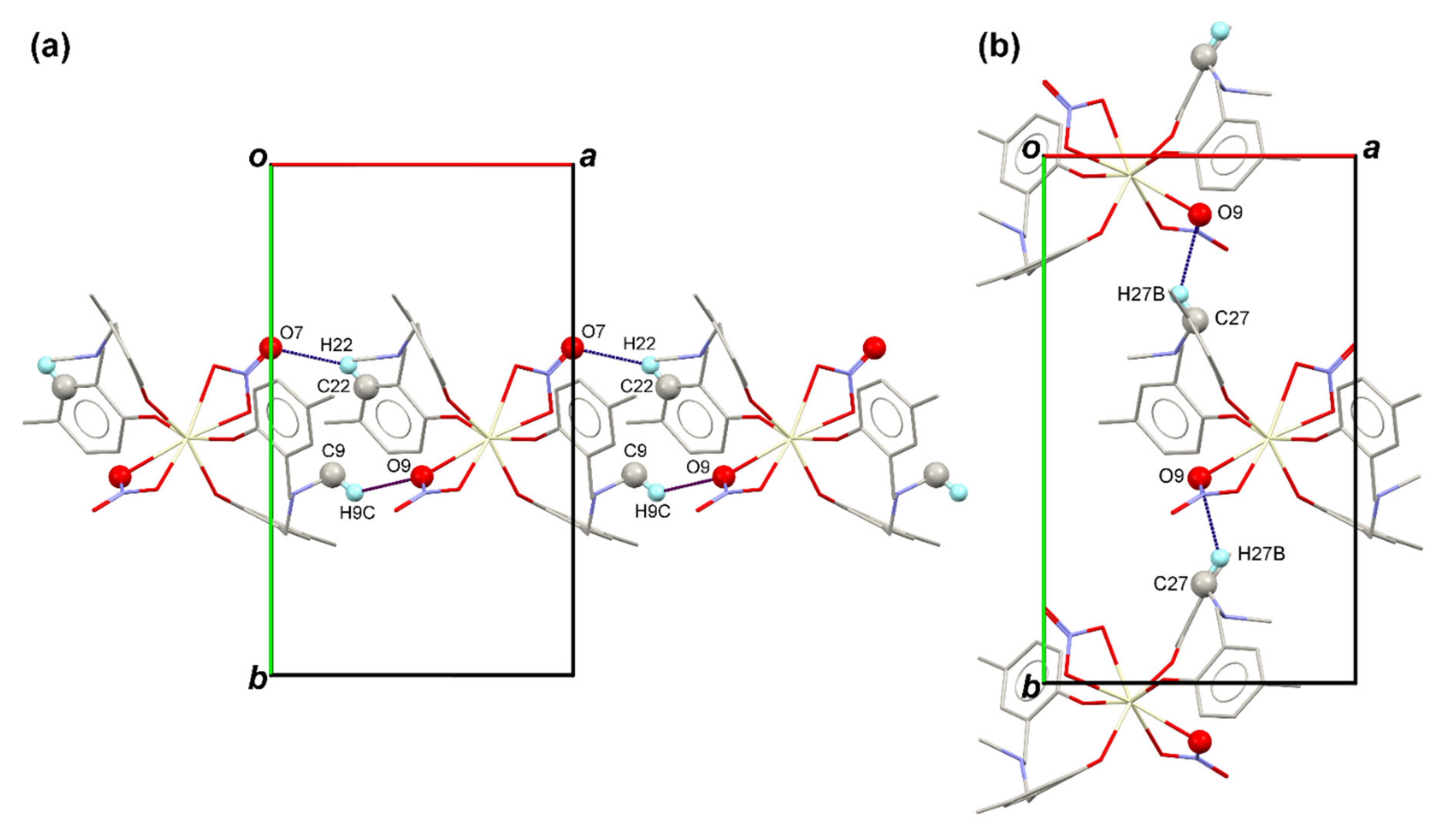
| C9–H9C⋯O9 i | 0.96 | 2.541 | 3.242 | 130 |
| C22–H22⋯O7 i | 0.93 | 2.660 | 3.421 | 140 |
| C27–H27B⋯O9 ii | 0.97 | 2.672 | 3.417 | 134 |
| C–H⋯π interactions | ||||
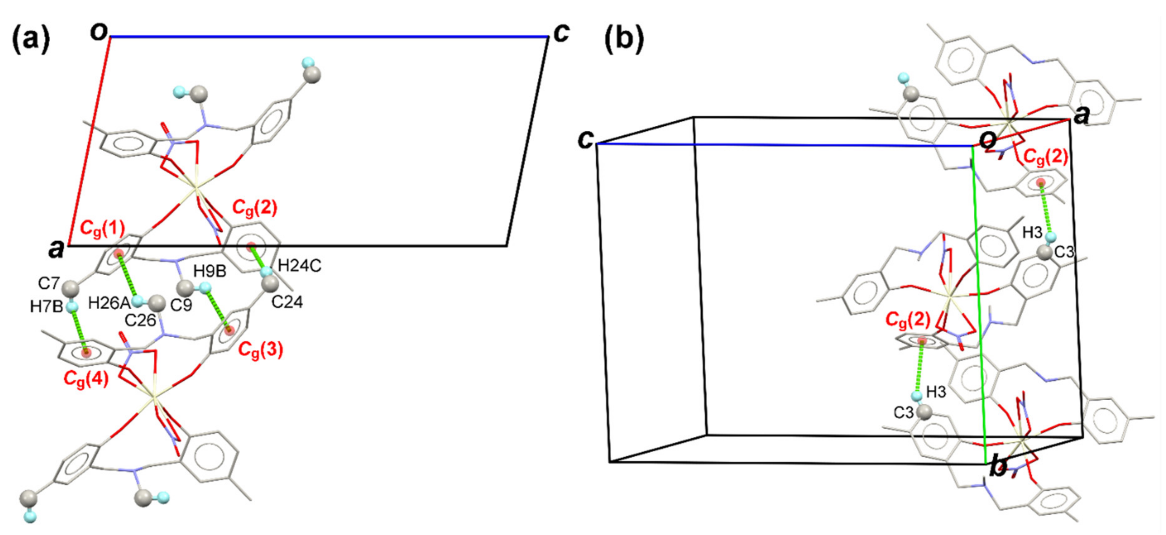
| C26–H26A⋯Cg(1) i | 0.96 | 2.908 | 3.502 | 121 |
| C24–H24C⋯Cg(2) i | 0.96 | 2.840 | 3.790 | 171 |
| C9–H9B⋯Cg(3) i | 0.96 | 2.864 | 3.423 | 118 |
| C7–H7B⋯Cg(4) i | 0.96 | 3.015 | 3.481 | 111 |
| C3–H3⋯Cg(2) iii | 0.93 | 2.920 | 3.727 | 146 |
| C7–H7C⋯Cg(1) iv | 0.96 | 2.634 | 3.543 | 158 |
Publisher’s Note: MDPI stays neutral with regard to jurisdictional claims in published maps and institutional affiliations. |
© 2021 by the authors. Licensee MDPI, Basel, Switzerland. This article is an open access article distributed under the terms and conditions of the Creative Commons Attribution (CC BY) license (https://creativecommons.org/licenses/by/4.0/).
Share and Cite
Wattanathana, W.; Suetrong, N.; Kongsamai, P.; Chansaenpak, K.; Chuanopparat, N.; Hanlumyuang, Y.; Kanjanaboos, P.; Wannapaiboon, S. Crystallographic and Spectroscopic Investigations on Oxidative Coordination in the Heteroleptic Mononuclear Complex of Cerium and Benzoxazine Dimer. Molecules 2021, 26, 5410. https://doi.org/10.3390/molecules26175410
Wattanathana W, Suetrong N, Kongsamai P, Chansaenpak K, Chuanopparat N, Hanlumyuang Y, Kanjanaboos P, Wannapaiboon S. Crystallographic and Spectroscopic Investigations on Oxidative Coordination in the Heteroleptic Mononuclear Complex of Cerium and Benzoxazine Dimer. Molecules. 2021; 26(17):5410. https://doi.org/10.3390/molecules26175410
Chicago/Turabian StyleWattanathana, Worawat, Natapol Suetrong, Peetikamol Kongsamai, Kantapat Chansaenpak, Nutthawat Chuanopparat, Yuranan Hanlumyuang, Pongsakorn Kanjanaboos, and Suttipong Wannapaiboon. 2021. "Crystallographic and Spectroscopic Investigations on Oxidative Coordination in the Heteroleptic Mononuclear Complex of Cerium and Benzoxazine Dimer" Molecules 26, no. 17: 5410. https://doi.org/10.3390/molecules26175410