
Role of Honey in Advanced Wound Care
by
 , , , , and
Hana Scepankova
1,† ,
,
Patricia Combarros-Fuertes
2,†,
José María Fresno
2,
María Eugenia Tornadijo
2,
Miguel Sousa Dias
3,
Carlos A. Pinto
1,
Jorge A. Saraiva
1 and
Letícia M. Estevinho
3,* 1
LAQV-REQUIMTE, Department of Chemistry, University of Aveiro, 3810-193 Aveiro, Portugal
2
Department of Food Hygiene and Technology, Faculty of Veterinary Science, Campus de Vegazana, University of León, 24071 León, Spain
3
CIMO, Mountain Research Center, Polytechnic Institute of Bragança, Campus Santa Apolónia, 5301-855 Bragança, Portugal
*
Author to whom correspondence should be addressed.
†
These authors contributed equally to this work.
Molecules 2021, 26(16), 4784; https://doi.org/10.3390/molecules26164784
Submission received: 13 July 2021
/
Revised: 29 July 2021
/
Accepted: 29 July 2021
/
Published: 7 August 2021
(This article belongs to the Special Issue Bioproducts for Health II)
Abstract
:Honey is a natural product rich in several phenolic compounds, enzymes, and sugars with antioxidant, anticarcinogenic, anti-inflammatory, and antimicrobial potential. Indeed, the development of honey-based adhesives for wound care and other biomedical applications are topics being widely investigated over the years. Some of the advantages of the use of honey for wound-healing solutions are the acceleration of dermal repair and epithelialization, angiogenesis promotion, immune response promotion and the reduction in healing-related infections with pathogenic microorganisms. This paper reviews the main role of honey on the development of wound-healing-based applications, the main compounds responsible for the healing capacity, how the honey origin can influence the healing properties, also highlighting promising results in in vitro and in vivo trials. The challenges in the use of honey for wound healing are also covered and discussed. The delivery methodology (direct application, incorporated in fibrous membranes and hydrogels) is also presented and discussed.
1. Introduction
The skin is composed of three layers (epidermis, dermis, and hypodermis), and is considered the first physical barrier against external infectious agents. Wounds are defined as the disruption in the continuity of the skin, induced by mechanical, chemical, or thermal harms, and resulting in the loss of the defensive functions of this tissue [1].
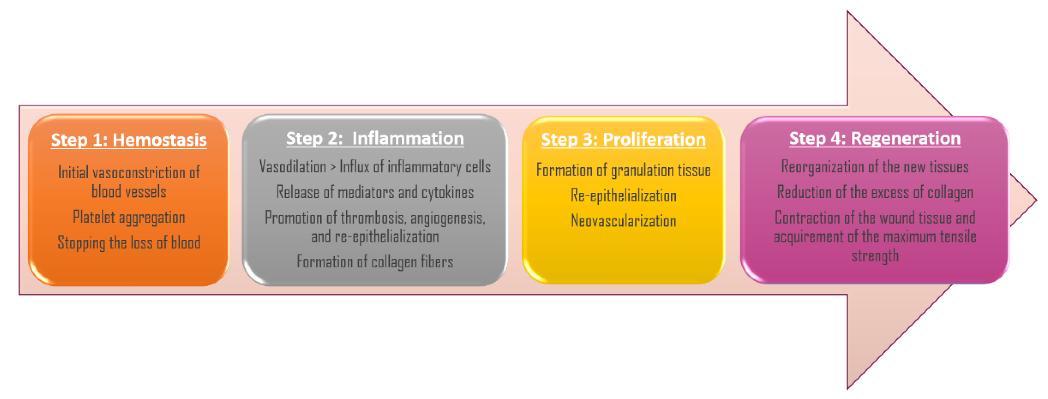
The wound-healing process has the purpose of recovering the integrity of the damaged tissue and the regeneration of the epithelium that was lost, and it is a dynamic and complex process that globally occurs in four overlapping steps: hemostasis, inflammation, tissue proliferation, and regeneration [1,2] (Figure 1).
The cascade of initial vasoconstriction of blood vessels and platelet aggregation play a key role in stopping the loss of blood. The initial vasoconstriction is followed by a vasodilation, which allows an influx of a variety of inflammatory cells which release several types of mediators and cytokines to promote thrombosis, angiogenesis, and re-epithelialization. In addition, the fibroblasts release extracellular components which initiate the formation of collagen fibers that will serve as scaffolding [1,3]. During the inflammatory phase the hemostasis, chemotaxis, and the increased vascular permeability limit further damage, close the wound, remove cellular debris and bacteria, and promote cellular migration [3]. Afterwards, the proliferative phase implicates the formation of granulation tissue, re-epithelialization, and neovascularization, a process that can last from several weeks until some months, or even more time in the case of the existence of some co-morbidities or particular patient situations [4]. In the end, during the maturation period, the new tissues are remodeled, the excess of collagen is reduced, and the wound contracts and reaches the maximum tensile strength [1].
Acute wounds derived from unexpected accidents or surgical injury commonly heal within a predictable period depending on the size, depth, and extent of damage. Nevertheless, deficiencies in the wound-healing process cause more than 38 million patients with chronic wounds worldwide, which reaches epidemic proportions and causes a large economic weight on healthcare systems [5].
Prolonged chronicity of wounds is normally related to a bacterial injured-tissue colonization, which can progress into a bacterial resistance to topical and systemic antimicrobial agents, or into biofilm development, which complicates, in both cases, their treatment [6]. In the end, this type of wound can cause sepsis and inflammation in organs and lead to increased morbidity and mortality.
The clinical considerations in wound-healing management include preventing and controlling the infection and/or contamination, maintaining the adequate moisture environment, treating edema, and preventing further injury. Conventional chronic wound care involves debridement to remove non-viable tissue and bacterial biofilms, followed by wound dressing. The common wound dressings consist of a standard cotton bandage or highly absorbent dressings, such as collagen and alginate, or hydrocolloids; however, this procedure of wound care is often ineffective. Due to this need for new, efficient, and improved therapies, there has been a revived interest in alternative treatment approaches, such as honey. The management of wound healing has become the primary field of therapeutic application of this natural product [7,8].
Honey has been used for wound healing since ancient times, mainly due to its antimicrobial activity. In addition to the broad spectrum of antibacterial activity against common wound-infecting microorganisms, honey has been demonstrated to be effective against antibiotic-resistant bacteria and was able to restore the efficacy of some antibiotics against bacteria with previously acquired resistance [8,9,10]. Furthermore, due to its several antimicrobial components and its different antibacterial action mechanisms, the development of bacterial resistance to honey is unlikely [8,11,12]. Moreover, the wound-healing ability of honey is also related to its anti-inflammatory and antioxidant activity, as well as its capacity to promote re-epithelialization and angiogenesis and stimulate skin and immune cells [13,14]. All these mechanisms act together favoring the regeneration process of the damaged tissue.
Several case studies and randomized controlled trials provide considerable evidence of the effectiveness of honey in healing different types of wounds, such as amputation wounds, burns, skin grafting sites, skin lesions, or skin ulcers including leg, varicose, malignant, diabetic, and sickle cell ulcers [15,16,17,18].
The resolution time of wounds using honey or honey-derivates varies from some days to several months depending on the type, the depth, the anatomical location, and the chronicity of the wound [17,19]. Due to its osmotic effect, honey creates a liquid layer between the dressing and the wound bed. This liquid layer is an advantage in the removal and change of wound-dressings by reducing or eliminating the pain of this process and avoiding damage of the newly grown tissue, reducing the healing time [20].
The use of honey obtained a remarkable improvement of recalcitrant wounds, and has demonstrated even more efficacy than conventional treatments using commercial wound dressings or antibiotics (systemic and topical) [19,21,22]. Honey rapidly replaces sloughs with granulation tissue and promotes a quick epithelialization and absorption of edema from around the ulcer margins, significantly reducing the healing time [19,23]. In addition, honey-based products showed excellent cytocompatibility with tissue cell cultures when compared with conventional treatments, such as silver dressings [24].
Moreover, some studies demonstrated that the combination of honey with other compounds or in combination with conventional treatment can be beneficial for diabetic foot ulcer healing, reducing the wound-resolution time, the cost of the hospital stay, and the rate of amputation when compared to other conventional treatment [17].
However, the use of honey by itself might present some limitations which are being overcome with the development of different honey formulations and honey wound dressings.
This review aims to highlight the mechanism of honey’s action in wound healing and gather the literature available regarding the use of honey and modern engineering templates for promoting modern solutions for wound and skin healing and regeneration.
2. The Mechanisms of Honey in Wound and Burn Healing
Honey is a natural and greatly complex substance with hundreds of compounds in its composition [8]. Honey bioactivity, and in consequence, its wound-healing potential, will be influenced by its composition, which depends mainly on the floral source and other factors, such as seasonal, environmental, as well as processing, manipulation, packaging, and storage conditions [25,26]. In addition to some inherent characteristics, such as the acidity and the osmotic pressure, the healing properties of honey in diverse types of wounds and burns have also been attributed to other components which act through different mechanisms that work together to restore the structural integrity of the damaged tissues [6,18,27] (Figure 2).
2.1. Antibacterial Effects
Honey has been traditionally used in the prevention and treatment of wound infections [28,29]. However, with the arrival of antibiotics, the use of honey gradually decreased. Nowadays, microbial drug-resistance has become an increasingly common concern, and honey has regained the scientific interest as an antibacterial agent [8,12,13]. Intrinsic characteristics of honey, such as high osmolarity, low water activity, and acidity, as well as some compounds, such as hydrogen peroxide, phenolic compounds, methylglyoxal, or bee defensin-1 peptide, directly affect the bacterial growth and survival [30,31,32]. In addition, honey shows an indirect antimicrobial action which involves the promotion of lymphocyte and antibody production, cytokines and immunomodulation, and nitric oxide (NO) [6,33,34,35].
Non-healing wounds, as well as burns, present an elevated risk of infection, which might increase morbidity and mortality derived from sepsis and inflammation in organs [13]. In addition, drug-resistant infections and wounds with biofilms are particularly difficult to treat, since bacteria do not respond to the therapy or are protected by a self-produced matrix of polysaccharide material [36].
Several studies have demonstrated, in vitro and in vivo, the efficacy of different varieties of honey against a broad spectrum of bacteria, including those that commonly caused wound and burn infections, such as Staphylococcus aureus, Pseudomonas aeruginosa, Escherichia coli, Acinetobacter baumannii, or Staphylococcus epidermidis [6,18,37,38,39,40]. In addition, honey has also been demonstrated to be effective against antibiotic-resistant bacteria [19,41,42,43], as well as against biofilms by preventing the formation and the development of the biofilm [31,44,45,46,47], by reducing the metabolic activity of already formed biofilms [44,48], or by altering the gene expression of different genes related to the formation and the development of biofilms [48,49], and is related to the bacterial quorum sensing [49,50].
Moreover, some studies demonstrated that manuka honey acts synergistically with several antibiotics, reducing the doses required to inhibit bacterial growth or reverting the antibiotic resistance previously acquired [9,10,51,52]. These results suggest a potential application of a combined therapy of honey and antibiotics.
2.2. Anti-Inflammatory Effects
Inflammation is the response of a living tissue to a local injury and plays a fundamental role as a defense and protection mechanism to avoid infections and to repair the affected tissue. The inflammatory phase is a necessary part of wound healing; however, when this response is not adequate, an overproduction of inflammatory mediators by immune cells, which do not respond to initial triggers, might be produced, becoming a problem for wound resolution [53]. The anti-inflammatory activity of honey is a consequence of different mechanisms.
During the inflammatory phase, the affected tissues release a high concentration of free radicals. The antioxidant compounds in honey act synergistically and can reduce the damage caused by these radicals, and therefore prevent tissue necrosis [14].
In addition, in vitro and in vivo studies have demonstrated that honey reduces the activity of cyclooxygenases 1 and 2 (COX1 and COX2) that intervene in the synthesis of prostaglandins [54,55]. Prostaglandins participate in the inflammatory response by producing vasodilation, increasing the permeability of blood vessels and allowing the passage of leukocytes, acting as an antiplatelet agent, and stimulating the nerve endings of pain. The reduction in prostaglandin concentration in plasma may induce a diminution of inflammation, edema, and pain [56].
Moreover, honey can inhibit the expression of tumor necrosis factor (TNF-α) and reduce the concentration of pro-inflammatory cytokines through the attenuation of nuclear factor kappa B (NF-κB) [54]. Furthermore, NF-κB is involved in the activation of the inducible NO synthase enzyme (iNO). During inflammation, iNO is induced by cytokines, TNF-α, interleukins, and bacterial endotoxins, producing NO.
NO is a free radical that acts as a mediator in acute and chronic inflammation and favors the healing process of tissues. However, an excess of NO or an overproduction at the wrong time can be detrimental and contribute to the development of pathologies related to inflammation [56].
Another advantage of the anti-inflammatory action of honey is the decrease in edema, thus reducing the pressure on the microvasculature of wound tissue that allows the availability of oxygen and nutrients required for growth of tissue and wound repair [20]. This effect also allows the control of the wounds’ exudate with an appropriate moisture balance, which is still a constant challenge in the healing processes [39].
The anti-inflammatory activity of honey has been mainly attributed to phenolic compounds [53,57]. However, until now, no correlation was found between the level of anti-inflammatory activity in different honey samples and the phenolic compound content [58], which might be due to the distinct types of interactions that can occur among these compounds and other compounds present in honey.
2.3. Antioxidant Activity
The antioxidant activity of honey is due to a wide variety of compounds, such as flavonoids, phenolic acids, tocopherols, ascorbic acid, and enzymes including catalase or superoxide dismutase [14,59,60]. In addition, melanoidins, products of the Maillard reaction, were described as the main components responsible for the radical-scavenging capacity of honey [61,62]. These substances reduce the adverse effects of reactive oxygen species (ROS) and reactive nitrogen species (RNS), inhibit the enzymes responsible for producing superoxide anions, act as metal chelators, and interfere in the chain reactions of free radicals and can play a preventative role in the process of their formation [63]. Through these antioxidant mechanisms, honey contributes to wound and burn healing by interfering with abnormal inflammatory response [6].
ROS act as messengers to give feedback amplification of the inflammatory response [20] and mediate TNF-α induced cytotoxicity [64]. Moreover, in chronic wounds, neutrophils and macrophages liberate high levels of ROS against invading bacteria [64]. The extended exposure to ROS causes cell damage of the tissue and might delay wound healing. In addition, the ROS formed in the inflammatory phase of wound healing stimulate the activity of the fibroblasts which produce the collagen fibers of scar tissue. If the inflammatory phase prolongates, it could induce hyper-granulation and fibrosis, so honey minimizes or prevents hypertrophic scarring [14,64]. In addition, flavonoids protect tissue against RNS, such as NO and peroxy-nitrite [65].
2.4. Debridement and Anti-Eschar Action
Wound debridement is essential in producing the functional process of tissue reparation. The conventional procedure is the surgical remotion of dead tissue, which is painful, may cause infections, and produces toxins that can destroy the surrounding tissues [6,14,27].
The moist environment produced by honey facilitates the wounds´ autolytic debridement process. The high osmotic pressure pulls out lymphatic fluid from the deeper zones, which automatically remove dead, damaged, or infected scar tissue [27,39]. In addition, lymph is a rich source of proteases that activated by the hydrogen peroxide produced when honey is diluted and assists in the debriding activity [6].
Additionally, honey inhibits the production of plasminogen activator inhibitor (PAI) by the macrophages derived from its anti-inflammatory activity [39]. PAI blocks the transformation of plasminogen, the enzymically inactive precursor of plasmin, into active plasmin. Plasmin is an enzyme that specifically digests fibrin attached to the wound surface, but does not digest the collagen matrix, which is necessary for tissue reparation, thus preventing eschar formation [20,66]. Inflammation increases the generation of PAI, so the mechanism through which honey decreases the production of PAI is probably related to its anti-inflammatory activity [20].
2.5. Angiogenesis Promoter
Angiogenesis occurs in the proliferative phase of wound healing. The development of new blood vessels from pre-existing ones supplies the required oxygen in the wound, which is an important stage in the healing process. This dynamic process is strongly regulated by signals from serum and the surrounding extracellular matrix environment [67]. Stimulation of angiogenesis by honey was demonstrated in an in vitro study with analogues of angiogenesis and an endothelial proliferation assay [68], and more recently, in another in vivo-model study [69].
Hydrogen peroxide (generated from glucose by the action of the enzyme glucose oxidase present in raw honey) induces the recruitment of leukocytes to wounds through a concentration gradient mechanism. Due to an oxidant induct, macrophages release vascular endothelial growth factor (VEGF), which stimulates angiogenesis [67]. In addition, the high concentration of sugars present in honey, as well as other minor constituents, such as amino acids, vitamins and trace elements, provide, in a moist environment, a local cellular energy source, which may improve local nutrition and endothelial cell proliferation [70,71,72].
On the contrary, another study has demonstrated the anti-angiogenic activity of honey is mediated by the modulation of prostaglandin E2 and VEGF production [73]. This disparity among studies might be explained by the honey concentration tested, since the highest pro-angiogenic effect was found in a low concentration of honey, whereas higher concentrations demonstrated anti-angiogenic activity [68].
2.6. Immune System Promoter
Some studies have also demonstrated the activity of honey in stimulating some immune system mediators. Honey can stimulate B- lymphocytes and T-lymphocytes and activate neutrophil phagocytosis in cell culture [20,74]. In addition, honey induces monocytes (MM6 cells) to secrete cytokines, tumor necrosis factor-α (TNF-α), interleukin-1 (IL-1), and interleukin-6 (IL-6), which activate the immune response to infection [33,34,35].
Moreover, honey stimulates antibody production during primary and secondary immune responses against thymus-dependent and thymus-independent antigens [75] and increases humoral immunity by the intrinsic NO, which activates specific signal transduction pathways in monocytes in a concentration-dependent manner [76,77].
2.7. Healing-Rate Promoter
In addition to all the honey effects previously described, honey acts in the regeneration of the new formed tissue, an essential step in the wound-healing process [78].
The acidification of the wound environment favors the action of macrophages, limits bacterial growth, and neutralizes the ammonia produced by bacterial metabolisms that could damage tissues [14]. However, the acidic pH of honey also limits the activity of proteases; these enzymes might inactivate the tissue growth factors and destroy the plasma fibronectin and the collagen matrix, which are necessary for fibroblast activity and tissue re-epithelialization [14,42]. In addition, the diminution of pH in the wound bed makes more oxygen available from hemoglobin in the blood [42].
Furthermore, all the nutritious components present in honey (sugars, amino acids, vitamins, and other trace elements) stimulate cell growth and the development of repair tissues [70,71,72,79,80]. In addition, re-epithelialization would also be promoted by the increment of TNF-α and interleukin-1β (IL-1β) levels. A concentration of 1% of honey has been found to stimulate the release of the cytokine TNF-α, IL-1β and IL-6 from monocytes [81], which induce the keratinocyte migration and proliferation, which are the major cellular components that are involved in the intricate mechanisms of initiation, maintenance, and completion of wound healing, and may induce collagen synthesis by fibroblasts [14,68,82].
3. Safety of Honey Used for Topical Treatment
The extensive scientific evidence proves that honey may offer distinct advantages over the chemotherapeutic substances currently used in the wound- and burn- healing processes. However, this natural product shows a series of limitations, and is not completely free from adverse effects.
The composition of honey is rather variable, depending primarily on the botanical origin, and secondarily on other factors such as geographical origin, or harvesting, processing, and storage conditions [38,39]. This variability determines its bioactive properties, and consequently influences the therapeutic efficacy of the wound treatment [39]. In addition, the absence of standardization and the incomplete knowledge of the active components, and the mechanisms through which they interact and act in wound healing, are the major limitations for the application of honey in medicine. For this reason, is essential to select the more appropriate varieties of honey, and it is recommended to carry out a previous screening [38].
In addition, other considerations must be considered before honey application in wounds. The low pH, derived from the presence of organic acids in honey, may contribute to a stinging or burning sensation when it is applied to a damaged tissue [83]. Besides this unpleasant sensation, it is necessary to consider that, although minor, there is a risk of wound infection, mainly related to the presence of clostridial spores which have occasionally been found in honey [39,84]. This risk can be reduced by using gamma-irradiation, which inactivates the spores without modifying the original biological activity [85,86]. Nevertheless, no cases of wound infection due to clostridial spores related to the use of non-irradiated honey on wounds have been reported to date.
Furthermore, the honey used for medical purposes must be free of any chemical contamination, such as pesticides, herbicides, or heavy metals. In this sense, to guarantee the maximum purity, honey should be collected in areas that meet the requirements for organic production, as well as following rigorous quality, processing and storage standards [8,19]. In addition, is necessary to consider that some varieties of honey might present toxic active compounds originating from the nectar of species such as rhododendron, oleanders, mountain and sheep laurels, or azaleas. However, these effects have been described by honey ingestion [87,88].
4. Biomedical Application of Honey in Advanced Wound Care
4.1. Medical Grade Honey and Honey Ointments for Topical Application
The safety threats previously described in wound treatment with honey are overcome by medical-grade honey approved for wound care [18,39]. Indeed, the medical-grade honeys are sterilized by gamma irradiation with the aim to kill Clostridium spores, produced under rigorous standards of hygiene, without pollutants or contaminating pesticides in its composition, and standardized under different defined criteria [8]. They have potent in vitro bactericidal activity against antibiotic-resistant bacteria and are approved for application in wound management. Having reproducible antibacterial activity, these honeys are produced under controlled conditions in greenhouses (i.e., Revamil source honey); medical grade Leptospermum-derived Manuka honeys are analyzed individually by each batch to assess the Unique Manuka Factor (UMF) that gives a number based on its bactericidal activity [89]. Unlike other varieties, the antibacterial activity of Manuka honey is based on its non-peroxide activity related to compounds which are mainly present in this variety, such as methylglyoxal, leptosperin, or methyl syringate [38].
Medical-grade honey can be directly applied to the wound bed and then covered with conventional dressing. However, in high-exuding wounds, honey can become less viscous and diluted. The liquid state of honey might complicate its application and its permanence on wound and burn environments, and in the treatment of excessively exudative lesions, honey might be diluted to concentrations that present minimal or no effects in short periods of time [7,39]. Nevertheless, according to [6], even when honey is heavily diluted by wound exudate, it will still have potent enough antibacterial activity to inhibit the growth of bacteria (the MIC values were found to be below 11%).
In clinical practice today, several honey-based wound-care commercial preparations in the form of gels, ointment, and dressings, are approved by the US Food and Drug Administration (FDA) and registered as medical devices [90]. Most of them are formulated with medical-grade Manuka honey, since it is one of the most studied varieties of honey in the world, and it was the first honey type to obtain the status of medical-grade honey. However, there are also alternatives that use other types of honey, such as buckwheat, multifloral, and Revamil source honey, among others [8]. For instance, a honey-based gel formula can be prepared with 100 % medical-grade honey without the addition of other ingredient (Table 1) or can be mixed with other agents(s) such as natural emollients (e.g., lanolin, polyethylene glycol, glycerine, myristil myristrate) or different plant waxes and gelling agents [90].
The FDA-approved honey-based devices are indicated in the treatment of different types of wounds, such as low and moderate-to-heavy exuding wounds, diabetic foot ulcers, leg ulcers, pressure ulcers, burns, traumatic wounds, surgical wounds, chronic wounds, or colonized acute wounds, among other indications [18]. Despite the availability of these products, their use in medical practice is still limited, probably due to the misconception that there is no evidence to support the use of honey with therapeutic purposes, as well as the scarce promotion and diffusion of honey products for wound care [20].
Nevertheless, effectiveness of the MedihoneyTM Antibacterial Wound Gel has been evaluated in eight post-coronary artery bypass graft (CABG) patients. The gel was selected as the active primary product of choice for all the graft wounds (seven of the eight patients had wound infections). The honey-based gel was applied directly onto the wound bed and covered with an adhesive-bordered non-adherent gauze dressing. The wounds all reduced in size and there was a significant reduction in pain, odor, and exudate. Moreover, the wound gel had reduced the bioburden of the wounds enabling them to progress to healing. Finally, the use of medical honey became a regular dressing choice within the authors’ cardiothoracic unit [91]. Moreover, the combined treatment with honey-based gels (L-Mesitran Soft) and ointment (L-Mesitran Ointment) has been successfully applied for treatment of infected ulcers in diabetics patients [19]. Indeed, the use of ointments help to entrap water, keep the skin moist, and provide an emollient protective film, which are all crucial elements for wound healing [19,90], while the layer of gel fights infections and optimizes wound healing [19,90].
The medical-grade honey, as well as the ointments and gels, is applied to the wound bed and requires a secondary conventional dressing (e.g., cotton wool bandage) to contain the honey in the wound bed environment, which on removal cause pain [92].
4.2. Honey-Based Advanced Wound Care Products
Many years ago, wound management was based on covering the wound using conventional dressings (i.e., gauzes, absorbent cotton, bandages). However, they are limited in terms of influencing/accelerating wound healing and preventing/treating infections. Currently, wound management has been updated due to a greater understanding of the molecular and cellular processes involved in wound healing. Additionally, with advancements in technology, the design and functionality of wound dressings has advanced in the direction of multi-functionality [96]. The modern dressings are designed to maintain the moist wound environment and promote healing [97]. Moreover, the critical necessities of modern wound dressings include biocompatibility, no cytotoxic effects, a rate of biodegradability directly proportional with the rate of formation of new tissue, a release of incorporated bioactive compounds (drugs), and the control of possible infections [96].
Tissue engineering has recently introduced wound dressings/scaffolds as an alternative treatment of wounds with advanced properties, suitable for keeping a moist environment while absorbing exudates, creating a barrier against pathogens, and facilitating drug delivery systems [96].
The recent in vitro and in vivo research demonstrated that honey is a valuable addition to many tissue-engineering templates in eliminating bacterial infection, aiding in inflammation resolution and improving tissue integration with the template (Table 2 and Table 3) [28]. Currently, hydrogels and electrospun nanofibers are the most researched types of honey-incorporated scaffolds [98].
4.2.1. Honey-Based Hydrogels
Hydrogels are high-water-content materials prepared from cross-linked polymers, such as chitosan, and can provide sustained, local delivery of a variety of therapeutic agents [106]. Incorporation of honey into the hydrogel system can beneficially affect the water absorption capacity of the polymer (of the hydrogel) and increase the antibacterial activity of the scaffold [99].
Noori, Kokabi, and Hassan (2018) investigated a honey-loaded PVA/chitosan/montmorillonite nanocomposite (PCMH) hydrogel dressing as a drug model for wound healing. Results demonstrated the ability of the PCMH nanocomposite hydrogel to smart release honey against pH and temperature changes. The maximum release of honey from the hydrogel occurred at pH 7, while the minimum was at pH 2. Independently of the pH, increasing temperature caused higher honey release from the hydrogel matrix. However, the addition of the nanoclay (montmorillonite) to the hydrogel decreases the hydrogel swelling and delays the honey release. Nevertheless, the authors suggested that the honey-loaded nanocomposite hydrogel could be used in low exudate wounds to supply optimized humidity in the wound bed [102].
Different honey concentrations have different efficacies in the scavenging of free radicals and promoting epithelial cell proliferation [119]. It has been shown that the release of honey increases when increasing its concentration in hydrogel, regardless of the polymeric hydrogel used in the formulation. For instance, the honey 75%-chitosan formula showed the best healing properties (regeneration of the epidermis tissue and the formation of new blood capillaries) compared to honey hydrogel formulae with a lower concentration of honey (up to 50%, w/w). Similarly, an 80%-Manuka/PVA wound dressing hybrid hydrogel showed sustained release of honey over 24 h with progressively low adhesion to the wound bed that protects new epithelialization and promotes cell proliferation. Both dressings demonstrate the high value of cell viability and proliferation and promoted antibacterial activity, being suitable for wounds with moderate to relatively high exudate [100].
Further, one of the advantages of using hydrogels instead of conventional designs is their transparency, which allows us to observe the status of the burn or wound without removal of the dressing [103]. In vivo study showed that burns treated with honey hydrogel sheets were completely healed after 12 days with intact epidermis and topical proliferation of hair follicles. In contrast, burns treated with commercial ointment (MEBO-treated burns) and non-treated burns presented 15% and 63% unclosed wound areas, respectively [104].
In addition, hydrogel wound scaffolds containing honey do not function merely as coverage to provide a clean, moist environment for healing, but also directly contribute to enhanced tissue regeneration and recovery [104].
4.2.2. Honey-Based Electrospun Nanofiber Scaffolds
In biomedical applications, the nanofiber membranes prepared by electro-spinning are used in wound dressings, biosensing, tissue engineering scaffolds, artificial organs, and drug delivery [114]. The formulation of the electrospun nanofiber scaffolds contain protein-based polymers, such as collagen, gelatin, and silk, or/and polysaccharide-based polymers such as chitosan, hyaluronic acid, and alginate. Prepared stirred polymeric solution is loaded into a syringe that is attached to a needle of which the tip exhibits voltage [120].
The advantageous properties of nanofibers are a large surface area to volume ratio, high porosity, and a very small pore size, which lead to high exudate absorption, better wound permeation, and prevention of further infection [112].
The fabrication of honey-based electrospun nanofibers increases interest due to the enhanced activity realized upon combining the advantages of the nanofibrous structure, primarily the increased surface-to-volume ratio with the advantageous properties of honey (Table 3) [112].
Wound healing scaffolds are expected to absorb body fluids and maintain hydration, but without increasing infection of the biofilm. Indeed, wounds with biofilm fail to re-epithelialize, show vascular granulation tissue, and consist of recalcitrant microbes. Honey/PVA nanofiber membranes were found to effectively decrease the biofilm formation [113]. Manuka honey is effective against both Gram-positive and Gram-negative bacteria; however, results of honey scaffolds containing 1–20% Manuka/PCL nanofibers indicated that the controlled release of smaller amounts of honey by the scaffold is more effective against Gram-negative-bacteria-infected wounds. Interestingly, opposite results have been found for high-concentration honey chitosan electrospun nanofibers. The honey/PVA/chitosan membrane (30%:7%:3.5%) enhanced antibacterial activity against S. aureus (complete inhibition after 48 h with 30%: 7%:5.5 %) and showed poor antibacterial activity against the Gram-negative E. coli. Moreover, the scaffold showed high biocompatibility and low cytotoxicity effects [108].
5. Conclusions
The use of honey for biomedical applications has gained special focus over the years, with the development of novel applications for this natural product, taking advantage of its unique chemical characteristics. Due to its characteristics, namely, low pH and water activity, it presents a good microbiological, enzymatic and (bio)chemical stability, which can be lost if not properly processed for safe use by means of keeping both functionality and microbiological safety. Thus, the design of proper honey processing methodologies is of utmost importance for its use. As reviewed, honey presents a very promising potential to be used in wound-healing processes, either by direct application, incorporated in fibrous membranes, or in hydrogel, with very promising results in either in vitro and in vivo trials. Nonetheless, further research is needed to overcome the main challenges on the use of honey for biomedical applications.
Author Contributions
Conceptualization, writing—original draft preparation, H.S., P.C.-F.; writing—review and editing, H.S., P.C.-F., J.M.F., M.E.T., M.S.D., C.A.P., L.M.E.; visualization, C.A.P.; Project administration, supervision, J.A.S., L.M.E. All authors have read and agreed to the published version of the manuscript.
Funding
This research received no external funding.
Institutional Review Board Statement
Not applicable.
Informed Consent Statement
Not applicable.
Data Availability Statement
The data presented in this study are available on request from the corresponding author.
Acknowledgments
Thanks are due to the University of Aveiro and FCT/MCT for the financial support of LAQV-REQUIMTE research Unit (UIDB/50006/2020) through national funds and, where applicable, co-financed by the FEDER within the PT2020 Partnership Agreement, and for financing the PhD grant of Hana Scepankova (SFRH/BD/88133/2012) and Carlos A. Pinto (SFRH/BD/137036/2018).
Conflicts of Interest
The authors declare no conflict of interest.
References
- Kus, K.J.B.; Ruiz, E.S. Wound Dressings—A Practical Review. Curr. Dermatol. Rep. 2020, 9, 298–308. [Google Scholar] [CrossRef]
- Khaleghverdi, S.; Karimi, A.; Soltani, R.; Zare, R. The Effect of Myrtus, Honey, Aloe Vera and Pseudomonas Phage Treatment on Infected Second Degree Burns: In Vivo Study. Biointerface Res. Appl. Chem. 2021, 11, 7422–7430. [Google Scholar] [CrossRef]
- Bowden, L.G.; Byrne, H.M.; Maini, P.K.; Moulton, D.E. A Morphoelastic Model for Dermal Wound Closure. Biomech. Model. Mechanobiol. 2016, 15, 663–681. [Google Scholar] [CrossRef] [PubMed]
- Anderson, K.; Hamm, R.L. Factors That Impair Wound Healing. J. Am. Coll. Clin. Wound Spec. 2012, 4, 84–91. [Google Scholar] [CrossRef] [PubMed] [Green Version]
- Olsson, M.; Järbrink, K.; Divakar, U.; Bajpai, R.; Upton, Z.; Schmidtchen, A.; Car, J. The Humanistic and Economic Burden of Chronic Wounds: A Systematic Review. Wound Repair Regen. 2019, 27, 114–125. [Google Scholar] [CrossRef] [Green Version]
- Molan, P.; Rhodes, T. Honey: A Biologic Wound Dressing. Wounds 2015, 27, 141–151. [Google Scholar] [PubMed]
- Rossi, M.; Marrazzo, P. The Potential of Honeybee Products for Biomaterial Applications. Biomimetics 2021, 6, 6. [Google Scholar] [CrossRef]
- Combarros-Fuertes, P.; Fresno, J.M.; Estevinho, M.M.; Sousa-Pimenta, M.; Tornadijo, M.E.; Estevinho, L.M. Honey: Another Alternative in the Fight against Antibiotic-Resistant Bacteria? Antibiotics 2020, 9, 774. [Google Scholar] [CrossRef]
- Campeau, M.E.M.; Patel, R. Antibiofilm Activity of Manuka Honey in Combination with Antibiotics. Int. J. Bacteriol. 2014, 2014, 795281. [Google Scholar] [CrossRef] [Green Version]
- Hayes, G.; Wright, N.; Gardner, S.L.; Telzrow, C.L.; Wommack, A.J.; Vigueira, P.A. Manuka Honey and Methylglyoxal Increase the Sensitivity of Staphylococcus Aureus to Linezolid. Lett. Appl. Microbiol. 2018, 66, 491–495. [Google Scholar] [CrossRef]
- Lu, J.; Cokcetin, N.N.; Burke, C.M.; Turnbull, L.; Liu, M.; Carter, D.A.; Whitchurch, C.B.; Harry, E.J. Honey Can Inhibit and Eliminate Biofilms Produced by Pseudomonas Aeruginosa. Sci. Rep. 2019, 9, 18160. [Google Scholar] [CrossRef] [Green Version]
- Maddocks, S.E.; Jenkins, R.E. Honey: A Sweet Solution to the Growing Problem of Antimicrobial Resistance? Future Microbiol. 2013, 8, 1419–1429. [Google Scholar] [CrossRef]
- McLoone, P.; Tabys, D.; Fyfe, L. Honey Combination Therapies for Skin and Wound Infections: A Systematic Review of the Literature. Clin. Cosmet. Investig. Dermatol. 2020, 13, 875–888. [Google Scholar] [CrossRef] [PubMed]
- Oryan, A.; Alemzadeh, E.; Moshiri, A. Biological Properties and Therapeutic Activities of Honey in Wound Healing: A Narrative Review and Meta-Analysis. J. Tissue Viability 2016, 25, 98–118. [Google Scholar] [CrossRef]
- Jull, A.; Walker, N.; Parag, V.; Molan, P.; Rodgers, A. Randomized Clinical Trial of Honey-Impregnated Dressings for Venous Leg Ulcers. Br. J. Surg. 2008, 95, 175–182. [Google Scholar] [CrossRef]
- Smaropoulos, E.; Cremers, N.A.J. Treating Severe Wounds in Pediatrics with Medical Grade Honey: A Case Series. Clin. Case Rep. 2020, 8, 469–476. [Google Scholar] [CrossRef] [PubMed] [Green Version]
- Alam, F.; Islam, M.A.; Gan, S.H.; Khalil, M.I. Honey: A Potential Therapeutic Agent for Managing Diabetic Wounds. Evid. Based Complement. Altern. Med. 2014, 2014, 169130. [Google Scholar] [CrossRef] [PubMed] [Green Version]
- Hixon, K.R.; Klein, R.C.; Eberlin, C.T.; Linder, H.R.; Ona, W.J.; Gonzalez, H.; Sell, S.A. A Critical Review and Perspective of Honey in Tissue Engineering and Clinical Wound Healing. Adv. Wound Care 2019, 8, 403–415. [Google Scholar] [CrossRef]
- Nair, H.K.R.; Tatavilis, N.; Pospíšilová, I.; Kučerová, J.; Cremers, N.A.J. Medical-Grade Honey Kills Antibiotic-Resistant Bacteria and Prevents Amputation in Diabetics with Infected Ulcers: A Prospective Case Series. Antibiotics 2020, 9, 529. [Google Scholar] [CrossRef]
- Molan, P.C. The Evidence and the Rationale for the Use of Honey. Wound Pract. Res. 2011, 19, 204–220. [Google Scholar]
- Güneş, Ü.Y.; Eşer, I. Effectiveness of a Honey Dressing for Healing Pressure Ulcers. J. Wound Ostomy Cont. Nurs. 2007, 34, 184–190. [Google Scholar] [CrossRef]
- Kamaratos, A.V.; Tzirogiannis, K.N.; Iraklianou, S.A.; Panoutsopoulos, G.I.; Kanellos, I.E.; Melidonis, A.I. Manuka Honey-Impregnated Dressings in the Treatment of Neuropathic Diabetic Foot Ulcers. Int. Wound J. 2012, 11, 259–263. [Google Scholar] [CrossRef] [PubMed]
- Efem, S.E.E. Clinical Observations on the Wound Healing Properties of Honey. Br. J. Surg. 1988, 75, 679–681. [Google Scholar] [CrossRef] [PubMed]
- Du Toit, D.F.; Page, B.J. An in Vitro Evaluation of the Cell Toxicity of Honey and Silver Dressings. J. Wound Care 2009, 18, 383–389. [Google Scholar] [CrossRef] [PubMed] [Green Version]
- Silvia, P.M.D.; Gauche, C.; Gonzaga, L.V.; Costa, A.C.O.; Fett, R. Honey: Chemical Composition, Stability and Authenticity. Food Chem. 2016, 196, 309–323. [Google Scholar] [CrossRef]
- Combarros-Fuertes, P.; Valencia-Barrera, R.M.; Estevinho, L.M.; Dias, L.G.; Castro, J.M.; Tornadijo, M.E.; Fresno, J.M. Spanish Honeys with Quality Brand: A Multivariate Approach to Physicochemical Parameters, Microbiological Quality, and Floral Origin. J. Apic. Res. 2019, 58, 92–103. [Google Scholar] [CrossRef] [Green Version]
- Yilmaz, A.C.; Aygin, D. Honey dressing in wound treatment: A systematic review. Complement. Ther. Med. 2020, 51, 102388. [Google Scholar] [CrossRef]
- Martinotti, S.; Ranzato, E. Honey, Wound Repair and Regenerative Medicine. J. Funct. Biomater. 2018, 9, 34. [Google Scholar] [CrossRef] [Green Version]
- Mcloone, P.; Warnock, M.; Fyfe, L. Honey: A Realistic Antimicrobial for Disorders of the Skin. J. Microbiol. Immunol. Infect. 2016, 49, 161–167. [Google Scholar] [CrossRef] [Green Version]
- Albaridi, N.A. Antibacterial Potency of Honey. Int. J. Microbiol. 2019, 2019, 2464507. [Google Scholar] [CrossRef]
- Proaño, A.; Coello, D.; Villacrés-Granda, I.; Ballesteros, I.; Debut, A.; Vizuete, K.; Brenciani, A.; Álvarez-Suarez, J.M. The Osmotic Action of Sugar Combined with Hydrogen Peroxide and Bee-Derived Antibacterial Peptide Defensin-1 Is Crucial for the Antibiofilm Activity of Eucalyptus Honey. LWT 2021, 136, 110379. [Google Scholar] [CrossRef]
- Peršurić, Ž.; Pavelić, S.K. Bioactives from Bee Products and Accompanying Extracellular Vesicles as Novel Bioactive Components for Wound Healing. Molecules 2021, 26, 3770. [Google Scholar] [CrossRef] [PubMed]
- Abuharfeil, N.; Al-Oran, R.; Abo-Shehada, M. Food and Agricultural Immunology The Effect of Bee Honey on the Proliferative Activity of Human B-and T-Lymphocytes and the Activity of Phagocytes. Food Agric. Immunol. 1999, 11, 169–177. [Google Scholar] [CrossRef]
- Tonks, A.J.; Cooper, R.A.; Jones, K.P.; Blair, S.; Parton, J.; Tonks, A. Honey Stimulates Inflammatory Cytokine Production from Monocytes. Cytokine 2003, 21, 242–247. [Google Scholar] [CrossRef]
- Tonks, A.; Cooper, R.A.; Price, A.J.; Molan, P.C.; Jones, K.P. Stimulation of TNF-α Release in Monocytes by Honey. Cytokine 2001, 14, 240–242. [Google Scholar] [CrossRef]
- Vestby, L.K.; Grønseth, T.; Simm, R.; Nesse, L.L. Bacterial Biofilm and Its Role in the Pathogenesis of Disease. Antibiotics 2020, 9, 59. [Google Scholar] [CrossRef] [PubMed] [Green Version]
- Molan, P.C. Honey: Antimicrobial Actions and Role in Disease Management. In New Strategies Combating Bacterial Infection; Wiley-VCH Verlag GmbH & Co. KGaA: Weinheim, Germany, 2009; pp. 229–253. ISBN 9783527322060. [Google Scholar]
- Combarros-Fuertes, P.; Estevinho, L.M.; Dias, L.G.; Castro, J.M.; Tomás-Barberán, F.A.; Tornadijo, M.E.; Fresno-Baro, J.M. Bioactive Components and Antioxidant and Antibacterial Activities of Different Varieties of Honey: A Screening Prior to Clinical Application. J. Agric. Food Chem. 2019, 67, 688–698. [Google Scholar] [CrossRef] [Green Version]
- Krishnakumar, G.S.; Mahendiran, B.; Gopalakrishnan, S.; Muthusamy, S.; Malarkodi Elangovan, S. Honey Based Treatment Strategies for Infected Wounds and Burns: A Systematic Review of Recent Pre-Clinical Research. Wound Med. 2020, 30, 100188. [Google Scholar] [CrossRef]
- Sukur, S.M.; Halim, A.S.; Singh, K.K.B. Evaluations of Bacterial Contaminated Full Thickness Burn Wound Healing in Sprague Dawley Rats Treated with Tualang Honey. Indian J. Plast. Surg. 2011, 44, 112–117. [Google Scholar] [CrossRef] [Green Version]
- Natarajan, S.; Williamson, D.; Grey, J.; Harding, K.G.; Cooper, R.A. Healing of an MRSA-Colonized, Hydroxyurea-Induced Leg Ulcer with Honey. J. Dermatol. Treat. 2001, 12, 33–36. [Google Scholar] [CrossRef]
- Gethin, G.; Cowman, S. Bacteriological Changes in Sloughy Venous Leg Ulcers Treated with Manuka Honey or Hydrogel: An RCT. J. Wound Care 2008, 17, 241–247. [Google Scholar] [CrossRef] [Green Version]
- Johnson, D.W.; Van Eps, C.; Mudge, D.W.; Wiggins, K.J.; Armstrong, K.; Hawley, C.M.; Campbell, S.B.; Isbel, N.M.; Nimmo, G.R.; Gibbs, H. Randomized, Controlled Trial of Topical Exit-Site Application of Honey (Medihoney) versus Mupirocin for the Prevention of Catheter-Associated Infections in Hemodialysis Patients. J. Am. Soc. Nephrol. 2005, 16, 1456–1462. [Google Scholar] [CrossRef] [PubMed] [Green Version]
- Sindi, A.; Chawn, M.V.B.; Hernandez, M.E.; Green, K.; Islam, M.K.; Locher, C.; Hammer, K. Anti-Biofilm Effects and Characterisation of the Hydrogen Peroxide Activity of a Range of Western Australian Honeys Compared to Manuka and Multifloral Honeys. Sci. Rep. 2019, 9, 1–7. [Google Scholar] [CrossRef]
- Cooper, R.; Jenkins, L.; Rowlands, R. Inhibition of Biofilms through the Use of Manuka Honey. Wounds 2011, 7, 24–32. [Google Scholar]
- Maddocks, S.E.; Lopez, M.S.; Rowlands, R.S.; Cooper, R.A. Manuka Honey Inhibits the Development of Streptococcus Pyogenes Biofilms and Causes Reduced Expression of Two Fibronectin Binding Proteins. Microbiology 2012, 158, 781–790. [Google Scholar] [CrossRef] [PubMed] [Green Version]
- Merckoll, P.; Jonassen, T.Ø.; Vad, M.E.; Jeansson, S.L.; Melby, K.K. Bacteria, Biofilm and Honey: A Study of the Effects of Honey on “planktonic” and Biofilm-Embedded Chronic Wound Bacteria. Scand. J. Infect. Dis. 2009, 41, 341–347. [Google Scholar] [CrossRef] [PubMed]
- Kot, B.; Sytykiewicz, H.; Sprawka, I.; Witeska, M. Effect of Manuka Honey on Biofilm-Associated Genes Expression during Methicillin-Resistant Staphylococcus Aureus Biofilm Formation. Sci. Rep. 2020, 10, 13552. [Google Scholar] [CrossRef]
- Wasfi, R.; Elkhatib, W.F.; Khairalla, A.S. Effects of Selected Egyptian Honeys on the Cellular Ultrastructure and the Gene Expression Profile of Escherichia Coli. PLoS ONE 2016, 11, e0150984. [Google Scholar] [CrossRef]
- Truchado, P.; López-Gálvez, F.; Gil, M.I.; Tomás-Barberán, F.A.; Allende, A. Quorum Sensing Inhibitory and Antimicrobial Activities of Honeys and the Relationship with Individual Phenolics. Food Chem. 2009, 115, 1337–1344. [Google Scholar] [CrossRef]
- Jenkins, R.; Cooper, R. Improving Antibiotic Activity against Wound Pathogens with Manuka Honey In Vitro. PLoS ONE 2012, 7, e45600. [Google Scholar] [CrossRef] [PubMed] [Green Version]
- Jenkins, R.E.; Cooper, R. Synergy between Oxacillin and Manuka Honey Sensitizes Methicillin-Resistant Staphylococcus Aureus to Oxacillin. J. Antimicrob. Chemother. 2012, 67, 1405–1407. [Google Scholar] [CrossRef] [Green Version]
- Silva, B.; Biluca, F.C.; Gonzaga, L.V.; Fett, R.; Dalmarco, E.M.; Caon, T.; Costa, A.C.O. In Vitro Anti-Inflammatory Properties of Honey Flavonoids: A Review. Food Res. Int. 2021, 141, 110086. [Google Scholar] [CrossRef]
- Yadav, A.; Verma, S.; Keshri, G.K.; Gupta, A. Combination of Medicinal Honey and 904 nm Superpulsed Laser-Mediated Photobiomodulation Promotes Healing and Impedes Inflammation, Pain in Full-Thickness Burn. J. Photochem. Photobiol. B Biol. 2018, 186, 152–159. [Google Scholar] [CrossRef] [PubMed]
- Nooh, H.Z.; Nour-Eldien, N.M. The Dual Anti-Inflammatory and Antioxidant Activities of Natural Honey Promote Cell Proliferation and Neural Regeneration in a Rat Model of Colitis. Acta Histochem. 2016, 118, 588–595. [Google Scholar] [CrossRef] [PubMed]
- Kassim, M.; Achoui, M.; Mansor, M.; Mohd, K. Fitoterapia The Inhibitory Effects of Gelam Honey and Its Extracts on Nitric Oxide and Prostaglandin E 2 in in Fl Ammatory Tissues. Fitoterapia 2010, 81, 1196–1201. [Google Scholar] [CrossRef]
- Kassim, M.; Achoui, M.; Mustafa, M.R.; Mohd, M.A.; Yusoff, K.M. Ellagic Acid, Phenolic Acids, and Flavonoids in Malaysian Honey Extracts Demonstrate in Vitro Anti-Inflammatory Activity. Nutr. Res. 2010, 30, 650–659. [Google Scholar] [CrossRef]
- Leong, A.G.; Herst, P.M.; Harper, J.L. Indigenous New Zealand Honeys Exhibit Multiple Anti-Inflammatory Activities. Innate Immun. 2011, 18, 459–466. [Google Scholar] [CrossRef] [Green Version]
- Al-Mamary, M.; Al-Meeri, A.; Al-Habori, M. Antioxidant Activities and Total Phenolics of Different Types of Honey. Nutr. Res. 2002, 22, 1041–1047. [Google Scholar] [CrossRef]
- Gheldof, N.; Engeseth, N.J. Antioxidant Capacity of Honeys from Various Floral Sources Based on the Determination of Oxygen Radical Absorbance Capacity and Inhibition of in Vitro Lipoprotein Oxidation in Human Serum Samples. J. Agric. Food Chem. 2002, 50, 3050–3055. [Google Scholar] [CrossRef]
- Brudzynski, K.; Miotto, D. The Relationship between the Content of Maillard Reaction-like Products and Bioactivity of Canadian Honeys. Food Chem. 2011, 124, 869–874. [Google Scholar] [CrossRef]
- Brudzynski, K.; Miotto, D. The Recognition of High Molecular Weight Melanoidins as the Main Components Responsible for Radical-Scavenging Capacity of Unheated and Heat-Treated Canadian Honeys. Food Chem. 2011, 125, 570–575. [Google Scholar] [CrossRef]
- Pyrzynska, K.; Biesaga, M. Analysis of Phenolic Acids and Flavonoids in Honey. TrAC Trends Anal. Chem. 2009, 28, 893–902. [Google Scholar] [CrossRef]
- Majtan, J. Honey: An Immunomodulator in Wound Healing. Wound Repair Regen. 2014, 22, 187–192. [Google Scholar] [CrossRef]
- Alvarez-suarez, J.M.; Giampieri, F.; Battino, M. Honey as a Source of Dietary Antioxidants: Structures, Bioavailability and Evidence of Protective Effects against Human Chronic Diseases. Curr. Med. Chem. 2013, 20, 621–638. [Google Scholar] [CrossRef]
- Subrahmanyam, M. A Prospective Randomised Clinical and Histological Study of Superficial Burn Wound Healing with Honey and Silver Sulfadiazine. Burns 1998, 24, 157–161. [Google Scholar] [CrossRef]
- Kumar, P.; Kumar, S.; Udupa, E.P.; Kumar, U.; Rao, P.; Honnegowda, T. Role of Angiogenesis and Angiogenic Factors in Acute and Chronic Wound Healing. Plast. Aesthetic Res. 2015, 2, 243. [Google Scholar] [CrossRef] [Green Version]
- Rossiter, K.; Cooper, A.J.; Voegeli, D.; Lwaleed, B.A. Honey Promotes Angiogeneic Activity in the Rat Aortic Ring Assay. J. Wound Care 2010, 19, 440–446. [Google Scholar] [CrossRef] [Green Version]
- Chaudhary, A.; Bag, S.; Banerjee, P.; Chatterjee, J. Wound Healing Efficacy of Jamun Honey in Diabetic Mice Model through Reepithelialization, Collagen Deposition and Angiogenesis. J. Tradit. Complement. Med. 2020, 10, 529–543. [Google Scholar] [CrossRef]
- Eteraf-oskouei, T.; Najafi, M. Traditional and Modern Uses of Natural Honey in Human Diseases: A Review. Iran. J. Basic Med Sci. 2013, 16, 731–742. [Google Scholar]
- Stewart, J.A.; McGrane, O.L.; Wedmore, I.S. Wound Care in the Wilderness: Is There Evidence for Honey? Wilderness Environ. Med. 2014, 25, 103–110. [Google Scholar] [CrossRef] [Green Version]
- Gethin, G.T.; Cowman, S.; Conroy, R.M. The Impact of Manuka Honey Dressings on the Surface PH of Chronic Wounds. Proc. Int. Wound J. 2008, 5, 185–194. [Google Scholar] [CrossRef]
- Eteraf-Oskouei, T.; Najafi, M.; Gharehbagheri, A. Natural Honey: A New and Potent Anti-Angiogenic Agent in the Air-Pouch Model of Inflammation. Drug Res. 2013, 64, 530–536. [Google Scholar] [CrossRef] [PubMed]
- Barui, A.; Mandal, N.; Majumder, S.; Das, R.K.; Sengupta, S.; Banerjee, P.; Ray, A.K.; Roychaudhuri, C.; Chatterjee, J. Assessment of Molecular Events during in Vitro Re-Epithelialization under Honey-Alginate Matrix Ambience. Mater. Sci. Eng. C 2013, 33, 3418–3425. [Google Scholar] [CrossRef] [PubMed]
- Tonks, A.; Dudley, E.; Porter, N.G.; Parton, J.; Brazier, J.; Smith, E.L.; Tonks, A. A 5.8-kDa Component of Manuka Honey Stimulates Immune Cells via TLR4. J. Leukoc. Biol. 2007, 82, 1147–1155. [Google Scholar] [CrossRef] [PubMed]
- Al-Waili, N.S.; Haq, A. Effect of Honey on Antibody Production against Thymus-Dependent and Thymus-Independent Antigens in Primary and Secondary Immune Responses. J. Med. Food 2004, 7, 491–494. [Google Scholar] [CrossRef]
- Wink, D.A.; Hines, H.B.; Cheng, R.Y.S.; Switzer, C.H.; Flores-Santana, W.; Vitek, M.P.; Ridnour, L.A.; Colton, C.A. Nitric Oxide and Redox Mechanisms in the Immune Response. J. Leukoc. Biol. 2011, 89, 873–891. [Google Scholar] [CrossRef] [Green Version]
- Al-Waili, N.S. Identification of Nitric Oxide Metabolites in Various Honeys: Effects of Intravenous Honey on Plasma and Urinary Nitric Oxide Metabolites Concentrations. J. Med. Food 2003, 6, 359–364. [Google Scholar] [CrossRef]
- Azmi, M.F.; Abd Ghafar, N.; Che Hamzah, J.; Chua, K.H.; Ng, S.L. The Role of Gelam Honey in Accelerating Reepithelialization of Ex Vivo Corneal Abrasion Model. J. Food Biochem. 2021, 45, e13645. [Google Scholar] [CrossRef]
- Angioi, R.; Morrin, A.; White, B. The Rediscovery of Honey for Skin Repair: Recent Advances in Mechanisms for Honey-Mediated Wound Healing and Scaffolded Application Techniques. Appl. Sci. 2021, 11, 5192. [Google Scholar] [CrossRef]
- Majtan, J.; Kumar, P.; Majtan, T.; Walls, A.F.; Klaudiny, J. Effect of Honey and Its Major Royal Jelly Protein 1 on Cytokine and MMP-9 MRNA Transcripts in Human Keratinocytes. Exp. Dermatol. 2010, 19, e73–e79. [Google Scholar] [CrossRef]
- Molan, P.C. Re-Introducing Honey in the Management of Wounds and Ulcers—Theory and Practice. Ostomy/Wound Manag. 2002, 48, 28–40. [Google Scholar]
- Oluwatosin, O.M.; Olabanji, J.K.; Oluwatosin, O.A.; Tijani, L.A.; Onyechi, H.U. A Comparison of Topical Honey and Phenytoin in the Treatment of Chronic Leg Ulcers. Afr. J. Med. Med Sci. 2000, 29, 31–34. [Google Scholar] [PubMed]
- Kwakman, P.H.S.; Zaat, S.A.J. Antibacterial Components of Honey. IUBMB Life 2012, 64, 48–55. [Google Scholar] [CrossRef]
- Horniackova, M.; Bucekova, M.; Valachova, I.; Majtan, J. Effect of Gamma Radiation on the Antibacterial and Antibiofilm Activity of Honeydew Honey. Eur. Food Res. Technol. 2017, 243, 81–88. [Google Scholar] [CrossRef]
- Molan, P.C.; Allen, K.L. The Effect of Gamma-Irradiation on the Antibacterial Activity of Honey. J. Pharm. Pharmacol. 1996, 48, 1206–1209. [Google Scholar] [CrossRef]
- Gunduz, A.; Turedi, S.; Oksuz, H. The Honey, the Poison, the Weapon. Wilderness Environ. Med. 2011, 22, 182–184. [Google Scholar] [CrossRef] [Green Version]
- Gunduz, A.; Turedi, S.; Russell, R.M.; Ayaz, F.A. Clinical Review of Grayanotoxin/Mad Honey Poisoning Past and Present. Clin. Toxicol. 2008, 46, 437–442. [Google Scholar] [CrossRef]
- Scepankova, H.; Saraiva, J.A.; Estevinho, L.M. Honey health benefits and uses in medicine. In Bee Products—Chemical and Biological Properties; Springer International Publishing: Cham, Switzerland, 2017; pp. 83–96. [Google Scholar]
- Hossain, L.; Lim, L.Y.; Hammer, K.; Hettiarachchi, D.; Locher, C. Honey-Based Medicinal Formulations: A Critical Review. Appl. Sci. 2021, 11, 5159. [Google Scholar] [CrossRef]
- Bateman, S.; Graham, T. The Use of MedihoneyTM Antibacterial Wound Gel on Surgical Wounds Post-CABG. Wounds 2007, 3, 76–83. [Google Scholar]
- Rezvani Ghomi, E.; Khalili, S.; Nouri Khorasani, S.; Esmaeely Neisiany, R.; Ramakrishna, S. Wound Dressings: Current Advances and Future Directions. J. Appl. Polym. Sci. 2019, 136, 1–12. [Google Scholar] [CrossRef] [Green Version]
- Rajput, M.; Mandal, M.; Anura, A.; Mukhopadhyay, A.; Subramanian, B.; Paul, R.R.; Chatterjee, J. Honey Loaded Silk Fibroin 3D Porous Scaffold Facilitates Homeostatic Full-Thickness Wound Healing. Materialia 2020, 12, 100703. [Google Scholar] [CrossRef]
- Datta, S.; Sarkar, R.; Vyas, V.; Bhutoria, S.; Barui, A.; Roy Chowdhury, A.; Datta, P. Alginate-Honey Bioinks with Improved Cell Responses for Applications as Bioprinted Tissue Engineered Constructs. J. Mater. Res. 2018, 33, 2029–2039. [Google Scholar] [CrossRef]
- Schuhladen, K.; Mukoo, P.; Liverani, L.; Nescakova, Z.; Boccaccini, A.R. Manuka Honey and Bioactive Glass Impart Methylcellulose Foams with Antibacterial Effects for Wound-Healing Applications. Biomed. Mater. 2020, 15, 065002. [Google Scholar] [CrossRef] [Green Version]
- Negut, I.; Dorcioman, G.; Grumezescu, V. Scaffolds for Wound Healing Applications. Polymers 2020, 12, 2010. [Google Scholar] [CrossRef]
- Öhnstedt, E.; Lofton Tomenius, H.; Vågesjö, E.; Phillipson, M. The Discovery and Development of Topical Medicines for Wound Healing. Expert Opin. Drug Discov. 2019, 14, 485–497. [Google Scholar] [CrossRef]
- Minden-Birkenmaier, B.A.; Bowlin, G.L. Honey-Based Templates in Wound Healing and Tissue Engineering. Bioengineering 2018, 5, 46. [Google Scholar] [CrossRef] [Green Version]
- El-Kased, R.F.; Amer, R.I.; Attia, D.; Elmazar, M.M. Honey-Based Hydrogel: In Vitro and Comparative in Vivo Evaluation for Burn Wound Healing. Sci. Rep. 2017, 7, 9692. [Google Scholar] [CrossRef] [Green Version]
- Mohd Zohdi, R.; Abu Bakar Zakaria, Z.; Yusof, N.; Mohamed Mustapha, N.; Abdullah, M.N.H. Gelam (Melaleuca Spp.) Honey-Based Hydrogel as Burn Wound Dressing. Evid.-Based Complement. Altern. Med. 2012, 2012, 843025. [Google Scholar] [CrossRef] [Green Version]
- Noori, S.; Kokabi, M.; Hassan, Z.M. Poly(Vinyl Alcohol)/Chitosan/Honey/Clay Responsive Nanocomposite Hydrogel Wound Dressing. J. Appl. Polym. Sci. 2018, 135, 46311. [Google Scholar] [CrossRef]
- Tavakoli, J.; Tang, Y. Honey/PVA Hybrid Wound Dressings with Controlled Release of Antibiotics: Structural, Physico-Mechanical and in-Vitro Biomedical Studies. Mater. Sci. Eng. C 2017, 77, 318–325. [Google Scholar] [CrossRef]
- Afshari, M.J.; Sheikh, N.; Afarideh, H. PVA/CM-Chitosan/Honey Hydrogels Prepared by Using the Combined Technique of Irradiation Followed by Freeze-Thawing. Radiat. Phys. Chem. 2015, 113, 28–35. [Google Scholar] [CrossRef]
- Wang, T.; Zhu, X.K.; Xue, X.T.; Wu, D.Y. Hydrogel Sheets of Chitosan, Honey and Gelatin as Burn Wound Dressings. Carbohydr. Polym. 2012, 88, 75–83. [Google Scholar] [CrossRef]
- Zhang, Q.; Lin, Z.; Zhang, W.; Huang, T.; Jiang, J.; Ren, Y.; Zhang, R.; Li, W.; Zhang, X.; Tu, Q. Fabrication of Green Poly(Vinyl Alcohol) Nanofibers Using Natural Deep Eutectic Solvent for Fast-Dissolving Drug Delivery. RSC Adv. 2020, 11, 1012–1021. [Google Scholar] [CrossRef]
- Croitoru, A.M.; Ficai, D.; Ficai, A.; Mihailescu, N.; Andronescu, E.; Turculet, C.F. Nanostructured Fibers Containing Natural or Synthetic Bioactive Compounds in Wound Dressing Applications. Materials 2020, 13, 2407. [Google Scholar] [CrossRef]
- Abou Zekry, S.S.; Abdellatif, A.; Azzazy, H.M.E. Fabrication of Pomegranate/Honey Nanofibers for Use as Antibacterial Wound Dressings. Wound Med. 2020, 28, 100181. [Google Scholar] [CrossRef]
- Sarkar, R.; Ghosh, A.; Barui, A.; Datta, P. Repositing Honey Incorporated Electrospun Nanofiber Membranes to Provide Anti-Oxidant, Anti-Bacterial and Anti-Inflammatory Microenvironment for Wound Regeneration. J. Mater. Sci. Mater. Med. 2018, 29, 31. [Google Scholar] [CrossRef]
- Sarhan, W.A.; Azzazy, H.M.E. High Concentration Honey Chitosan Electrospun Nanofibers: Biocompatibility and Antibacterial Effects. Carbohydr. Polym. 2015, 122, 135–143. [Google Scholar] [CrossRef]
- Nazeri, S.; Ardakani, E.M.; Babavalian, H.; Latifi, A.M. Evaluation of Effectiveness of Honey-Based Alginate Hydrogel on Wound Healing in Rat Model. J. Appl. Biotechnol. Rep. 2015, 2, 293–297. [Google Scholar]
- Shamloo, A.; Aghababaie, Z.; Afjoul, H.; Jami, M.; Bidgoli, M.R.; Vossoughi, M.; Ramazani, A.; Kamyabhesari, K. Fabrication and Evaluation of Chitosan/Gelatin/PVA Hydrogel Incorporating Honey for Wound Healing Applications: An in Vitro, in Vivo Study. Int. J. Pharm. 2021, 592, 120068. [Google Scholar] [CrossRef]
- Giusto, G.; Vercelli, C.; Comino, F.; Caramello, V.; Tursi, M.; Gandini, M. A New, Easy-to-Make Pectin-Honey Hydrogel Enhances Wound Healing in Rats. BMC Complement. Altern. Med. 2017, 17, 266. [Google Scholar] [CrossRef] [Green Version]
- Sarhan, W.A.; Azzazy, H.M.E.; El-Sherbiny, I.M. The Effect of Increasing Honey Concentration on the Properties of the Honey/Polyvinyl Alcohol/Chitosan Nanofibers. Mater. Sci. Eng. C 2016, 67, 276–284. [Google Scholar] [CrossRef] [PubMed]
- Sarhan, W.A.; Azzazy, H.M. Apitherapeutics and Phage-Loaded Nanofibers as Wound Dressings with Enhanced Wound Healing and Antibacterial Activity. Nanomedicine 2017, 12, 2055–2067. [Google Scholar] [CrossRef] [PubMed]
- Amin, M.A.; Abdel-Raheem, I.T. Accelerated wound healing and anti-inflammatory effects of physically cross linked polyvinyl alcohol-chitosan hydrogel containing honey bee venom in diabetic rats. Arch. Pharm. Res. 2014, 37, 1016–1031. [Google Scholar] [CrossRef] [PubMed]
- Maleki, H.; Gharehaghaji, A.A.; Dijkstra, P.J. A Novel Honey-Based Nanofibrous Scaffold for Wound Dressing Application. J. Appl. Polym. Sci. 2013, 127, 4086–4092. [Google Scholar] [CrossRef]
- Khan, M.Q.; Lee, H.; Khatri, Z.; Kharaghani, D.; Khatri, M.; Ishikawa, T.; Im, S.S.; Kim, I.S. Fabrication and Characterization of Nanofibers of Honey/Poly(1,4-Cyclohexane Dimethylene Isosorbide Trephthalate) by Electrospinning. Mater. Sci. Eng. C 2017, 81, 247–251. [Google Scholar] [CrossRef]
- Hixon, K.R.; Lu, T.; Mcbride-gagyi, S.H.; Janowiak, B.E.; Sell, S.A. A Comparison of Tissue Engineering Scaffolds Incorporated with Manuka Honey of Varying UMF. BioMed Res. Int. 2017, 2017, 4843065. [Google Scholar] [CrossRef] [PubMed] [Green Version]
- Yang, X.; Fan, L.; Ma, L.; Wang, Y.; Lin, S.; Yu, F.; Pan, X.; Luo, G.; Zhang, D.; Wang, H. Green Electrospun Manuka Honey/Silk Fibroin Fibrous Matrices as Potential Wound Dressing. Mater. Des. 2017, 119, 76–84. [Google Scholar] [CrossRef]
- Minden-Birkenmaier, B.A.; Neuhalfen, R.M.; Janowiak, B.E.; Sell, S.A. Preliminary Investigation and Characterization of Electrospun Polycaprolactone and Manuka Honey Scaffolds for Dermal Repair. J. Eng. Fibers Fabr. 2015, 10, 155892501501000. [Google Scholar] [CrossRef]
Figure 1.
Stages of wound-healing process.

Figure 2.
Wound healing mechanisms described for honey, different effects related to each mechanism, and the compound(s) or honey characteristics associated with the mechanism.
Figure 2.
Wound healing mechanisms described for honey, different effects related to each mechanism, and the compound(s) or honey characteristics associated with the mechanism.


Table 1.
Commercially available medical-grade honey and honey-based gels and ointments for wound healing.
Table 1.
Commercially available medical-grade honey and honey-based gels and ointments for wound healing.
| Product Name | Product Type | Composition |
|---|---|---|
| Manuka Guard® Medical Grade Manuka Honey | Honey | 100% Manuka honey |
| Manuka Fill® | Honey | 100% Manuka honey |
| Ectocare® Manuka FillTM | Honey | 100% Manuka honey |
| ManukaDress–T | Honey | 100% Manuka honey |
| Activon® Tube | Paste formula | 100% Manuka honey |
| Manuka Health® Wound Gel | Gel formula | 94% Manuka honey with natural gelling agents |
| Medihoney® Barrier Cream | Cream formula | 30% Manuka honey, other non-described components |
| Medihoney® Gel Wound & Burn Dressing | Gel formula | 100% Manuka honey in a hydrocolloidal suspension |
| Melladerm® Plus | Gel formula | 45% medical-grade multi-flower honey, other non-described components |
| Melloxy® | Gel formula | 40% medical-grade multi-flower honey, 11% ozonated vegetable olive oil, other non-described components |
| MANUKApli® | Gel formula | 100% Manuka honey |
| L-Mesitran® Soft | Gel formula | 40% medical-grade honey (not Manuka) with lanolin, polyethylene glycol, and vitamins C and E |
| L-Mesitran® Ointment | Gel formula | 48% medical-grade honey (not Manuka), lanolin, cod liver oil, sunflower oil, calendula, aloe vera, zinc oxide, and vitamins C and E |
| Revamil Gel® | Gel formula | 100% medical-grade honey (not Manuka) |
| Revamil Balm® | Balm formula | 25% medical-grade honey (not Manuka), arachis oleum, cera alba, glyceryl oleate, aqua. |
| Surgihoney™RO® | Gel formula | Mixture of medical-grade honey from various sites/floral sources engineered to produce hydrogen peroxide and reactive oxygen species when diluted in water |
| Therahoney® Gel | Gel formula | 100% Manuka honey |
Table 2.
Examples of honey hydrogel-based wound dressings, methods of formation, evaluated models and main findings.
Table 2.
Examples of honey hydrogel-based wound dressings, methods of formation, evaluated models and main findings.
| Honey Hydrogel Formulation | In Vivo Wound and Burn Model | Findings | References | ||||
|---|---|---|---|---|---|---|---|
| Incorporated Honey | Hydrogel Matrix | Method of Hydrogel Formation | Type of Mice | Location of Lesson | Type of Wound/Burn | ||
| 6% (v/v) Gelam (Melaleuca Apis melífera) honey (Malaysia) | PVP 15% (w/v), PEG 1% (v/v), protein-free agar solution 1% (w/v). | Electron beam irradiation (25 kGy) | 96 male Sprague Dawley rats | Dorsum of rat | Deep partial thickness burn wounds | Good transparency; slightly acidic (pH 4.3); high capabilities in absorbing fluid. Significant acceleration of dermal repair and advanced re-epithelialization. Modulation of proinflammatory cytokines in wound healing. Synergistic effect of hydrogel matrix and incorporated honey. | [99] |
| Up to 3.5 % (w/w) Iran honey | PVA 10 % (w/w), CM-chitosan up to 3.5 % (w/w), water 85 % (w/w). | 60Co Gamma-ray (radiation method) up to 40 kGy; 60Co Gamma-ray followed by 3 cycles of freeze–thawing (combinational method). | male NMRI mice | Dorsum of mice | 0.7 cm × 0.7 cm wound | Acceptable swelling degree, transparency and inhibition of the growth of E. coli bacteria. The hydrogel containing more honey in its formulation has a more effective action in the wound healing process of the mouse. The mechanical strength of the hydrogels prepared by the combinational method was higher than by radiation method. | [100] |
| 70% honey-based alginate hydrogel | Alginate hydrogel | - | 20 male Wistar rats | Side of vertebral column between the ears | Full-thickness wound (1 cm × 1 cm) | Epiderm growth (after 21 days) and collagen synthesis (after 14 days). Wound-healing influences were attributed to the synergistic effect of the alginate hydrogel and the incorporated honey. | [101] |
| 15 wt% PVA/chitosan nanoclay hydrogel | PVA 10% (w/w), Chitosan 2% (w/w), TPP (chitosan crosslinking agent), Montmorillonite up to 3 wt%, Acetic acid solution 2% (v/v) | Freeze–thawing method (freezing at −15 °C/24 h and subsequently thawing at room temperature for 24 h). | 15 female mice | Dorsum | Full-thickness wound (1 cm × 1 cm) | Honey-loaded hydrogel nanocomposite wound dressings (PCMH) had better wound-healing ability than nanoclay hydrogel without honey (PCM) and hydrogel without nanoclay and honey (control group). The wound size reduction at the third post-operation day was: 39.62% (control), 39.62% (PCM), and 39.62% (PCMH); at the sixth day: 55.23% (control), 58.38% (PCM), and 72.60% (PCMH). | [102] |
| 10% and 20% (v/v) Chicory honey (Iran) | Chitosan, gelatin 5% (w/v), PVA 10% (w/v), acetic acid 3% (w/v); Ratio of 2:1:1 (v/v/v) chitosan, PVA, gelatin solution | 3 cycles of freeze–thawing (freezing at −20 °C for 20 h, and subsequently thawing at 25 °C for 4 h) | 18 male Wistar rats | Back of rats | Full thickness excisional wounds (2 cm × 2 cm) | The higher concentration of honey in the hydrogel facilitated the wound-healing process from inflammation to proliferation, and finally, to the maturation phase. Almost 50% wound closure was observed after 4 days (20% v/v honey); and 95% after 12 days (10 and 20% v/v honey). | [103] |
| 20 g of diluted (50:50 w/w) raw sunflower honey (China) | Chitosan, gelatin, honey (ratio of 0.5:20:20, w/w/w). Distilled water up to 100 g (final volume). | Standing and cooling to room temperature. Sterilization of hydrogel sheets with UV rays for 45 min. | 4 male rabbits | Dorsum | Second degree burn wound (3 cm × 3 cm) | On day 12, the burns treated with honey hydrogel sheets (HS) were completely healed with intact epidermis and topical proliferation of hair follicles. In contrast, MEBO-treated burns (commercial ointment) and non-treated burns presented 15% and63% unclosed wound area, respectively. | [104] |
| Solution (1:1, v/v) of liquid Manuka honey (New Zealand) | pectin | Hot-air-dried at 40 °C and conditioned in air drier at 25 °C for 5 days. Sterilization by gamma-irradiation at 25 kGray | 36 male Sprague Dawley rats | Dorsum | Full thickness excisional wounds (2 cm × 2 cm) | Topical administration of pectin and pectin-honey hydrogels accelerate wound healing in rats. On the 23rd day, the entire surface of the lesion treated with the dressing was covered with hair follicles and matured fibrous tissue. | [105] |
| Honey (Egypt) Up to 75% (w/w) | Carbopol 934, Chitosan, Methyl paraben, TEA, GAA, purified water. | Cold mechanical method (placed in refrigerator) | 10 albino mice | Dorsum | Third-degree burn type with a focal wide area of necrosis in the epidermis | Honey 75%-chitosan formula showed the best healing properties (regeneration of the epidermis tissue and the formation of new blood capillaries) compared to the pure honey and the commercial product tested (silver sulphadiazine). | [106] |
| Manuka honey 80% w/w) | PVA, borax (crosslinking agent) | Solution was molded in Petri dishes and kept at 50 °C overnight. | - | - | - | A wound dressing hybrid hydrogel with sustained release of honey over 24 h and with progressively low adhesion to the wound bed that protects new epithelialization and promotes cell proliferation. Antibacterial activity observed against the tested S. aureus. | [107] |
Table 3.
Examples of honey electrospun nanofibrous scaffolds, obtention methods and main findings.
| Composition | Crosslinking of Fibres | Findings | ||||||
|---|---|---|---|---|---|---|---|---|
| Nanofibrous Honey Scaffold | Honey | Material | Method | Conditions | Wound Healing Properties | Characteristics | References | |
| Honey/PVA/ Chitosan | ||||||||
| High concentration honey chitosan electrospun nanofibers (HP-chitosan) | Honey (20–40%) | chitosan (1.5% to 5.5%), PVA. | Chemical crosslink Physical crosslink-heating and freeze/thawing | GA vapors heating (under vacuum in an oven, up to 110°/up to 24 h) and freezing (in liquid nitrogen)/thawing (at room temperature. | In vitro | HP–chitosan; (30%:7%:3.5%) enhanced antibacterial activity against S. aureus (complete inhibition after 48 h with 30%:7%:5.5 %), poor antibacterial activity against E. coli. | HP–chitosan (30%:7%:3.5%) upon aging for more than 2 days acquired the optimum viscosity required for easy spinning and formation of uniform nanofiber. Effective biocompatible wound dressing. | [108] |
| Honey /PVA/chitosan nanofibers | Honey (10–30%) | chitosan (3.5%), PVA (7%), acetic acid (1%). | Chemical crosslink Heating | GA vapors 40 °C | In vitro | Enhanced antibacterial activity against Gram-positive S. aureus over the Gram-negative E.coli | Increase in fiber diameter; Large pore diameter reaching 140 μm (10, 30% honey); Degradation decreased with crosslinking of the fibre mats. | [109] |
| Honey-PVA-chitosan nanofibers green wound dressing (bio-compatible apitherapeutic nanofibers international patent (2006.01) | Honey (25–50%) | PVA, chitosan (1.5–11%) bee venom, propolis, garlic (2–30%), bacteriophage | In vitro | Honey-PVA-chitosan nanofibers loaded with bee venom/bacteriophage exhibited potent antibacterial activity against Gram-positive and Gram-negative strains and achieved nearly complete killing of multidrug-resistant Pseudomonase aeruginosa. Enhanced wound healing and improved biocompatibility | [110,111] | |||
| Honey/PVA | ||||||||
| Honey, pomegranate peel extract and bee venomnanofibrous wound dressing | Manuka honey (MH) (10–25%), lyophilized multiflora honey powder (25 %) | PVA (up to 12%), bee venom (BV, 0.01%), methanolic pomegranate peel extract (PPP, up to 2.5%). | Chemical crosslink Heating | 25% GH vapours in a vacuum oven at 40 °C/24 h | In vivo | MH/PPP/BV/PVA (25%/2.5%/0.01%/9.7%) close resemblance to normal skin at day 10; | No cytotoxic (100 % viability, tested on L929 fibroblast cells) | [112] |
| In vitro | effective inhibition of bacterial growth for S. aureus and E. coli. | |||||||
| PVA/honey nanofibers | Iran-Tabriz honey (up to 40%) | PVA, dexamethasone sodium phosphate (anti-inflammatory drug loaded up to 15%) | Only electro-spinning | Decreased diameter of electrospun fibers caused by increasing honey concentration. | [104] | |||
| PVA/honey nanofibrous scaffolds (with low honey concentration for internal tissue regeneration) | Dabur honey (India) (0.2–1% w/v) | PVA 12% (w/v) | Chemical crosslink | GA vapours (2 M) for 24 h. | In vitro | Drastically reduced biofilm Nanofiber membranes with 0.5% honey loading can be suggested as optimum concentration | Minimal weight loss of fibers for 10 days. | [113] |
| PVA–DES–honey nanofibers | Acacia honey (China) (5%, w/v) | PVA (8%, w/v), DES (5%, w/v) | Only electro-spinning | In vitro | Possess excellent antimicrobial activity (E. coli, S. aureus) total bacterial reduction of 37.0% and 37.9% against E. coli and S. aureus, respectively, after 6 hour incubation in bacterial cultures; excellent cytocompatibility, non-toxic | The nanofiber materials dissolved rapidly in artificial saliva solutions, suggesting potential use of materials for fast-dissolving drug delivery in oral cavities | [114] | |
| In vivo | PVA–DES–honey nanofibers accelerated the wound healing process, and improved the wound healing rate on rat skin to 85.2% after 6 days of surgery, when compared to the control PVA (68.2%) and PVA–DES (76.3%) nanofibe | |||||||
| Honey/PICT | ||||||||
| PICT/honey nanofibrous | Pakistan forest honey (10–20%) | PICT | Good elastic behavior and tensile strength (PICT/honey nanofibers containing 15% honey); good releasing efficiency, complete release of honey in 15 min, the maximum release in 10 min (72 mg/L, 56% of honey). | [115] | ||||
| Honey/silk fibroin | ||||||||
| Honey-silk fibroin (SF) electrospun scaffold | Medical grade Manuka honey (Melita) (5% of 5 and 20 UMF) | Lyophilized SF (5%) | In vitro | Tissue engineered scaffold could be incorporated with MH of any UMF, resulting in the same bactericidal outcome | No significant difference in porosity, bacterial clearance and adhesion, glucose release, or proliferation of cells as effected by the incorporation of 5 versus 20 UMF MH. | [116] | ||
| MH/SF composite fibrous matrices manufactured by green electrospinning | Manuka honey (UMF 5+) 0, 10%, 30%, 50%, 70% | SF 20% (wt/v) and 2% (wt/v) PEO | In vivo | The addition of MH improved the wound healing rate of the SF fibrous matrices; wound treated with MH (70%)/SF showed a similar healing effect as the AquacelAg dressing. | Excellent biocompatibility m (incorporation of MH could further improve the affinity of SF fibrous matrices for cells) | [117] | ||
| In vitro | Increase in the bacterial inhibition efficacy with increasing the content of MH. | |||||||
| Honey/PCL | ||||||||
| Manuka honey-PCL nanofiber scaffolds | 1, 5, 10, and 20% v/v Manuka honey solutions | 15 wt% of PCL (Polycaprolactone) | Honey scaffolds demonstrated significant clearance in only the Gram-negative E. coli | Lower elasticity and strength with honey incorporation, but showed no notable change in material degradation rate with the presence of honey over a 28 day PBS soak. | [118] | |||
Publisher’s Note: MDPI stays neutral with regard to jurisdictional claims in published maps and institutional affiliations. |
© 2021 by the authors. Licensee MDPI, Basel, Switzerland. This article is an open access article distributed under the terms and conditions of the Creative Commons Attribution (CC BY) license (https://creativecommons.org/licenses/by/4.0/).
Share and Cite
MDPI and ACS Style
Scepankova, H.; Combarros-Fuertes, P.; Fresno, J.M.; Tornadijo, M.E.; Dias, M.S.; Pinto, C.A.; Saraiva, J.A.; Estevinho, L.M. Role of Honey in Advanced Wound Care. Molecules 2021, 26, 4784. https://doi.org/10.3390/molecules26164784
AMA Style
Scepankova H, Combarros-Fuertes P, Fresno JM, Tornadijo ME, Dias MS, Pinto CA, Saraiva JA, Estevinho LM. Role of Honey in Advanced Wound Care. Molecules. 2021; 26(16):4784. https://doi.org/10.3390/molecules26164784
Chicago/Turabian StyleScepankova, Hana, Patricia Combarros-Fuertes, José María Fresno, María Eugenia Tornadijo, Miguel Sousa Dias, Carlos A. Pinto, Jorge A. Saraiva, and Letícia M. Estevinho. 2021. "Role of Honey in Advanced Wound Care" Molecules 26, no. 16: 4784. https://doi.org/10.3390/molecules26164784