Article

Effect of Oligosaccharide Degree of Polymerization on the Induction of Xylan-Degrading Enzymes by Fusarium oxysporum f. sp. Lycopersici

Biochemical Process Engineering, Division of Chemical Engineering, Department of Civil, Environmental and Natural Resources Engineering, Luleå University of Technology, 97187 Luleå, Sweden
*
Author to whom correspondence should be addressed.
Molecules 2020, 25(24), 5849; https://doi.org/10.3390/molecules25245849
Submission received: 17 November 2020 / Revised: 8 December 2020 / Accepted: 9 December 2020 / Published: 11 December 2020
(This article belongs to the Special Issue Advances in Conversion of Biomass and Waste to Chemicals and Fuels)

Abstract

:
Xylan is one of the most abundant carbohydrates on Earth. Complete degradation of xylan is achieved by the collaborative action of endo-β-1,4-xylanases and β-d-xylosidases and a number of accessories enzymes. In filamentous fungi, the xylanolytic system is controlled through induction and repression. However, the exact mechanism remains unclear. Substrates containing xylan promote the induction of xylanases, which release xylooligosaccharides. These, in turn, induce expression of xylanase-encoding genes. Here, we aimed to determine which xylan degradation products acted as inducers, and whether the size of the released oligomer correlated with its induction strength. To this end, we compared xylanase production by different inducers, such as sophorose, lactose, cellooligosaccharides, and xylooligosaccharides in Fusarium oxysporum f. sp. lycopersici. Results indicate that xylooligosaccharides are more effective than other substrates at inducing endoxylanase and β-xylosidases. Moreover, we report a correlation between the degree of xylooligosaccharide polymerization and induction efficiency of each enzyme. Specifically, xylotetraose is the best inducer of endoxylanase, xylohexaose of extracellular β-xylosidase, and xylobiose of cell-bound β-xylosidase.

1. Introduction

Xylan forms part of hemicellulose and constitutes nearly 1/3 of plant biomass, making it one of the most abundant carbohydrates on the planet [1]. Hemicellulose, cellulose, and lignin represent the three main components of the plant cell wall. They are bound to each other by covalent and non-covalent bonds, which provide plants with cell wall integrity and fiber cohesion [2]. Xylan is linked to other polysaccharides through hydrogen bonds and to ferulic and coumaric acid units of lignin through covalent linkage of arabinofuranosyl side chains [3]. It is located mainly in secondary cell walls [4] of hardwoods, softwoods, and annual plants [5]. In hardwoods, hemicellulose consists mostly of glucuronoxylan, whereas in softwoods, it comprises mainly glucomannan and to a smaller extent glucuronoxylan. Depending on the plant species, its structure and composition can differ significantly [6].
Although xylans with a linear unsubstituted structure can be found in esparto grass [7], tobacco [8], and some marine algae [9], they are found mainly as heteropolysaccharides with a backbone of 1,4-linked β-d-xylopyranosyl units and different side groups, including glucuronopyranosyl, 4-O-methyl-d-glucuronopyranosyl, α-l-arabinofuranosyl, acetyl, feruloyl, and/or p-coumaroyl. In hardwoods, xylan is found mostly as O-acetyl-4-O-methylglucuronoxylan, while in softwoods and annual plants it is in the form of arabino-4-O-methylglucuronoxylan and arabinoglucuronoxylan, respectively [10]. Several enzymes with different specific activities are required to completely break up such complex polymers.
Xylanases represent a group of glycosyl-hydrolases (GHs) responsible for the hydrolysis of the xylan backbone. Fungal xylanases belong mainly to CAZy GH families 10 and 11, with the former releasing products with lower molecular weight [3]. Xylanases include mainly endo-β-1,4-xylanases (EC 3.2.1.8) and β-d-xylosidases (EC 3.2.1.37), as well as a recently reported group of GH30 enzymes exhibiting xylobiohydrolase activity [11]. To achieve better xylan degradation, endoxylanases rely on the synergistic action of side-chain removing enzymes [3,12,13], with other ones being required depending on xylan chain length, presence of side-chain substituents, and degree of branching [10,14,15]. Endo-β-1,4-xylanase hydrolyzes the glycosidic bond of xylan and produces xylooligosaccharides, which are subsequently hydrolyzed by β-xylosidase [16]. Endoxylanases are sensitive to the presence of adjacent substituents, linkage type, degree of xylan branching, and type of branched sugar [17]. β-xylosidases are categorized in 11 GH families, with fungal β-xylosidases belonging to GH3 and GH43 CAZy families [10]. They release xylose either by cleaving xylobiose or by attacking the non-reducing end of short-chain xylooligosaccharides to generate monomers [18,19]. This step is crucial to prevent end-product inhibition of endoxylanases by accumulated xylobiose [10].
Xylanases are of increasing importance in food, wastewater treatment, single-cell proteins production, biofuel, pulp, and paper industries [2,6]. One of their main applications is as a starting material for the production of xylitol from wood residue [6].
Xylanases are produced by a variety of organisms, such as marine algae, protozoa, crustaceans, insects, snails, seeds of land plants, bacteria, and filamentous fungi, actinomycetes, and yeasts [20,21,22,23]. Filamentous fungi secrete large amounts of biomass-degrading enzymes including xylanases [19]. The genera Trichoderma, Aspergillus, Fusarium, and Thermomyces are good candidates for industrial applications [24,25]. Fusarium oxysporum is a plant pathogen that causes significant economic loss due to its ability to infect and induce rotting of the roots of more than 100 plant species [26]. Not surprisingly, this species possesses a wide collection of plant cell wall-degrading enzymes, many of which are secreted. From an industrial point of view, F. oxysporum is of great interest due to its ability to hydrolyze both cellulose [27] and hemicellulose [28], against which it has evolved at least four different xylanases [29], converting the resulting sugars to ethanol in a single consolidated step [30,31]. Moreover, one of the most important genera able to develop diseases in plants is Fusarium which not only produces losses by the fungal presence but also mycotoxin production harmful to human and animal consumers [32] and xylanases have been shown to play an important role during pathogenesis [33]. Understanding of the plant-pathogen interaction at enzyme induction levels would help in developing strategies to combat Fusarium disease.
The xylanolytic system in filamentous fungi responds to induction and repression [6,34]. Complex xylan-containing substrates, such as wheat bran [35] and corn cobs [36], are good inducers of xylanases. These enzymes release xylooligosaccharides, which are then transported inside the cells of microorganisms, where they are further broken down by intracellular xylanases. The final products are believed to induce xylanase-producing genes [37,38]. However, it remains to be determined which of the released oligomers behaves as an inducer, and if there is any relationship between oligomer size and induction efficiency. Addition of glucose, xylose, sucrose, fructose, and xylan to Trichoderma asperellum grown on oil palm empty fruit bunches significantly decreased xylanase production through catabolic repression of xylanase genes, with monosaccharides exhibiting the strongest repressive effect and xylan the weakest [39]. The same study found also that sucrose repressed the xylanase genes more effectively than cellobiose, which could be explained by its easier degradation to glucose and fructose. These results contradict a study on Aspergillus niger B03, in which xylan acted as an inducer [40,41]. Because xylose and xylan serve as repressors in filamentous fungi, it is likely that the derivatives of xylan hydrolysis (e.g., xylobiose, xylotriose, and other xylooligosaccharides) act as inducers. Other studies have shown that xylan in F. oxysporum and xylose and arabinose in A. niger are inducers of xylan-degrading enzymes [42,43]. Xylose and lactose are known to be potential inducers in some microorganisms, such as Trichoderma longibrachiatum and Aspergillus fischeri [44,45], but are ineffective in F. oxysporum [36]. Two low-molecular weight endoxylanases (I and II) isolated from F. oxysporum F3 were found capable of binding to crystalline cellulose but not insoluble xylan [46]. All of these results suggest a complex induction and repression mechanism of xylanases.
Such complexity can be attributed to the different physiological and metabolic properties of microorganisms, fermentation type (solid state or submerged fermentation), composition of culture medium, and carbon source (pure sugars or lignocellulosic substrates) [6,39]. In most instances where xylose and xylan induced xylanases, these substrates were added as pure carbon sources in submerged fermentation. However, in media composed of lignocellulosic substrates, the addition of easily-metabolized sugars at concentrations above 2% and 3% w/w represses these genes [24,47]. Hence, the concentration of the compound also dictates whether it acts as inducer or not. This was confirmed in A. niger, whereby a low concentration of d-xylose induced xylanases, whereas a higher one repressed them through CreA [48]. CreA is a C2H2 finger domain that functions as a transcription repressor of CAZymes in filamentous fungi, such as A. niger and Aspergillus nidulans [49,50]. Cre1 is its ortholog and has similar function in Neurospora crassa and Trichoderma ressei [49,51,52]. Stability and function of CreA depend on the CreB–CreC deubiquitination complex, which contributes to the carbon catabolite repression of CreA [49,53,54,55].
In this study, we aimed to elucidate the mechanism underlying induction of xylan-degrading enzymes in response to different inducers, such as xylooligosaccharides, cellooligosaccharides, sophorose, and lactose. Applying inducers with different degrees of polymerization and concentration allowed us to determine a correlation between these factors and their induction strength.

2. Results

2.1. Effect of Inducers on Enzyme Activity

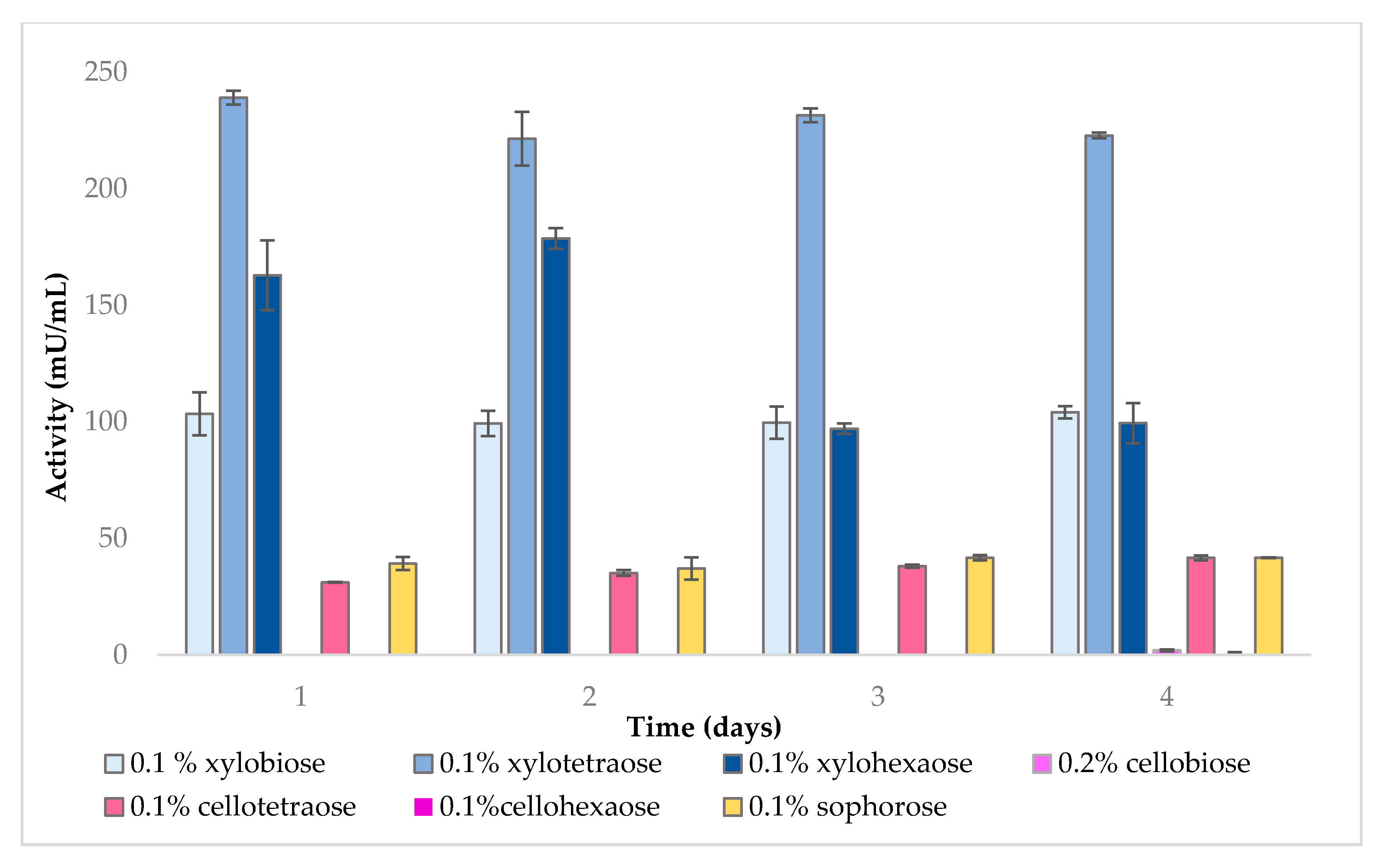
2.1.1. Endo-β-1,4-Xylanase

Endo-β-1,4-xylanase activity was measured in the presence of different inducers over a period of 4 days. As shown in Figure 1, xylooligosaccharides were more efficient than other types of sugars at inducing endo-β-1,4-xylanase activity. Maximum induction was achieved with xylotetraose, followed by xylohexaose and xylobiose.
While no activity was detected upon addition of lactose, control and cellobiose, endo-β-1,4-xylanase was induced to a lesser extent by sophorose and cellotetraose. Interestingly, 0.1% cellotetraose led to considerably higher enzyme induction compared to 0.2% cellobiose or 0.1% cellohexaose. This result is in contrast with a previous study, whereby 1% (w/v) cellobiose and cellulose could induce xylanase in F. oxysporum [36], but can be explained by the much lower concentration of cellobiose used here.

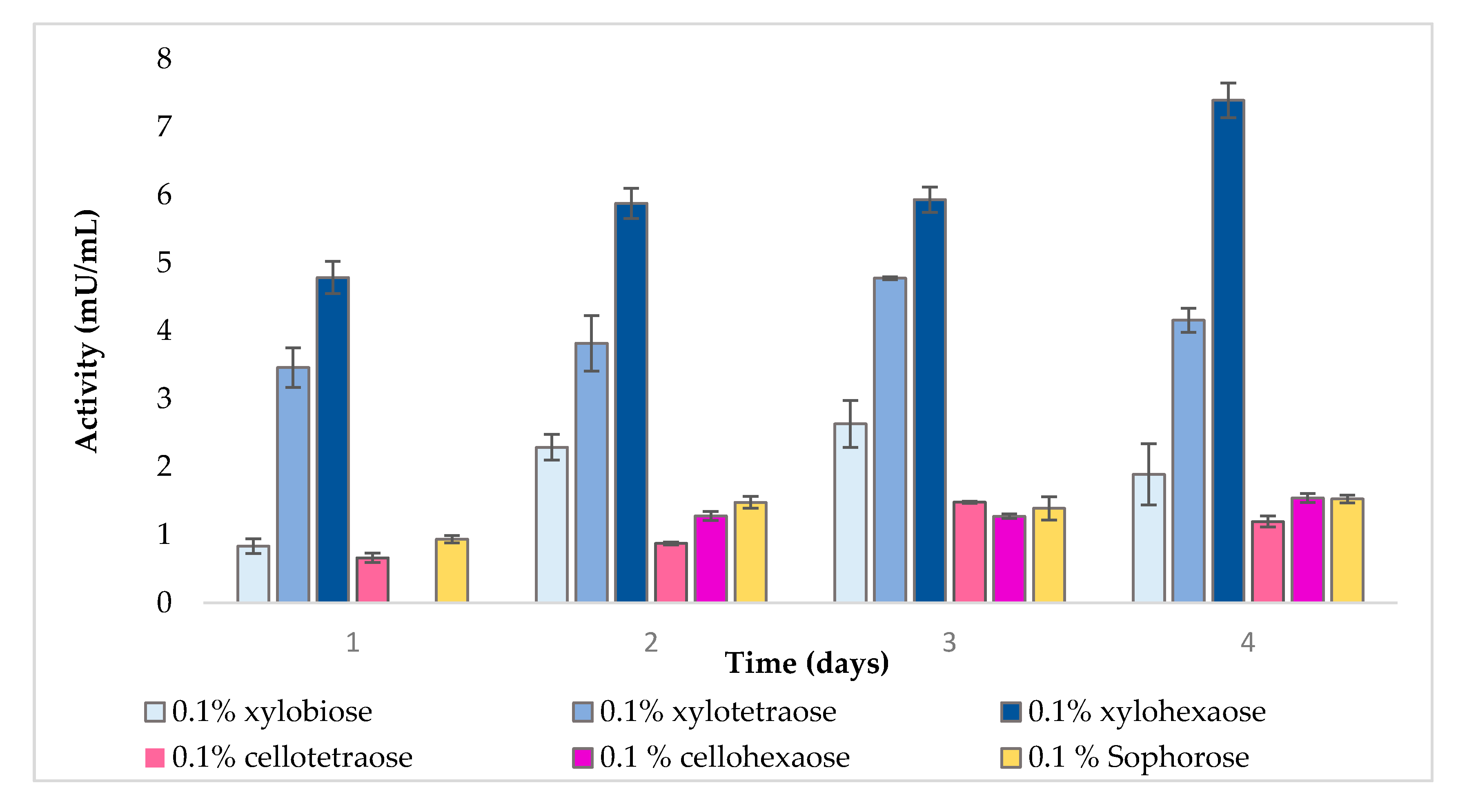
2.1.2. Extracellular β-Xylosidase

As shown in Figure 2, xylooligomers appeared to be considerably better inducers of extracellular β-xylosidase compared to other compounds. Moreover, there was a positive correlation between the length of the inducer and its induction strength. Hence, xylohexaose was the best inducer, followed by xylotetraose and xylobiose. Sophorose, cellotetraose, and cellohexaose achieved similar low extracellular β-xylosidase induction, whereas the control showed no activity.

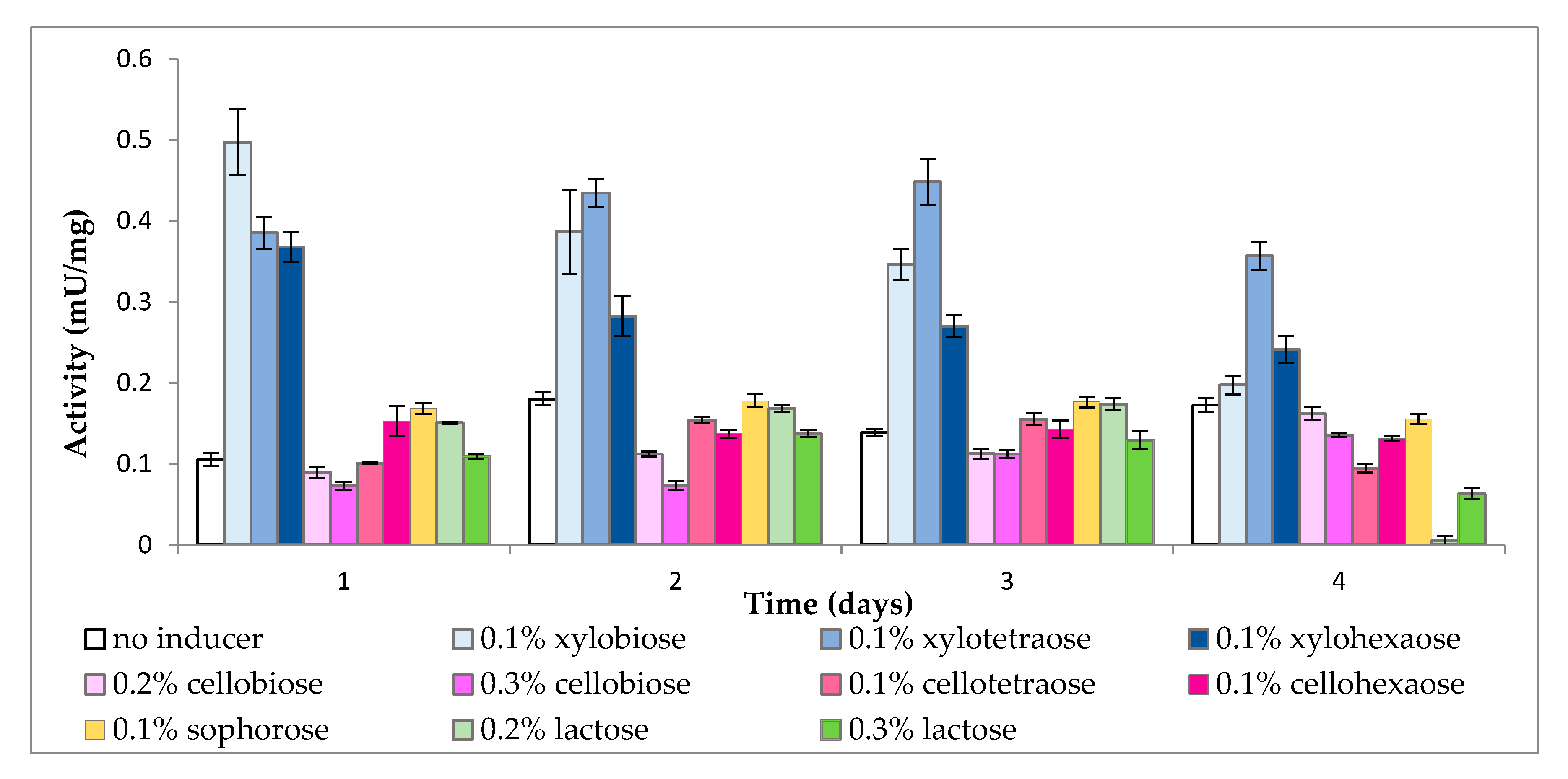
2.1.3. Cell-Bound β-Xylosidase

As is evident from Figure 3, all samples, even the control, demonstrated some cell-bound β-xylosidase activity, which could be ascribed to the presence of transporters with β-1,4-cleaving capacity. Again, xylooligomers were the most effective inducers, while the remaining sugars failed to increase enzyme activity beyond that of the control. However, the length of xylooligomers had an inverse effect on cell wall-bound β-xylosidase activity, with xylobiose exerting stronger induction at the beginning and xylotetraose later on, while xylohexaose was generally weaker than the other two.

2.2. Uptake Experiment

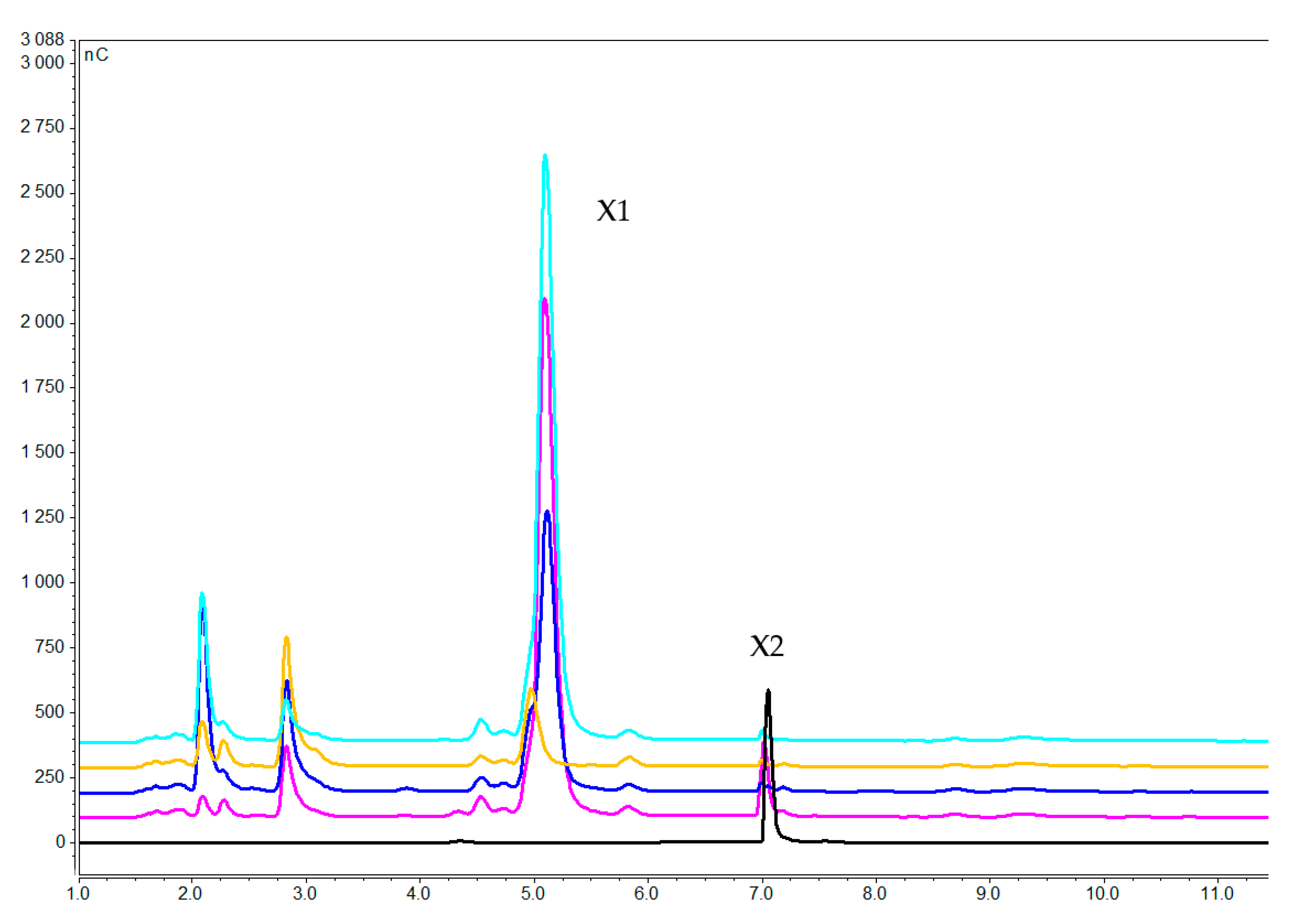
2.2.1. Concentration of Xylooligosaccharides in the Medium

Analysis of the medium revealed degradation of xylooligosaccharides in all samples between 1 and 7 h. Xylobiose as inducer was nearly complete degraded to xylose after 3 h (Figure 4); whereas xylotetraose and xylohexaose were apparently broken up immediately as they could not be detected after as little as 1 h (Figure 5 and Figure 6) generating mainly xylose, xylobiose and xylotriose. This is in agreement with previous reports that F. oxysporum xylanolytic system has an endo character and clearly attacks mainly the internal glycosidic bonds, releasing xylobiose and xylotriose [46,56].

2.2.2. Intracellular Concentration of Xylooligosaccharides

Extracts of samples induced by xylobiose, xylotetraose, and xylohexaose revealed the presence of only xylobiose in cellular fractions A and B (Figure 7, Figure 8 and Figure 9), which are the cell extracts after 10 min and 4 h of boiling. This finding suggests that xylobiose was the most probable xylooligosaccharide degradation product to enter the cell and induce expression of genes related to xylan break up.

3. Discussion

Substrates containing easily metabolized carbon sources, such as xylose, lactose, sophorose, and xylan are excellent candidates for industrial production of xylanases. Xylose and xylan [6,40], as well as simple sugars derived from lignocellulosic substrates are effective xylanase inducers. While a mixture of these sugars was suggested to potentially improve production of xylanases [39], it remains unclear how a microorganism reacts to each individual inducer [24,39,47]. Here, we evaluated the effect of different xylooligosaccharides and cellooligosaccharides on induction of xylanolytic enzymes by F. oxysporum. Moreover, their effect was compared to sophorose, which is the most powerful xylanase inducer, and lactose, which is employed for industrial production of hydrolytic enzymes [57,58,59]. Furthermore, due to the complicated process of induction, the process was simplified by using a linear substrate rather than hemicellulosic substrates with side-chains to focus on the correlation between degree of polymerization and the induction power of inducers.
Generally, enzyme production is a reflection of gene regulation. Constitutive xylanases degrade xylan and produce xylose, xylobiose, xylotriose, as well as other oligosaccharides [60,61], which can further induce xylanase production. Unlike xylose, which can easily enter microbial cells [40,62,63], larger products require more energy to be transported [61,64]. This can occur via the release of xylooligomers by xylanases, followed by transport to the cell matrix and degradation by intracellular β-xylosidase to produce xylose [65,66]. Alternatively, transporters with β-1,4-cleaving activity hydrolyze oligomers to monomers during transport from the cell wall to the cell matrix [40].
Present results provide novel insights into the link between length of the inducer and induction of sugar-degrading enzymes. Accordingly, xylotetraose appeared to be a better endoxylanase inducer than xylohexaose or xylobiose. As the latter contains only two xylose subunits, the cell needs to secrete less endoxylanase or β-xylosidase compared to xylotetraose or xylohexaose, and can instead guide the energy toward producing more cell-bound β-xylosidase.
The uptake results suggest that xylobiose is the main product of endoxylanase-mediated degradation and its intracellular detection indicates that xylobiose is the imported saccharide into the cell. Thus, the synergistic action of endoxylanase and β-xylosidase leads to xylobiose release and transportation into the cell.
Long-chain xylooligosaccharides such as xylohexaose cannot be transported easily into the cell, and need to be first broken up in smaller residues. Compared to xylotetraose and particularly xylobiose, xylohexaose is broken up into more residues and for that requires more extracellular β-glucosidase to convert xylotriose to xylobiose. Therefore, xylohexaose induces the highest amount of extracellular β-glucosidase, followed by xylotetraose and xylobiose.
The key step in biomass degradation is appropriate sensing of substrate in the medium, which then triggers the enzymatic induction cascade [67]. Transporters play an important role in this context by sensing and taking up soluble sugars inside the cell, as well as by enabling communication between the cell and the surrounding environment [67]. Transporters are known to influence CAZymes-encoding genes by carrying small inducers from the environment into the cell [67,68,69,70]. The major facilitator superfamily (MFS) and ATP binding cassette (ABC) transporters are the most studied examples in fungi [71,72,73].
ABC transporters make up the largest family of membrane transport proteins. They are actively involved in the transport of heavy metals, sugars, lipids, drugs, and secondary metabolites, cell signaling, as well as resistance to toxic compounds [74,75,76]. The ability to export toxic chemicals out of the cell allows fungi to fend off toxins in the soil or plant defense compound [74]. ABC transporters comprise 45 known families [77] found mainly in prokaryotes, with some of them acting also as sugar transporters [73,78].
The genome of filamentous fungi encodes numerous MFS transporters [79,80] divided across 17 families. Comprising of a single polypeptide, families 1, 5, and 7 transport sugars, such as pentoses and hexoses inside the cell [81,82,83] through a mechanism that uses free energy obtained from a chemiosmotic ion gradient [77,84]. MFS transporters translocate also nucleosides, lipids, amino acids, peptides, and ions [84]. The main feature of MFS transporters in filamentous fungi is their ability to sense and carry more than one kind of sugar [67]. The type of carbon source controls transcriptional regulation of sugar transporters in fungi [74,85,86]. For example, the MFS family CDT2 hexose transporter in N. crassa [73,87] collaborates with the GH43-2/7 family of CAZymes to promote xylobiose, xylotriose, and xylotetraose, consumption [73,88]. In our experiment, transporters carried xylobiose inside the cell to trigger expression of xylan-degrading enzymes. A similar result has been observed in Aspergillus fumigatus Z5, whereby xylan induced endoxylanases and β-xylosidases to produce xylobiose, while specific sugar transporters translocated it into the cell [68,89]. There, it was further broken down to xylose through induction of intracellular β-xylosidases [90].
In summary, the present results have identified the products of xylooligomer degradation responsible for induction of xylanolytic enzymes in F. oxysporum and uncovered how their degree of polymerization affected the strength of induction.

4. Materials and Methods

4.1. Microorganism and Pre-Cultures

All chemicals were purchased from Sigma-Aldrich (St. Louis, MO, USA) unless otherwise stated. F. oxysporum f. sp. lycopersici (CBS 123668) was obtained from Centraal bureau voor Schimmelcultures (Utrecht, The Netherlands) and maintained on potato dextrose agar (39 g/L). The fungus was initially grown for 2 days in pre-culture medium containing yeast extract (20 g/L) and malt extract (5 g/L) at 29 °C in a shaking incubator (190 rpm).

4.2. Enzyme Induction Trials

To study the production of xylanolytic enzymes, F. oxysporum was first grown in medium containing 0.2 M sodium phosphate buffer, 0.3 g/L MgSO4·7H2O, 1 g/L KH2PO4, 10 g/L (NH4)2HPO4, and 1% w/v sucrose. The medium was inoculated with 6% v/v of pre-culture and incubated for 2 days in a shaking incubator. Cells were harvested through a sterile vacuum filter (0.45 µm, Sarstedt, Nümbrecht, Germany), washed with sterile milliQ water, and transferred to the same medium as above, but without sucrose. Cells were incubated for 24 h to ensure that the mycelium was free of any residual sugars. Then, individual compounds with potential inducer activity were added at the following final concentrations: 0.1% w/v sophorose (Megazyme, Wicklow, Ireland); 0.2 and 0.3% w/v cellobiose; 0.2 and 0.3% w/v lactose; 0.1% w/v xylobiose, xylotetraose, and xylohexaose (Megazyme); and 0.1% w/v cellotetraose and cellohexaose (Megazyme). The fungal culture was incubated at 29 °C and 190 rpm. To minimize errors during sampling, each sample point consisted of a separate flask, whose entire content was harvested and filtered. The supernatant and biomass fractions were used to determine extracellular and cell-bound xylanase activity, respectively, as described below.

4.3. Enzyme Activity Assessment

The cell filtrate (supernatant) was used to determine extracellular endoxylanase and β-xylosidase enzyme activities. Specifically, for endo-β-1,4-xylanase activity, a solution of 1% w/v xylan from birch wood dissolved in phosphate-citrate buffer (pH 5) was used as substrate and 200 µL of this solution was incubated with 50 µL enzyme at 40 °C for 30 min. The released reducing sugars were measured by the dinitrosalicylic acid method, using a xylose standard curve [91]. For β-xylosidase activity determination, 1 mM p-nitrophenyl-β-d-xylopyranoside was used as substrate and 900 µL of this solution was incubated with 100 µL of the enzyme at 40 °C for 30 min. After the reaction was stopped by adding 200 mL 30% Na2CO3, the released p-nitrophenol was measured at 410 nm and compared to a standard curve prepared under similar conditions [28]. One unit (U) was defined as the amount of enzyme that released one μmole of product (glucose equivalent or p-nitrophenol) per minute from each substrate.
Cell-bound β-xylosidase activity was assessed following the same procedure as described above for extracellular β-xylosidase, with the difference that 1.5 mg of air-dried biomass was used as substrate. Enzyme activity was expressed in mU/mL and mU/mg for extracellular and cell-bound enzymes, respectively.

4.4. Xylooligosacharides Uptake Experiment

Xylooligosaccharides in the medium were separated by high-performance anion-exchange chromatography (HPAEC), after 1, 3, and 7 h of induction with different oligosaccharides. Upon repeating the experiment, the culture filtrates were analyzed by Dionex ICS-5000 system (Thermo Scientific, Waltham, MA USA) with a pulsed amperometric detector equipped with a disposable electrochemical gold electrode, using a CarboPac PA1 4 × 250 mm analytical column and a CarboPac PA1 4 × 50 mm guard column, at 30 °C.
The presence of xylooligosaccharides inside the cells after 1 h of induction with different oligosaccharides was measured by the same system. The culture was vacuum-filtered, and the towel-dried biomass was ground immediately in liquid nitrogen, washed with 5 mL water, boiled for 10 min, and centrifuged. The supernatant was aspirated to a separate tube and labeled as fraction A. Separately, 3 mL water was added to the precipitated biomass, which was then boiled for 4 h and the ensuing supernatant was labeled as fraction B.

Author Contributions

Conceptualization, U.R. and P.C.; methodology, N.N., L.M., U.R., and P.C.; investigation, N.N.; writing—original draft preparation, N.N.; writing—review and editing, L.M., U.R., and P.C. All authors have read and agreed to the published version of the manuscript.

Funding

This research was funded by Swedish research council (VR), grant number 2014-05906.

Acknowledgments

The authors greatly acknowledge Anthi Karnaouri for her advice and comments that greatly improved the manuscript.

Conflicts of Interest

The authors declare no conflict of interest.

References

  1. Prade, R.A. Xylanases: From Biology to BioTechnology. Biotechnol. Genet. Eng. Rev. 1996, 13, 101–132. [Google Scholar] [CrossRef] [PubMed]
  2. Beg, Q.K.; Kapoor, M.; Mahajan, L.; Hoondal, G.S. Microbial xylanases and their industrial applications: A review. Appl. Microbiol. Biotechnol. 2001, 56, 326–338. [Google Scholar] [CrossRef] [PubMed]
  3. Wyman, C.; Decker, S.; Himmel, M.; Brady, J.; Skopec, C.; Viikari, L. Hydrolysis of Cellulose and Hemicellulose. Polysaccharides 2004, 1, 1023–1062. [Google Scholar]
  4. Wong, K.K.; Tan, L.U.; Saddler, J.N. Multiplicity of β-1,4-xylanase in microorganisms: Functions and applications. Microbiol. Rev. 1988, 52, 305–317. [Google Scholar] [CrossRef] [PubMed]
  5. Singh, S.; Madlala, A.M.; Prior, B.A. Thermomyces lanuginosus: Properties of strains and their hemicellulases. FEMS Microbiol. Rev. 2003, 27, 3–16. [Google Scholar] [CrossRef] [Green Version]
  6. Kulkarni, N.; Shendye, A.; Rao, M. Molecular and biotechnological aspects of xylanases. FEMS Microbiol. Rev. 1999, 23, 411–456. [Google Scholar] [CrossRef]
  7. Chanda, S.K.; Hirst, E.L.; Jones, J.K.N.; Percival, E.G.V. 262. The constitution of xylan from esparto grass (stipa tenacissima, L.). J. Chem. Soc. 1950, 1289–1297. [Google Scholar] [CrossRef]
  8. Eda, S.; Ohnishi, A.; Kato, K. Xylan Isolated from the Stalk of Nicotiana tabacum. Agric. Biol. Chem. 1976, 40, 359–364. [Google Scholar] [CrossRef]
  9. Kenneth, J.H. Average Gestation Period and n π. Nat. Cell Biol. 1940, 146, 620. [Google Scholar] [CrossRef]
  10. Knob, A.; Terrasan, C.R.F.; Carmona, E.C. β-Xylosidases from filamentous fungi: An overview. World J. Microbiol. Biotechnol. 2010, 26, 389–407. [Google Scholar] [CrossRef]
  11. Katsimpouras, C.; Dedes, G.; Thomaidis, N.S.; Topakas, E. A novel fungal GH30 xylanase with xylobiohydrolase auxiliary activity. Biotechnol. Biofuels 2019, 12, 1–14. [Google Scholar] [CrossRef] [PubMed] [Green Version]
  12. Gielkens, M.M.C.; Visser, J.; De Graaff, L.H. Arabinoxylan degradation by fungi: Characterization of the arabinoxylan-arabinofuranohydrolase encoding genes from Aspergillus niger and Aspergillus tubingensis. Curr. Genet. 1997, 31, 22–29. [Google Scholar] [CrossRef] [PubMed]
  13. Poutanen, K.; Sundberg, M.; Korte, H.; Puls, J. Deacetylation of xylans by acetyl esterases of Trichoderma reesei. Appl. Microbiol. Biotechnol. 1990, 33, 506–510. [Google Scholar] [CrossRef]
  14. Reilly, P.J. Xylanases: Structure and Function. Trends Biol. Ferment. Fuels Chem. 1981, 18, 111–129. [Google Scholar] [CrossRef]
  15. Li, W.; Wang, J.; Coluccio, L.M.; Matsudaira, P.; Grand, R.J. Brush border myosin I (BBMI): A basally localized transcript in human jejunal enterocytes. J. Histochem. Cytochem. 2000, 48, 89–94. [Google Scholar] [CrossRef] [PubMed] [Green Version]
  16. Bajpai, P. Microbial Xylanolytic Systems and Their Properties. Xylanolytic Enzym. 2014, 19–36. [Google Scholar] [CrossRef]
  17. Poutanen, K.; Sundberg, M. An acetyl esterase of Trichoderma reesei and its role in the hydrolysis of acetyl xylans. Appl. Microbiol. Biotechnol. 1988, 28, 419–424. [Google Scholar] [CrossRef]
  18. Sørensen, H.R.; Meyer, A.S.; Pedersen, S. Enzymatic hydrolysis of water-soluble wheat arabinoxylan. 1. Synergy between α-l-arabinofuranosidases, endo-1,4-β-xylanases, and β-xylosidase activities. Biotechnol. Bioeng. 2003, 81, 726–731. [Google Scholar] [CrossRef]
  19. Rasmussen, L.E.; Sørensen, H.R.; Vind, J.; Viksø-Nielsen, A. Mode of action and properties of the β-xylosidases fromTalaromyces emersonii andTrichoderma reesei. Biotechnol. Bioeng. 2006, 94, 869–876. [Google Scholar] [CrossRef]
  20. Sunna, A.; Antranikian, G. Xylanolytic Enzymes from Fungi and Bacteria. Crit. Rev. Biotechnol. 1997, 17, 39–67. [Google Scholar] [CrossRef]
  21. Hrmova, M.; Biely, P.; Vršanská, M.; Petráková, E. Induction of cellulose- and xylan-degrading enzyme complex in the yeast Trichosporon cutaneum. Arch. Microbiol. 1984, 138, 371–376. [Google Scholar] [CrossRef]
  22. Liu, W.; Zhu, W.; Lu, Y.; Kong, J.; Ma, G. Production, partial purification and characterization of xylanase from Trichosporon cutaneum SL409. Process. Biochem. 1998, 33, 331–336. [Google Scholar] [CrossRef]
  23. Gilbert, J.; Hazlewood, P. Review Article Bacterial cellulases and xylanases. J. Gen. Microbiol. 1993, 1, 187–194. [Google Scholar] [CrossRef] [Green Version]
  24. Lakshmi, G.S.; Bhargavi, P.L.; Prakasham, R.S. Sustainable Bioprocess Evaluation for Xylanase Production by Isolated Aspergillus terreus and Aspergillus fumigatus Under Solid—State Fermentation Using Oil Palm Empty Fruit Bunch Fiber. Curr. Trends Biotechnol. Pharm. 2011, 5, 1434–1444. [Google Scholar]
  25. Pathak, P.; Bhardwaj, N.K.; Singh, A.K. Production of Crude Cellulase and Xylanase From Trichoderma harzianum PPDDN10 NFCCI-2925 and Its Application in Photocopier Waste Paper Recycling. Appl. Biochem. Biotechnol. 2014, 172, 3776–3797. [Google Scholar] [CrossRef] [PubMed]
  26. Agrios, G.N. Chapter Fourteen—Plant Diseases Caused by Viruses. In Agrios GNBT-PP, 5th ed.; Academic Press: San Diego, CA, USA, 2005; pp. 723–824. [Google Scholar] [CrossRef]
  27. Bertonha, L.C.; Neto, M.L.; Garcia, J.A.A.; Vieira, T.F.; Castoldi, R.; Bracht, A.; Peralta, R.M. Screening of Fusarium sp. for xylan and cellulose hydrolyzing enzymes and perspectives for the saccharification of delignified sugarcane bagasse. Biocatal. Agric. Biotechnol. 2018, 16, 385–389. [Google Scholar] [CrossRef]
  28. Panagiotou, G.; Kekos, D.; Macris, B.J.; Christakopoulos, P. Production of cellulolytic and xylanolytic enzymes by Fusarium oxysporum grown on corn stover in solid state fermentation. Ind. Crop. Prod. 2003, 18, 37–45. [Google Scholar] [CrossRef]
  29. Calero-Nieto, F.; Di Pietro, A.; Roncero, M.I.G.; Hera, C. Role of the Transcriptional Activator XlnR of Fusarium oxysporum in Regulation of Xylanase Genes and Virulence. Mol. Plant Microbe Interact. 2007, 20, 977–985. [Google Scholar] [CrossRef] [Green Version]
  30. Linko, M.; Viikari, L.; Suihko, M.-L. Hydrolysis of xylan and fermentation of xylose to ethanol. Biotechnol. Adv. 1984, 2, 233–252. [Google Scholar] [CrossRef]
  31. Christakopoulos, P.; Macris, B.; Kekos, D. Direct fermentation of cellulose to ethanol by Fusarium oxysporum. Enzym. Microb. Technol. 1989, 11, 236–239. [Google Scholar] [CrossRef]
  32. Dinolfo, M.I.; Eliana, C.; Stenglein, S.A. Fusarium–plant interaction: State of the art—A review. Plant Prot. Sci. 2017, 53, 61–70. [Google Scholar] [CrossRef] [Green Version]
  33. Paccanaro, M.C.; Sella, L.; Castiglioni, C.; Giacomello, F.; Martínez-Rocha, A.L.; D’Ovidio, R.; Schã Fer, W.; Favaron, F. Synergistic Effect of Different Plant Cell Wall–Degrading Enzymes Is Important for Virulence of Fusarium graminearum. Mol. Plant Microbe Interact. 2017, 30, 886–895. [Google Scholar] [CrossRef] [PubMed] [Green Version]
  34. De Vries, R.P.; Visser, J.; De Graaff, L.H. CreA modulates the XlnR-induced expression on xylose of Aspergillus niger genes involved in xylan degradation. Res. Microbiol. 1999, 150, 281–285. [Google Scholar] [CrossRef]
  35. Kuhad, R.C.; Manchanda, M.; Singh, A. Optimization of xylanase production by a hyperxylanolytic mutant strain of Fusarium oxysporum. Process. Biochem. 1998, 33, 641–647. [Google Scholar] [CrossRef]
  36. Christakopoulos, P.; Mamma, D.; Nerinckx, W.; Kekos, D.; Macris, B.; Claeyssens, M. Production and partial characterization of xylanase from Fusarium oxysporum. Bioresour. Technol. 1996, 58, 115–119. [Google Scholar] [CrossRef]
  37. Fontes, C.M.G.A.; Gilbert, H.J.; Hazlewood, G.P.; Clarke, J.; Prates, J.A.M.; McKie, V.A.; Nagy, T.; Fernandes, T.H.; Ferreira, L.M.A. A novel Cellvibrio mixtus family 10 xylanase that is both intracellular and expressed under non-inducing conditions The GenBank accession numbers for the sequences described in this paper are AF049493 and AF168359 for xynC and xynG, respectively. Microbiology 2000, 146, 1959–1967. [Google Scholar] [CrossRef] [Green Version]
  38. Teplitsky, A.; Shulami, S.; Moryles, S.; Shoham, Y.; Shoham, G. Crystallization and preliminary X-ray analysis of an intracellular xylanase from Bacillus stearothermophilus T-6. Acta Crystallogr. Sect. D Biol. Crystallogr. 2000, 56, 181–184. [Google Scholar] [CrossRef]
  39. Kamoldeen, A.A.; Leh, C.P.; Abdullah, W.N.W.; Lee, C.K. Assessment of the Effect of Easily-metabolised Carbon Supplements on Xylanase Production by Newly Isolated Trichoderma asperellum USM SD4 Cultivated on Oil Palm Empty Fruit Bunches. Bioresources 2016, 11, 9611–9627. [Google Scholar] [CrossRef] [Green Version]
  40. Subramaniyan, S.; Prema, P. Biotechnology of Microbial Xylanases: Enzymology, Molecular Biology, and Application. Crit. Rev. Biotechnol. 2002, 22, 33–64. [Google Scholar] [CrossRef]
  41. Dobrev, G.T.; Pishtiyski, I.G.; Stanchev, V.S.; Mircheva, R. Optimization of nutrient medium containing agricultural wastes for xylanase production by Aspergillus niger B03 using optimal composite experimental design. Bioresour. Technol. 2007, 98, 2671–2678. [Google Scholar] [CrossRef]
  42. De Vries, R.; van de Vondervoort, P.; Hendriks, L.; Van de Belt, M.; Visser, J. Regulation of the α-glucuronidase-encoding gene (aguA) from Aspergillus niger. Mol. Genet. Genom. 2002, 268, 96–102. [Google Scholar] [CrossRef] [PubMed]
  43. Martiín-Urdiíroz, M.C.; Di Pietro, A.; Huertas-González, M.D.; Roncero, M.I. Two xylanase genes of the vascular wilt pathogen Fusarium oxysporum are differentially expressed during infection of tomato plants. Mol. Genet. Genom. 1999, 261, 530–536. [Google Scholar] [CrossRef]
  44. Royer, J.; Nakas, J. Xylanase production by Trichoderma longibrachiatum. Enzym. Microb. Technol. 1989, 11, 405–410. [Google Scholar] [CrossRef]
  45. Raj, K.C.; Chandra, T.S. A cellulase-free xylanase from alkali-tolerantAspergillus fischeri Fxn1. Biotechnol. Lett. 1995, 17, 309–314. [Google Scholar] [CrossRef]
  46. Christakopoulos, P.; Nerinckx, W.; Kekos, D.; Macris, B.; Claeyssens, M. Purification and characterization of two low molecular mass alkaline xylanases from Fusarium oxysporum F3. J. Biotechnol. 1996, 51, 181–189. [Google Scholar] [CrossRef]
  47. Gaffney, M.; Doyle, S.; Murphy, R. Optimization of Xylanase Production byThermomyces lanuginosusin Solid State Fermentation. Biosci. Biotechnol. Biochem. 2009, 73, 2640–2644. [Google Scholar] [CrossRef] [Green Version]
  48. Khosravi, C.; Kowalczyk, J.E.; Chroumpi, T.; Battaglia, E.; Pontes, M.-V.A.; Peng, M.; Wiebenga, A.; Ng, V.; Lipzen, A.; He, G.; et al. Transcriptome analysis of Aspergillus niger xlnR and xkiA mutants grown on corn Stover and soybean hulls reveals a highly complex regulatory network. BMC Genom. 2019, 20, 1–16. [Google Scholar] [CrossRef]
  49. Zhang, T.; Liu, H.; Lv, B.; Li, C. Regulating Strategies for Producing Carbohydrate Active Enzymes by Filamentous Fungal Cell Factories. Front. Bioeng. Biotechnol. 2020, 8, 691. [Google Scholar] [CrossRef]
  50. Dowzer, C.E.A.; Kelly, J.M. Cloning of the creA gene from Aspergillus nidulans: A gene involved in carbon catabolite repression. Curr. Genet. 1989, 15, 457–459. [Google Scholar] [CrossRef]
  51. Strauss, J.; Mach, R.L.; Zeilinger, S.; Hartler, G.; Stöffler, G.; Wolschek, M.; Kubicek, C.P. Crel, the carbon catabolite repressor protein from Trichoderma reesei. FEBS Lett. 1995, 376, 103–107. [Google Scholar] [CrossRef] [Green Version]
  52. De La Serna, I.; Ng, D.; Tyler, B.M. Carbon Regulation of Ribosomal Genes in Neurospora crassa Occurs by a Mechanism Which Does Not Require Cre-1, the Homologue of the Aspergillus Carbon Catabolite Repressor, CreA. Fungal Genet. Biol. 1999, 26, 253–269. [Google Scholar] [CrossRef] [PubMed]
  53. Todd, R.B.; Lockington, R.A.; Kelly, J.M. The Aspergillus nidulans creC gene involved in carbon catabolite repression encodes a WD40 repeat protein. Mol. Genet. Genom. 2000, 263, 561–570. [Google Scholar] [CrossRef] [PubMed]
  54. Lockington, R.A.; Kelly, J.M. The WD40-repeat protein CreC interacts with and stabilizes the deubiquitinating enzyme CreB in vivo in Aspergillus nidulans. Mol. Microbiol. 2002, 43, 1173–1182. [Google Scholar] [CrossRef] [PubMed] [Green Version]
  55. Ries, L.N.A.; Beattie, S.R.; Espeso, E.A.; Cramer, R.A.; Goldman, G.H. Diverse Regulation of the CreA Carbon Catabolite Repressor in Aspergillus nidulans. Genetics 2016, 203, 335–352. [Google Scholar] [CrossRef] [Green Version]
  56. Christakopoulos, P.; Nerinckx, W.; Kekos, D.; Macris, B.; Claeyssens, M. The alkaline xylanase III from Fusarium oxysporum F3 belongs to family F/10. Carbohydr. Res. 1997, 302, 191–195. [Google Scholar] [CrossRef]
  57. Chaudhuri, B.; Sahai, V. Comparison of growth and maintenance parameters for cellulase biosynthesis by Trichoderma reesei-C5 with some published data. Enzym. Microb. Technol. 1994, 16, 1079–1083. [Google Scholar] [CrossRef]
  58. Xu, J.; Nogawa, M.; Okada, H.; Morikawa, Y. Regulation of xyn3 gene expression in Trichoderma reesei PC-3-7. Appl. Microbiol. Biotechnol. 2000, 54, 370–375. [Google Scholar] [CrossRef]
  59. Xiong, H.; Turunen, O.; Pastinen, O.; Leisola, M.; Von Weymarn, N. Improved xylanase production by Trichoderma reesei grown on l-arabinose and lactose or d-glucose mixtures. Appl. Microbiol. Biotechnol. 2004, 64, 353–358. [Google Scholar] [CrossRef]
  60. Kang, S.-W.; Kim, S.W.; Lee, J.-S. Production of cellulase and xylanase in a bubble column using immobilizedAspergillus Niger KKS. Appl. Biochem. Biotechnol. 1995, 53, 101–106. [Google Scholar] [CrossRef]
  61. Wang, P.; Mason, J.C.; Broda, P. Xylanases from Streptomyces cyaneus: Their production, purification and characterization. J. Gen. Microbiol. 1993, 139, 1987–1993. [Google Scholar] [CrossRef] [Green Version]
  62. Zhao, Y.; Chany, C.J.; Sims, P.F.; Sinnott, M.L. Definition of the substrate specificity of the ‘sensing’ xylanase of Streptomyces cyaneus using xylooligosaccharide and cellooligosaccharide glycosides of 3,4-dinitrophenol. J. Biotechnol. 1997, 57, 181–190. [Google Scholar] [CrossRef]
  63. Biely, P. Microbial xylanolytic systems. Trends Biotechnol. 1985, 3, 286–290. [Google Scholar] [CrossRef]
  64. Gomes, D.; Gomes, J.; Steiner, W. Factors influencing the induction of endo-xylanase by Thermoascus aurantiacus. J. Biotechnol. 1994, 33, 87–94. [Google Scholar] [CrossRef]
  65. Rapp, P.; Wagner, F. Production and Properties of Xylan-Degrading Enzymes from Cellulomonas uda. Appl. Environ. Microbiol. 1986, 51, 746–752. [Google Scholar] [CrossRef] [Green Version]
  66. La Grange, D.C.; Claeyssens, M.; Pretorius, I.S.; Van Zyl, W.H. Coexpression of the Bacillus pumilus β-xylosidase ( xynB ) gene with the Trichoderma reesei β-xylanase 2 ( xyn2 ) gene in the yeast Saccharomyces cerevisiae. Appl. Microbiol. Biotechnol. 2000, 54, 195–200. [Google Scholar] [CrossRef]
  67. De Paula, R.G.; Antoniêto, A.C.C.; Ribeiro, L.F.C.; Carraro, C.B.; Nogueira, K.M.V.; Lopes, D.C.B.; Silva, A.C.; Zerbini, M.T.; Pedersoli, W.R.; Costa, M.D.N.; et al. New Genomic Approaches to Enhance Biomass Degradation by the Industrial Fungus Trichoderma reesei. Int. J. Genom. 2018, 2018, 1–17. [Google Scholar] [CrossRef] [Green Version]
  68. Cai, P.; Gu, R.; Wang, B.; Li, J.; Wan, L.; Tian, C.; Ma, Y. Evidence of a Critical Role for Cellodextrin Transporte 2 (CDT-2) in Both Cellulose and Hemicellulose Degradation and Utilization in Neurospora crassa. PLoS ONE 2014, 9, e89330. [Google Scholar] [CrossRef]
  69. Porciuncula, J.D.O.; Furukawa, T.; Shida, Y.; Mori, K.; Kuhara, S.; Morikawa, Y.; Ogasawara, W. Identification of Major Facilitator Transporters Involved in Cellulase Production during Lactose Culture ofTrichoderma reeseiPC-3-7. Biosci. Biotechnol. Biochem. 2013, 77, 1014–1022. [Google Scholar] [CrossRef] [Green Version]
  70. Ivanova, C.; Bååth, J.A.; Seiboth, B.; Kubicek, C.P. Systems Analysis of Lactose Metabolism in Trichoderma reesei Identifies a Lactose Permease That Is Essential for Cellulase Induction. PLoS ONE 2013, 8, e62631. [Google Scholar] [CrossRef] [Green Version]
  71. Chaudhary, N.; Kumari, I.; Sandhu, P.; Ahmed, M.; Akhter, Y. Proteome scale census of major facilitator superfamily transporters in Trichoderma reesei using protein sequence and structure based classification enhanced ranking. Gene 2016, 585, 166–176. [Google Scholar] [CrossRef]
  72. Schmoll, M.; Dattenböck, C.; Carreras-Villaseñor, N.; Mendoza-Mendoza, A.; Tisch, D.; Alemán, M.I.; Baker, S.E.; Brown, C.; Cervantes-Badillo, M.G.; Cetz-Chel, J.; et al. The Genomes of Three Uneven Siblings: Footprints of the Lifestyles of Three Trichoderma Species. Microbiol. Mol. Biol. Rev. 2016, 80, 205–327. [Google Scholar] [CrossRef] [PubMed] [Green Version]
  73. Nogueira, K.M.V.; Mendes, V.; Carraro, C.B.; Taveira, I.C.; Oshiquiri, L.H.; Gupta, V.K.; Silva, R.N. Sugar transporters from industrial fungi: Key to improving second-generation ethanol production. Renew. Sustain. Energy Rev. 2020, 131, 109991. [Google Scholar] [CrossRef]
  74. Fravel, D.; Moravec, B.; Jones, R.W.; Costanzo, S. Characterization of two ABC transporters from biocontrol and phytopathogenic Fusarium oxysporum. Physiol. Mol. Plant Pathol. 2008, 73, 2–8. [Google Scholar] [CrossRef]
  75. Atanasova, L.; Le Crom, S.; Gruber, S.; Coulpier, F.; Seidl-Seiboth, V.; Kubicek, C.P.; Druzhinina, I.S. Comparative transcriptomics reveals different strategies of Trichoderma mycoparasitism. BMC Genom. 2013, 14, 121. [Google Scholar] [CrossRef] [PubMed] [Green Version]
  76. Kang, J.; Hwang, J.-U.; Lee, M.; Kim, Y.-Y.; Assmann, S.M.; Martinoia, E.; Lee, Y. PDR-type ABC transporter mediates cellular uptake of the phytohormone abscisic acid. Proc. Natl. Acad. Sci. USA 2010, 107, 2355–2360. [Google Scholar] [CrossRef] [Green Version]
  77. Henderson, R.K.; Fendler, K.; Poolman, B. Coupling efficiency of secondary active transporters. Curr. Opin. Biotechnol. 2019, 58, 62–71. [Google Scholar] [CrossRef]
  78. Baral, B. Chapter Four—Evolutionary Trajectories of Entomopathogenic Fungi ABC Transporters. In Goodwin SFBT-A in G; Friedmann, T., Dunlap, J.C., Eds.; Academic Press: Cambridge, MA, USA, 2017; pp. 117–154. [Google Scholar] [CrossRef]
  79. Vongsangnak, W.; Salazar, M.; Hansen, K.; Nielsen, J. Genome-wide analysis of maltose utilization and regulation in aspergilli. Microbiology 2009, 155, 3893–3902. [Google Scholar] [CrossRef] [Green Version]
  80. Galagan, J.E.; Calvo, S.E.; Borkovich, K.A.; Selker, E.U.; Read, N.D.; Jaffe, D.; Fitzhugh, W.; Ma, L.-J.; Smirnov, S.; Purcell, S.; et al. The genome sequence of the filamentous fungus Neurospora crassa. Nature 2003, 422, 859–868. [Google Scholar] [CrossRef]
  81. Saier, M.H.; Beatty, J.T.; Goffeau, A.; Harley, K.T.; Heijne, W.H.M.; Huang, S.C.; Jack, D.L.; Jähn, P.S.; Lew, K.; Liu, J.; et al. The major facilitator superfamily. J. Mol. Microbiol. Biotechnol. 1999, 1, 257–279. [Google Scholar]
  82. Law, C.J.; Maloney, P.C.; Wang, D. Ins and Outs of Major Facilitator Superfamily Antiporters. Annu. Rev. Microbiol. 2008, 62, 289–305. [Google Scholar] [CrossRef] [Green Version]
  83. Yan, N. Structural advances for the major facilitator superfamily (MFS) transporters. Trends Biochem. Sci. 2013, 38, 151–159. [Google Scholar] [CrossRef] [PubMed]
  84. Wang, S.C.; Davejan, P.; Hendargo, K.J.; Javadi-Razaz, I.; Chou, A.; Yee, D.C.; Ghazi, F.; Lam, K.J.K.; Conn, A.M.; Madrigal, A.; et al. Expansion of the Major Facilitator Superfamily (MFS) to include novel transporters as well as transmembrane-acting enzymes. Biochim. Biophys. Acta Biomembr. 2020, 1862, 183277. [Google Scholar] [CrossRef] [PubMed]
  85. Castro, S.; Pedersoli, W.R.; Cristina, A.; Antoniêto, C.; Steindorff, A.S.; Silva-rocha, R.; Martinez-Rossi, N.M.; Rossi, A.; Brown, N.A.; Goldman, G.H.; et al. Comparative metabolism of cellulose, sophorose and glucose in Trichoderma reesei using high-throughput genomic and proteomic analyses. Biotechnol. Biofuels 2014, 7, 41. [Google Scholar] [CrossRef] [PubMed] [Green Version]
  86. Antoniêto, A.C.C.; De Paula, R.G.; Castro, L.D.S.; Silva-Rocha, R.; Persinoti, G.F.; Silva, R.N. Trichoderma reesei CRE1-mediated Carbon Catabolite Repression in Response to Sophorose Through RNA Sequencing Analysis. Curr. Genom. 2016, 17, 119–131. [Google Scholar] [CrossRef] [PubMed] [Green Version]
  87. Galazka, J.M.; Tian, C.; Beeson, W.T.; Martinez, B.; Glass, N.L.; Cate, J.H. Cellodextrin transport in yeast for improved biofuel production. Science 2010, 330, 84–86. [Google Scholar] [CrossRef]
  88. Li, X.; Yu, V.Y.; Lin, Y.; Chomvong, K.; Estrela, R.; Park, A.; Liang, J.M.; Znameroski, E.; Feehan, J.; Kim, S.R.; et al. Expanding xylose metabolism in yeast for plant cell wall conversion to biofuels. eLife 2015, 4, 1–55. [Google Scholar] [CrossRef]
  89. Miao, Y.; Li, J.; Xiao, Z.; Shen, Q.; Zhang, R. Characterization and identification of the xylanolytic enzymes from Aspergillus fumigatus Z5. BMC Microbiol. 2015, 15, 126. [Google Scholar] [CrossRef] [Green Version]
  90. Znameroski, E.A.; Coradetti, S.T.; Roche, C.M.; Tsai, J.C.; Iavarone, A.T.; Cate, J.H.D.; Glass, N.L. Induction of lignocellulose-degrading enzymes in Neurospora crassa by cellodextrins. Proc. Natl. Acad. Sci. USA 2012, 109, 6012–6017. [Google Scholar] [CrossRef] [Green Version]
  91. Miller, G.L. Use of Dinitrosalicylic Acid Reagent for Determination of Reducing Sugar. Anal. Chem. 1959, 31, 426–428. [Google Scholar] [CrossRef]
Figure 1. Endo-β-1,4-xylanase activity in the presence of different inducers.
Molecules 25 05849 g001
Figure 2. Extracellular β-xylosidase activity in the presence of different inducers.
Molecules 25 05849 g002
Figure 3. Cell-bound β-xylosidase activity in the presence of different inducers.
Molecules 25 05849 g003
Figure 4. Xylooligosacharides concentration in the medium after 1 h (pink), 3 h (dark blue), and 7 h (orange) of induction with xylobiose. Blue and black lines correspond to pure xylose and xylobiose respectively (X1 = xylose, X2 = xylobiose).
Molecules 25 05849 g004
Figure 5. Xylooligosaccharides concentration in the medium after 1 h (pink), 3 h (orange), and 7 h (green) of induction with xylotetraose. Blue, black and dark blue lines correspond to pure xylose, xylobiose and xylotetraose, respectively (X1 = xylose, X2 = xylobiose, X3 = xylotriose, X4 = xylotetraose).
Molecules 25 05849 g005
Figure 6. Xylooligosaccharides concentration in the medium after 1 h (pink), 3 h (orange), and 7 h (green) of induction with xylohexaose. Blue, black, dark blue and red lines correspond to pure xylose, xylobiose, xylotetraose, and xylohexaose respectively (X1 = xylose, X2 = xylobiose, X3 = xylotriose, X4 = xylotetraose; X6 = xylohexaose).
Molecules 25 05849 g006
Figure 7. Intracellular composition after 1 h of induction with xylobiose. (a) Pure xylobiose. (b) Pure xylose (c) Cell extract fraction A. (d) Cell extract fraction B (X1 = xylose, X2 = xylobiose).
Molecules 25 05849 g007
Figure 8. Intracellular composition after 1 h of induction with xylotetraose. (a) Pure xylobiose. (b) Pure xylotetraose. (c) Pure xylose (d) Cell extract fraction A. (e) Cell extract fraction B (X1 = xylose; X2 = xylobiose, X4 = xylotetraose).
Molecules 25 05849 g008
Figure 9. Intracellular composition after 1 h of induction with xylohexaose. (a) Pure xylobiose. (b) Pure xylotetraose. (c) Pure xylose (d) Cell extract fraction A. (e) Cell extract fraction B (f) pure xylohexaose (X1 = xylose, X2 = xylobiose, X4 = xylotetraose, X6 = xylohexaose).
Molecules 25 05849 g009
Sample Availability: Samples of the compounds are not available from the authors.
Publisher’s Note: MDPI stays neutral with regard to jurisdictional claims in published maps and institutional affiliations.

