Niosomal Nanocarriers for Enhanced Skin Delivery of Quercetin with Functions of Anti-Tyrosinase and Antioxidant

Abstract
: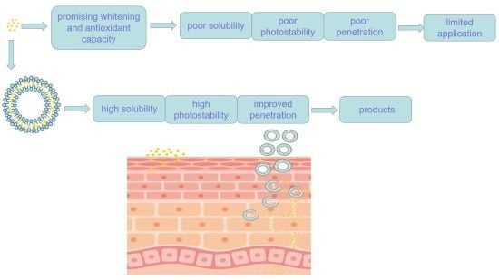
1. Introduction
2. Results
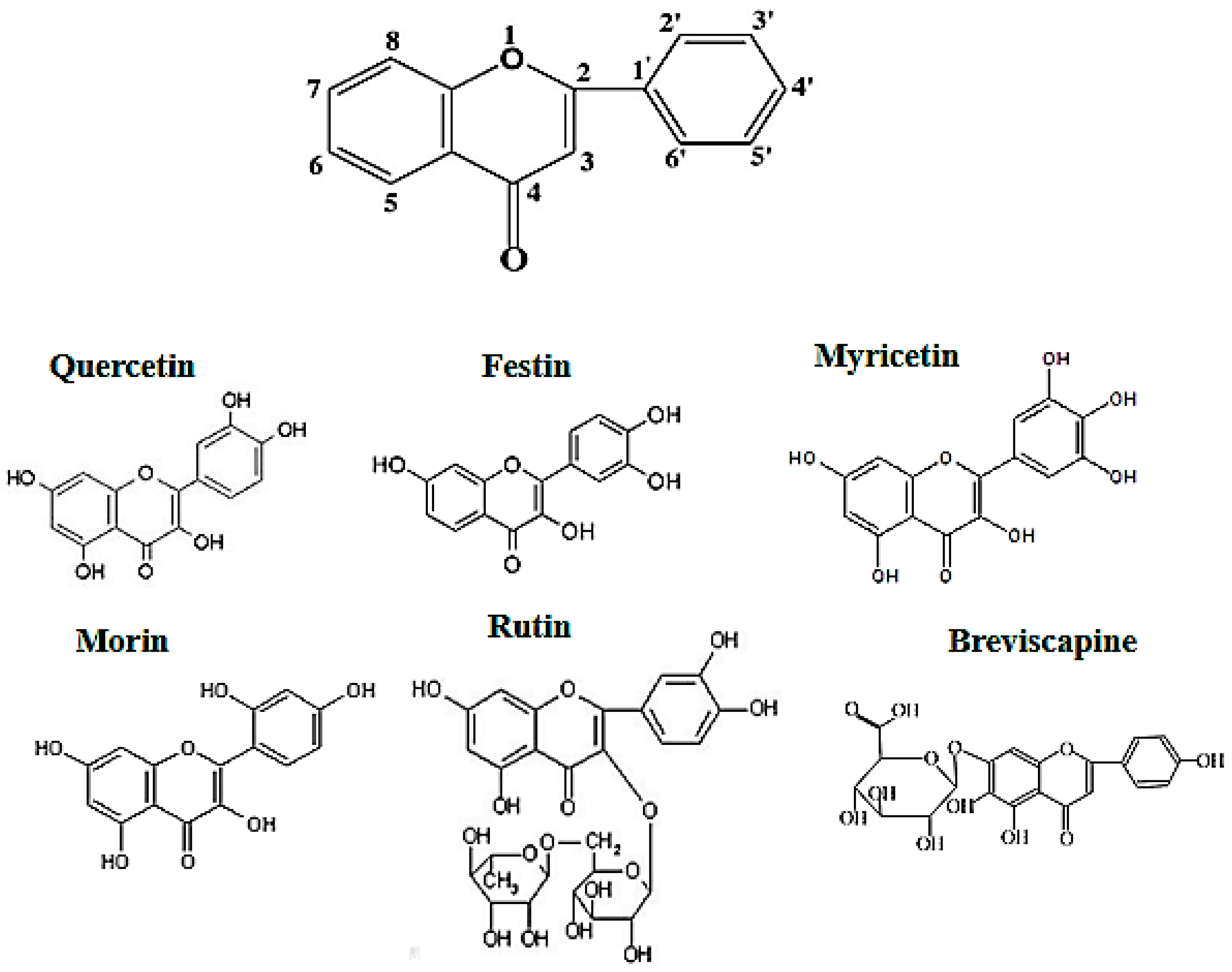
2.1. Tyrosinase Inhibition of Flavonoids
2.2. Antioxidant Activity of Flavonoids
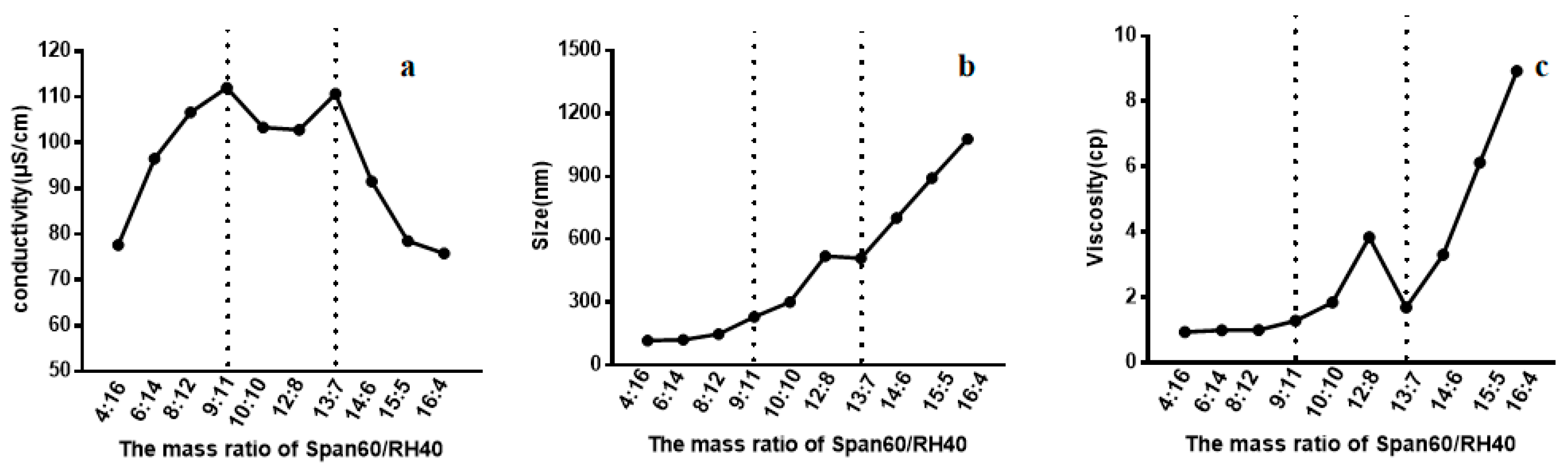
2.3. Formulation of Niosomes
2.3.1. Conductivity of the Span60-RH40 System
2.3.2. Size of the Span60-RH40 System
2.3.3. Viscosity of the Span60-RH40 System
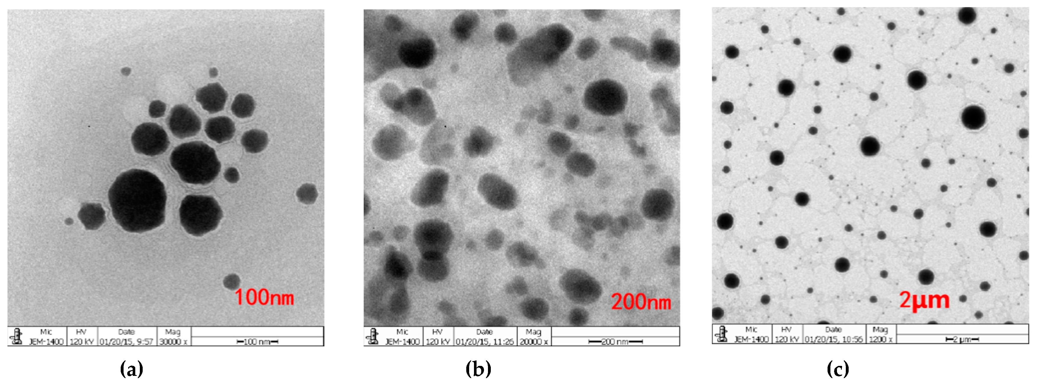
2.4. Size, Zeta Potential, Morphology and Entrapment Efficiency of Quercetin-Niosomes
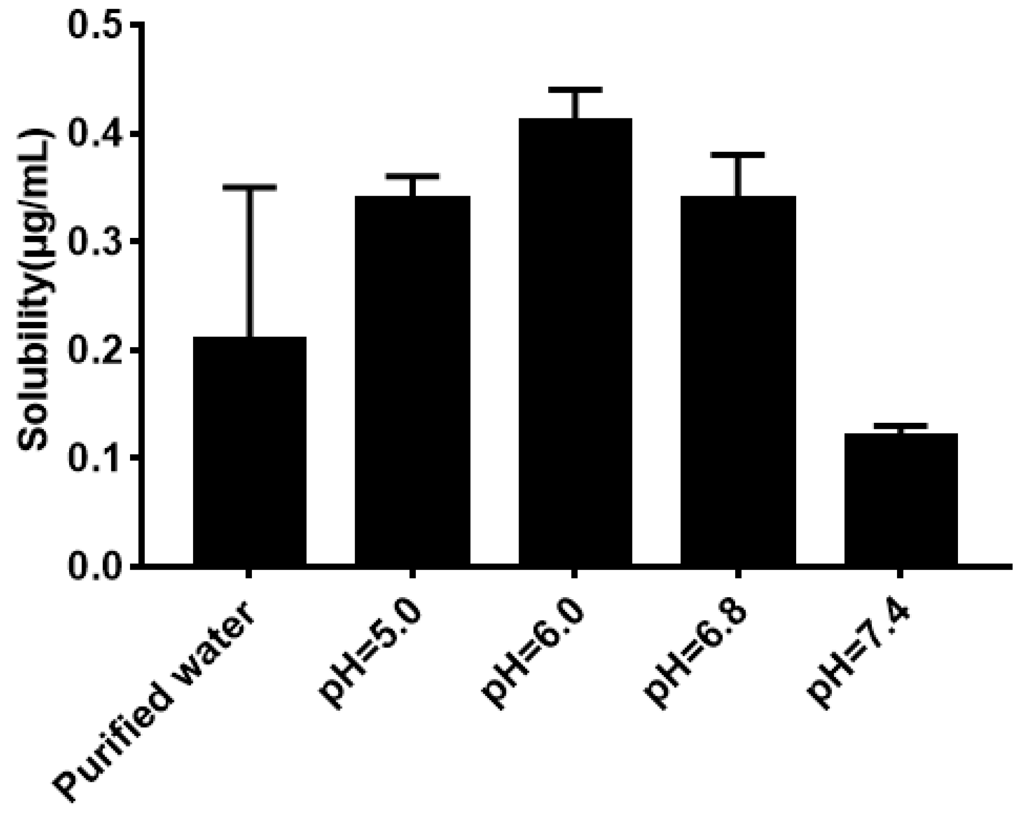
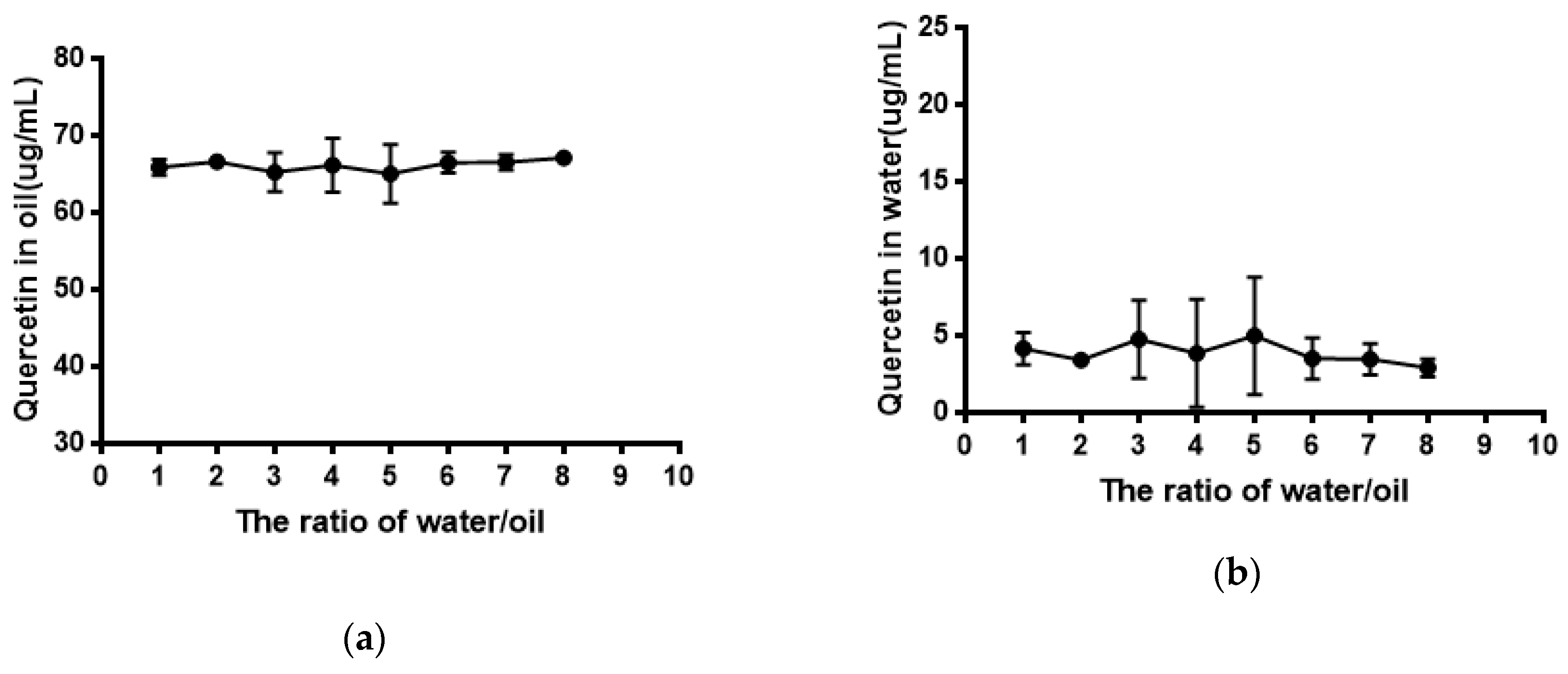
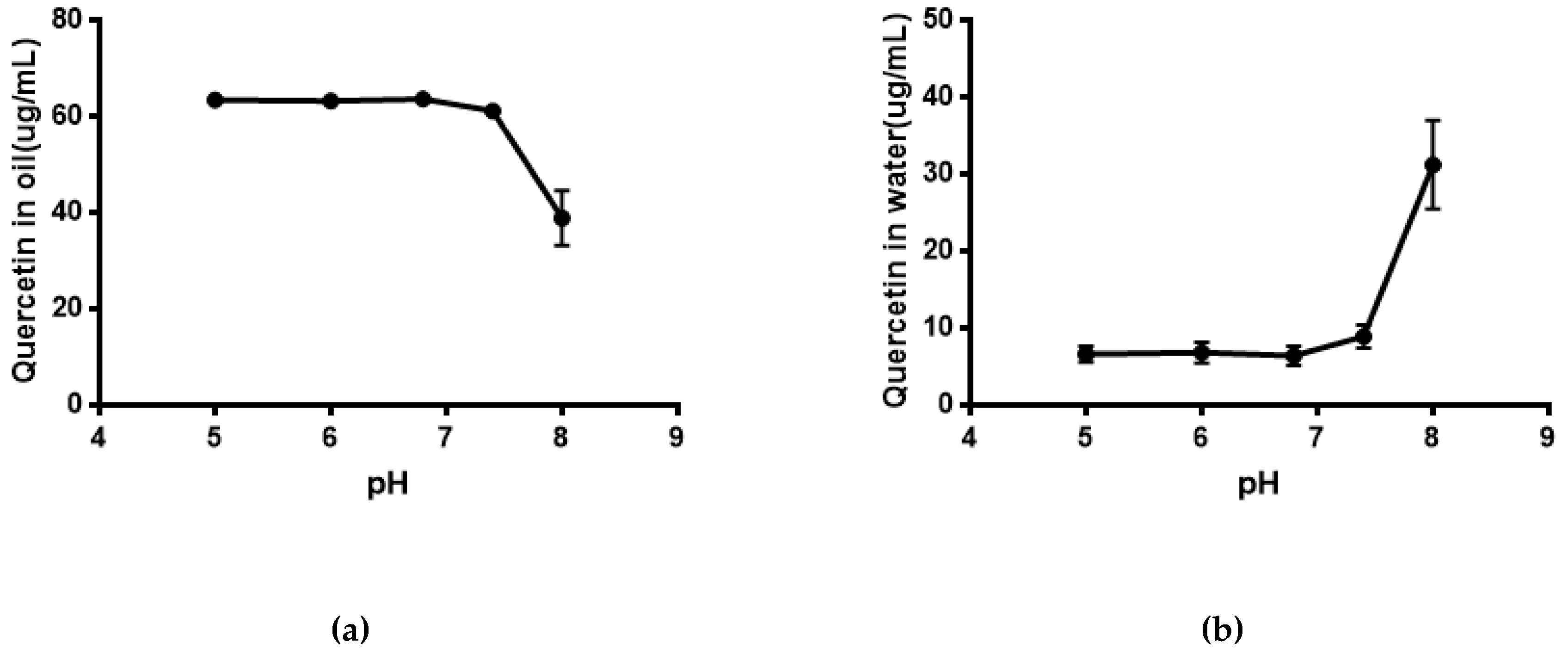
2.5. Water Solubility and Equilibrium Solubility of Quercetin
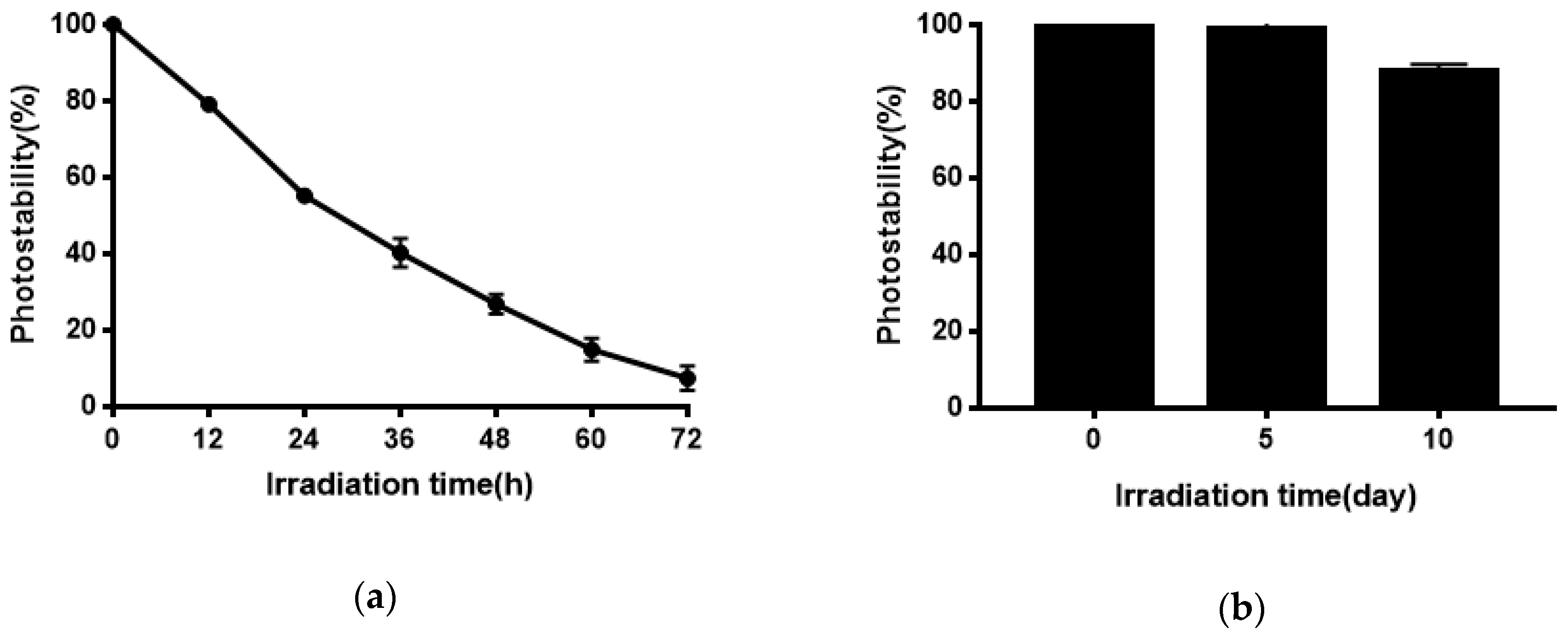
2.6. Photostability of Quercetin-Niosomes
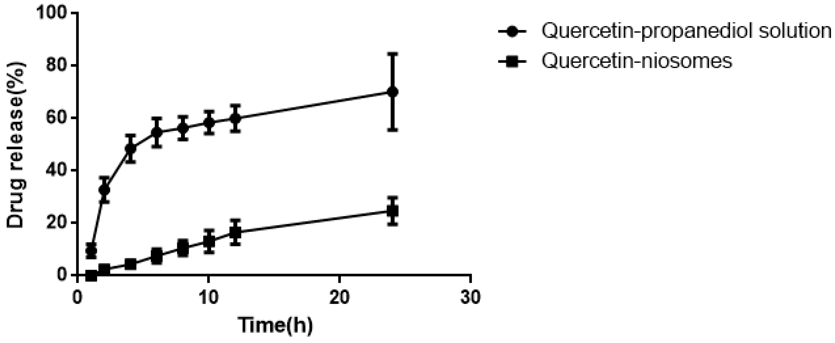
2.7. In Vitro Release Behavior of Quercetin-Niosomes
2.8. Ex Vivo Permeation and Skin Retention Studies
3. Discussion
4. Materials and Methods
4.1. Materials
4.2. In Vitro Tyrosinase Inhibition Activity of Flavonoids
4.3. In Vitro Antioxidant Activity of Flavonoids
4.4. Formulation of Niosomes
4.5. Preparation and Evaluation of Quercetin-Loaded Niosomes
4.5.1. Preparation of Quercetin-Loaded Niosomes
4.5.2. Size, Zeta Potential and Morphology of Quercetin-Loaded Niosomes
4.5.3. Drug Entrapment Efficiency
4.6. Water Solubility and Equilibrium Solubility of Quercetin
4.7. Photostability of Quercetin-Niosomes
4.8. In Vitro Release Study
4.8.1. The Solubility of Quercetin in Different Release Medium
4.8.2. Drug Release by Dialysis
4.9. Ex Vivo Drug Permeation and Skin Retention Studies
4.10. HPLC Method
4.11. Statistical Analysis
5. Conclusions
Author Contributions
Funding
Conflicts of Interest
References
- Chhikara, N.; Kaur, R.; Jaglan, S.; Sharma, R.; Gat, Y.; Panghal, A. Bioactive compounds and pharmacological and food applications of Syzygium cumini—A review. Food Funct. 2018, 9, 6096. [Google Scholar] [CrossRef] [PubMed]
- Carol López de Dicastillo Carol López Bustos, F.; Valenzuela, X.; López-Carballo, G.; Vilariño, J.M.; Galotto, M.J. Chilean berry, Ugni molinae, Turcz. Fruit and leaves extracts with interesting antioxidant, antimicrobial and tyrosinase inhibitory properties. Food Res. Int. 2017, 102, 119–128. [Google Scholar]
- Chiari, M.E.; Vera, D.M.; Palacios, S.M.; Carpinella, M.C. Tyrosinase inhibitory activity of a 6-isoprenoid-substituted flavanone isolated from Dalea elegans. Bioorg. Med. Chem. 2011, 19, 3474–3482. [Google Scholar] [CrossRef] [PubMed]
- Sirat, H.M.; Rezali, M.F.; Ujang, Z. Isolation and Identification of Radical Scavenging and Tyrosinase Inhibition of Polyphenols from Tibouchina semidecandra. J. Agric. Food Chem. 2010, 58, 10404–10409. [Google Scholar] [CrossRef] [PubMed]
- Xiong, Z.; Liu, W.; Zhou, L.; Chen, J. Mushroom (Agaricus bisporus) polyphenoloxidase inhibited by apigenin: Multi-spectroscopic analyses and computational docking simulation. Food Chem. 2016, 203, 430–439. [Google Scholar] [CrossRef] [PubMed]
- Yan-Zhen, Z.; Da-Fu, C.; Geng, D.; Guo, R. The Substituent Effect on the Radical Scavenging Activity of Apigenin. Molecules 2018, 23, 1989. [Google Scholar] [Green Version]
- Handjani-Vila, R.M.; Ribier, A.; Rondot, B.; Vanlerberghie, B. Dispersions of lamellar phases of non-ionic lipids in cosmetic products. Int. J. Cosmet. Sci. 1979, 1, 303–314. [Google Scholar] [CrossRef] [PubMed]
- Yoshida, H.; Lehr, C.M.; Kok, W.; Junginger, H.M.; Verhoef, J.C.; Bouwstra, J.A. Niosomes for oral delivery of peptide drugs. J. Control. Release 1992, 21, 145–153. [Google Scholar] [CrossRef]
- Uchegbu, I.F.; Double, J.A.; Kelland, L.R.; Turton, J.A.; Florence, A.T. The Activity of Doxorubicin Niosomes against an Ovarian Cancer Cell Line and Three in Vivo Mouse Tumour Models. J. Drug Target. 1996, 3, 399–409. [Google Scholar] [CrossRef]
- Alsarra, I.A.; Bosela, A.A.; Ahmed, S.M.; Mahrous, G.M. Proniosomes as a drug carrier for transdermal delivery of ketorolac. Eur. J. Pharm. Biopharm. 2005, 59, 485–490. [Google Scholar] [CrossRef]
- Hong, M.; Zhu, S.; Jiang, Y.; Tang, G.; Pei, Y. Efficient tumor targeting of hydroxycamptothecin loaded PEGylated niosomes modified with transferrin. J. Control. Release 2009, 133, 96–102. [Google Scholar] [CrossRef] [PubMed]
- Tavano, L.; Muzzalupo, R.; Picci, N.; de Cindio, B. Co-encapsulation of antioxidants into niosomal carriers: Gastrointestinal release studies for nutraceutical applications. Colloid Surf. B Biointerface 2014, 114, 82–88. [Google Scholar] [CrossRef] [PubMed]
- Sohrabi, S.; Haeri, A.; Mahboubi, A.; Mortazavi, A.; Dadashzadeh, S. Chitosan gel-embedded moxifloxacin niosomes: An efficient antimicrobial hybrid system for burn infection. Int. J. Biol. Macromol. 2016, 85, 625–633. [Google Scholar] [CrossRef] [PubMed]
- Kumar, N.; Goindi, S. Statistically designed nonionic surfactant vesicles for dermal delivery of itraconazole: Characterization and in vivo evaluation using a standardized Tinea pedis infection model. Int. J. Pharm. 2014, 472, 224–240. [Google Scholar] [CrossRef] [PubMed]
- Cerqueira-Coutinho, C.; Dos Santos, E.P.; Mansur, C.R. Niosomes as Nano-Delivery Systems in the Pharmaceutical Field. Crit. Rev. Ther. Drug Carr. Syst. 2016, 33, 195. [Google Scholar] [CrossRef]
- Yeh, M.I.; Huang, H.C.; Liaw, J.H.; Huang, M.C.; Huang, K.F.; Hsu, F.L. Dermal delivery by niosomes of black tea extract as a sunscreen agent. Int. J. Dermatol. 2013, 52, 239–245. [Google Scholar] [CrossRef] [PubMed]
- Manconi, M.; Sinico, C.; Valenti, D.; Loy, G.; Fadda, A.M. Niosomes as carriers for tretinoin. I. Preparation and properties. Int. J. Pharm. 2002, 234, 237–248. [Google Scholar] [CrossRef]
- Manconi, M.; Valenti, D.; Sinico, C.; Lai, F.; Loy, G.; Fadda, A.M. Niosomes as carriers for tretinoin: II. Influence of vesicular incorporation on tretinoin photostability. Int. J. Pharm. 2003, 260, 261–272. [Google Scholar] [CrossRef]
- Kunitake, T.; Okahata, Y. A totally synthetic bilayer membrane. J. Am. Chem. Soc. 1977, 99, 3860–3861. [Google Scholar] [CrossRef]
- Brady, J.E.; Evans, D.F.; Kachar, B.; Ninham, B.M. Spontaneous vesicles. J. Am. Chem. Soc. 1984, 106, 4279–4280. [Google Scholar] [CrossRef]
- Kaler, E.; Murthy, A.; Rodriguez, B.; Zasadzinski, J. Spontaneous vesicle formation in aqueous mixtures of single-tailed surfactants. Science 1989, 245, 1371–1374. [Google Scholar] [CrossRef] [PubMed]
- Kaler, E.W.; Herrington, K.L.; Murthy, A.K.; Zasadzinski, J.A.N. Phase behavior and structures of mixtures of anionic and cationic surfactants. J. Phys. Chem. 1992, 96, 6698–6707. [Google Scholar] [CrossRef]
- Uchegbu, I.F.; Vyas, S.P. Non-ionic surfactant based vesicles (niosomes) in drug delivery. Int. J. Pharm. 1998, 172, 33–70. [Google Scholar] [CrossRef]
- Mahale, N.B.; Thakkar, P.D.; Mali, R.G.; Walunj, D.R.; Chaudhari, S.R. Niosomes: Novel sustained release nonionic stable vesicular systems-An overview. Adv. Colloid Interface Sci. 2012, 183–184, 46–54. [Google Scholar] [CrossRef] [PubMed]
- Udupa, N.; Chandraprakash, K.S.; Umadevi, P.; Pillai, G.K. Formulation and Evaluation of Methotrexate Niosomes. Drug Dev. Commun. 1993, 19, 12. [Google Scholar] [CrossRef]
- Uchegbu, I.F.; Florence, A.T. Non-ionic surfactant vesicles (niosomes): Physical and pharmaceutical chemistry. Adv. Colloid Interface Sci. 1995, 58, 1–55. [Google Scholar] [CrossRef]
- Yoshioka, T.; Sternberg, B.; Florence, A.T. Preparation and properties of vesicles (niosomes) of sorbitan monoesters (Span 20, 40, 60 and 80) and a sorbitan triester (Span 85). Int. J. Pharm. 1994, 105, 1–6. [Google Scholar] [CrossRef]
- Manosroi, A.; Wongtrakul, P.; Manosroi, J.; Sakai, H.; Sugawara, F.; Yuasa, M.; Abe, M. Characterization of vesicles prepared with various non-ionic surfactants mixed with cholesterol. Colloid Surf. B Biointerface 2003, 30, 129–138. [Google Scholar] [CrossRef]
- Alsarra, I.A. Evaluation of proniosomes as an alternative strategy to optimize piroxicam transdermal delivery. J. Microencapsul. 2009, 26, 272–278. [Google Scholar] [CrossRef]
- Sharma, S.; Das, A.; Dasgupta, A.; Lee, W.M.; Kim, J.O.; Oh, D.H.; Kim, D.D.; Kim, J.S.; Yoo, B.Y.; Choi, H.G.; et al. Formulation and in vitro assessment of minoxidil niosomes for enhanced skin delivery. Int. J. Pharm. 2009, 377, 1–8. [Google Scholar]
- Junyaprasert, V.B.; Singhsa, P.; Suksiriworapong, J.; Chantasart, D. Physicochemical properties and skin permeation of Span 60/Tween 60 niosomes of ellagic acid. Int. J. Pharm. 2012, 423, 303. [Google Scholar] [CrossRef] [PubMed]
- Manosroi, A.; Khanrin, P.; Lohcharoenkal, W.; Götz, F.; Manosroi, W.; Manosroi, J. Transdermal absorption enhancement through rat skin of gallidermin loaded in niosomes. Int. J. Pharm. 2010, 392, 304–310. [Google Scholar] [CrossRef] [PubMed]
- Auda, S.H.; Fathalla, D.; Fetih, G.; El-Badry, M.; Shakeel, F. Niosomes as transdermal drug delivery system for celecoxib: In vitro and in vivo studies. Polym. Bull. 2016, 73, 1229–1245. [Google Scholar] [CrossRef]
- Manosroi, A.; Jantrawut, P.; Manosroi, J. Anti-inflammatory activity of gel containing novel elastic niosomes entrapped with diclofenac diethylammonium. Int. J. Pharm. 2008, 360, 156–163. [Google Scholar] [CrossRef] [PubMed]
- Shahiwala, A.; Misra, A. Studies in topical application of niosomally entrapped Nimesulide. J. Pharm. Pharm. Sci. 2002, 5, 220–225. [Google Scholar] [PubMed]
- Pardakhty, A.; Varshosaz, J.; Rouholamini, A. In vitro study of polyoxyethylene alkyl ether niosomes for delivery of insulin. Int. J. Pharm. 2007, 328, 130–141. [Google Scholar] [CrossRef]
- Abdelbary, G.; El-Gendy, N. Niosome-Encapsulated Gentamicin for Ophthalmic Controlled Delivery. Aaps Pharmscitech 2008, 9, 740–747. [Google Scholar] [CrossRef] [PubMed]
- Wadda Abbad, S.; Yu, F.; Munyendo, W.L.L.; Wang, J.; Lv, H.; Zhou, J. Formulation, characterization and pharmacokinetics of Morin hydrate niosomes prepared from various non-ionic surfactants. Int. J. Pharm. 2013, 456, 446–458. [Google Scholar] [CrossRef]
- Mishra, J.; Swain, J.; Mishra, A.K. Probing the Temperature Dependent Changes of the Interfacial Hydration and Viscosity of Tween20: Cholesterol (1:1) Niosome Membrane using Fisetin as a Fluorescent Molecular Probe. Phys. Chem. Chem. Phys. 2018, 20, 13279–13289. [Google Scholar] [CrossRef]
- Procházková, D.; Boušová, I.; Wilhelmová, N. Antioxidant and prooxidant properties of flavonoids. Fitoterapia 2011, 82, 513–523. [Google Scholar] [CrossRef]
- Cherrak, S.A.; Mokhtari-Soulimane, N.; Berroukeche, F.; Bensenane, B.; Cherbonnel, A.; Merzouk, H.; Elhabiri, M. In Vitro Antioxidant versus Metal Ion Chelating Properties of Flavonoids: A Structure-Activity Investigation. PLoS ONE 2016, 11, e0165575. [Google Scholar] [CrossRef] [PubMed]
- Amic, D.; Davidovic-Amic, D.; Beslo, D.; Rastija, V.; Lucic, B.; Trinajstic, N. SAR and QSAR of the Antioxidant Activity of Flavonoids. Curr. Med. Chem. 2007, 14, 827–845. [Google Scholar] [CrossRef] [PubMed]
- Machado, N.D.; Fernando, S.O.; De Rossi, R.H.; Fernandez, M.A. Cyclodextrin modified niosomes to encapsulate hydrophilic compounds. RSC Adv. 2018, 8, 29909–29916. [Google Scholar] [CrossRef] [Green Version]
- Imran, M.; Shah, M.R.; Ullah, F.; Ullah, S.; Elhissi, A.M.A.; Nawaz, W.; Ahmad, F.; Sadiq, A.; Ali, I. Glycoside-based niosomal nanocarrier for enhanced in-vivo performance of Cefixime. Int. J. Pharm. 2016, 505, 122–132. [Google Scholar] [CrossRef] [PubMed]
- Hao, Y.; Zhao, F.; Li, N.; Yang, Y.; Li, K. Studies on a high encapsulation of colchicine by a niosome system. Int. J. Pharm. 2002, 244, 73–80. [Google Scholar] [CrossRef]
- Anandam, S.; Selvamuthukumar, S. Fabrication of cyclodextrin nanosponges for Quercetin delivery: physicochemical characterization, photostability, and antioxidant effects. J. Mater. Sci. 2014, 49, 8140–8153. [Google Scholar] [CrossRef]
- Lv, X.; Liu, T.; Ma, H.; Tian, Y.; Li, L.; Li, Z.; Gao, M.; Zhang, J.; Tang, Z. Preparation of Essential Oil-Based Microemulsions for Improving the Solubility, pH Stability, Photostability, and Skin Permeation of Quercetin. AAPS PharmSciTech 2017, 18, 3097–3104. [Google Scholar] [CrossRef]
- Azzi, J.; Jraij, A.; Auezova, L.; Fourmentin, S.; Greige-Gerges, H. Novel findings for Quercetin encapsulation and preservation with cyclodextrins, liposomes, and drug-in-cyclodextrin-in-liposomes. Food HydroColloid 2018, 81, 328–340. [Google Scholar] [CrossRef]
- Smith, G.J.; Thomsen, S.J.; Markham, K.R.; Andary, C.; Cardon, D. The photostabilities of naturally occurring 5-hydroxyflavones, flavonols, their glycosides and their aluminium complexes. J. Photochem. Photobiol. A Chem. 2000, 136, 87–91. [Google Scholar] [CrossRef]
- Kubo, I.; Kinsthori, I. Flavonols from saffron flower: Tyrosinase inhibitory activity and inhibition mechanism. J. Agric. Food Chem. 1999, 47, 4121–4125. [Google Scholar] [CrossRef]
- Kim, Y.J.; Uyama, H. Tyrosinase inhibitors from natural and synthetic sources: Structure, inhibition mechanism and perspective for the future. Cell. Mol. Life Sci. 2005, 62, 1707–1723. [Google Scholar] [CrossRef] [PubMed]
- Kim, D.; Park, J.; Kim, J.; Han, C.; Yoon, J.; Kim, N.; Seo, J.; Lee, C. Flavonoids as Mushroom Tyrosinase Inhibitors: A Fluorescence Quenching Study. J. Agric. Food Chem. 2006, 54, 935–941. [Google Scholar] [CrossRef] [PubMed]
- Fan, M.; Zhang, G.; Hu, X.; Xu, X.; Gong, D. Quercetin as a tyrosinase inhibitor: Inhibitory activity, conformational change and mechanism. Food Res. Int. 2017, 100, 226–233. [Google Scholar] [CrossRef] [PubMed]
- Didem Şöhretoğlu Suat, S.; Burak, B.; Özel, A. Tyrosinase inhibition by some flavonoids: Inhibitory activity, mechanism by in vitro and in silico studies. Bioorg. Chem. 2018, 81, 168–174. [Google Scholar] [CrossRef] [PubMed]
- Gopinath, D.; Ravi, D.; Karwa, R.; Rao, B.; Shashank, A.; Rambhau, D. Pharmacokinetics of Zidovudine Following Intravenous Bolus Administration of a Novel Niosome Preparation devoid of Cholesterol. Arzneimittelforschung 2011, 51, 924–930. [Google Scholar] [CrossRef] [PubMed]
- Ghosh, S.; Ghatak, C.; Banerjee, C.; Mandal, S.; Kuchlyan, J.; Sarkar, N. Spontaneous Transition of Micelle-Vesicle-Micelle in a Mixture of Cationic Surfactant and Anionic Surfactant-like Ionic Liquid: A Pure Nonlipid Small Unilamellar Vesicular Template Used for Solvent and Rotational Relaxation Study. Langmuir 2013, 29, 10066–10076. [Google Scholar] [CrossRef]
- Sakai, T.; Ikoshi, R.; Toshida, N.; Kagaya, M. Thermodynamically Stable Vesicle Formation and Vesicle-to-Micelle Transition of Single-Tailed Anionic Surfactant in Water. J. Phys. Chem. B 2013, 117, 5081. [Google Scholar] [CrossRef]
- Ghosh, S.; Mondal, S.; Pan, A.; Mitra, R.K.; Ghosh, S. Ionic liquid mediated micelle to vesicle transition of a cationic gemini surfactant: A spectroscopic investigation. Soft Matter 2018, 14, 4185–4193. [Google Scholar]
- Mukherjee, B.; Patra, B.; Layek, B.; Mukherjee, A. Sustained release of acyclovir from nano-liposomes and nano-niosomes: An in vitro study. Int. J. Nanomed. 2007, 2, 213–225. [Google Scholar]
- Blois, M.S. Antioxidant Determinations by the Use of a Stable Free Radical. Nature 1958, 181, 1199–1200. [Google Scholar] [CrossRef]
- Rui, Y.; Ying, G.; Weixin, W.; Chen, H.; He, Z.; Jia, A.-Q. Antioxidant capacity of phenolics in Camellia nitidissima Chi flowers and their identification by HPLC Triple TOF MS/MS. PLoS ONE 2018, 13, e0195508. [Google Scholar]
Sample Availability: Samples of the compounds are not available from the authors. |


| Component | IC50 (μg/mL) 1 |
|---|---|
| Arbutin | 98.14 ± 1.45 |
| Quercetin | 1.59 ± 0.38 |
| Morin | 4.26 ± 0.47 |
| Festin | 23.10 ± 0.32 |
| Rutin | >1000 |
| Breviscapine | >1000 |
| Myricetin | — |
| Component | EC50 (μg/mL) 1 |
|---|---|
| Butylated hydroxytoluene (BHT) | 358.15 ± 1.43 |
| Quercetin | 80.75 ± 0.26 |
| Festin | 83.19 ± 0.33 |
| Myricetin | 118.63 ± 0.57 |
| Morin | 123.06 ± 0.48 |
| Rutin | 188.28 ± 0.68 |
| Breviscapine | 368.08 ± 1.04 |
| Sample | Size (nm) | Zeta Potential (mV) |
|---|---|---|
| Empty niosomes | 146.4 ± 5.4 | −41.2 ± 0.6 |
| Quercetin-niosomes | 97.6 ± 3.1 | −31.1 ± 0.9 |
| Medium | Solubility (μg/mL) |
|---|---|
| Water | 0.21 ± 0.14 |
| 0.6% Tween80 | 56.35 ± 1.42 * |
| 0.6% Sodium dodecyl sulfate(SDS) | 6.48 ± 0.44 * |
| Niosomes | 261.9 ± 4.8 * |
| Tween80 Aqueous Solution (μg/mL) | pH-0.8%Tween80 Aqueous Solution (μg/mL) | SDS Aqueous Solution (μg/mL) | |||
|---|---|---|---|---|---|
| 0.1% | 12.19 ± 1.05 | pH 5.0 | 79.49 ± 0.41 | 0.2% | 4.58 ± 0.04 |
| 0.2% | 24.72 ± 2.15 | ||||
| 0.3% | 31.37 ± 0.73 | pH 6.8 | 52.36 ± 0.71 | 0.4% | 4.22 ± 0.43 |
| 0.4% | 41.34 ± 0.19 | ||||
| 0.6% | 56.35 ± 1.42 | pH 7.4 | 55.02 ± 6.60 | 0.6% | 6.48 ± 0.44 |
| 0.8% | 76.07 ± 3.11 | ||||
| Quercetin Formulation | Skin Retention (μg/cm2) |
|---|---|
| Quercetin-propanediol | 0.65 ± 0.10 |
| Quercetin-niosomes | 1.92 ± 0.74 * |
| Quercetin-niosome-1%propanediol Quercetin-transfersomes | 2.34 ± 0.40 * 2.53 ± 0.40 * |
| Reaction System | Tube Label and Volume of Reaction Solution | |||
|---|---|---|---|---|
| A | B | C | D | |
| Phosphate buffer (mL) | 1.7 | 2.2 | 1.5 | 2 |
| Tyrosinase solution (mL) | 0.5 | 0 | 0.5 | 0 |
| Arbutin solution/Flavonoid solution (mL) | 0 | 0 | 0.5 | 0.5 |
| l-tyrosine (mL) | 0.5 | 0.5 | 0.5 | 0.5 |
| Dimethyl sulfoxide (DMSO) (mL) | 0.3 | 0.3 | - | - |
© 2019 by the authors. Licensee MDPI, Basel, Switzerland. This article is an open access article distributed under the terms and conditions of the Creative Commons Attribution (CC BY) license (http://creativecommons.org/licenses/by/4.0/).
Share and Cite
Lu, B.; Huang, Y.; Chen, Z.; Ye, J.; Xu, H.; Chen, W.; Long, X. Niosomal Nanocarriers for Enhanced Skin Delivery of Quercetin with Functions of Anti-Tyrosinase and Antioxidant. Molecules 2019, 24, 2322. https://doi.org/10.3390/molecules24122322
Lu B, Huang Y, Chen Z, Ye J, Xu H, Chen W, Long X. Niosomal Nanocarriers for Enhanced Skin Delivery of Quercetin with Functions of Anti-Tyrosinase and Antioxidant. Molecules. 2019; 24(12):2322. https://doi.org/10.3390/molecules24122322
Chicago/Turabian StyleLu, Banyi, Yanting Huang, Zhongyun Chen, Jingyi Ye, Hongyu Xu, Wenrong Chen, and Xiaoying Long. 2019. "Niosomal Nanocarriers for Enhanced Skin Delivery of Quercetin with Functions of Anti-Tyrosinase and Antioxidant" Molecules 24, no. 12: 2322. https://doi.org/10.3390/molecules24122322