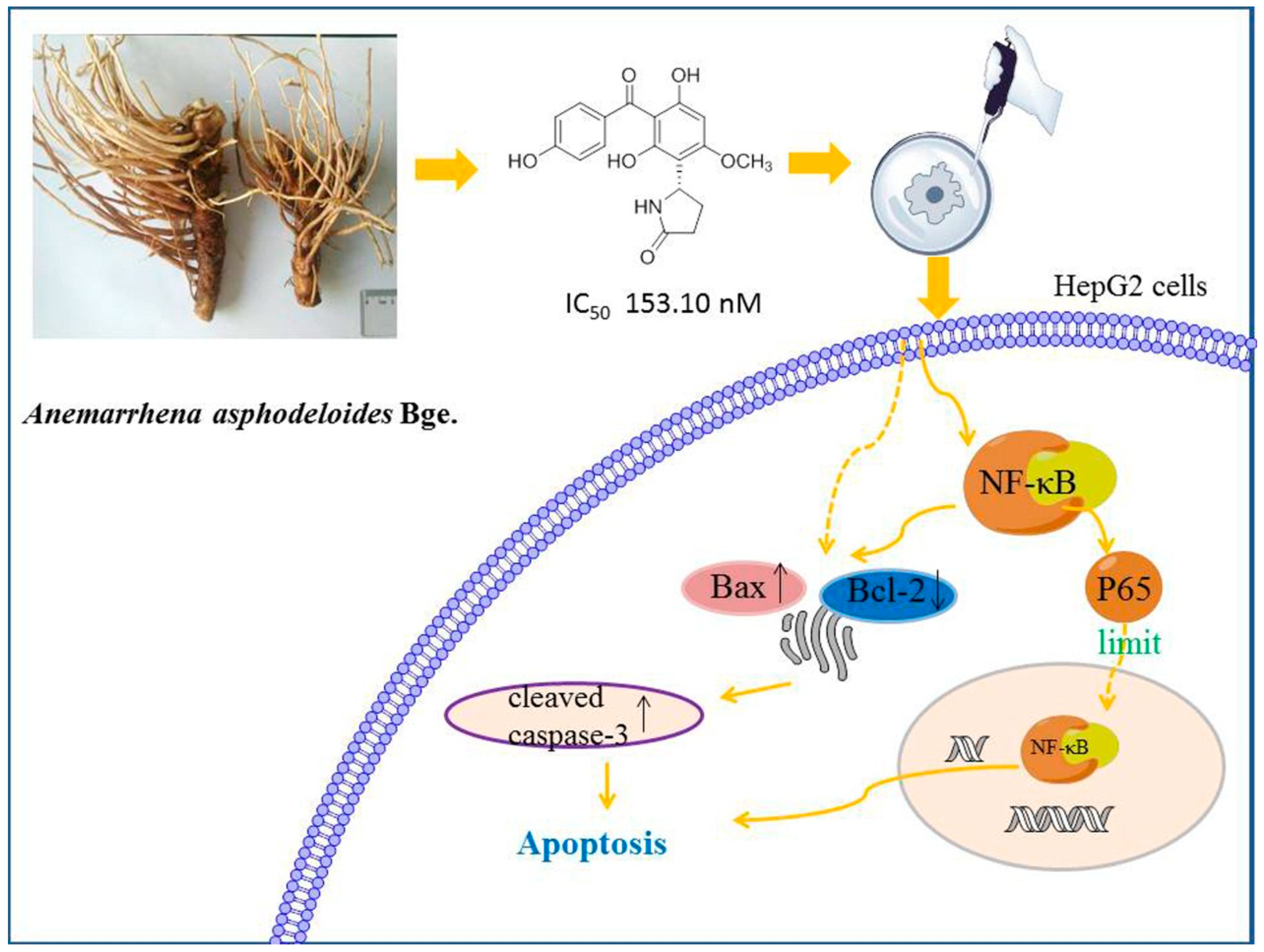
Benzophenones from Anemarrhena asphodeloides Bge. Exhibit Anticancer Activity in HepG2 Cells via the NF-κB Signaling Pathway

 and
and
Abstract
:1. Introduction
2. Results
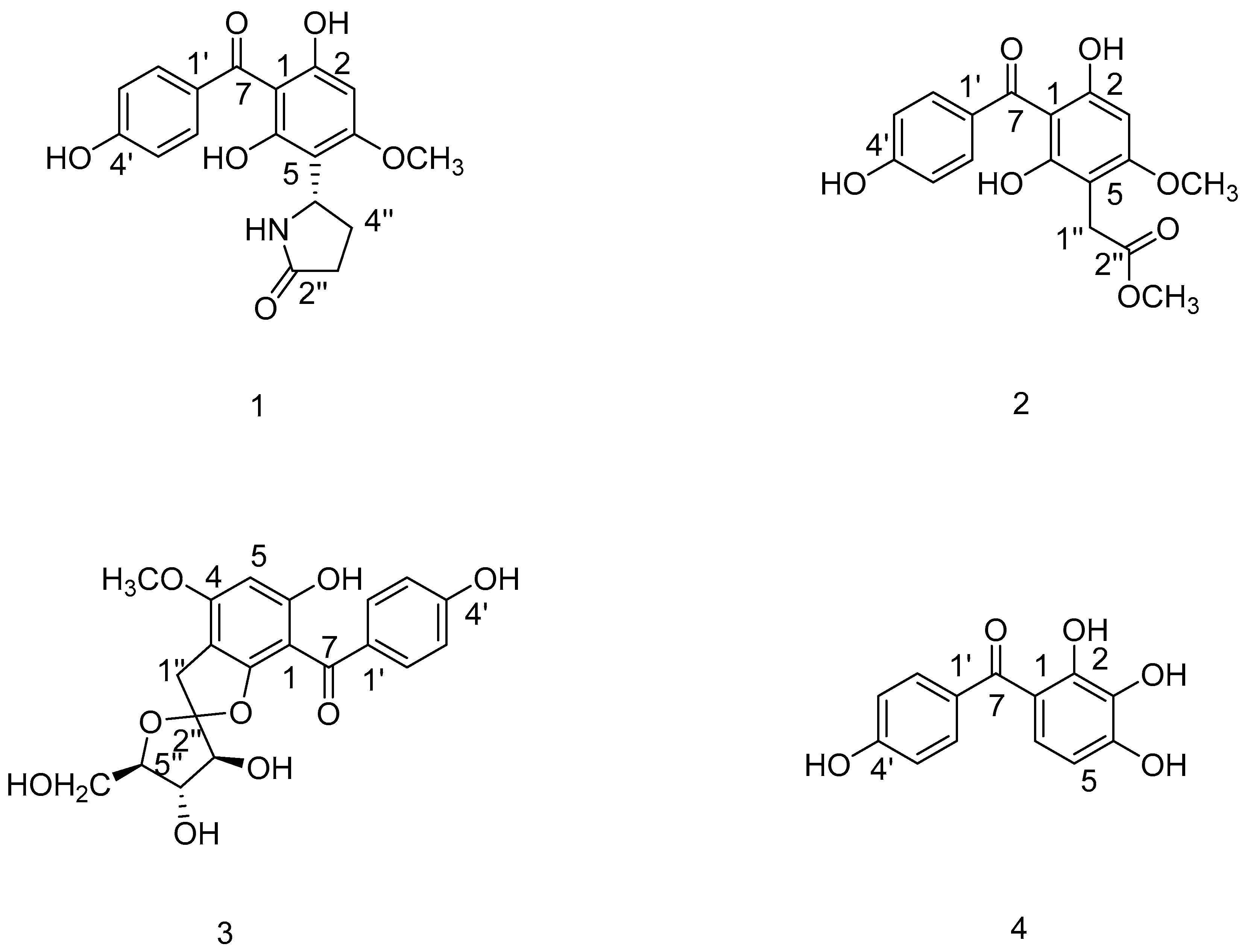
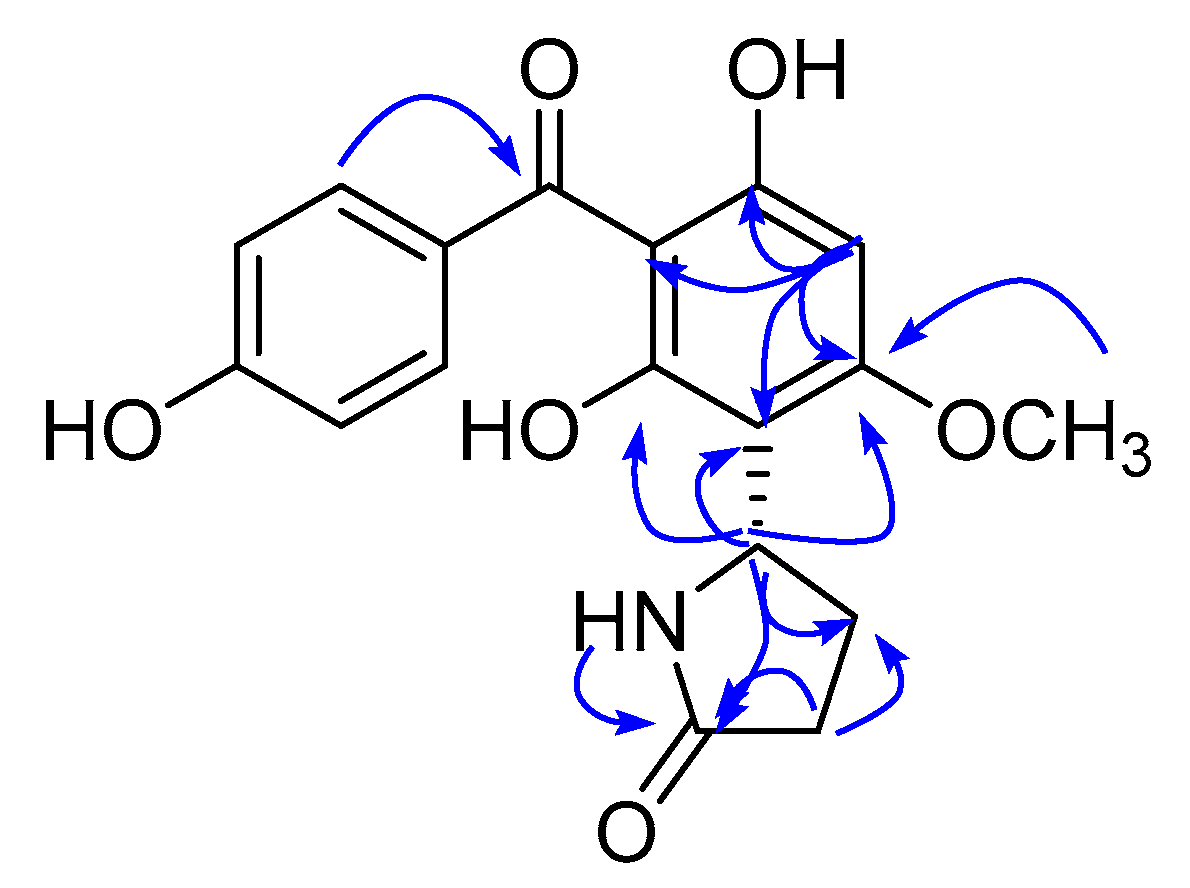
2.1. Identification of Compounds 1–4 from the Fibrous Roots of A. asphodeloides Bge.
2.2. Cytotoxicity In Vitro
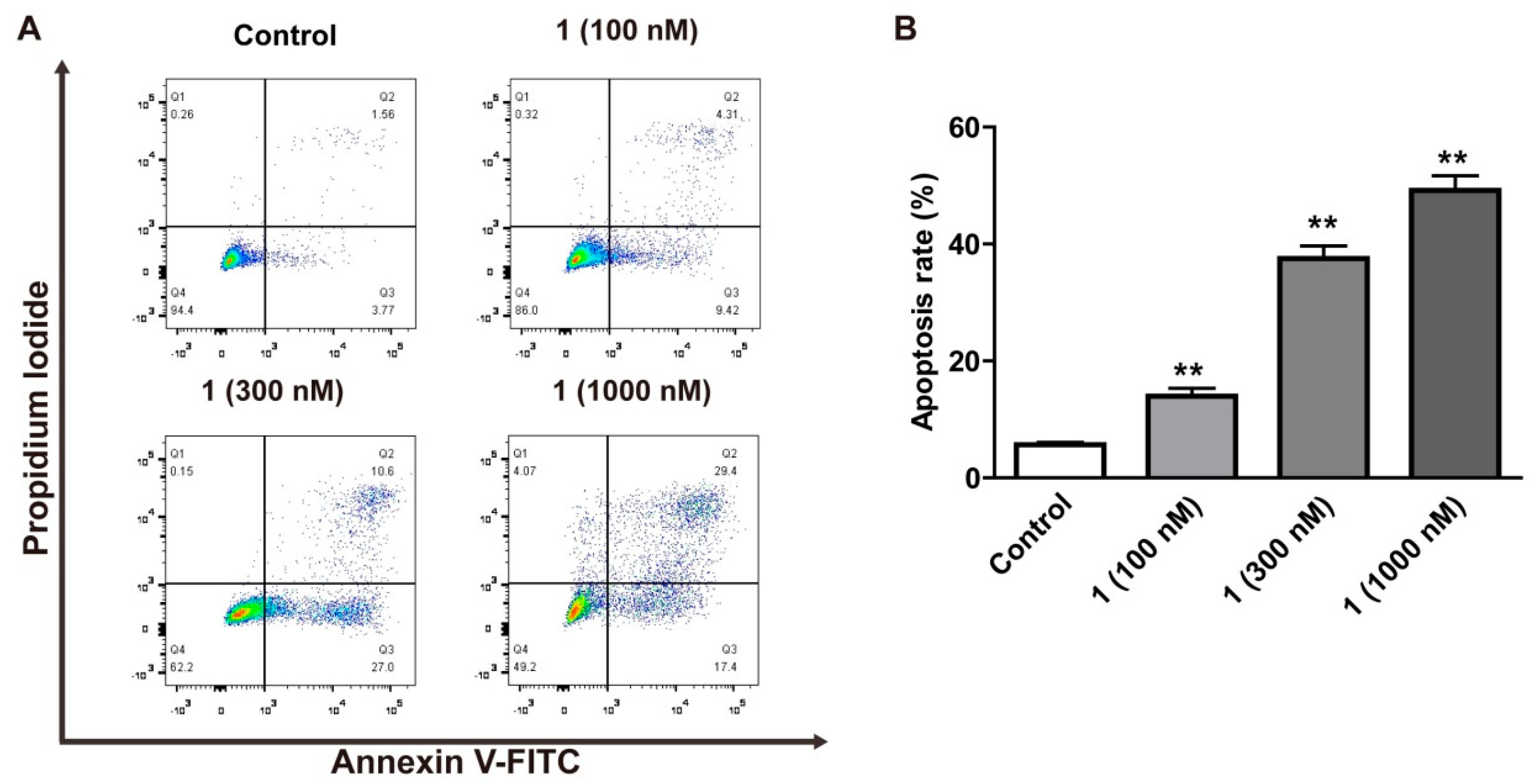
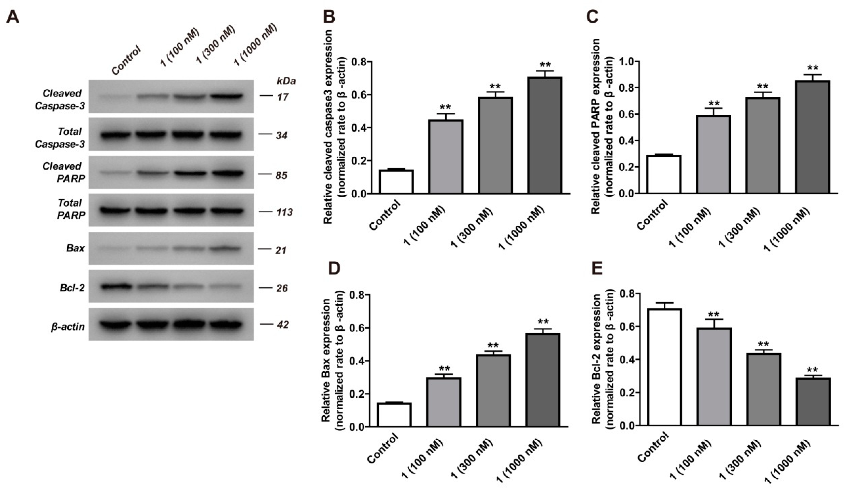
2.3. Apoptosis Study
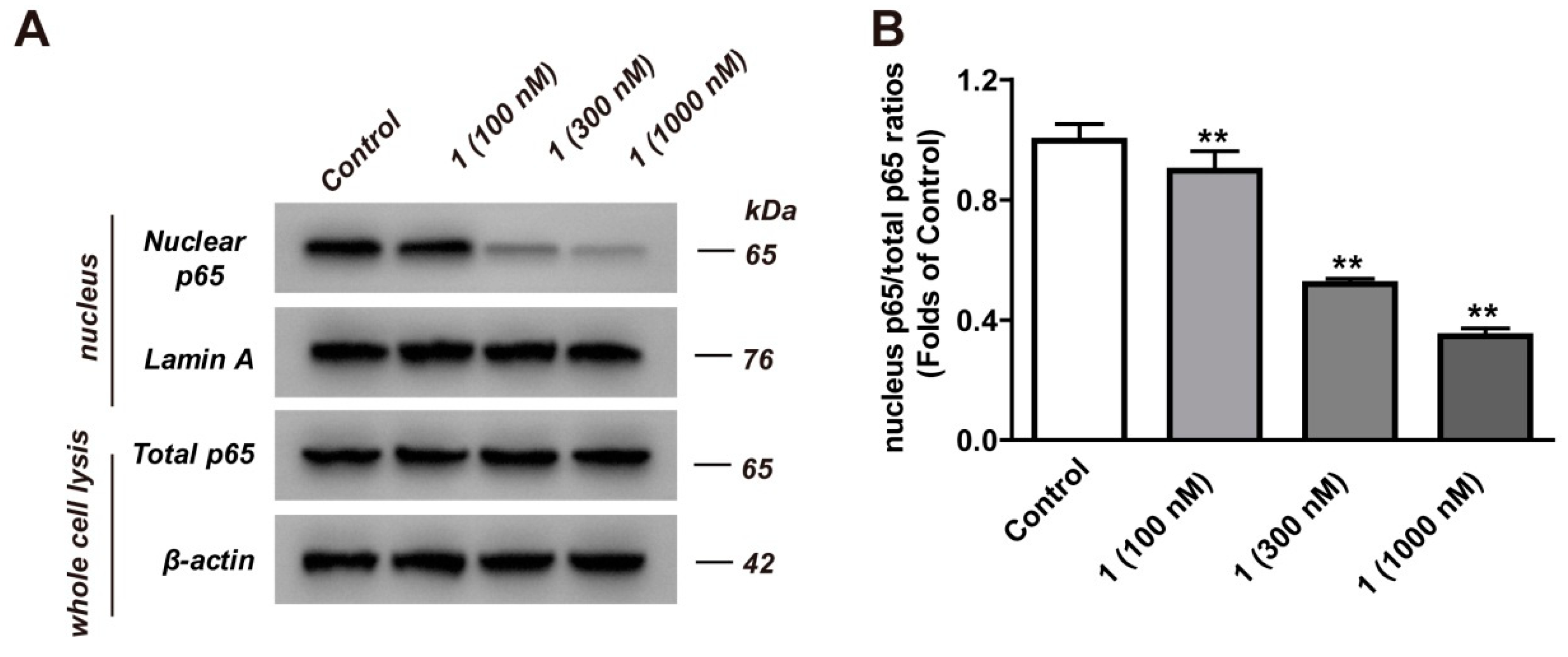
2.4. Inhibition Analysis of NF-κB
3. Discussion
4. Materials and Methods
4.1. General Experimental Procedures
4.2. Plant Material
4.3. Extraction and Isolation
4.4. Characterization of Compound 1
4.5. Optical Rotation Calculations
4.6. Cell Culture
4.7. MTT Assay
4.8. Flow Cytometric Analysis
4.9. Western Blot Analysis
4.10. Statistical Analysis
5. Conclusions
Supplementary Materials
Author Contributions
Funding
Conflicts of Interest
References
- Kim, S.; Yang, Y.; Seki, E. Inflammation and Liver Cancer: Molecular Mechanisms and Therapeutic Targets. Semin. Liver Dis. 2019, 39, 26–42. [Google Scholar]
- Saber, S.; Mahmoud, A.A.A.; Goda, R.; Helal, N.S.; El-ahwany, E.; Abdelghany, R.H. Perindopril, fosinopril and losartan inhibited the progression of diethylnitrosamine-induced hepatocellular carcinoma in mice via the inactivation of nuclear transcription factor kappa-B. Toxicol. Lett. 2018, 295, 32–40. [Google Scholar] [CrossRef] [PubMed]
- Mohan, C.D.; Anilkumar, N.C.; Rangappa, S.; Shanmugam, M.K.; Mishra, S.; Chinnathambi, A.; Alharbi, S.A.; Bhattacharjee, A.; Sethi, G.; Kumar, A.P.; et al. Novel 1,3,4-Oxadiazole Induces Anticancer Activity by Targeting NF-kappaB in Hepatocellular Carcinoma Cells. Front. Oncol. 2018, 8, 42. [Google Scholar] [CrossRef] [PubMed]
- Li, W.; Tan, D.; Zenali, M.J.; Brown, R.E. Constitutive activation of nuclear factor-kappa B (NF-κB) signaling pathway in fibrolamellar hepatocellular carcinoma. Int. J. Clin. Exp. Pathol. 2010, 3, 238–243. [Google Scholar]
- Wan, L.F.; Shen, J.J.; Wang, Y.H.; Zhao, W.; Fang, N.Y.; Yuan, X.; Xue, B.Y. Extracts of Qizhu decoction inhibit hepatitis and hepatocellular carcinoma in vitro and in C57BL/6 mice by suppressing NF-κB signaling. Sci. Rep. 2019, 9, 1415. [Google Scholar] [CrossRef] [PubMed]
- Bettermann, K.; Vucur, M.; Haybaeck, J.; Koppe, C.; Janssen, J.; Heymann, F.; Weber, A.; Weiskirchen, R.; Liedtke, C.; Gassler, N.; et al. TAK1 Suppresses a NEMO-Dependent but NF-κB-Independent Pathway to Liver Cancer. Cancer Cell 2010, 17, 481–496. [Google Scholar] [CrossRef]
- Dong, M.; Miao, L.; Zhang, F.; Li, S.; Han, J.; Yu, R.; Qie, S. Nuclear factor-&kappa B p65 regulates glutaminase 1 expression in human hepatocellular carcinoma. Oncotargets Ther. 2018, 11, 3721–3729. [Google Scholar]
- Li, Q.; Zhang, Y.; Sun, J.; Bo, Q. Paeonol-mediated apoptosis of hepatocellular carcinoma cells by NF-κB pathway. Oncol. Lett. 2019, 17, 1761–1767. [Google Scholar] [CrossRef]
- Van Thanh, N.; Jang, H.J.; Vinh, L.B.; Linh, K.T.P.; Huong, P.T.T.; Cuong, N.X.; Nam, N.H.; Van Minh, C.; Kim, Y.H.; Yang, S.Y. Chemical constituents from Vietnamese mangrove Calophyllum inophyllum and their anti-inflammatory effects. Bioorganic Chem. 2019, 88, 102921. [Google Scholar] [CrossRef]
- Yao, G.D.; Sun, Q.; Song, X.Y.; Huang, X.X.; Zhang, Y.; Song, S.J. 1,3-Diphenylpropanes from Daphne giraldii induced apoptosis in hepatocellular carcinoma cells through nuclear factor kappa-B inhibition. Bioorganic Chem. 2018, 77, 619–624. [Google Scholar] [CrossRef]
- Tian, J.L.; Yao, G.D.; Zhang, Y.Y.; Lin, B.; Zhang, Y.; Li, L.Z.; Huang, X.X.; Song, S.J. Pyran-2-one derivatives from Croton crassifolius as potent apoptosis inducers in HepG2 cells via p53-mediated Ras/Raf/ERK pathway. Bioorganic Chem. 2018, 79, 355–362. [Google Scholar] [CrossRef] [PubMed]
- Han, F.Y.; Song, X.Y.; Chen, J.J.; Yao, G.D.; Song, S.J. Timosaponin AIII: A novel potential anti-tumor compound from Anemarrhena asphodeloides. Steroids 2018, 140, 125–130. [Google Scholar] [CrossRef] [PubMed]
- Zhao, Y.F.; Zhang, Y.W.; Wang, Y.; Morris-Natschke, S.L.; Liu, W.; Shang, T.T.; Yin, H.; Lee, K.H.; Huang, X.F. New transformation pathway and cytotoxic derivatives from the acid hydrolysis of timosaponin B III. Nat. Prod. Res. 2018, 1–7. [Google Scholar] [CrossRef] [PubMed]
- Liao, Z.D.; Chen, F.F.; Xu, F.Q.; Zhang, W.; Ren, Y.S.; Wang, C.C.; Wu, D.L. One new benzophenone and one new 1,3-diphenylpropane from the fibrous roots of Anemarrhena asphodeloides Bge. and their cytotoxicity. Nat. Prod. Res. 2019, 1–7. [Google Scholar] [CrossRef] [PubMed]
- Zhao, J.; Yang, T.; Ji, J.; Li, C.; Li, Z.; Li, L. Garcinol exerts anti-cancer effect in human cervical cancer cells through upregulation of T-cadherin. Biomed. Pharmacother. Biomed. Pharmacother. 2018, 107, 957–966. [Google Scholar] [CrossRef] [PubMed]
- Kim, S.; Seo, S.; Min, K.J.; Woo, S.; Nam, J.O.; Kubatka, P.; Kim, S.; Park, J.W.; Kwon, T. Garcinol Enhances TRAIL-Induced Apoptotic Cell Death through Up-Regulation of DR5 and Down-Regulation of c-FLIP Expression. Molecules 2018, 23, 1614. [Google Scholar] [CrossRef] [PubMed]
- Kokotkiewicz, A.; Luczkiewicz, M.; Sowinski, P.; Glod, D.; Gorynski, K.; Bucinski, A. Isolation and structure elucidation of phenolic compounds from Cyclopia subternata Vogel (honeybush) intact plant and in vitro cultures. Food Chem. 2012, 133, 1373–1382. [Google Scholar] [CrossRef]
- Li, Y.; Zhang, F.; Wu, Z.H.; Zeng, K.W.; Zhang, C.; Jin, H.W.; Zhao, M.B.; Jiang, Y.; Li, J.; Tu, P.F. Nitrogen-containing bibenzyls from Pleione bulbocodioides: Absolute configurations and biological activities. Fitoterapia 2015, 102, 120–126. [Google Scholar] [CrossRef] [PubMed]
- Liao, Z.D.; Xu, F.Q.; Wu, D.L.; Zhang, W.; Huang, Q. A new benzophenone isolated from fibrous roots of Anemarrhena asphodeloides. China J. Chin. Mater. Med. 2019, 44, 1392–1396. [Google Scholar]
- Pan, J.; Yi, X.; Wang, Y.; Chen, G.; He, X. Benzophenones from Mango Leaves Exhibit alpha-Glucosidase and NO Inhibitory Activities. J. Agric. Food Chem. 2016, 64, 7475–7480. [Google Scholar] [CrossRef] [PubMed]
- Cetinkaya, Y.; Gocer, H.; Menzek, A.; Gulcin, I. Synthesis and antioxidant properties of (3,4-dihydroxyphenyl)(2,3,4-trihydroxyphenyl)methanone and its derivatives. Arch. Pharm. 2012, 345, 323–334. [Google Scholar] [CrossRef] [PubMed]
- Zhang, H.; Li, J.C.; Luo, H.; Zhao, L.; Zhang, Z.D.; Shen, X.F. Pseudolaric acid B exhibits anti-cancer activity on human hepatocellular carcinoma through inhibition of multiple carcinogenic signaling pathways. Phytomed. Int. J. Phytother. Phytopharm. 2018, 59, 152759. [Google Scholar] [CrossRef] [PubMed]
- Valizadeh, A.; Majidinia, M.; Samadi-Kafil, H.; Yousefi, M.; Yousefi, B. The roles of signaling pathways in liver repair and regeneration. J. Cell. Physiol. 2019. [Google Scholar] [CrossRef] [PubMed]
- Moles, A.; Butterworth, J.A.; Sanchez, A.; Hunter, J.E.; Leslie, J.; Sellier, H.; Tiniakos, D.; Cockell, S.J.; Mann, D.A.; Oakley, F.; et al. A RelA(p65) Thr505 phospho-site mutation reveals an important mechanism regulating NF-kappaB-dependent liver regeneration and cancer. Oncogene 2016, 35, 4623–4632. [Google Scholar] [CrossRef] [PubMed]
- Sahu, S.K.; Chawla, Y.K.; Dhiman, R.K.; Singh, V.; Duseja, A.; Taneja, S.; Kalra, N.; Gorsi, U. Rupture of Hepatocellular Carcinoma: A Review of Literature. J. Clin. Exp. Hepatol. 2019, 9, 245–256. [Google Scholar] [CrossRef] [PubMed]
- Jiang, X.; Tan, H.Y.; Teng, S.; Chan, Y.T.; Wang, D.; Wang, N. The Role of AMP-Activated Protein Kinase as a Potential Target of Treatment of Hepatocellular Carcinoma. Cancers 2019, 11, 647. [Google Scholar] [CrossRef]
- Tsuchiya, N.; Sawada, Y.; Endo, I.; Saito, K.; Uemura, Y.; Nakatsura, T. Biomarkers for the early diagnosis of hepatocellular carcinoma. World J. Gastroenterol. 2015, 21, 10573–10583. [Google Scholar] [CrossRef]
- Baig, B.; Halim, S.A.; Farrukh, A.; Greish, Y.; Amin, A. Current status of nanomaterial-based treatment for hepatocellular carcinoma. Biomed. Pharmacother. 2019, 116, 108852. [Google Scholar] [CrossRef]
- Dimitroulis, D.; Damaskos, C.; Valsami, S.; Davakis, S.; Garmpis, N.; Spartalis, E.; Athanasiou, A.; Moris, D.; Sakellariou, S.; Kykalos, S.; et al. From diagnosis to treatment of hepatocellular carcinoma: An epidemic problem for both developed and developing world. World J. Gastroenterol. 2017, 23, 5282. [Google Scholar] [CrossRef]
- Han, M.; Gao, H.; Xie, J.; Yuan, Y.P.; Yuan, Q.; Gao, M.Q.; Liu, K.L.; Chen, X.H.; Han, Y.T.; Han, Z.W. Hispidulin induces ER stress-mediated apoptosis in human hepatocellular carcinoma cells in vitro and in vivo by activating AMPK signaling pathway. Acta Pharmacol. Sin. 2019, 40, 666–676. [Google Scholar] [CrossRef]
- Harvey, A.L.; Edrada-Ebel, R.; Quinn, R.J. The re-emergence of natural products for drug discovery in the genomics era. Nat. Rev. Drug Discov. 2015, 14, 111–129. [Google Scholar] [CrossRef] [PubMed] [Green Version]
- Ee, G.C.L.; Teh, S.S.; Kwong, H.C.; Mah, S.H.; Lim, Y.M.; Rahmani, M. A new benzophenone from Mesua congestiflora, an inhibitor against human B lymphocyte cancer cell line. Phytochem. Lett. 2012, 5, 545–548. [Google Scholar] [CrossRef]
- Kalantzopoulos, G.N.; Lundvall, F.; Checchia, S.; Lind, A.; Wragg, D.S.; Fjellvag, H.; Arstad, B. In Situ Flow MAS NMR Spectroscopy and Synchrotron PDF Analyses of the Local Response of the Bronsted Acidic Site in SAPO-34 during Hydration at Elevated Temperatures. Chemphyschem A Eur. J. Chem. Phys. Phys. Chem. 2018, 19, 519–528. [Google Scholar] [CrossRef] [PubMed]
- Li, M.; Zhang, M.; Zhang, Z.L.; Liu, N.; Han, X.Y.; Liu, Q.C.; Deng, W.J.; Liao, C.X. Induction of Apoptosis by Berberine in Hepatocellular Carcinoma HepG2 Cells via Downregulation of NF-kappaB. Oncol. Res. 2017, 25, 233–239. [Google Scholar] [CrossRef] [PubMed]
- Yao, B.R.; Sun, Y.; Chen, S.L.; Suo, H.D.; Zhang, Y.L.; Wei, H.; Wang, C.H.; Zhao, F.; Cong, W.; Xin, W.Y.; et al. Dissymmetric pyridyl-substituted 3,5-bis(arylidene)-4-piperidones as anti-hepatoma agents by inhibiting NF-κB pathway activation. Eur. J. Med. Chem. 2019, 167, 187–199. [Google Scholar] [CrossRef] [PubMed]
- Luedde, T.; Schwabe, R.F. NF-κB in the liver—Linking injury, fibrosis and hepatocellular carcinoma. Nat. Rev. Gastroenterol. Hepatol. 2011, 8, 108–118. [Google Scholar] [CrossRef]
- Park, J.S.; Choi, H.I.; Kim, D.H.; Kim, C.S.; Bae, E.H.; Ma, S.K.; Kim, S.W. Alpha-lipoic acid attenuates p-cresyl sulfate-induced renal tubular injury through suppression of apoptosis and autophagy in human proximal tubular epithelial cells. Biomed. Pharmacother. 2019, 112, 108679. [Google Scholar] [CrossRef]
- Liu, H.; Li, G.; Zhang, B.; Sun, D.; Wu, J.; Chen, F.; Kong, F.; Luan, Y.; Jiang, W.; Wang, R.; et al. Suppression of the NF-kappaB signaling pathway in colon cancer cells by the natural compound Riccardin D from Dumortierahirsute. Mol. Med. Rep. 2018, 17, 5837–5843. [Google Scholar]
- Li, J.; Wang, Q.; Wang, Z.; Cui, N.; Yang, B.; Niu, W.; Kuang, H. Tetrandrine inhibits colon carcinoma HT-29 cells growth via the Bcl-2/Caspase 3/PARP pathway and G1/S phase. Biosci. Rep. 2019, 39. [Google Scholar] [CrossRef]
- Yan, Z.Y.; Chen, J.J.; Duan, Z.K.; Yao, G.D.; Lin, B.; Wang, X.B.; Huang, X.X.; Song, S.J. Racemic phenylpropanoids from the root barks of Ailanthus altissima (Mill.) Swingle with cytotoxicity against hepatoma cells. Fitoterapia 2018, 130, 234–240. [Google Scholar] [CrossRef]
- Park, H.S.; Jo, E.; Han, J.H.; Jung, S.H.; Lee, D.H.; Park, I.; Heo, K.S.; Na, M.; Myung, C.S. Hepatoprotective effects of an Acer tegmentosum Maxim extract through antioxidant activity and the regulation of autophagy. J. Ethnopharmacol. 2019, 239, 111912. [Google Scholar] [CrossRef] [PubMed]
Sample Availability: Samples of the compounds are available from the authors. |


| Positions | δH (J in Hz) | δC |
|---|---|---|
| 1 | - | 106.8 |
| 2 | - | 158.3 |
| 3 | 6.08 (1H, s, H-3) | 91.5 |
| 4 | - | 161.5 |
| 5 | - | 108.6 |
| 6 | - | 157.8 |
| 7 | - | 195.8 |
| 1′ | - | 130.8 |
| 2′, 6′ | 7.53 (2H, d, J = 8.7 Hz) | 131.6 |
| 3′, 5′ | 6.78 (2H, d, J = 8.7 Hz) | 114.6 |
| 4′ | - | 161.8 |
| 1″ | - | - |
| 2″ | - | 176.8 |
| 3″ | 2.32 (1H, m) 2.16 (1H, m) | 30.7 |
| 4″ | 2.28 (1H, m) 2.01 (1H, m) | 25.8 |
| 5″ | 5.11 (1H, d, J = 4.9, 9.3 Hz) | 46.9 |
| 4-OCH3 | 3.73 (3H, s) | 55.5 |
© 2019 by the authors. Licensee MDPI, Basel, Switzerland. This article is an open access article distributed under the terms and conditions of the Creative Commons Attribution (CC BY) license (http://creativecommons.org/licenses/by/4.0/).
Share and Cite
Wu, D.-L.; Liao, Z.-D.; Chen, F.-F.; Zhang, W.; Ren, Y.-S.; Wang, C.-C.; Chen, X.-X.; Peng, D.-Y.; Kong, L.-Y. Benzophenones from Anemarrhena asphodeloides Bge. Exhibit Anticancer Activity in HepG2 Cells via the NF-κB Signaling Pathway. Molecules 2019, 24, 2246. https://doi.org/10.3390/molecules24122246
Wu D-L, Liao Z-D, Chen F-F, Zhang W, Ren Y-S, Wang C-C, Chen X-X, Peng D-Y, Kong L-Y. Benzophenones from Anemarrhena asphodeloides Bge. Exhibit Anticancer Activity in HepG2 Cells via the NF-κB Signaling Pathway. Molecules. 2019; 24(12):2246. https://doi.org/10.3390/molecules24122246
Chicago/Turabian StyleWu, De-Ling, Zhen-Dong Liao, Fang-Fang Chen, Wei Zhang, Ya-Shuo Ren, Can-Can Wang, Xiao-Xiao Chen, Dai-Yin Peng, and Ling-Yi Kong. 2019. "Benzophenones from Anemarrhena asphodeloides Bge. Exhibit Anticancer Activity in HepG2 Cells via the NF-κB Signaling Pathway" Molecules 24, no. 12: 2246. https://doi.org/10.3390/molecules24122246