Article

Antioxidant and Hepatoprotective Effect of Penthorum chinense Pursh Extract against t-BHP-Induced Liver Damage in L02 Cells

State Key Laboratory of Quality Research in Chinese Medicine, Institute of Chinese Medical Sciences, University of Macau, Macau 999078, China
*
Authors to whom correspondence should be addressed.
These authors contributed equally to this work.
Molecules 2015, 20(4), 6443-6453; https://doi.org/10.3390/molecules20046443
Submission received: 13 February 2015 / Revised: 21 March 2015 / Accepted: 26 March 2015 / Published: 10 April 2015
(This article belongs to the Section Natural Products Chemistry)

Abstract

:
Penthorum chinense Pursh (P. chinense), a traditional Chinese medicine used by the Chinese Miao minority, has been used to treat liver diseases for a long time. However, the mechanism behind the liver protective effects of P. chinense remains unclear so far. The aim of the present study was to investigate the hepatoprotective effect of P. chinense and its possible mechanism(s). Immortalized normal human normal liver L02 cells were used to evaluate the protective effect of P. chinense aqueous extract against tert-butyl hydroperoxide (t-BHP)-induced liver cell damage. Treatment with P. chinense aqueous extract significantly protected L02 cells from t-BHP-induced cytotoxicity, prevented t-BHP-induced reactive oxygen species (ROS) generation and decreased the percentage of apoptosis by inhibiting the mitochondrial apoptotic pathway. This study demonstrates that P. chinense is a potential hepatoprotective agent in t-BHP-induced liver cell damage, which may benefit the further application of P. chinense in the clinic.

1. Introduction

Penthorum chinense Pursh (P. chinense), called Ganhuangcao in Chinese, belongs to the family Saxifragaceae and is widely distributed in eastern Asia, including China, Japan, Korea, and eastern Russia [1]. Daily consumption of P. chinense is recorded to be related with a decreased of liver diseases in Gulin country, Sichuan Province. Its favorable therapeutic effects in treating jaundice, cholecystitis, edema, traumatic injury and infectious hepatitis are also confirmed. Therefore, Gansu granules, a Chinese formulation, is prepared from P. chinense extracts and has been used in the clinic as a remedy for chronic hepatitis B and acute virus hepatitis for its action against the hepatitis B, C, and D viruses [2]. However, P. chinense has not been subjected to any detailed chemical constitution analysis and the mechanism of the liver protective effect of P. chinense remains unclear.
Production of reactive oxygen species (ROS) is implicated in normal aerobic cellular metabolism [3]. Generally, ROS production is counterbalanced by the antioxidant defense system to maintain an appropriate redox balance [4,5]. Oxidative stress, which is a physiological status whereby intracellular free radicals exceed the antioxidant abilities, has been recognized as a key factor in the pathogenesis of several chronic liver diseases, such as hepatitis, alcoholic and non-alcoholic fatty liver diseases [6,7]. The liver’s unique metabolic functions and relationships to the gastrointestinal tract make it vulnerable to the toxicity of drugs and xenobiotics [8,9]. Therefore, antioxidant therapy may be one of the strategies to correct the imbalance between oxidants and antioxidants in development of these liver diseases and prevent hepatocytes from excessive exposure to oxidative stress.
tert-Butyl hydroperoxide (t-BHP) is commonly used to induce oxidative stress in vitro and in vivo, and the induction of apoptosis in hepatocytes is a critical feature of t-BHP [10]. It can be metabolized to free radical intermediates, which further cause lipid peroxidation, GSH depletion and DNA damage [11]. Though P. chinense was reported to possess potent antioxidant capabilities [2,12,13], it still remains unclear whether the hepatoprotective effect of P. chinense could be verified by an ability to decrease t-BHP-induced oxidative stress and consequent apoptosis. Therefore, the objective of the present study was to investigate the potential protective effect and mechanism of P. chinense against oxidative stress induced by t-BHP in human liver L02 cells.

2. Results and Discussion

Oxidative stress is a situation whereby intracellular ROS/reactive nitrogen species (RNS) levels overwhelm the cellular antioxidant abilities and is reported to be associated with more than 100 diseases [3,8]. Basically, a normal ROS/RNS level and oxidative stress is essential for cell survival, while high levels of ROS/RNS or severe oxidative stress will inevitably weaken the cells’ self-repair capacity and lead to cell death thereof [3,14]. As the major organ responsible for the regulation of extensive physiological processes, the liver is a representative and target organ of the toxicity of drugs and xenobiotics due to its continuous exposure to these toxicants [9,15]. Most hepatotoxic chemicals can increase the production of free radicals that cause oxidative stress, which is considered as one of the important mechanisms that mediate liver toxicity [16]. P. chinense, a Miao ethnic medicine, has been applied for treatment of liver diseases for many years. We postulated that P. chinense could protect the liver from cell injury induced by t-BHP, a well-known ROS inducer, based on previous studies indicating that P. chinense possesses ROS scavenging activity [2,12].

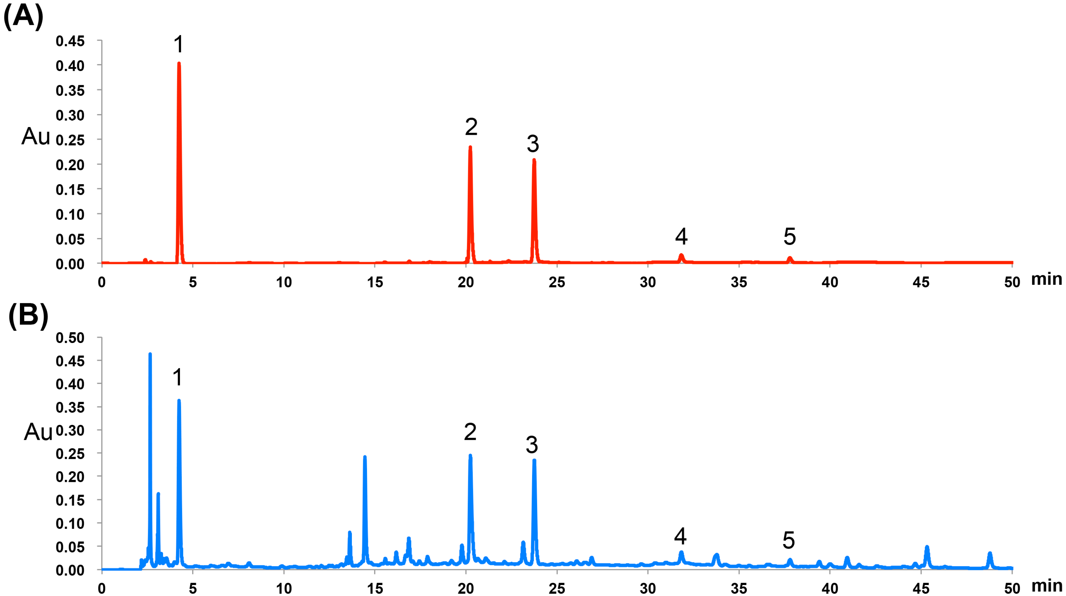
2.1. Chemical Characteristics of P. chinense Extract

Previous studies showed that P. chinense is rich in polyphenols, which possess strong antioxidant activities [12,17]. In order to investigate the chemical characteristics of P. chinense extract used in present study, HPLC analysis of P. chinense extract was carried out and confirmed the dominant presence of polyphenols in the P. chinense extract as anticipated (Figure 1). The polyphenols were identified by comparison of the retention times with authentic mixed polyphenol standards. Five peaks were identified as gallic acid, isoquercitrin, quercitrin, quercetin and kaempferol, which is consistent with previous reports [17]. The contents of the five compounds were quantified using corresponding chemical standards. Specifically, the contents of gallic acid, isoquercitrin, quercitrin, quercetin and kaempferol in P. chinense extract were 5.50, 14.1, 10.4, 0.8 and 0.1 mg/g, respectively.
Figure 1. Representative HPLC-UV chromatograms of mixed standards (A) and P. chinense extract (B). Gallic acid (1), isoquercitrin (2), quercitrin (3), quercetin (4) and kaemferol (5).
Molecules 20 06443 g001

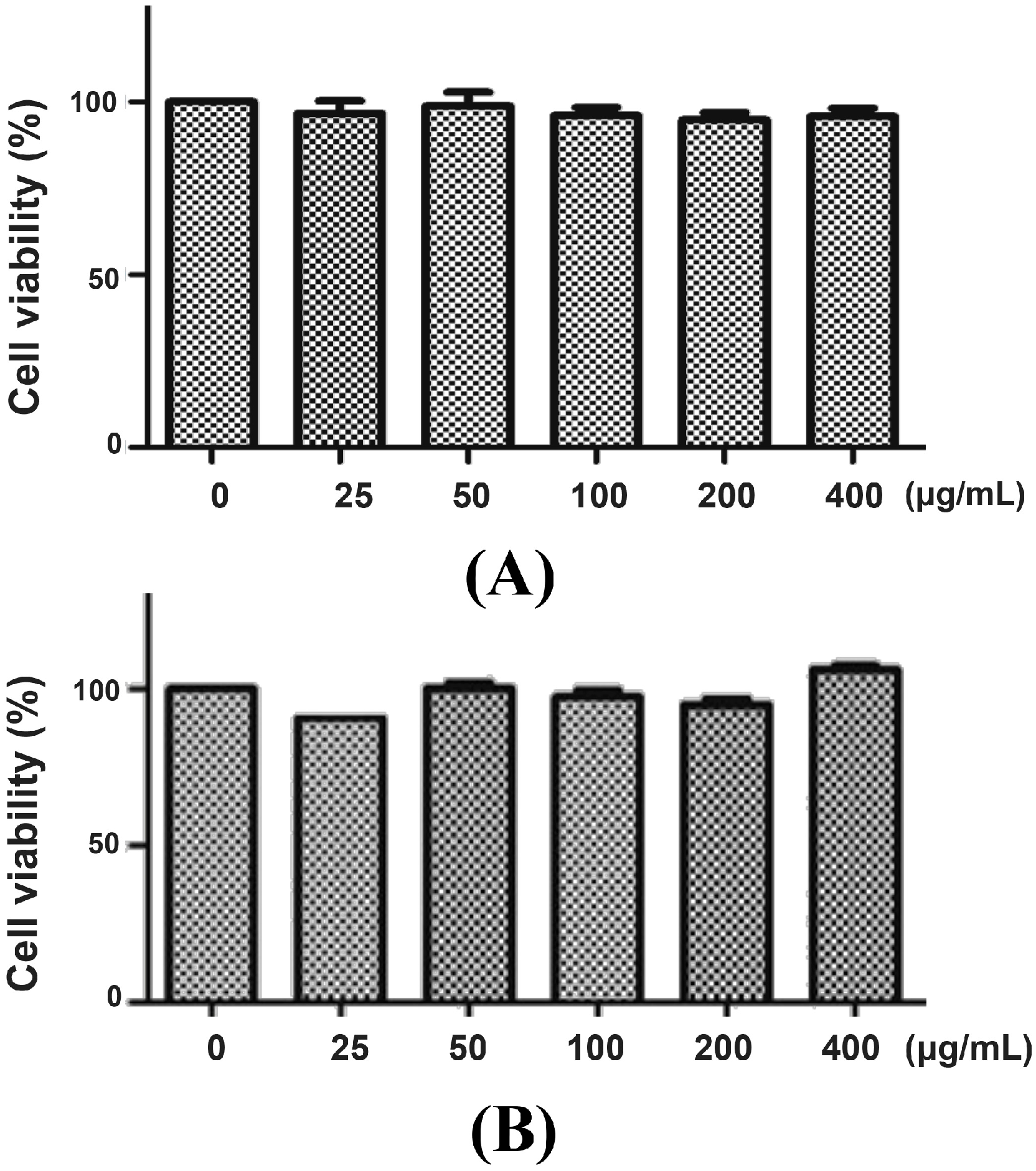
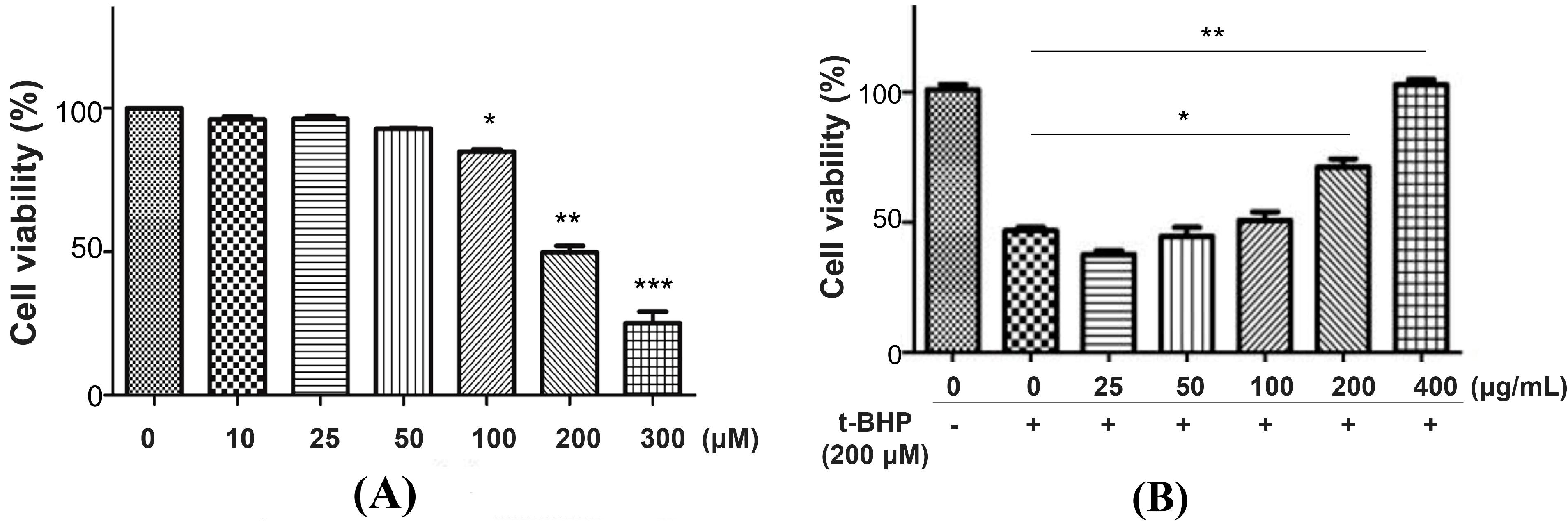
2.2. Protective Effect of P. chinense Extract on t-BHP-Induced Cytotoxicity in L02 Cells

We first determined the cytotoxicity of P. chinense extract in L02 cells. After 12 h (Figure 2A) and 24 h (Figure 2B) treatment, P. chinense extract showed negligible toxic effect on L02 cells even at high concentration (400 µg/mL). Then, t-BHP, a potent pro-oxidant, was used to induce oxidative damage in L02 cells [18]. As shown in Figure 3A, t-BHP (25 to 400 µM) treatment for 6 h evoked the decrease of L02 cell viability in a concentration-dependent manner, with an IC50 value of 205.8 µM. A 6 h treatment of 200 µM t-BHP, which induced almost 50% decrease of cell viability, was used to evaluate the potential hepatoprotective ability of P. chinense extract in the subsequent studies.
Figure 2. Cytotoxicity of P. chinense extract on L02 cells. L02 cells were treated with different concentrations of P. chinense extract for 12 h (A) and 24 h (B), then cell viability were assessed by MTT assay. Data are expressed as means ± SEM of at least three independent experiments.
Molecules 20 06443 g002
Pretreatment of L02 cells with 25, 50 and 100 µg/mL P. chinense extract for 12 h showed weak protective effect on t-BHP-induced cytotoxicity, while 200 µg/mL of P. chinense extract significantly reduced cell damage. A higher dose of P. chinense extract (400 µg/mL) further reduced cell damage to values similar to those of control cells (Figure 3B), indicating the strong protective activity of P. chinense extract against t-BHP-induced cytotoxicity in L02 cells.
Figure 3. The protective effects of P. chinense extract on t-BHP-induced cytotoxicity in L02 cells. (A) L02 cells were treated with different concentrations of t-BHP for 6 h. * p < 0.05, ** p < 0.01 and *** p < 0.001 as compared to untreated control cells. (B) L02 cells were pretreated with different concentrations of P. chinense extract for 12 h, followed by treatment with 200 µM t-BHP for another 6 h. Cell viability was assessed by MTT assay. Data are expressed as means ± SEM of at least three independent experiments. Con indicates control. * p < 0.05 and ** p < 0.01.
Molecules 20 06443 g003

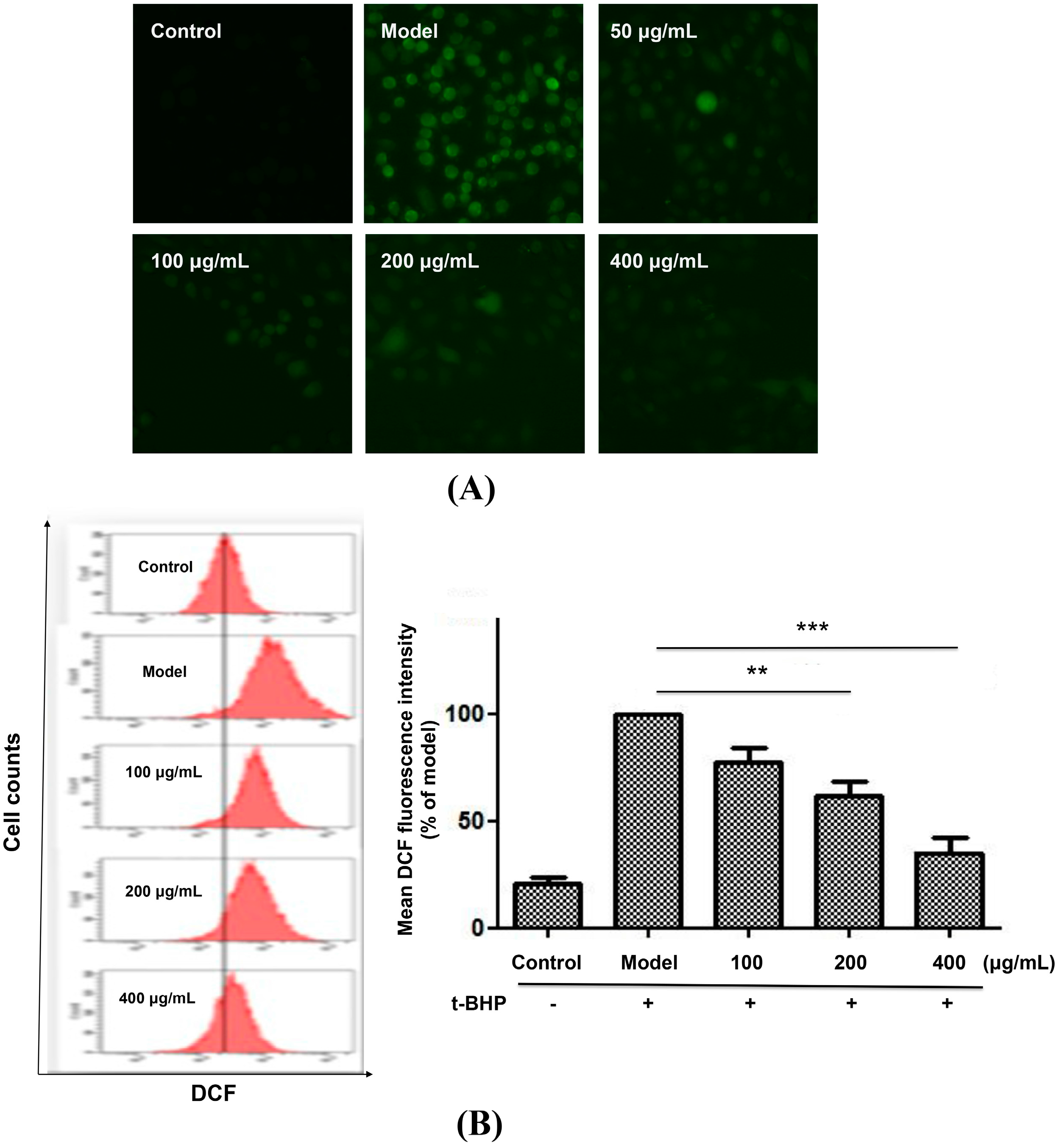
2.3. P. chinense Extract Decreased ROS Generation in L02 Cells

It is well known that ROS play an important role in cell apoptosis [19]. Measurement of intracellular ROS levels is a common indication of the oxidative damage to living cells. To investigate whether the protective effect of P. chinense extract on t-BHP-induced cytotoxicity was attributed to the intracellular ROS levels, we further determined the intracellular ROS levels using a ROS-specific dye (CM-H2DCFDA). The regulation of ROS generation was evaluated in L02 cells treated by t-BHP with or without P. chinense extract pretreatment. As shown in Figure 4A, t-BHP treatment significantly increased the intracellular ROS level compared with that of untreated controls, while pretreated with P. chinense extract greatly decreased ROS production to those of untreated cells.
Figure 4. P. chinense extract attenuated t-BHP-induced ROS generation in L02 cells. L02 cells were pretreated with different concentrations of P. chinense extract for 12 h, followed by exposition to t-BHP for 30 min, and then incubated with 1 µM (CM-H2DCF) for another 30 min. The intracellular ROS levels were measured by In Cell Analyzer 2000 (A) and flow cytometry (B). Data are expressed as means ± SEM of at least three independent experiments. ** p < 0.01 and *** p < 0.001. Magnification 200×.
Molecules 20 06443 g004
Flow cytometry studies further indicated that, compared with model group, pretreated with 100, 200 and 400 µg/mL P. chinense extract obviously decreased the ROS levels to 77.48%, 61.90% and 35.25% of the model cells (Figure 4B), respectively. These results clearly showed that P. chinense extract strongly inhibits the generation of ROS induced by t-BHP, thus preventing or delaying conditions that favor oxidative stress in liver cells.

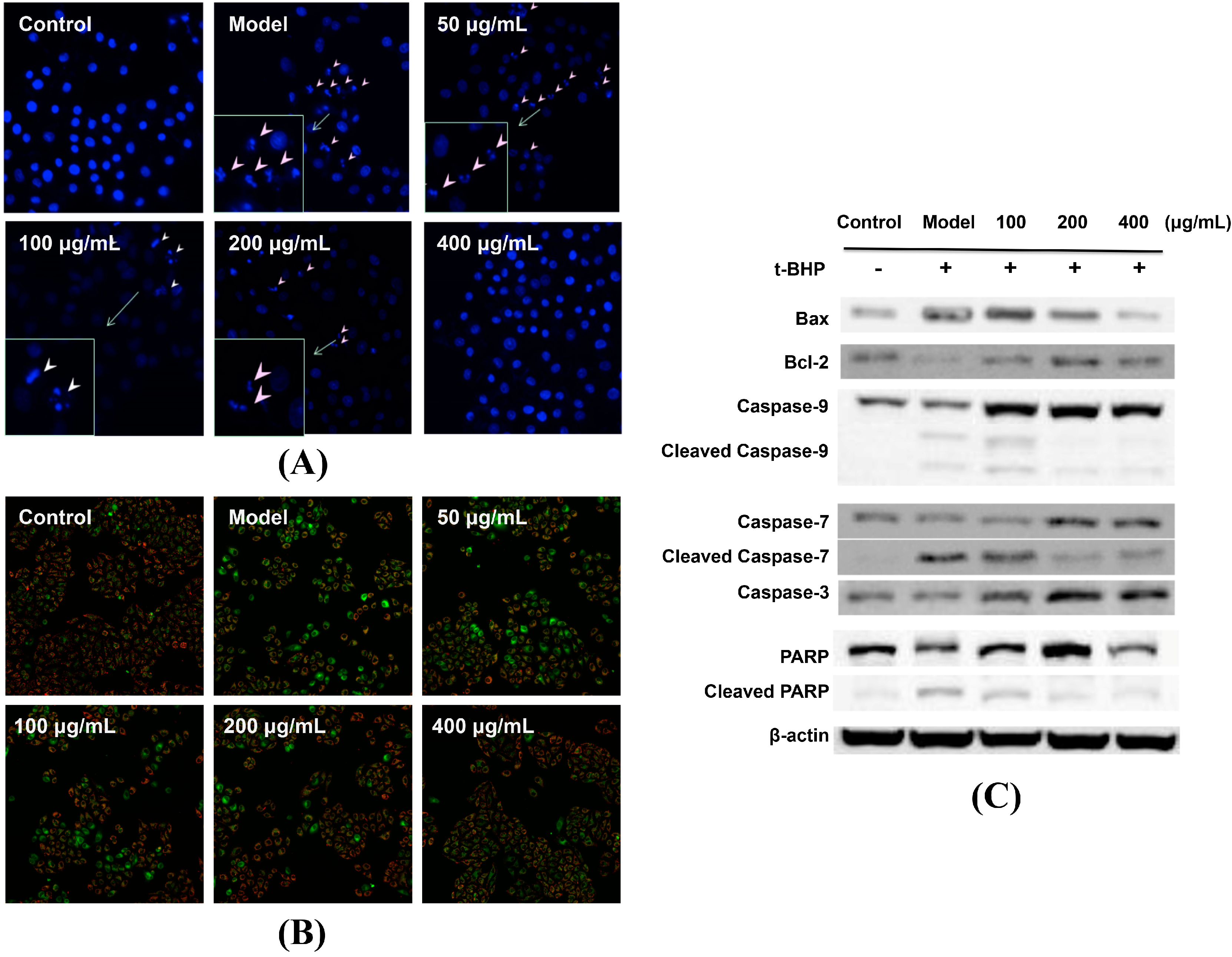
2.4. P. chinense Extract Attenuated t-BHP-Induced Apoptosis

The induction of apoptosis in hepatocytes by t-BHP is a critical feature of oxidative agents [20]. We further evaluate the effect of P. chinense extract on the apoptosis of L02 cells induced by t-BHP. The depolarization of MMP is an essential event for detecting initial apoptosis [21]. In this study, we used JC-1 as the fluorescent dye and estimated the change of MMP through the intensity weaken of JC-1 aggregates, which may reduce as MMP decreases. As shown in Figure 5A, t-BHP treatment greatly increased the green florescence, suggesting the significant decrease of MMP. However, cells pretreated with P. chinense extract inhibited the decrease of MMP by a concentration-dependent manner (Figure 5A). Hochest 33342 staining also showed that L02 cells pretreated with P. chinense extract reduced condensed, fragmented or degraded cell nuclei as compared to t-BHP-treated group (Figure 5B).
Figure 5. P. chinense extract attenuated t-BHP-induced apoptosis by inhibiting mitochondrial apoptotic pathway in L02 cells. (A) L02 cells were pretreated with different concentrations of P. chinense extract for 12 h, followed by exposition to t-BHP for 30 min, and then incubated with JC-1 dye for another 30 min. The cell MMP was measured using In Cell Analyzer 2000. (B) L02 cells were treated as described above and cell nuclei were stained with Hoechst 33342 and imaged using an In Cell Analyzer 2000. (C) L02 cells were treated as described above and were harvested for western blotting analysis using indicated antibodies. Similar results were obtained from two or three independent experiments. Magnification 200×.
Molecules 20 06443 g005
In order to investigate the protective mechanisms of P. chinense extract, the expression of several apoptosis-related proteins were determined by western blotting in L02 cells. Caspases as potent executors of apoptosis exist as zymogens in normal cells and undergo proteolytic processing via several pathways [22]. PARP is involved in many cellular processes including DNA repair and cell apoptosis [23], and cleaved PARP is recognized as a marker of apoptosis. Generally, apoptosis pathways can be divided into death receptors pathway and mitochondria pathway. ROS are predominantly produced in mitochondria. It has been reported that the mitochondria-dependent apoptotic pathway is involved in t-BHP-induced cytotoxicity, in which the Bcl-2 family proteins play a key role [24]. The anti-apoptotic Bcl-2 bind to the outer membrane of the mitochondria can stabilize the membrane permeability and prevent the release of cytochrome c [25], while the pro-apoptotic members like Bax are responsible for permeabilizing membrane under cellular stress [26,27]. t-BHP treatment increased the expression of Bax and cleaved products of caspase-9, caspase-7 and PARP, while decreased the level of Bcl-2 (Figure 5C). We found that P. chinense extract pretreatment could counteract t-BHP-induced upregulation of Bax and cleaved products of caspase-9, caspase-7 and PARP, as well as downregulated Bcl-2 expression (Figure 5C), suggesting that P. chinense extract may attenuate t-BHP-induced apoptosis by inhibiting mitochondrial apoptotic pathway in L02 cells.

3. Experimental Section

3.1. Chemicals and Reagents

Phosphate-buffered saline (PBS) powder, RPMI 1640 medium, penicillin-streptomycin (PS), 0.25% (w/v) trypsin/1 mM EDTA, fetal bovine serum (FBS), CM-H2DCF-DA and JC-1 dyes were purchased from Life Technologies (Grand Island, NY, USA). tert-Butyl hydroperoxide (t-BHP) and 3-[4,5-dimethylthiazol-2-yl]-2,5-diphenyl-2H-tetrazolium bromide (MTT) were purchased from Sigma-Aldrich (St. Louis, MO, USA). All primary antibodies and the secondary antibodies were purchased from Cell Signaling Technology (Danvers, MA, USA). The chemical standards including gallic acid, isoquercitrin, quercitrin, quercetin and kaemferol were obtained from Dalian Meilun Biology Technology Co., Ltd. (Dalian, Liaoning, China).

3.2. Preparation of P. chinense Extract

The dried P. chinense, obtained from Luzhou city (Sichuan, China), was provided by Neautus Traditional Chinese Medicine Co. (Sichuan, China). Briefly, the sliced crude herb materials were extracted for three times by decoction, 1 h each time. After filtration, the solution was evaporated under reduced pressure to obtain an extract at a yield of 11.6% (w/w) and stored at −20 °C for further use.

3.3. HPLC Analysis of P. chinense Extract

The P. chinense extract (100 mg) was suspended in distilled water (100 mL) and sonicated for 30 min. After centrifugation at 3000 rpm for 10 min, the supernatant was filtered through a 0.45 µm membrane. The subsequent filtrate was then analyzed using an e2695 HPLC system (Waters, Milford, MA, USA) equipped with a UV detector. HPLC was achieved using a Zorbax SB-C18 column (250 mm × 4.6 mm, 5 µm; Agilent, Santa Clara, CA, USA) with a gradient elution program at a flow rate of 1.0 mL/min. The mobile phase was composed of 0.01% aqueous trifluoroacetic acid (A) and acetonitrile (B), and the following gradient was applied: 0–6 min, 95% A; 6–12 min, 95%–85% A; 12–55 min, 85%–50% A. The column temperature was maintained at 35 °C, the detection wavelength was set at 254 nm, and the sample injection volume was 20 µL. Representative chromatograms of the P. chinense extract were analyzed using a Waters Empower system.

3.4. Cells and Cell Culture

The immortalized normal human liver cell line L02 was obtained from Shanghai Institute of Biochemistry and Cell Biology (Shanghai, China). Cells were cultured in RPMI 1640 medium supplemented with 10% FBS, 100 U/mL penicillin and 100 mg/mL streptomycin at 37 °C in a 5% CO2 humidified environment.

3.5. Cell Viability Assay

The cell viability was estimated by MTT assay as described previously [28]. L02 cells were seeded in 96-well plates at a density of 5 × 10³ cells/well. After reaching approximately 70%–80% confluence, cells were treated with indicated drugs. Cell viability was determined by being incubated with medium containing MTT (1 mg/mL) for 4 h, followed by dissolving the formazan crystals with DMSO. The absorbance at 570 nm was determined by a microplate reader (SpectraMax M5, Molecular Devices, CA, USA) and presented as relative cell viability. The results were analyzed based on at least three independent experiments.

3.6. Measurement of Reactive Oxygen Species

A fluorescent probe, 5-(and-6)-chloromethyl-2',7'-dichlorodihydrofluorescein diacetate (CM-H2DCFDA, Life Technologies, East Rutherford, NJ, USA), was used for determination of intracellular ROS levels. Cells were pretreated with different concentrations of P. chinense extract for 12 h and then refreshed the medium and incubated with t-BHP for another 30 min. After staining with CM-H2DCF-DA (1 µM) for 15 min, cells were gently washed with PBS and harvested for analysis by flow cytometry (Becton Dickinson, Franklin Lakes, NJ, USA).

3.7. Mitochondrial Membrane Potential (MMP) Assay

The mitochondrial membrane potential was monitored using fluorescent probe JC-1 staining. Briefly, L02 cells were pretreated with different concentrations of P. chinense extract for 12 h, followed by refreshing the medium and treating with t-BHP for 30 min. The cells were stained with JC-1 (1 μg/mL) for another 10 min. The fluorescence of JC-1 was observed with In Cell Analyzer 2000 (GE Healthcare Life Sciences, Logan, UT, USA).

3.8. Hochest 33342 Staining

Hochest 33342 staining was performed as previously reported [28]. In brief, L02 cells were treated with different concentrations of P. chinense extract for 12 h and then refreshed the medium and incubated with t-BHP for another 6 h. The cells were fixed with 4% paraformaldehyde for 30 min at room temperature and stained with Hochest 33342 (1 µg/mL) for 10 min. The apoptotic morphology of cell nuclei was observed using In Cell Analyzer 2000 (GE Healthcare Life Sciences, Logan, UT, USA).

3.9. Western Blot

Cells were treated as described above, and western blot analyses were conducted as previously described [29]. Briefly, the total protein was extracted using RIPA lysis buffer containing 1% phenylmethanesulfonylfluoride (PMSF) and 1% protease inhibitor cocktail. Protein concentrations were quantified with a BCA protein assay kit (Life Technologies, Grand Island, NY, USA) and equivalent amounts of proteins from each group were separated by SDS-PAGE, followed by transferring onto PVDF membranes. After blocking with 5% nonfat milk at room temperature for 1 h, the membrane was incubated with specific primary antibodies and corresponding secondary antibodies. Specific protein bands were visualized using an ECL advanced western blotting blot kit (GE Healthcare Life Sciences). Band detection was calculated with the Quantity one software (Bio-Rad, Hercules, CA, USA).

3.10. Data Analysis

All the data expressed in the mean of three separately performed experiments and were presented as mean ± SEM. Statistical analysis was carried out using GraphPad Prism 5 software (GraphPad Software, San Diego, CA, USA). One-way ANOVA was used for statistical comparison, and p-values less than 0.05 were considered statistically significant.

4. Conclusions

Taken together, our results indicate that the aqueous extract of P. chinense, which does not show significant toxicity up to 400 μg/mL, could serve as a candidate with strong hepatoprotective effects in t-BHP-induced liver cell damage, by mediating ROS scavenging and inhibition of the mitochondrial apoptotic pathway. These findings may benefit the further application of P. chinense in the clinic. However, although oxidative stress and ROS were implicated in the hepatoprotective effect P. chinense in this study, future metabolomics and/or transcriptomics studies are still required to investigate the specific metabolic or signaling transduction pathways involved in this process.

Acknowledgments

This study was supported by the Macao Science and Technology Development Fund (077/2011/A3) and the Research Fund of the University of Macau (MYRG 208 (Y3-L4)-ICMS11-WYT, MYRG2014-00033-ICMS-QRCM, MYRG2014-00051-ICMS-QRCM, MRG012/WYT/2013/ICMS, MYRG2014-00020-ICMS-QRCM). We also thank Yun Jiang, from Neautus Traditional Chinese Medicine Company (Sichuan, China), for providing the dried Penthorum chinense Pursh in the study.

Author Contributions

Y.H., S.W. and A.W. prepared the extract and contributed to evaluation of bioactivity. S.W., M.C., L.L. prepared the manuscript and Y.W. supervised whole research project.

Conflicts of Interest

The authors declare no conflict of interest.

References

  1. Feng, H.; Wang, Z.M.; Dong, G.Y.; Wu, Z. Studies on chemical constitutents from Penthorum chinense Pursh. Zhongguo Zhong Yao Za Zhi 2001, 26, 260–262. [Google Scholar] [PubMed]
  2. Lu, Q.; Jiang, M.H.; Jiang, J.G.; Zhang, R.F.; Zhang, M.W. Isolation and identification of compounds from Penthorum chinense Pursh with antioxidant and antihepatocarcinoma properties. J. Agric. Food Chem. 2012, 60, 11097–11103. [Google Scholar] [CrossRef] [PubMed]
  3. Yan, L.J. Positive oxidative stress in aging and aging-related disease tolerance. Redox Biol. 2014, 2C, 165–169. [Google Scholar] [CrossRef]
  4. Kruidenier, L.; Verspaget, H.W. Review article: Oxidative stress as a pathogenic factor in inflammatory bowel disease—Radicals or ridiculous? Aliment. Pharmacol. Ther. 2002, 16, 1997–2015. [Google Scholar] [CrossRef] [PubMed]
  5. Hu, S.Q.; Han, R.W.; Mak, S.H.; Han, Y.F. Protection against 1-methyl-4-phenylpyridinium ion (MPP+)-induced apoptosis by water extract of ginseng (Panax ginseng CA Meyer) in SH-SY5Y cells. J. Ethnopharmacol. 2011, 135, 34–42. [Google Scholar] [CrossRef] [PubMed]
  6. Tang, W.; Jiang, Y.F.; Ponnusamy, M.; Diallo, M. Role of Nrf2 in chronic liver disease. World J. Gastroenterol. 2014, 20, 13079–13087. [Google Scholar] [CrossRef] [PubMed]
  7. Kaplowitz, N. Mechanisms of liver cell injury. J. Hepatol. 2000, 32, 39–47. [Google Scholar] [CrossRef] [PubMed]
  8. Mandelker, L. Introduction to oxidative stress and mitochondrial dysfunction. Vet. Clin. North Am. Small Anim. Pract. 2008, 38, 1–30. [Google Scholar] [CrossRef] [PubMed]
  9. Pineiro-Carrero, V.M.; Pinero, E.O. Liver. Pediatrics 2004, 113, 1097–1106. [Google Scholar] [PubMed]
  10. Rush, G.F.; Gorski, J.R.; Ripple, M.G.; Sowinski, J.; Bugelski, P.; Hewitt, W.R. Organic hydroperoxide-induced lipid-peroxidation and cell-death in isolated hepatocytes. Toxicol. Appl. Pharmacol. 1985, 78, 473–483. [Google Scholar] [CrossRef] [PubMed]
  11. Kim, Y.S.; Hwang, J.W.; Sung, S.H.; Jeon, Y.J.; Jeong, J.H.; Jeon, B.T.; Moon, S.H.; Park, P.J. Antioxidant activity and protective effect of extract of Celosia cristata L. flower on tert-butyl hydroperoxide-induced oxidative hepatotoxicity. Food Chem. 2015, 168, 572–579. [Google Scholar]
  12. Zeng, Q.H.; Zhang, X.W.; Xu, X.L.; Jiang, M.H.; Xu, K.P.; Piao, J.H.; Zhu, L.; Chen, J.; Jiang, J.G. Antioxidant and anticomplement functions of flavonoids extracted from Penthorum chinense Pursh. Food Funct. 2013, 4, 1811–1818. [Google Scholar] [CrossRef] [PubMed]
  13. Cao, Y.; Jiang, Y.; Zhang, D.; Wang, M.; Chen, W.; Su, H.; Wang, Y.; Wan, J. Protective effects of Penthorum chinense Pursh against chronic ethanol-induced liver injury in mice. J. Ethnopharmacol. 2015, 161, 92–98. [Google Scholar] [CrossRef] [PubMed]
  14. Sena, L.A.; Chandel, N.S. Physiological roles of mitochondrial reactive oxygen species. Mol. Cell 2012, 48, 158–167. [Google Scholar] [CrossRef] [PubMed]
  15. Cho, B.O.; Ryu, H.W.; So, Y.; Jin, C.H.; Baek, J.Y.; Park, K.H.; Byun, E.H.; Jeong, I.Y. Hepatoprotective effect of 2,3-dehydrosilybin on carbon tetrachloride-induced liver injury in rats. Food Chem. 2013, 138, 107–115. [Google Scholar] [CrossRef] [PubMed]
  16. Mates, J.M. Effects of antioxidant enzymes in the molecular control of reactive oxygen species toxicology. Toxicology 2000, 153, 83–104. [Google Scholar] [CrossRef] [PubMed]
  17. Zhang, T.; Chen, Y.M.; Zhang, G.L. Novel neolignan from Penthorum chinense. J. Integr. Plant Biol. 2007, 49, 1611–1614. [Google Scholar] [CrossRef]
  18. Alia, M.; Ramos, S.; Mateos, R.; Granado-Serrano, A.B.; Bravo, L.; Goya, L. Quercetin protects human hepatoma HepG2 against oxidative stress induced by tert-butyl hydroperoxide. Toxicol. Appl. Pharmacol. 2006, 212, 110–118. [Google Scholar] [CrossRef] [PubMed]
  19. Raj, L.; Ide, T.; Gurkar, A.U.; Foley, M.; Schenone, M.; Li, X.Y.; Tolliday, N.J.; Golub, T.R.; Carr, S.A.; Shamji, A.F.; et al. Selective killing of cancer cells by a small molecule targeting the stress response to ROS. Nature 2011, 475, 231–234. [Google Scholar]
  20. Shen, C.H.; Tung, S.Y.; Huang, W.S.; Lu, C.C.; Lee, K.C.; Hsieh, Y.Y.; Chang, P.J.; Liang, H.F.; Chen, J.H.; Lin, T.H.; et al. Exploring the effects of tert-butylhydroperoxide induced liver injury using proteomic approach. Toxicology 2014, 316, 61–70. [Google Scholar]
  21. Sievers, C.; Platt, N.; Perry, V.H.; Coleman, M.P.; Conforti, L. Neurites undergoing Wallerian degeneration show an apoptotic-like process with Annexin V positive staining and loss of mitochondrial membrane potential. Neurosci. Res. 2003, 46, 161–169. [Google Scholar] [CrossRef] [PubMed]
  22. Kan, S.F.; Huang, W.J.; Lin, L.C.; Wang, P.S. Inhibitory effects of evodiamine on the growth of human prostate cancer cell line LNCaP. Int. J. Cancer 2004, 110, 641–651. [Google Scholar] [CrossRef] [PubMed]
  23. Lazebnik, Y.A.; Kaufmann, S.H.; Desnoyers, S.; Poirier, G.G.; Earnshaw, W.C. Cleavage of poly(ADP-ribose) polymerase by a proteinase with properties like ICE. Nature 1994, 371, 346–347. [Google Scholar] [CrossRef] [PubMed]
  24. Lin, W.; Wang, S.; Ho, Y.; Kuo, H.; Lee, Y.; Tseng, T. Ethyl acetate extract of Wedelia chinensis inhibits tert-butyl hydroperoxide-induced damage in PC12 cells and D-galactose-induced neuronal cell loss in mice. BMC Complement. Altern. Med. 2014, 14, 491. [Google Scholar] [PubMed]
  25. Yang, J.; Liu, X.S.; Bhalla, K.; Kim, C.N.; Ibrado, A.M.; Cai, J.Y.; Peng, T.I.; Jones, D.P.; Wang, X.D. Prevention of apoptosis by Bcl-2: Release of cytochrome c from mitochondria blocked. Science 1997, 275, 1129–1132. [Google Scholar] [CrossRef] [PubMed]
  26. Green, D.R.; Chipuk, J.E. Apoptosis: Stabbed in the BAX. Nature 2008, 455, 1047–1049. [Google Scholar] [CrossRef] [PubMed]
  27. Ewings, K.E.; Wiggins, C.M.; Cook, S.J. Bim and the pro-survival Bcl-2 proteins: Opposites attract, ERK repels. Cell Cycle 2007, 6, 2236–2240. [Google Scholar] [CrossRef] [PubMed]
  28. Wang, S.; Zhong, Z.; Wan, J.; Tan, W.; Wu, G.; Chen, M.; Wang, Y. Oridonin induces apoptosis, inhibits migration and invasion on highly-metastatic human breast cancer cells. Am. J. Chin. Med. 2013, 41, 177–196. [Google Scholar] [CrossRef] [PubMed]
  29. Zhong, Z.F.; Chen, X.P.; Tan, W.; Xu, Z.T.; Zhou, K.Y.; Wu, T.; Cui, L.; Wang, Y.T. Germacrone inhibits the proliferation of breast cancer cell lines by inducing cell cycle arrest and promoting apoptosis. Eur. J. Pharmacol. 2011, 667, 50–55. [Google Scholar] [CrossRef] [PubMed]
  • Sample Availability: Samples of the water extract of P. chinense is available from the authors.

