Characterization and Bioactivity of Polysaccharides Obtained from Pine Cones of Pinus koraiensis by Graded Ethanol Precipitation

Abstract
:1. Introduction
2. Results and Discussion
2.1. Isolation, Purification and Composition of the Polysaccharides

| PKP-A | PKP-B | PKP-C | PKP-D | PKP-E | |
|---|---|---|---|---|---|
| Yield (%) | 11.53 | 2.08 | 4.71 | 7.34 | 3.33 |
| Molecular Weight (kDa) | 6881.6 | 6267.2 | 5753.5 | 5518.5 | 5120.1 |
| Neutral Sugar (%) | 44.33 ± 0.15 | 42.67 ± 0.15 | 66.33 ± 0.15 | 58.33 ± 1.85 | 67.33 ± 1.23 |
| Uronic acid (%) | 87.87 ± 0.52 | 82.88 ± 0.78 | 53.27 ± 0.13 | 50.59 ± 0.78 | 45.06 ± 1.56 |
| Polyphenol (%) | 3.45 ± 0.10 | 1.20 ± 0.05 | 0.43 ± 0.02 | 0.43 ± 0.00 | 0.82 ± 0.09 |
| Protein (%) | 2.10 ± 0.15 | 1.03 ± 0.03 | 0.48 ± 0.02 | 0.50 ± 0.01 | 0.76 ± 0.06 |
| PKP-A | PKP-B | PKP-C | PKP-D | PKP-E | |
|---|---|---|---|---|---|
| Asp | 1.400 | 0.353 | Not detected | Not detected | Not detected |
| Thr | 1.023 | 0.433 | 0.957 | 1.027 | 1.347 |
| Ser | 1.291 | 0.503 | 0.882 | 1.275 | 1.527 |
| Glu | 1.479 | 0.432 | 0.079 | 0.100 | 0.119 |
| Gly | 0.846 | 0.282 | 0.233 | 0.255 | 0.363 |
| Ala | 0.684 | 0.231 | 0.305 | 0.448 | 0.547 |
| Cys | 1.254 | 0.362 | 0.349 | 0.215 | 0.234 |
| Val | 1.131 | 1.285 | 0.656 | 1.032 | 1.244 |
| Met | 1.416 | 0.071 | 0.813 | 0.076 | 0.046 |
| Ile | 0.439 | 0.138 | 0.047 | 0.084 | 0.112 |
| Leu | 0.622 | 0.154 | 0.073 | 0.068 | 0.099 |
| Tyr | 0.804 | 0.241 | 0.215 | 0.120 | 0.161 |
| Phe | 0.597 | 0.163 | 0.082 | 0.063 | 0.071 |
| Lys | 0.313 | 0.127 | 0.137 | 0.121 | 0.134 |
| His | 0.196 | 0.050 | 0.044 | 0.032 | 0.048 |
| Arg | 0.250 | 0.066 | 0.044 | 0.030 | 0.068 |

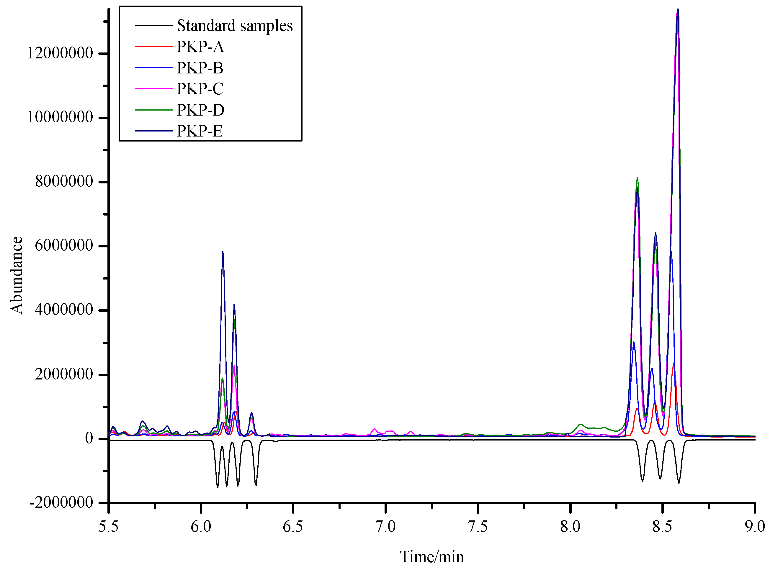
| Retention Time (%) | PKP-A | PKP-B | PKP-C | PKP-D | PKP-E | |
|---|---|---|---|---|---|---|
| d-ribose | 6.09 | 0.30 | 0.30 | 0.39 | 0.41 | 0.78 |
| l-rhamnose | 6.13 | 0.98 | 0.98 | 3.84 | 3.77 | 11.84 |
| l-arabinose | 6.20 | 1.96 | 1.85 | 5.01 | 8.70 | 9.84 |
| d-xylose | 6.29 | 0.40 | 0.53 | 0.12 | 1.84 | 1.96 |
| d-mannose | 8.39 | 2.16 | 7.77 | 21.98 | 24.47 | 23.60 |
| d-glucose | 8.48 | 2.75 | 4.80 | 16.02 | 15.41 | 16.59 |
| d-galactose | 8.58 | 5.11 | 12.75 | 36.93 | 36.69 | 36.95 |
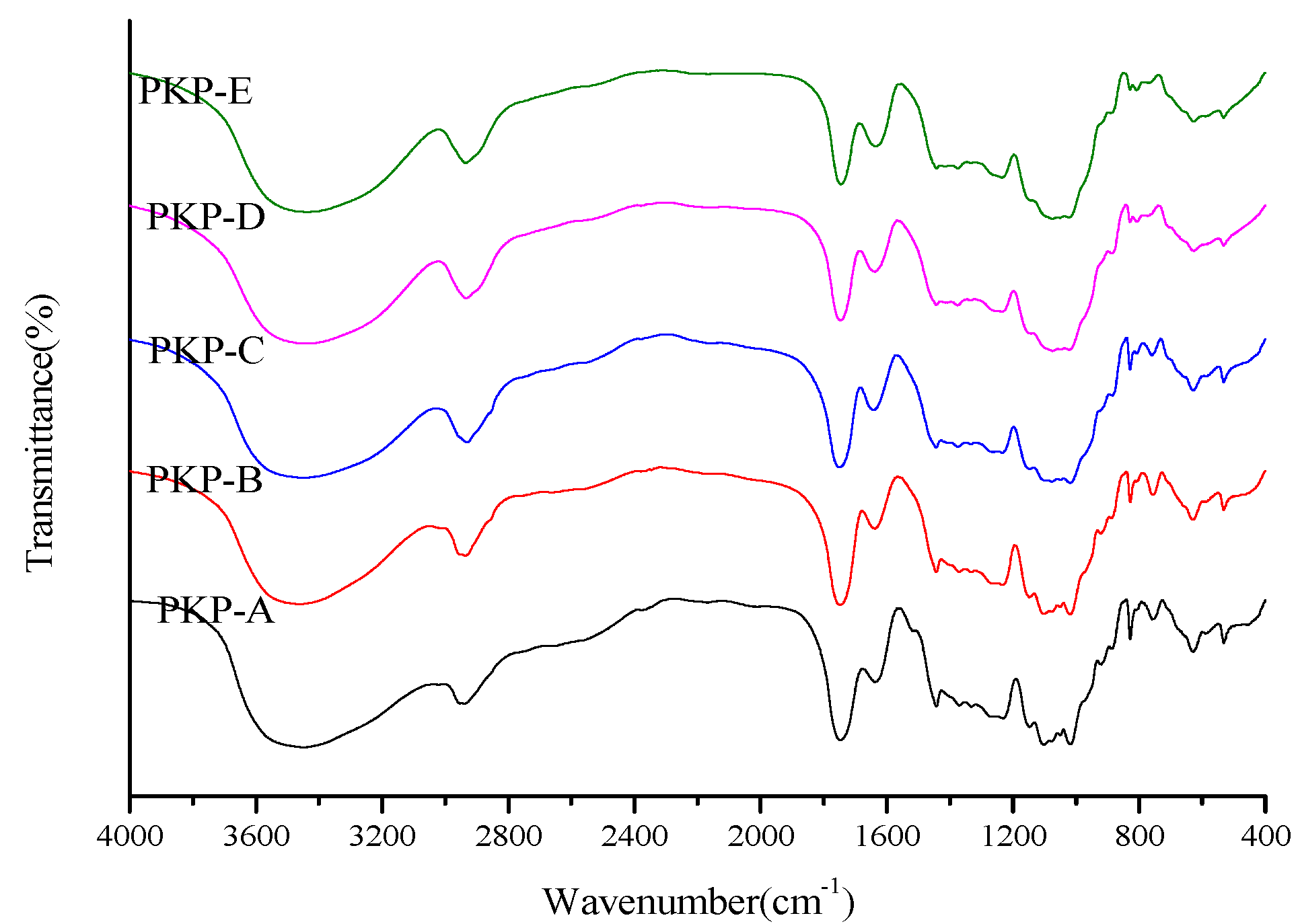
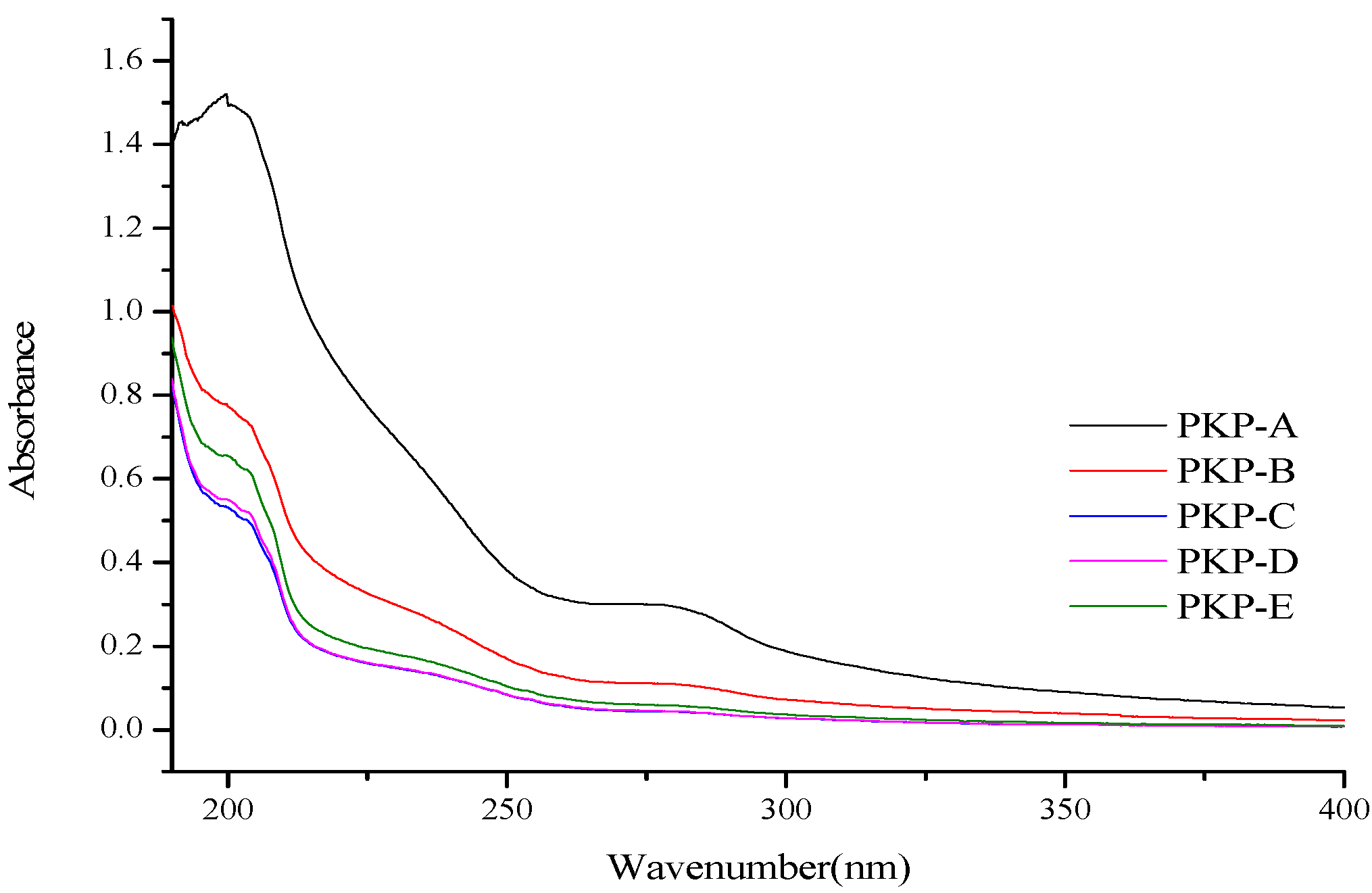
2.2. FT-IR and UV Spectra


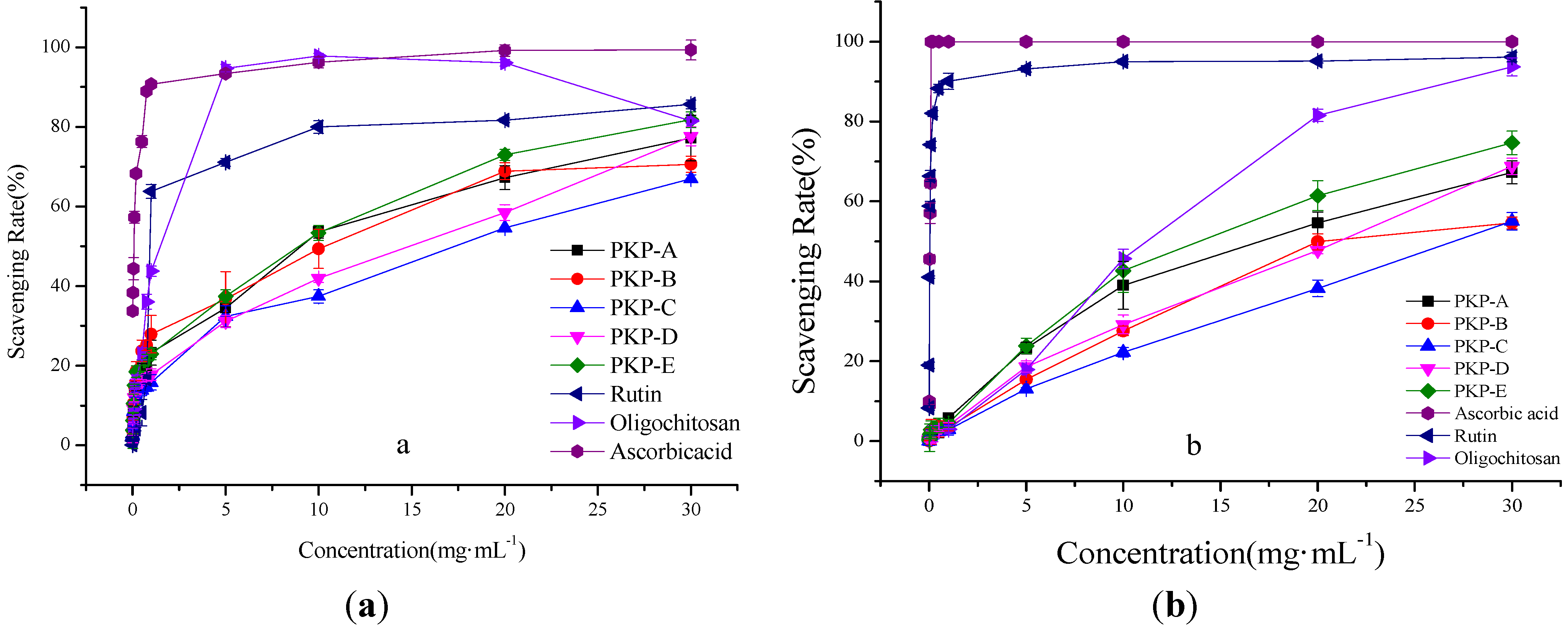
2.3. Antioxidant Activity
2.3.1. Scavenging Effects on Hydroxyl Radical

2.3.2. Scavenging Effects on ABTS Radical
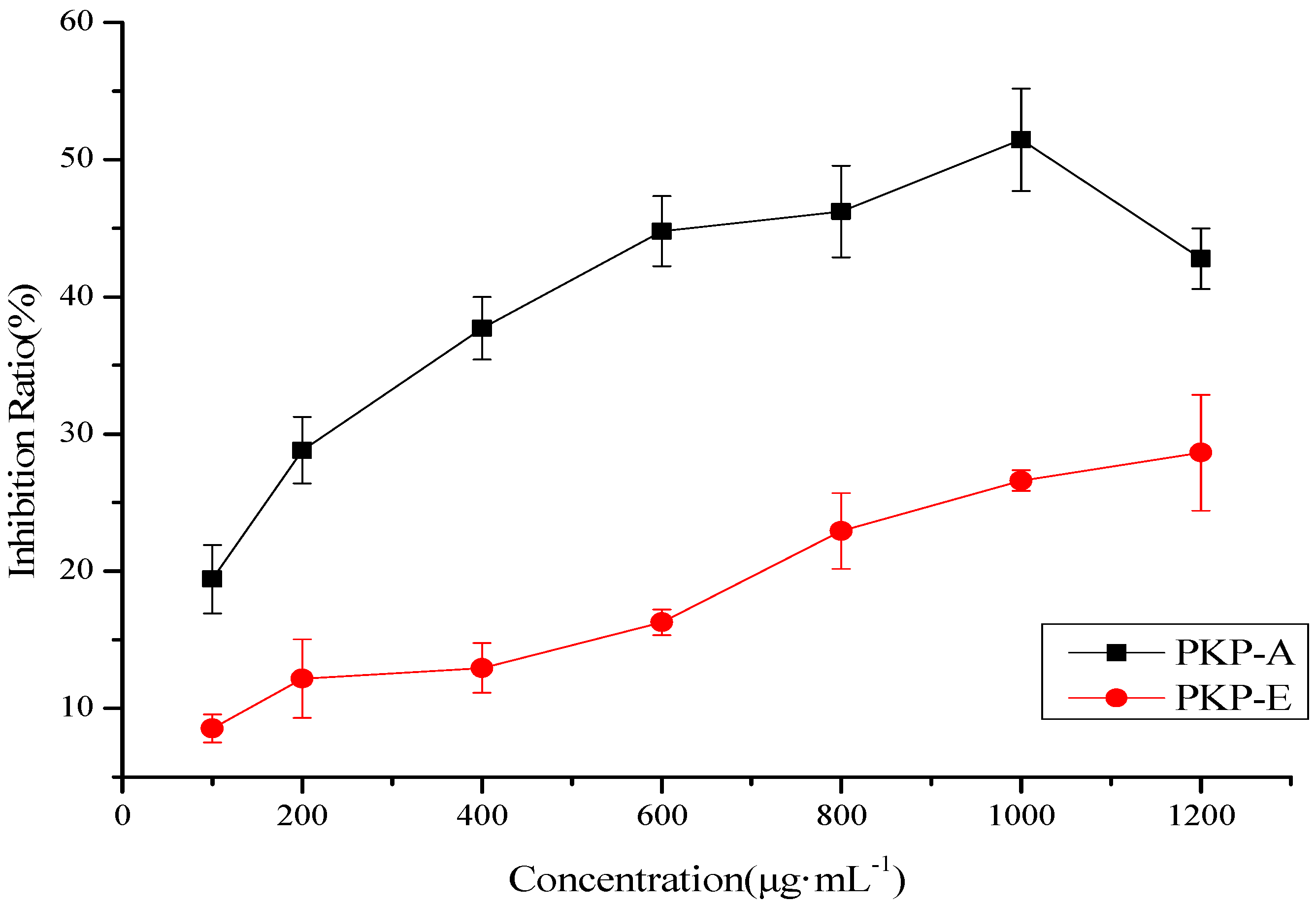
2.4. Inhibition Effects of the Polysaccharides on HepG2

3. Experimental
3.1. Materials and Chemicals
3.2. Extraction of Polysaccharides
3.3. Isolation and Purification of the Polysaccharides
3.3.1. Removal of Protein from the Crude Polysaccharides
3.3.2. Graded Purification of the Polysaccharides
3.4. Determination of Molecular Weight
3.5. Monosaccharide Composition and Properties
3.5.1. Hydrolysis of the Five Polysaccharides
3.5.2. Derivatization of the Monosaccharides
3.5.3. GC-MS Analysis
3.6. Neutral Sugar, Uronic Acid, Polyphenol, Protein and Amino Acid Analysis
3.7. FT-IR and UV Spectroscopy
3.8. Antioxidant Activity of the Polysaccharides
3.8.1. Hydroxyl radical scavenging assay
3.8.2. ABTS Radical Scavenging Assay
3.9. Anti-Hepatoma Activity Assay
3.9.1. Cell Culture
3.9.2. Preliminary MTT Analysis
3.9.3. Further MTT Assay
4. Conclusions
Acknowledgments
Conflicts of Interest
References
- Ofomaja, A.E.; Naidoo, E.B. Biosorption of copper from aqueous solution by chemically activated pine cone: A kinetic study. Chem. Eng. J. 2011, 175, 260–270. [Google Scholar] [CrossRef]
- Rezzi, S.; Bighelli, A.; Castola, V.; Casanova, J. Composition and chemical variability of the oleoresin of Pinus nigra ssp. laricio from Corsica. Ind. Crops Prod. 2005, 21, 71–79. [Google Scholar] [CrossRef]
- Sakagami, H.; Kawazoe, Y.; Komatsu, N.; Simpson, A.; Nonoyama, M.; Konno, K.; Yoshida, T.; Kuroiwa, Y.; Tanuma, S.I. Antitumor, Antiviral and immunopotentiating activities of pine cone extracts: potential medicinal efficacy of natural and synthetic lignin-related material. Anticancer Res. 1991, 11, 881–888. [Google Scholar]
- Sakagami, H.; Takeda, K.; Makino, Y.; Konno, K. Partical purification of novel differentiation-inducing substance(s) from hot water extract of Japanese pine cone. Jpn. J. Cancer Res. 1986, 77, 59–64. [Google Scholar]
- Patrick, K.; Lai, J.D.; Takayama, H.; Sakagami, H.; Tanaka, A.; Konno, K.; Nonoyama, M. Modification of human immunodeficiency viral relication by pine cone extracts. Aids Res. Hum. Retrov. 1990, 6, 205–217. [Google Scholar] [CrossRef]
- Li, W.J.; Yang, X.; Wang, P. Studies on antioxidant activity of polysaccharides from pine cones of Pinus koraiensis. China Beet Sugar. 2012, 2, 26–40. [Google Scholar]
- Xu, R.B.; Yang, X.; Wang, J.; Zhao, H.T.; Lu, W.H.; Cui, J.; Cheng, C.L.; Zou, P.; Huang, W.W.; Wang, P.; et al. Chemical Composition and Antioxidant Activities of Three Polysaccharide Fractions from Pine Cones. Int. J. Mol. Sci. 2012, 13, 14262–14277. [Google Scholar] [CrossRef]
- Zong, A.; Cao, H.; Wang, F. Anticancer polysaccharides from natural resources: A review of recent research. Carbohyd. Polym. 2012, 90, 1395–1410. [Google Scholar] [CrossRef]
- Yang, X.; Zhao, H.T.; Wang, J.; Meng, Q.; Zhang, H.; Yao, L.; Zhang, Y.C.; Dong, A.J.; Ma, Y.; Wang, Z.Y.; et al. Chemical composition and antioxidant activity of essential oil of pine cones of Pinus aramndii from the southwest region of China. J. Med. Plants Res. 2010, 4, 1668–1672. [Google Scholar]
- Meng, Q.; Yang, X.; Wang, J.; Ma, Y.; Ding, Y. Analysis of the Chemical Compostion of Pine Cone of Pinus Koraiensis with Solid Phase Microextraction and GC-MS Technique. Lishizhen Med. Mater. Med. Res. 2010, 21, 2762–2765. [Google Scholar]
- Yang, X.; Zhang, H.; Zhang, Y.; Ma, Y.; Wang, J. Two new diterpenoid acids from Pinus koraiensis. Fitoterapia 2008, 79, 179–181. [Google Scholar] [CrossRef]
- Yang, X.; Ding, Y.; Sun, Z.H.; Zhang, D.M. Studies on chemical constituents of Pinus armandii. Acta Pharm. Sin. 2005, 40, 435–437. [Google Scholar]
- Su, X. Y.; Wang, J.; Yang, X.; Cao, W.Q. Analysis of Volatile Oil Constituents in Pinecone of Pinus koraiensis by Gas Chromatography-Mass Spectrometry. Chin. J. Anal. Chem. 2006, 34, 217–219. [Google Scholar]
- Teleman, A.; Lundqvist, J.; Tjerneld, F.; Stalbrand, H.; Dahlman, O. Characterization of acetylated 4-o-methylglucuronoxylan isolated from aspen employing 1H and 13C NMR spectroscopy. Carbohyd. Res. 2000, 329, 807–815. [Google Scholar] [CrossRef]
- Xue, B.L.; Wen, J.L.; Xu, F.; Sun, R.C. Structural characterization of hemicelluloses fractionated by graded ethanol precipitation from Pinus yunnanensis. Carbohyd. Res. 2012, 352, 159–165. [Google Scholar] [CrossRef]
- Wang, X.M.; Sun, R.G.; Zhang, J.; Chen, Y.Y.; Liu, N.N. Structure and antioxidant activity of polysaccharide POJ-U1a extracted by ultrasound from Ophiopogon japonicus. Fitoterapia 2012, 83, 1576–1584. [Google Scholar] [CrossRef]
- Luo, A.; He, X.; Zhou, S.; Fan, Y.; Luo, A.; Chun, Z. Purification, composition analysis and antioxidant activity of the polysaccharides from Dendrobium nobile Lindl. Carbohyd. Polym. 2010, 79, 1014–1019. [Google Scholar] [CrossRef]
- Sun, Y.; Wang, S.; Li, T.; Li, X.; Jiao, L.; Zhang, L. Purification, structure and immunobiological activity of a new water-soluble polysaccharide from the mycelium of Polyporus albicans (Imaz.) Teng. Bioresour. Technol. 2008, 99, 900–904. [Google Scholar] [CrossRef]
- Sun, R.C.; Sun, X.F. Fractional and structural characterization of hemicelluloses isolated by alkali and alkaline peroxide from areley straw. Carbohyd. Polym. 2002, 49, 415–423. [Google Scholar] [CrossRef]
- Chen, Y.; Mao, W.; Gao, Y.; Teng, X.; Zhu, W.; Chen, Y.; Zhao, C.; Li, N.; Wang, C.; Yan, M.; et al. Structural elucidation of an extracellular polysaccharide produced by the marine fungus Aspergillus versicolor. Carbohyd. Polym. 2013, 93, 478–83. [Google Scholar] [CrossRef]
- Cai, W.; Xie, L.; Chen, Y.; Zhang, H. Purification, characterization and anticoagulant activity of the polysaccharides from green tea. Carbohyd. Polym. 2013, 92, 1086–1090. [Google Scholar] [CrossRef]
- Huang, D.J.; Ou, B.X.; Prior, R.L. The Chemisry behind antioxidant capacity assays. J. Agric. Food Chem. 2005, 53, 1841–1856. [Google Scholar] [CrossRef]
- Sun, Y.; Li, X.; Yang, J.; Liu, J.; Kennedy, J.F. Water-soluble polysaccharide from the fruiting bodies of Chroogomphis rutilus (Schaeff.: Fr.) O.K. Miller: Isolation, Structural features and its scavenging effect on hydroxyl radical. Carbohyd. Polym. 2010, 80, 720–724. [Google Scholar] [CrossRef]
- Ke, C.; Qiao, D.; Gan, D.; Sun, Y.; Ye, H.; Zeng, X. Antioxidant acitivity in vitro and in vivo of the capsule polysaccharides from Streptococcus equi subsp. zooepidemicus. Carbohyd. Polym. 2009, 75, 677–682. [Google Scholar] [CrossRef]
- Leong, L.P.; Shui, G. An investigation of antioxidant capacity of fruits in Singapore markets. Food Chem. 2002, 76, 69–75. [Google Scholar] [CrossRef]
- Fan, Y.; He, X.; Zhou, S.; Luo, A.; He, T.; Chun, Z. Composition analysis and antioxidant activity of polysaccharide from Dendrobium denneanum. Int. J. Biol. Macromol. 2009, 45, 169–173. [Google Scholar] [CrossRef]
- Wu, L.C.; Hsu, H.W.; Chen, Y.C.; Chiu, C.C.; Lin, Y.I.; Ho, J.A. Antioxidant and antiproliferative activities of red pitaya. Food Chem. 2006, 95, 319–327. [Google Scholar] [CrossRef]
- Han, J.; Weng, X.; Bi, K. Antioxidants from a Chinese medicinal herb—Lithospermum erythrorhizon. Food Chem. 2008, 106, 2–10. [Google Scholar] [CrossRef]
- Jemal, A.; Siegel, R.; Ward, E.; Hao, Y.; Xu, J.; Murray, T.; Thun, M.J. Cancer Statistics, 2008. CA- Cancer J. Clin. 2008, 58, 71–96. [Google Scholar] [CrossRef]
- Ali, B.H.; Ziada, A.; Blunden, G. Biological effects of gum arabic: A review of some recent research. Food Chem. Toxicol. 2009, 47, 1–8. [Google Scholar] [CrossRef]
- Wijesekara, I.; Pangestuti, R.; Kim, S.K. Biological activities and potential health benefits of sulfated polysaccharides derived from marine algae. Carbohyd. Polym. 2011, 84, 14–21. [Google Scholar] [CrossRef]
- Yu, Z.H.; Yin, L.H.; Qian, Y.; Yan, L. Effect of Lentinus edodes polysaccharide on oxidative stress, Immunity activity and oral ulceration of rats stimulated by phenol. Carbohyd. Polym. 2009, 75, 115–118. [Google Scholar] [CrossRef]
- Wang, M.; Jiang, C.; Ma, L.; Zhang, Z.; Cao, L.; Liu, J.; Zeng, X. Preparation, preliminary characterization and immunostimulatory activity of polysaccharide fractions from the peduncles of Hovenia dulcis. Food Chem. 2013, 138, 41–47. [Google Scholar] [CrossRef]
- Leung, M.Y.K.; Liu, C.; Zhu, L.F.; Hui, Y.Z.; Yu, B.; Fung, K.P. Chemical and biological characterization of a polysaccharide biological response modifier from Aloe vera L. var. chinesis (Haw.) Berg. Glycobiology 2004, 14, 501–510. [Google Scholar] [CrossRef]
- Xie, G.; Schepetkin, I.A.; Siemsen, D.W.; Kirpotina, L.N.; Wiley, J.A.; Quinn, M.T. Fractionation and characterization of biologically-active polysaccharides from Artemisia tripartita. Phytochemistry 2008, 69, 1359–1371. [Google Scholar] [CrossRef]
- Wang, J.H.; Luo, J.P.; Yang, X.F.; Zha, X.Q. Structural analysis of a rhamnoarabinogalactan from the stems of Dendrobium nobile Lindl. Food Chem. 2010, 122, 572–576. [Google Scholar] [CrossRef]
- Sun, Y.X.; Liu, J.C. Structural characterization of a water-soluble polysaccharide from the Roots of Codonopsis pilosula and its immunity activity. Int. J. Biol. Macromol. 2008, 43, 279–282. [Google Scholar] [CrossRef]
- Günter, E.A.; Popeiko, O.V.; Ovodov, Y.S. Isolation of polysaccharides from the callus culture of Lemna minor L. Appl. Biochem. Microbiol. 2004, 40, 80–83. [Google Scholar] [CrossRef]
- Rajalingam, D.; Loftis, C.; Xu, J.J.; Kumar, T.K.S. Trichloroacetic acid-induced protein precipitation involves the reversible association of a stable partially structured intermediate. Protein Sci. 2009, 18, 980–993. [Google Scholar] [CrossRef]
- Bian, J.; Peng, F.; Peng, P.; Xu, F.; Sun, R.C. Isolation and fractionation of hemicelluloses by graded ethanol precipitation from Caragana korshinskii. Carbohyd. Res. 2010, 345, 802–809. [Google Scholar] [CrossRef]
- Luo, D. Identification of structure and antioxidant activity of a fraction of polysaccharide purified from Dioscorea nipponica Makino. Carbohyd. Polym. 2008, 71, 544–549. [Google Scholar] [CrossRef]
- Zou, S.; Zhang, X.; Yao, W.; Niu, Y.; Gao, X. Structure characterization and hypoglycemic activity of a polysaccharide isolated from the fruit of Lycium barbarum L. Carbohyd. Polym. 2010, 80, 1161–1167. [Google Scholar] [CrossRef]
- Guadalupe, Z.; Martínez-Pinilla, O.; Garrido, Á.; Carrillo, J.D.; Ayestarán, B. Quantitative determination of wine polysaccharides by gas chromatography–mass spectrometry (GC–MS) and size exclusion chromatography (SEC). Food Chem. 2012, 131, 367–374. [Google Scholar] [CrossRef]
- Dubois, M.; Gilles, K.A.; Hamilton, J.K.; Rebers, P.A.; Smith, F. Colorimetri method for determination of sugars and related substances. Anal. Chem. 1956, 28, 350–356. [Google Scholar] [CrossRef]
- Blumenkrantz, N.; Aaboe-Hansen, G. New method for quantitative determination of uronic acids. Anal. Biochem. 1973, 54, 484–489. [Google Scholar] [CrossRef]
- Rajkumar, V.; Guha, G.; Kumar, R.A. Antioxidant and anti-neoplastic activities of Picrorhiza kurroa extracts. Food Chem. Toxicol. 2011, 49, 363–369. [Google Scholar] [CrossRef]
- Bradford, M.M. A rapid and sensitive method for the quantitation of microgram quantities of protein utilizing the principle of protein-dye binding. Anal. Biochem. 1976, 72, 248–254. [Google Scholar] [CrossRef]
- Li, H.; Ma, Y.; Li, Q.; Wang, J.; Cheng, J.; Xue, J.; Shi, J. The chemical composition and nitrogen distribution of chinese yak (maiwa) milk. Int. J. Mol. Sci. 2011, 12, 4885–4895. [Google Scholar] [CrossRef]
- Papageorgiou, S.K.; Kouvelos, E.P.; Favvas, E.P.; Sapalidis, A.A.; Romanos, G.E.; Katsaros, F.K. Metal–carboxylate interactions in metal–alginate complexes studied with FTIR spectroscopy. Carbohyd. Res. 2010, 345, 469–473. [Google Scholar] [CrossRef]
- Jen, J.F.; Leu, M.F.; Yang, T.C. Determination of hydroxyl radicals in an advanced oxidation process with salicylic acid trapping and liquid chromatography. J.Chromatogr. A 1998, 796, 283–288. [Google Scholar] [CrossRef]
- Zhou, H.C.; Lin, Y.M.; Li, Y.Y.; Li, M.; Wei, S.D.; Chai, W.M.; Tam, N.F.Y. Antioxidant properties of polymeric proanthocyanidins from fruit stones and pericarps of Litchi chinensis Sonn. Food Res. Int. 2011, 44, 613–620. [Google Scholar] [CrossRef]
- Zhang, M.; Cui, S.W.; Cheung, P.C.K.; Wang, Q. Antitumor polysaccharides from mushrooms: A review on their isolation process, Structural characteristics and antitumor activity. Trends Food Sci. Tech. 2007, 18, 4–19. [Google Scholar] [CrossRef]
- Hamid, R.; Rotshteyn, Y.; Rabadi, L.; Parikh, R.; Bullock, P. Comparison of alamar blue and MTT assays for high through-put screening. Toxicol. in Vitro 2004, 18, 703–710. [Google Scholar] [CrossRef]
- Young, F.M.; Phungtamdet, W.; Sanderson, B.J.S. Modification of MTT assay conditions to examine the cytotoxic effects of amitraz on the human lymphoblastoid cell line, WIL2NS. Toxicol. in Vitro. 2005, 19, 1051–1059. [Google Scholar] [CrossRef]
- Sample Availability: Samples of the compounds Pinus koraiensis polysaccharides (PKP-A~PKP-E) are available from the authors.
© 2013 by the authors; licensee MDPI, Basel, Switzerland. This article is an open access article distributed under the terms and conditions of the Creative Commons Attribution license (http://creativecommons.org/licenses/by/3.0/).
Share and Cite
Zou, P.; Yang, X.; Huang, W.-W.; Zhao, H.-T.; Wang, J.; Xu, R.-B.; Hu, X.-L.; Shen, S.-Y.; Qin, D. Characterization and Bioactivity of Polysaccharides Obtained from Pine Cones of Pinus koraiensis by Graded Ethanol Precipitation. Molecules 2013, 18, 9933-9948. https://doi.org/10.3390/molecules18089933
Zou P, Yang X, Huang W-W, Zhao H-T, Wang J, Xu R-B, Hu X-L, Shen S-Y, Qin D. Characterization and Bioactivity of Polysaccharides Obtained from Pine Cones of Pinus koraiensis by Graded Ethanol Precipitation. Molecules. 2013; 18(8):9933-9948. https://doi.org/10.3390/molecules18089933
Chicago/Turabian StyleZou, Pan, Xin Yang, Wei-Wei Huang, Hai-Tian Zhao, Jing Wang, Ren-Bo Xu, Xing-Long Hu, Si-Yan Shen, and Di Qin. 2013. "Characterization and Bioactivity of Polysaccharides Obtained from Pine Cones of Pinus koraiensis by Graded Ethanol Precipitation" Molecules 18, no. 8: 9933-9948. https://doi.org/10.3390/molecules18089933