Review

Biomineralization Forming Process and Bio-inspired Nanomaterials for Biomedical Application: A Review

1
School of Mechanical Engineering & Automation, Beihang University, Beijing 100191, China
2
Beijing Advanced Innovation Center for Biomedical Engineering, Beihang University, Beijing 100083, China
*
Authors to whom correspondence should be addressed.
Minerals 2019, 9(2), 68; https://doi.org/10.3390/min9020068
Submission received: 20 December 2018 / Revised: 11 January 2019 / Accepted: 17 January 2019 / Published: 23 January 2019
(This article belongs to the Special Issue Biomineral Crystal Structure)

Abstract

:
Biomineralization is a process in which organic matter and inorganic matter combine with each other under the regulation of living organisms. Because of the biomineralization-induced super survivability and retentivity, biomineralization has attracted special attention from biologists, archaeologists, chemists, and materials scientists for its tracer and transformation effect in rock evolution study and nanomaterials synthesis. However, controlling the biomineralization process in vitro as precisely as intricate biology systems still remains a challenge. In this review, the regulating roles of temperature, pH, and organics in biominerals forming process were reviewed. The artificially introducing and utilization of biomineralization, the bio-inspired synthesis of nanomaterials, in biomedical fields was further discussed, mainly in five potential fields: drug and cell-therapy engineering, cancer/tumor target engineering, bone tissue engineering, and other advanced biomedical engineering. This review might help other interdisciplinary researchers to bionic-manufacture biominerals in molecular-level for developing more applications of biomineralization.

1. Introduction

As a natural protective structure, biomineralization is a process in which organic matter and inorganic matter combine with each other under the regulation of living organisms [1]. Biomineralized crystals are different from the regular structure and surface of artificial crystals. According to the growing environment, biomineralized crystals exhibit distinct physical and chemical characteristics, which are difficult to simulate and manufacture in vitro, while the biomineralization forming process is much similar. As the development of image representation technology, the biomineralization forming mechanism has been found and defined, and their classification has been discussed in some review papers from different systems [2,3,4]. Based on this, many bio-inspired nanomaterials were created through simulating or mimicking the biomineralization forming process.
Biomineralization is a widespread phenomenon that leads to the formation of well-organized biominerals, which refers to the formation of inorganic minerals in organisms [3]. These inorganic minerals not only help to protect the living organisms, like mature mollusk shells [5], brachiopod shells [6], and crustacean cuticles [7], but also help to support organisms and sense signals from surroundings, like bones [8], teeth [9], echinoderm skeletons [10], coral skeleton [11,12], and sponge spicules [13]. C.J.G., et al. [14] have described the mechanism, structure, and bio-function of skeleton-shaped biomineralization in detail in a book. Biomineralization involves biology, chemistry, crystallography, materials science, mineralogy, and medicine, and other disciplines have also led to specialization in these fields. Studying the characteristics of biomineralization forming mechanism is not only helpful to the development and utilization of new nanomaterials, but also helps to treat abnormal mineralization that is caused by the human body disease, such as osteoporosis, osteomalacia, hypophosphatasia, kidney stones, and atherosclerosis. On the other hand, various nano-functional materials were synthesized and biomineralized with living characteristics. Because of the biomineralization-induced super survivability and retentivity, biomineralization has attracted special attention from biologists, archaeologists, chemists, and materials scientists for its tracer and transformation effect in rock evolution study and nanomaterials synthesis. However, controlling the biomineralization process in vitro as precisely as intricate biology systems still remains a challenge.
In this report, we first reviewed the biomineralization forming process in Section 2, including the general crystal growth type, the factors affecting biomineral formation, mechanism and approach of mineralization, and biomimetic biomineralization. In Section 3, the latest applications of biomineralization in biomedical engineering were summarized. Also, Section 4 discussed the future perspectives of biomineralization. This review might help other interdisciplinary researchers to simulate, and bionic-manufacture biominerals in molecular-level for developing more applications for biomineralization.

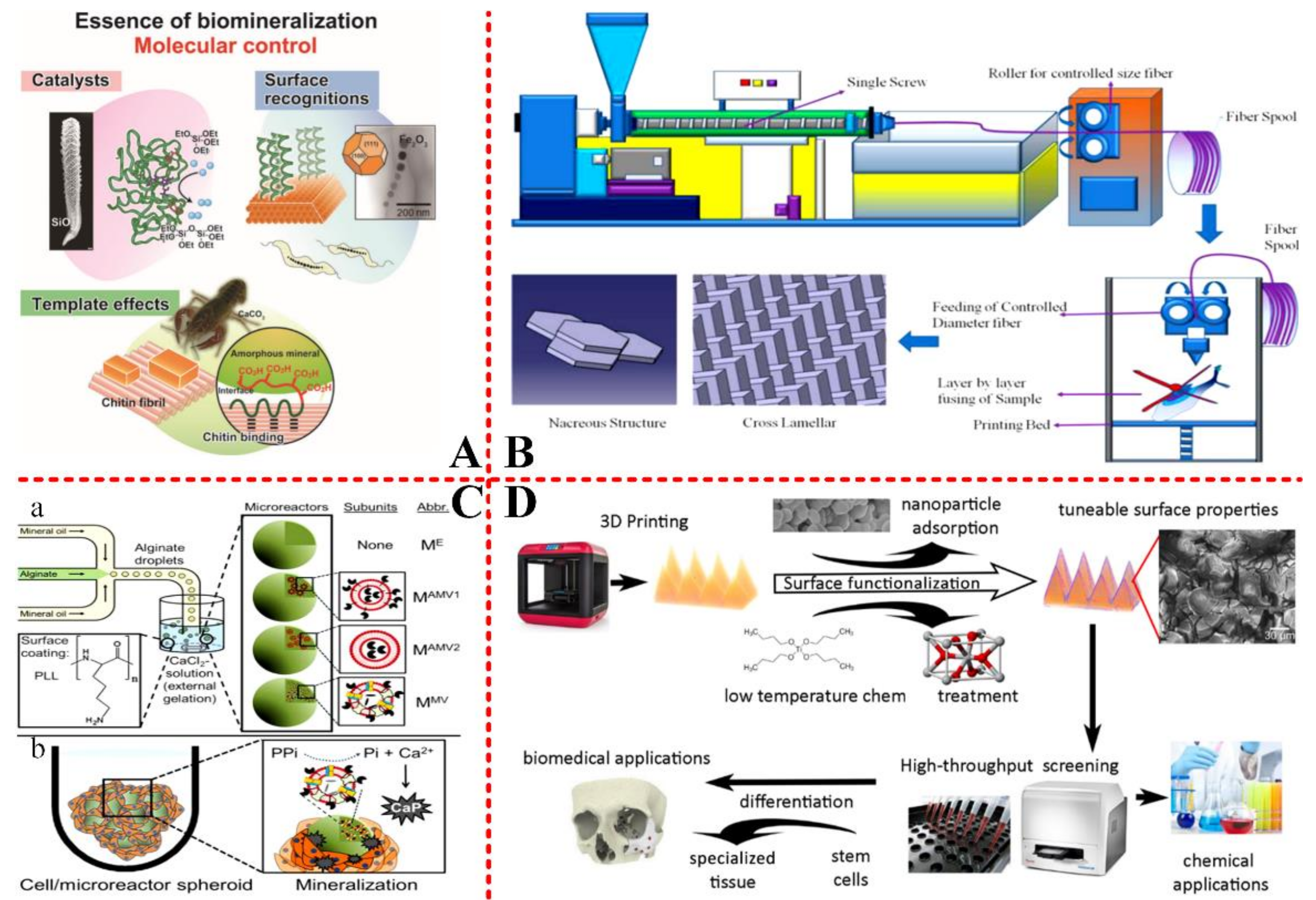
2. Biomineralization Forming Process

2.1. The Type of Mineralization

According to the degree of biological mineral control, biomineralization could be divided into two categories: biological induction and biological control. Bio-induced biomineralization is a process caused by physiological activities of organisms, such as metabolism, inhalation of oxygen, exhalation of carbon dioxide by respiration, as well as establishment of cell walls, which change the physical and chemical conditions of the surrounding environments. This mineralization is not guided by specific cell tissues or biological macromolecules, resulting in arbitrary orientation of crystals and lack of unique morphology. Negatively charged cell walls (containing carboxyl and phosphatidyl groups) combine Fe3+ ions by electrostatic interaction, and Fe3+ ions react with silicic acid to form iron silicate. This process is seldom controlled by cells, and its crystalline form is similar to that of iron silicate that is produced in inorganic solutions. Biologically controlled mineralization is a process that is caused by biological physiological activities and controlled by biology in three aspects: space, structure, and chemistry. It occurs in delineated confined spaces. Biomineral organic matter is formed with high content, unique crystallization habit, uniform size, uniform shape, and regular arrangement.
There are two forms of mineralization: one is normal mineralization, such as skeleton, teeth and shellfish, formation of shells, and so on. Another is abnormal mineralization, such as stones, dental stones and caries teeth, and so on. There are two theories to explain this biomineralization. Solution crystallization theory and polymer-induced liquid phase precursor mineralization theory. For solution crystallization theory, Type I collagen molecules self-assemble into a native “hole”, the “hole” contain negatively charged amino acids, the –COO– could bind to Ca2+, which lead to the deposition of Ca2+ in the “hole”[15]. After deposition, the calcium phosphate (Ca-Pi) is electrostatically formed through binding. The Ca–Pi nucleus is the basic building blocks of bone structure [16]. In addition, the “hole” is the nucleation site of hydroxyapatite crystals, which is another important component of bones [17]. For polymer-induced liquid phase precursor mineralization theory, highly hydrated amorphous calcium phosphate phase nanometer droplets penetrate into the pores and voids of collagen fibers the hydrates lose water and crystallize in the pores and voids of collagen fibers [18]. The calcification of atherosclerosis (AS) plaque is a typical sample of abnormal mineralization.

2.2. Factors Controlling Biomineralization

Mineralization in organisms can be divided into four stages. (1) Organic macromolecules are pre-assembled into ordered structures. Insoluble organic matter in organisms constructs an organized micro-reaction environment before mineral deposition, which determines the location of inorganic nucleation and the function of mineral formation. This stage is the precondition of biomineralization. (2) Molecular recognition of organic-inorganic interfaces controlling crystal nucleation and growth. (3) Growth regulation enabling the initial assembly of crystals to form subunits. (4) Cell prepressing, forming biominerals with multilevel structure by assembling subunit minerals. It is believed that biomineralization is controlled by varying factors. The formation of biominerals is often the result of the synergistic action of various factors, including pH, temperature, and matrix.

2.2.1. Temperature and pH

Temperature is an important factor affecting calcium carbonate deposition. The solubility of most salts in water will increase with the increase of temperature. However, calcium carbonate has abnormal solubility, the solubility will decrease when the temperature rises. That is to say, more calcium carbonate will deposit when the temperature rise. pH also has great influence on the solubility of carbonate. Reducing PH value will increase the solubility of carbonate. Furthermore, pH and temperatures can control calcium carbonate particles forming various crystal morphologies [19], such as, plate, rhombohedra, rectangles ellipsoid, cube, etc. Vaterite with specific morphologies was formed at low temperature, whereas needle-shaped aragonite crystals were obtained at high temperature [20,21].
There exists some controversy pertaining about the temperature effect on the biomineralization forming process. Some previous studies on the oysters C. gigas and C. virginica have found that raising temperature has stronger effects than moderate ocean acid on shell growth and metabolism [22,23]. The lesser pH effect demonstrated here agrees with D.G.H., et al. [24].
pH is the most important factor affecting the precipitation structure in Uranium (VI) biomineralization by Saccharomyces cerevisiae [25]. Some other reports have shown that there were no negative effects on the calcification rate, CaCO3 deposition, shell ultrastructure, and metabolic pathways in P. fucata exposed to warmed seawater [26]. If the thermal threshold is not breached, temperature elevation may promote CaCO3 deposition during biomineralization [26], as shown in Figure 1(A). Indeed, the effects of temperature rise on organic biomineralization vary from species to species. The effects of temperature and pH value on biomineralization are synergistic. It was also recognized that the Mg content in coralline carbonate varied seasonally experiments on synthetic Mg-calcite [27], which demonstrated a positive correlation between Mg content and temperature. Sometimes, they would need to improve the thermostability. It should be noted that egg cells with mineral shells are extremely thermostable. The stability of modified minerals could also be maintained at suitable pH [28] (Figure 1(B)) and temperature [29] (Figure 1(C)).
pH plays a significant role in controlling pores diameter on the surface of virus, which is usually used as an environment for biomimetic mineralization. The nano-in-micro composites were produced by coating oxidation-responsive NPs with chitosan and encapsulated in a pH-sensitive polymer. The microparticles maintain integrity at acidic pH, which could be used in drug release. The detrimental effects of ocean acid, pH altering, on biomineralization are a common response of marine organisms to environmental changes [30,31,32].

2.2.2. Guidance of Organic Matrix on Biominerals

The organic matrix in biominerals can be defined as any organic matrix localized at the surfaces of constituents, such as proteins, phospholipids, collagen and carbohydrates, and so on, which act as a mediator for the mineralization of biological systems. A large number of organic matrices, especially proteins, are involved in the formation of inorganic materials in the process of biomineralization, controlling the nucleation [33,34], growth, crystalline form, and trend of inorganic crystals. This process is also called molecular recognition. The macromolecules also play a role in initiating nucleation and directing crystal growth.

2.2.3. Additives

In recent years, the chemical composition and complex of additives have tremendously attracted scientific attention. Detailed controlling mechanism of additives on mineralization could be reviewed in recent Denis Gebauer’s paper [35]. Additives and matrix could lead to the crystal morphology modification. Insoluble and soluble organic additives provide a heterogeneous nucleation site and regulate crystal growth by their adsorption. Soluble organic substances distribution and incorporation level in the CaCO3 crystals can alter the growth kinetics and morphology of calcium carbonate. Insoluble matrices, as biomacromolecules, can form a three-dimensional framework for promoting the nucleation of CaCO3 crystals and thus lead to a finer crystallization of CaCO3 [36,37]. The inorganic ions have been demonstrated to be incorporated into the lattice of calcite, thus influencing the morphology of calcite crystals [38]. Citrate coated Au nanoparticles (CIT-Au NPs) and agarose gels were both introduced into the crystallization [39]. CIT, and the matrix, agarose gel, have synergistic effects on morphology regulation of synthetic calcite single-crystals. The agarose gel matrix is inert to the crystal morphology in the sense that the agarose gel grown calcite crystals maintain in characteristic rhombohedra. In contrast, CIT additives are active in crystal morphology modification and crystals begin to exhibit curved rough surfaces when grown in solutions. Apart from inorganic ions, the organic components, including amino acids, peptides, diblock copolymers, and proteins, which also contributed to shaping crystals while acquiring an in-depth understanding of crystal-additive interactions.
Polyacrylic acid (PAA) additives can control the morphologies of calcium carbonate crystal at a concentration dependency manner [19]. In the presence of cetyltrimethylammonium bromide (CTAB), various unusual calcium carbonate crystal morphologies, such as dendrite-shaped, flower-like, wheatgrass-like, needle-like, whiskers, and double-taper-like, can be obtained depending on the addition of various organic additives, such as glycol, glycerine, formaldehyde, acetaldehyde, glycol-methyl ether, and glycol-ethylether [40]. Calcite crystals gradually recovered their sharp rhombohedral morphology in the presence of Water soluble matrix (WSM) from irregular state [20]. One-dimensional Ca-deoxycholate fibers, as a novel insoluble organic polymorph controller, was demonstrated to be the key role in mediating the crystallization and controlling self-assembly processes of calcium carbonate [21]. A novel matrix protein Hic31 from the prismatic layer of Hyriopsis Cumingii displaying a collagen-like structure may play important roles in biomineralization of the pearl prismatic layer [41].
In summary, as shown in Figure 2, biomineralization could be divided into two types, normal mineralization and abnormal mineralization, and the forming process mainly regulated by the temperature, pH and additives.

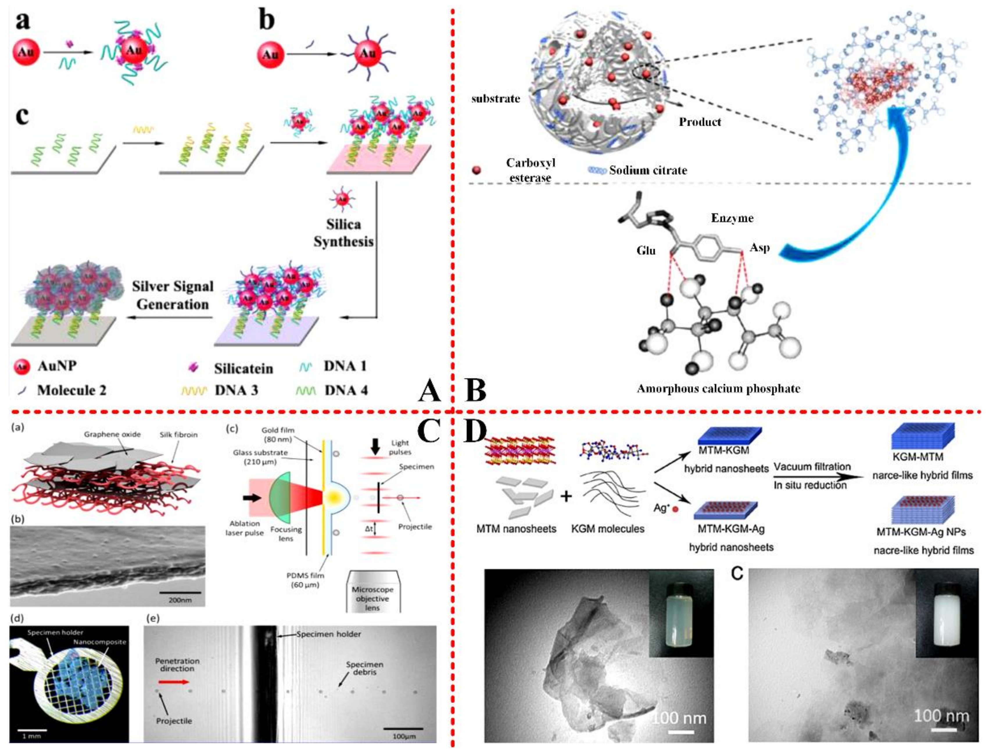
2.3. Mechanism and Approach of Mineralization

The mineralization process in organisms is very complex. Recognizing the formation mechanism of biominerals and matrix, cell and other symbiosis regulation of minerals and substances will be very helpful. Most of the research simply established simulation system and coordinated the regulation of biological macromolecules outside of the organism. There are very few studies on the control effect. Accordingly, we need to be closer to organisms. Further study on the specific regulation of biological macromolecules under environmental conditions function and synergistic regulation to further study organisms in matrix mineralization processes, cell-mineral interactions, to elucidate biominerals.
A deep molecular-level mechanistic understanding of how proteins participate in the nucleation and growth of inorganic crystals (both in vitro and in vivo) can be achieved by computational methods at the atomic scale, as shown in Figure 3(A) [42].
Michael S. Pacella et, al. compared the results of the Rosetta-Surface algorithm to an experimental benchmark of kinetic and thermodynamic measurements on peptide−biomineral interactions taken from atomic force microscopy, and successfully identified which mineral face and step edges will bind peptides the strongest [42]. The interaction effect between proteins and crystal can be illustrated using examples from biomineralization, as well as by the immune responses from pathological crystallizations to crystalline antigens, as shown in Figure 3(B). A.J., et al. [44] have employed self-assembled monolayers (SAMs) on Au or Ag, with disordered head groups, to induce the formation of an amorphous CaCO3 film. The mechanism of the molecular interactions occurred at the interface between the inorganic mineral and the macromolecules [45]. A form of additive manufacturing, selective laser sintering (SLS) uses a roller feed system, a heated chamber, and a laser to fuse plastic, ceramic powder layers or metal, together form a wide variety of solid objects [46]. As opposed to other printing methods that restrict model geometry or require removable, unfused powder provides support for parts. Together with the biocompatibility of the material feedstocks, this process also gives printed objects a rough surface of partially fused powder and provides a viable avenue for fabricating a wide variety of effective high-surface area substrates for tissue engineering and biomedical implants. Furthermore, the model of three-dimensional printed frameworks has tremendous potential for synthesis of biomaterials. It is especially effective when combined with post-synthesis chemical treatment, and this will be discussed in the later section.

2.4. Biomimetic Biomineralization

Traditional biomineralization studies emphasize the bionic design and preparation of materials by imitating nature, highlighting the regulation of inorganic crystallization by organic systems, thus improving the properties of materials. The new trend of biomineralization research is to use natural strategies for realizing the regulation of biological organisms through inorganic materials, highlighting the use of material systems to achieve bio-functional transformation.
In 2008, Tang Ruikang’s team successfully realized the mineralization transformation of yeast cells [47], and proposed that using the biomineralization strategy for more biological species could be endowed with new functions through material shells. For example, the preservation of vaccines is highly dependent on the cold chain. Through biomimetic mineralization, the team constructed a new vaccine-calcium phosphate complex, which greatly enhanced the thermal stability of vaccines on the basis of maintaining the original efficacy, and initially realized the construction of heat-stable vaccines independent of refrigerators. Biomimetic mineralization can not only enhance the structural stability of organisms through materials, but also change the original functions of organisms. By inducing the spontaneous accumulation of green algae through biomimetic mineralization of green algae, the group can activate hydrogenase while also ensuring the activity of photosynthetic system II, thus changing the photosynthetic pathway of green algae, and directly decompose water to produce hydrogen under natural conditions. Its hydrogen production efficiency is equal to the normal photosynthetic efficiencies.
When compared with natural or synthetic minerals, biominerals often exhibit excellent mechanical and other properties. The reason is that biominerals have multilevel ordered micro- and nano-structures with specific morphologies, which are also consistent with the macro-scale structure. The acquisition of multilevel ordered mineral structure depends on the synergistic effect of various biomass macromolecules in the process of biomineralization. Clearly, understanding this synergistic effect can better guide material chemists to make controllable composites to obtain excellent mechanical and other properties. However, it is difficult to observe the biomineralization process in situ, and the information that is provided by static biomineral microstructural analysis is relatively limited. Therefore, controllable biomimetic mineralization is still a major problem in the field of material synthesis. Up to now, biomimetic mineralization research is mostly based on empirical knowledge, and biomimetic mineralization research based on controllable route design is few and far between. Calcium carbonate films with shellfish-like prism structure were synthesized by the total synthesis method for the first time, as shown in Figure 4(A), and precise control of the micro-structure of bionic films was achieved, resulting in excellent mechanical properties [48].
Calcium oxalate needle crystals are formed plants using protein nanowires (14 kDa) embedded in needle crystals as templates in banana. Calcium oxalate nanospheres are induced to deposit orderly along the template and they eventually form hexagonal pyramidal mesoscopic structures driven by organic-inorganic self-assembly [49]. This study effectively reveals the structure-functional relationship between the chemical evolution of plant crystal morphology and the environment during the long evolutionary process, as shown in Figure 4(B). That is, in the physiological micro-environment of excessive calcium or oxalic acid metabolism (C4 plants), plants can maximize the capture of calcium and oxalic acid in the limited all oocyte, thus maintaining the metabolism of inorganic calcium and organic acid in plants. It is also of great significance to the development of bionic materials and the study of human pathological mineralization, such as kidney stones (calcium oxalate is also the main component).
Biomineralization, as a functional strategy in the process of biological evolution, can make organisms more adaptable to the environment and they produce evolutionary chains that are more conducive to their own development. It also provides a reference direction for human beings to realize the regulation of biological organisms through materials. By learning from nature, the study of biomineralization has realized the transformation from the regulation of material crystallization by biological system to the improvement of organisms by using materials, which provides a new direction for the sustainable development of human beings.
A rapid synthesis method of multifunctional macro-graphene composites was developed. Graphene oxide was added into biomimetic mineralized gel system to form graphene oxide, amorphous calcium carbonate nanoparticles, and polyacrylic acid cross-linked network structure. The soft and hard state of the graphene composite can be controlled by moisture content. It is also found that the material has excellent plasticity and self-healing ability, and it is expected to be used in multi-scale rapid processing and forming of graphene materials [50].

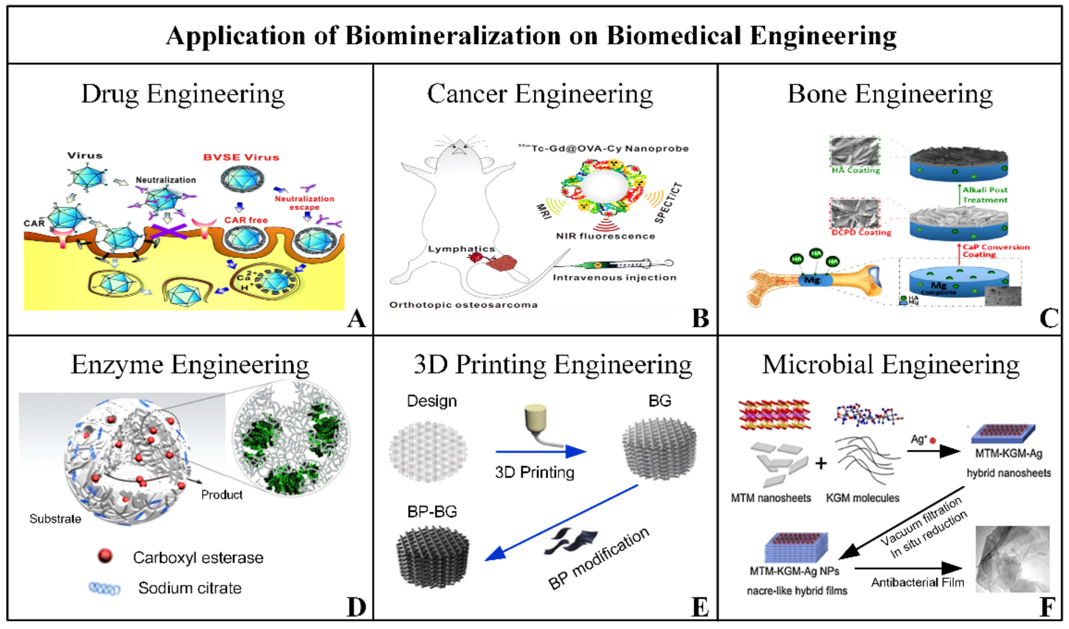
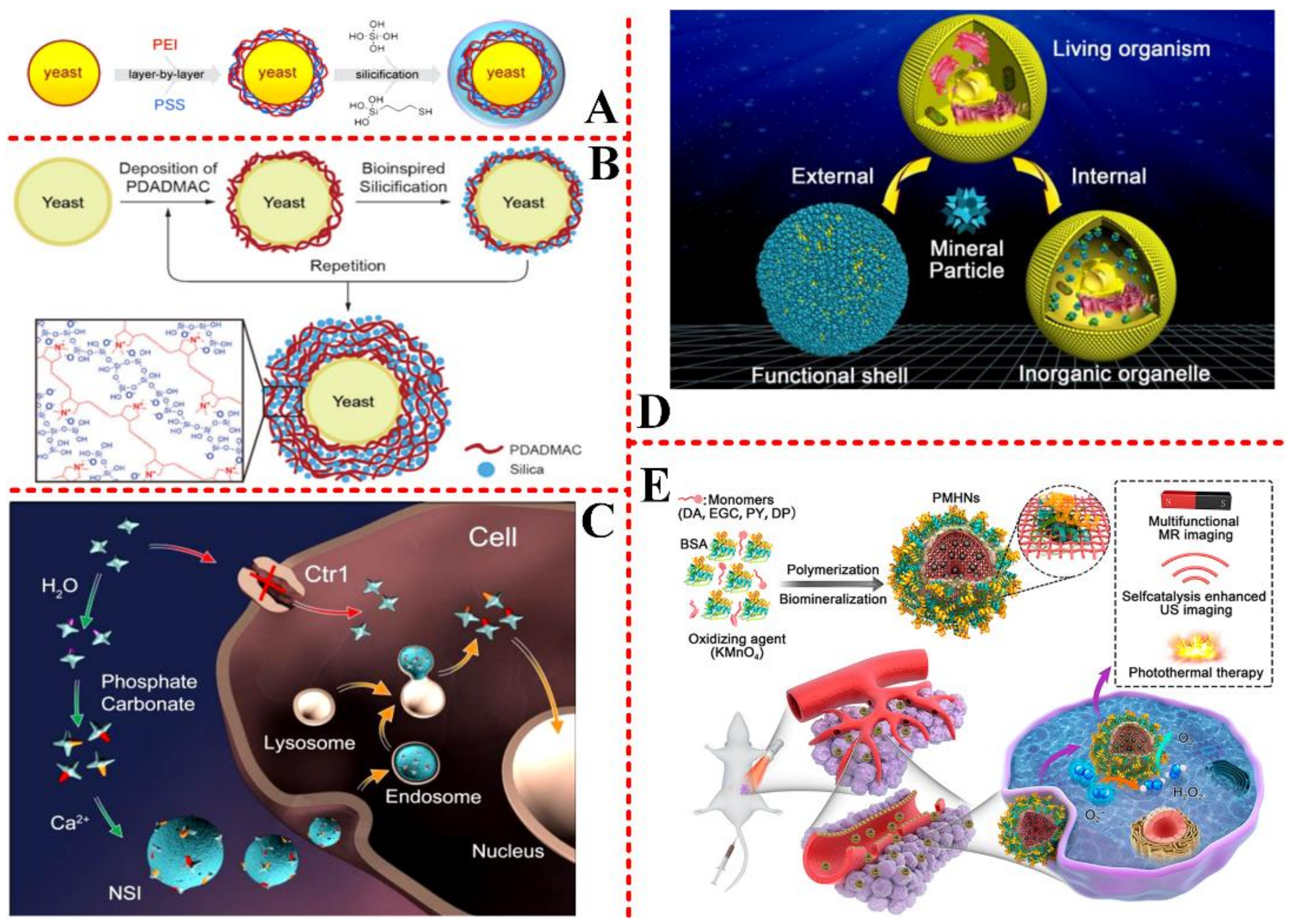
3. Application of Biomineralization in Biomedical Engineering

As a natural phenomenon, the forming process of biomineralization has been thoroughly studied and mimicked in vitro. Based on the mentioned forming process, incorporated with other rising technologies to synthesize new nanomaterials, biomineralization has penetrated into many fields around our life, for instance, mechanical engineering [51,52,53], electrical engineering [54,55,56], environmental engineering [57,58,59], as well as biomedical engineering [60,61,62]. As the pathfinder of human health and life, biomedical engineering has received specific attention and developed into a booming industry. Therefore, it becomes necessary to review the application of biomineralization in biomedical engineering. As shown in Figure 5, the application mainly includes these directions: drug engineering, cancer engineering, bone engineering, enzyme engineering, three-dimensional (3D) printing engineering, and microbial engineering. The detailed combination between biomineralization and biomedical engineering will be discussed in this section. Several important and interesting research were reviewed in the past eight years, and in order to elucidate revolution of these applications, we added the published year in the descriptions.

3.1. Drug and Cell-Therapy Engineering

As an organic regulated forming method of inorganic structure, biomineralization overcomes the problem of biocompatibility, offering an approach to protect the curing viral vectors from molecular recognition [63] on the application of drug and cell-therapy engineering. Inspired by the biomineralization of diatoms, Y.S.H., et al. (2011) [64] proposed a biomimetic method to silica-encapsulate yeast cells with surface functionalization, which firstly introduced the thiol functionalization into silica-encapsulation, as well as the fluorescein and streptavidin onto the silica-shells for better observation. The polyethyleneimine (PEI) and poly-sodium 4-styrenesulfonate (PSS), two biocompatible polyelectrolyte multilayers, were combined to perform as a catalytic template for yeast-surface silica-biomineralization, as shown in Figure 6(A). Following this mechanism, L.H., et al. (2014) [65] reported a biomineralization-based silica encapsulation of individual saccharomyces cerevisiae cell and realized the control of thickness of silica shells. The precise thickness regulation could better control the functional activities of internal drugs and metabolic activities of encapsulated cells, as well as increase the physicochemical stability of the nanomaterial. During fabrication, the layer-by-layer technology was combined with bioinspired silicification to form the living shell of cells, as shown in Figure 6(B).
C.W., et al. (2013) [71] reported a method to fabricate nano-solidified intermedia (NSI) of cisplatin based on biomineralization, and realize the carry and reverse of cancer drugs in vivo and in vitro, as shown in Figure 6(C). Biomineralization, used in the synthesis, not only enhanced the drug biocompatibility, but importantly overcame the drug resistance for better and longer cancer chemotherapy. The authors have made great progresses and results in the field of biomineralization-based nanomaterials on drug and cell-therapy engineering. In the next year, C.W., et al. (2014) [60] has particularly reviewed the biomineralization nanomodified of living organisms, and divided it into two typical categories, according to the location of biomineralization. One is the biological surface induced external mineral particle, which forms the functional shell. The other is the biological internal materials induced mineral particle, which forms the inorganic organelle, as shown in Figure 6(D). The location where biomineralization happened decides the structure of the novel organism, external shells, or internal scaffolds, which usually results from different processing methods. These novel living-mineral nanocomplexes have functional application on drug delivery, biological protection, and storage, etc.
Recently, X.B., et al. (2018) [72] proposed an albumin-template biomineralization, incorporating with oxidization polymerization, to synthesize nanotherapeutic agents for safer vascular drug delivery engineering. This technique was been demonstrated in several polymer monomers, including dopamine (DA), epigallocatechin (EGC), pyrrole (PY), as well as diamino-pyridine (DP). During the reaction, polymer monomers were suspended in bovine serum albumin (BSA) solution, and KMnO4 was chosen as the catalyst to induce the polymerization and biomineralization. After stirring at room temperature for 2h, the polymers and manganese dioxide hybrid nanoparticles (PMHNs) were synthesized. After intravenous injection into the mice, these PMHNs performed high sensitivity and resolution of imaging on magnetic resonance (MR) and ultrasonic detection. When combining with external precision control, these PMHNs could carry efficient drugs and pass through blood vessel wall to tumor sites, and further realize the in-situ ablation of tiny tumor, as shown in Figure 6(E).

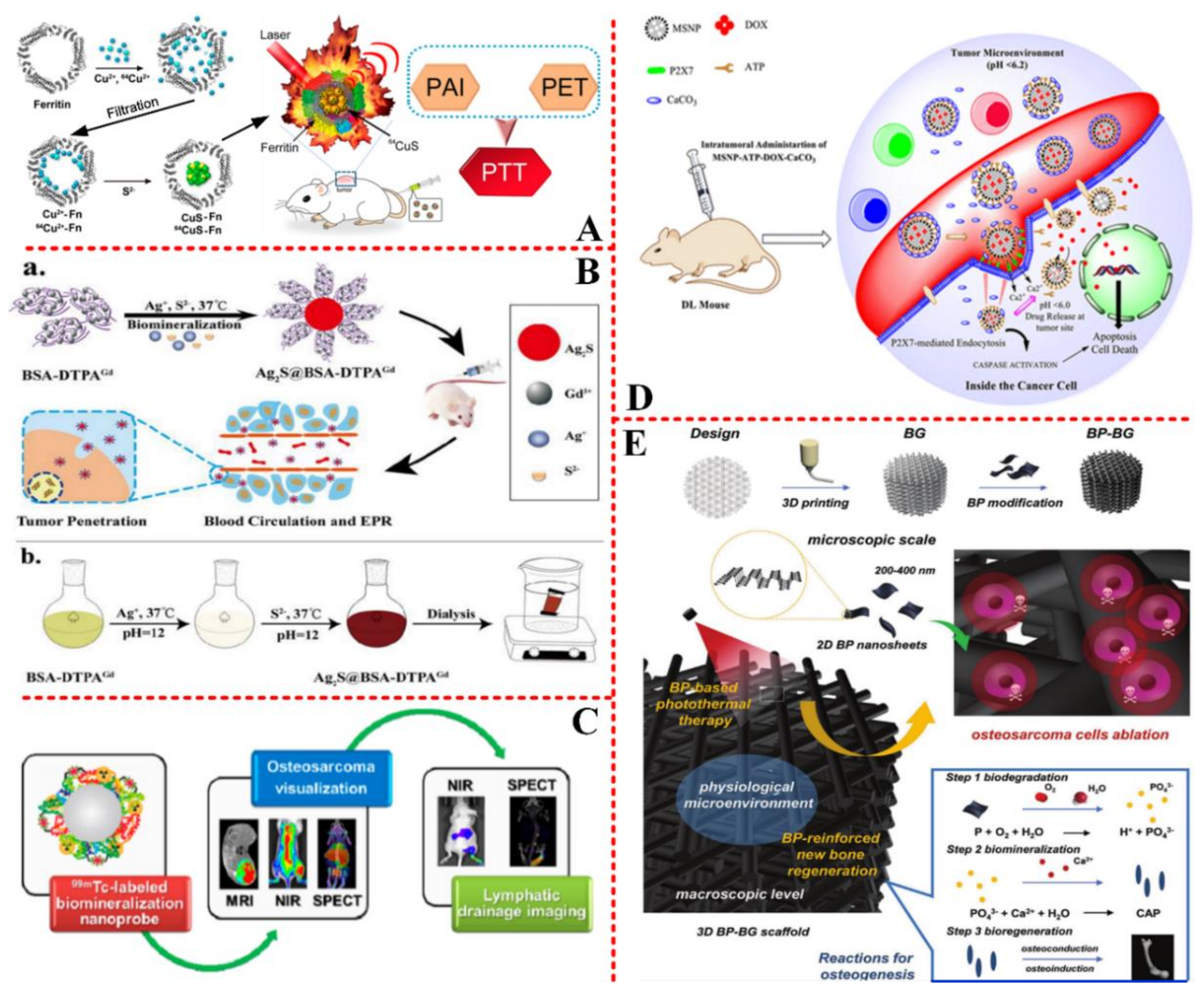
3.2. Cancer/Tumor Target Engineering

With the development of nanotechnology, various nanomaterials based therapeutic platforms have been developed for the diagnosis and treatment of cancer/tumor, which have been the biggest killer threatening human life and health. Except for the excellent biocompatibility, synthetic nanomaterials that are inspired by biomineralization have unique optical, magnetic, and thermal properties, which facilitate the construction of out-body control systems for better positioning, imaging, and final diagnosis of cancer/tumor. Inspired by the natural biomineralization of ferritin (Fn), W.Z.T., et al. (2016) [73] proposed a biomimetic synthesis method to synthesize Fn supported ultra-small copper sulfide (CuS) nanoparticles, which had high sensitivity and resolution on the photoacoustic imaging (PAI). When combined with position emission tomography (PEI), the quantitative PAI realized photothermal therapy (PTT) with superior therapeutic efficiency on cancer both in vitro and in vivo, as shown in Figure 7(A).
Z.J., et al. (2016) [74] also utilized a protein-platform to synthesize fluorescence contrast agents for MRI/NIR detection of cancer. The BSA, an easily decorated protein, was chosen as the platform for biomineralization to carry engineered Ag2S-pQDs with Gd3+ ions. After the intravenous injection into mice, the synthesized nanomaterial exhibited pronounced capability for tiny tumor targeting capability with high 3D resolution and sensitivity, as shown in Figure 7(B). Similarly, X.Z.M., et al. (2018) [66] produced a biomineralization nanoprobe, as a visualization tool, to realize the real-time imaging of multimodal detection during MRI/NIR on tumor diagnosis. On the basis of cypate-grafted biomineralization, ovalbumin was used to synthesize gadolinium oxide nanoparticles, after being labeled by a NIR fluorescence dye, the ovalbumin scaffold was intravenously injected into the mice to realize the real-time visualization of osteosarcoma and lymph nodes, as shown in Figure 7(C). Biomineralization enhanced the stability and biocompatibility of the nanoprobe, when comparing with previous colloidal nanoprobes, and the small size enables the nanoprobe to be cleared by the circulatory system.
Adenosine triphosphate (ATP), an existence form of energy, was found to mediate drug-liberating process during cancer therapy, and to be acted as an antineoplastic agent in vitro [76]. Based on this, S.P., et al. (2018) [75] synthesized an ATP-loaded nanocomposite with pronounced apoptosis in tumor cells. First, mesoporous silica nanoparticles (MSNPs), a biocompatible deliver, was decorated with ATP on the surface. Subsequently, the doxorubicin (DOX), an efficient cancer drug, was loaded into the poriferous nanoparticles. After biomineralization of ATP and CaCO3, the pores on the surface of NSNPs were closed and the DOX was encapsulated. Under an acidic environment, tumor location, CaCO3 was dissolved, ATP was exposed to bind with antigen on the surface of tumor, pores opened, and DOX could be released to effect on the tumor, as shown in Figure 7(D). When comparing with traditional drug therapy for tumor, this biomineralization-based synthesis of drug carrier nanomaterial protected ATP and DOX before they arrived tumor location, further reduced tumor burden, and increased the survival rate.
Y.B., et al. (2018) [69] reported a novel black-phosphorus (BP) supported biomineralization-based 3D printing scaffold for better treatment of osteosarcoma, as shown in Figure 7(E). The calcium-extracted biomineralization was induced in vivo, and the design and BP-modification of nanosheets were processed by 3D printing technology in vitro. Under the target physiological microenvironment, the excellent photothermal properties enable BP to induce in situ biomineralization of the nanosheets, and further promote the local bone tissue regeneration to repair the damage that is caused by osteosarcoma, an aggressive malignant neoplasm. Therefore, this technology could also be applied in bone tissue regeneration engineering.

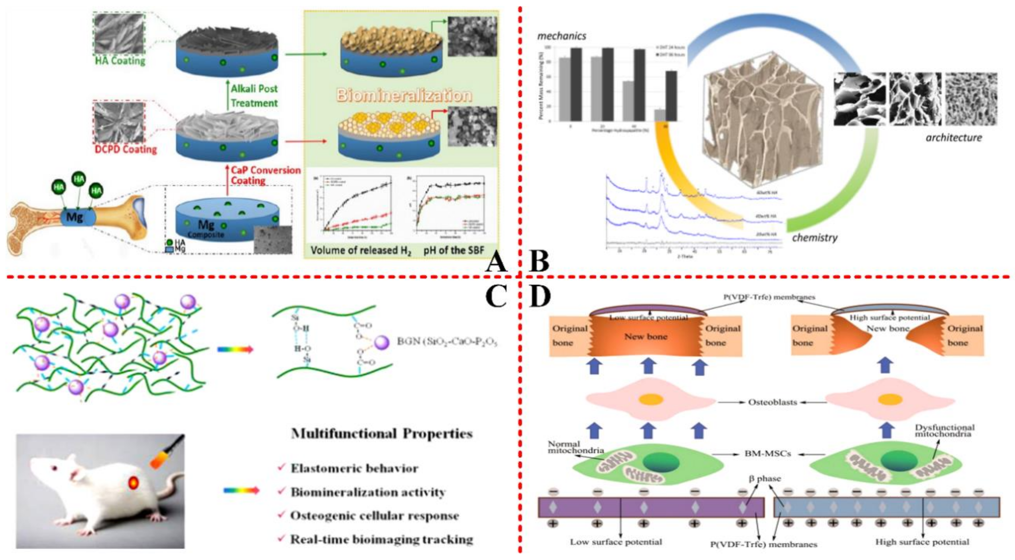
3.3. Bone Tissue Engineering

Except for the cell/drug-based disease therapy engineering, bone tissue engineering is another important area of the biomineralization application. S.Y.C., et al. (2016) [67] proposed a biomineralization-based conversion coating method to enhance the polarization resistance of coatings, as well as increase corrosion resistance and surface bioactivity. The synthesized nanocomposites were demonstrated to be well applied on the bone tissue engineering, as shown in Figure 8(A). Magnesium (Mg), a conventional orthopedic implant material, was used as the basic template to fabricate HA/Mg composites by authors themselves. After coating with hydroxyapatite (HA) and biocompatible dicalcium phosphate dihydrate (DCPD), biomineralization was induced on the template. Further electrochemical results were also carried out to prove the enhanced capability of corrosion resistance and biocompatibility of the novel synthesis.
The orthopedic implant material not only needs to have the capability of corrosion resistance, but it also needs to have the ability to enhance the expression of osteocalcin.P.K.M., et al. (2017) [77] reported a biomineralization-based fabrication of recombinant peptide scaffolds with enhanced expression of osteocalcin. As shown in Figure 8(B), a linear mineralized scaffold was fabricated by utilizing biomimetic nucleation, which had enhanced mechanical strength, increased HA content, as well as improved osteoblast mineralization. The all three scaffold signals, mechanics, chemistry, and architecture of the material, were optimized together, which is important and necessary for directing cell response in bone regeneration engineering. Based on this, L.Y., et al. (2018) [78] reported an elastomeric bioactive nanocomposite with multifunctional properties on biomineralization activity, elastomeric behavior, bioimaging tracking, osteogenic cellular response, as well as inflammatory response, as shown in Figure 8(C), these enhanced properties offered new design for better bone regeneration technology.
It has been proven that external electrical stimulation could be used to improve bone regeneration [79] and enhance bone healing [80]. However, how to control the electrical stimulation to fit the physiological potential is still not clear. Z.C.G., et al. (2018) [81] proposed a novel method to control surface potential of the bone membranes for enhancing and optimizing the regenerated bone tissue. Polyvinylidene fluoridetrifluoroethylene (PVDF-TRFE), a flexible and stable bone-healing material, was used as a template to investigate the relationship between osteogenic outcomes and surface potentials, as shown in Figure 8(D). It was found that bone-healing material with proper surface potential could be used as a promising periosteum substitutes in bone engineering.

3.4. Bionic and 3D Printing Engineering

Strictly speaking, all of the biomineralization-based synthesis of materials in vitro is called bionic manufacturing, because the fabrication method is mimicking the forming process of biominerals, which are organic/inorganic hybrid materials, the essential components of living organisms. Therefore, there is no doubt that biomineralization has many applications on bionic engineering. A.A., et al. (2015) [4] summarized the molecularly controlled processes of biomineralization and their application on bionic manufacturing. The forming process of biomineralization is controlled by three important factors, including catalysts, surface recognitions, and template effects, as shown in Figure 9(A). Controlling the composition of these three factors at the molecular level could result in distinct biomineralization with different structures, morphologies, functions, and performance.
Y.R., et al. (2018) [82] reported biomineralization-based bionic natural molluscan shell design and 3D printing, and layer-by-layer method was adopted, as shown in Figure 9(B). When comparing with nacre and foliated, the crossed lamellar microstructure was demonstrated to have high impact characteristics and improved wear rate. This microstructural dependent site-specific printing technology could serve for bionic and biomedical engineering on advanced functionalized materials. About the 3D printing of bionic nacre, Y.R., et al. (2017) [83] has made a good review. Similar to the unit assembly in 3D printing, I.F., et al. (2018) [84] proposed biomineralization-involved cell-microreactor assemblies to bio-mimic natural healing process of bone in vitro, as shown in Figure 9(C). First, alginate droplet was generated by a flow-focusing microfluidic chip, then it was coated with poly-lysine (PLL) in Ca-low-HEPES buffer, and the microreactor was fabricated. Altering the fluid materials and contents in microfluidic fabrication, four microreactors could be achieved. In the next step, the microreactor was biomineralized with SaOS-2 cells to form spheroids. During the reaction, under the induction of osteoconductive media, matrix vesicles (MVs), extracellular vesicles (EVs), as well as artificial MVs (AMVs) and organic pyrophosphates (PPi) was hydrolyzed by tissue-nonspecific alkaline phosphatases (TNAP) into inorganic phosphate (Pi), caused calcium carbonate to precipitate on the surface of spheroids. 3D printing technology has extended its application from mechanical materials to biomaterials [85]; however, biocompatibility is still a challenge when directly printing biological materials. J.R.J., et al. (2018) [86] proposed a method of combining 3D printing technology with biomineralization to optimize the biocompatibility of 3D-printed scaffolds, as shown in Figure 9(D). The 3D scaffolds were printed by polymer with porous structure, then the biomineralized materials could be grown on the surface, as well as the cells. It also demonstrated that the magnetic nanoparticles could be synthesized into the porous 3D surfaces to enable a chemical-free sterilization, which presented a great potential for application on bone grafting.

3.5. Other Biomedical Applications

Biomineralization is an ideal strategy to tailor and hidden the surface characteristics of virus, such that the virus could get into the biosystem without recognition as an exogenous organism by bodies [61], which is the fundamental purpose and main application target of biomineralization in tumor therapy and drug delivery engineering. As for the bone and 3D printing engineering, biomineralization is more like a modification method to improve the biocompatibility of artificial bone tissue for better bone tissue repairment. Except for these four fields, biomineralization has also been applied on other biomedical research, as follows. The biomineralization-based encapsulation of virus to synthesize novel vaccine with high targeting ability [29,87] is also an application direction of biomineralization in the biomedical field, which was not discussed here, because viruses are strongly influenced by temperature and pH, which have been discussed in previous forming mechanisms (Part 2).
Z.X., et al. (2010) [88] proposed a biomineralization-assisted amplification (BMA) method to realize the detection of DNA, as shown in Figure 10(A). It utilized the tandem reaction between two gold-nanoparticles composed probes, AuNP-O probe and AuNP-S probe, which were synthesized by biomineralization. AuNP probe has been demonstrated to generate a silver visual signal when contacting with other protein molecules [89], like DNA. After functional group modification by biomineralization, this technology had high sensitivity and selectivity of small DNA, which could be used on the future molecular diagnosis technologies. H.E.T., et al. (2013) [68] reported a novel method to fabricate amorphous calcium phosphate nanocomposite (ACP-NC) with the assistance of enzyme inside for regulating the functions of biomineralization. As shown in Figure 10(B), the enzyme was entrapped in the nanocomposites, and the loading method was easy via precipitation. The enzyme loading capacity could be improved up to 70%, and, even after repeated harsh shakings, 85% of the enzyme could retain their activity. This biomineralization-based enzyme loading technology enables enzymes to play an important role in biomedical engineering.
W.X., et al. (2018) [90] reported a biomineralization-based fabrication of nacre-mimetic nanofilm with application of body flexible armor, as shown in Figure 10(C). The graphene oxide and silk fibroin were spin-assisted layer-by-layer depicted together, like the brick and mortar, to form the nanofilm with precisely controlled thickness. The advance laser-induced projectile impact test of the nanocomposite demonstrated that the nanofilm had high stain rate (HSR) mechanical characterization, which could be applied on the armor materials in biomedical engineering, as well as military engineering. Similarly, W.Z., et al. (2018) [70] used montmorillonite (MTM) nanosheet as the brick, and konjac glucomannan (KGM) as the mortar, to synthesize nanocomposite membrane with enhanced light transmission performance. As shown in Figure 10(D), after the incorporation of Ag nanoparticles into the nanocomposite by means of in situ reducing, the nanocomposite membrane obtained a obvious antimicrobial effect, which could be potentially applied in biomedical engineering. Before this study, L.F., et al. (2013) [91] reported biomineralization-based bio-films growing with Au nanoparticles for the detection of bacteria.

4. Conclusions and Perspectives

In summary, most of the biomineralization happens in soft tissues, close to cells, thus cells and minerals utilize and interact with each other. Temperature, pH, and organics are the three important factors, regulating the forming process of biomineralization. Biomineralization not only offers an excellent support to synthesize various nanomaterials to protect drugs/cells/viruses from the physiological clearance during their transportation to target tumor/cancer, but also increases the thermal, magnetic, and optical properties of the nanomaterial for better real-time imaging diagnosis and treatment of diseases. More than that, biomineralization has been optimized by various researchers for more functional application, such as, drug and cell-therapy engineering, cancer/tumor targeting engineering, bone tissue engineering, and other advanced fields of biomedical engineering. However, the biological safety issue [92] must be addressed. As a modification method, biomineralized shell for viruses/cells/bacteria has been developed and applied, which helped to cure some disease and deliver drugs, at the mean time increase the survivability of living organisms. If these modified living organisms get back to the nature, they may interfere with the life cycle of other living organisms, and further break the ecological balance, leading to unimaginable disaster to humans and the earth. Therefore, efficient biomineralization removal technology should also be developed at the same time to restore the artificial virus to their natural form, maintaining the existing ecological order. As a modification strategy, biomineralization connects, as well as blurs the boundary of inorganic and organic matters. The nanomaterials that are produced by biomineralization possess the characteristics of life, which has brought a big revolution to biomedical engineering. As the development of nanotechnology and intelligent industry, the living nanomaterials would also bring a promising direction to intelligent bio-materials.

Author Contributions

Y.Y.C and Y.M.F equally contributed to this work; Y.Y.C, Y.M.F and L.F conceived and designed this paper; L.F amended this paper; Y.M.F wrote “Introduction and Part 2” of this paper; Y.Y.C wrote the other parts of this paper; G.J.D., F.S.C. and M.A.M helped to collect the paper materials. H.W.C and D.Y.Z helped to reviewed this paper.

Funding

This research received no external funding.

Acknowledgments

The authors gratefully acknowledge the financial support from the Natural Science Foundation of Beijing Municipality (Grant No. 17L20128) and Beihang University (No. ZG216S1751, ZG226S188D).

Conflicts of Interest

The authors declare no conflict of interest.

References

  1. Estroff, L.A. Introduction: Biomineralization. Chem. Rev. 2008, 108, 4329–4331. [Google Scholar] [CrossRef] [PubMed] [Green Version]
  2. Gal, A.; Weiner, S.; Addadi, L. A perspective on underlying crystal growth mechanisms in biomineralization: Solution mediated growth versus nanosphere particle accretion. CrystEngComm 2015, 17, 2606–2615. [Google Scholar] [CrossRef]
  3. Addadi, L.; Steve, W. Biomineralization: Mineral formation by organisms. Phys. Scr. 2014, 89, 098003. [Google Scholar] [CrossRef]
  4. Arakaki, A.; Shimizu, K.; Oda, M.; Sakamoto, T.; Nishimura, T.; Kato, T. Biomineralization-inspired synthesis of functional organic/inorganic hybrid materials: Organic molecular control of self-organization of hybrids. Org. Biomol. Chem. 2015, 13, 974–989. [Google Scholar] [CrossRef] [PubMed]
  5. DeVol, R.T.; Sun, C.Y.; Marcus, M.A.; Coppersmith, S.N.; Myneni, S.C.; Gilbert, P.U. Nanoscale Transforming Mineral Phases in Fresh Nacre. J. Am. Chem. Soc. 2015, 137, 13325–13333. [Google Scholar] [CrossRef] [PubMed]
  6. Pérez-Huerta, A.; Dauphin, Y.; Cusack, M. Biogenic calcite granules—Are brachiopods different? Micron 2013, 44, 395–403. [Google Scholar] [CrossRef] [PubMed]
  7. Matsumura, S.; Kajiyama, S.; Nishimura, T.; Kato, T. Formation of Helically Structured Chitin/CaCO3 Hybrids through an Approach Inspired by the Biomineralization Processes of Crustacean Cuticles. Small 2015, 11, 5127–5133. [Google Scholar] [CrossRef]
  8. Kokubo, T. Design of bioactive bone substitutes based on biomineralization process. Mater. Sci. Eng. C 2005, 25, 97–104. [Google Scholar] [CrossRef]
  9. Sone, E.D.; Weiner, S.; Addadi, L. Biomineralization of limpet teeth: A cryo-TEM study of the organic matrix and the onset of mineral deposition. J. Struct. Biol. 2007, 158, 428–444. [Google Scholar] [CrossRef]
  10. Märkel, K.; Röser, U.; Mackenstedt, U.; Klostermann, M. Ultrastructural investigation of matrix-mediated biomineralization in echinoids (Echinodermata, Echinoida). Zoomorphology 1986, 106, 232–243. [Google Scholar] [CrossRef]
  11. Cuif, J.P.; Dauphin, Y. The two-step mode of growth in the Scleractinian coral skeletons from the micrometre to the overall scale. J. Struct. Biol. 2005, 150, 319–331. [Google Scholar] [CrossRef] [PubMed]
  12. Ingalls, A.E.; Lee, C.; Druffel, E.R. Preservation of organic matter in mound-forming coral skeletons. Geochim. Cosmochim. Acta 2003, 67, 2827–2841. [Google Scholar] [CrossRef] [Green Version]
  13. Kopp, C.; Meibom, A.; Beyssac, O.; Stolarski, J.; Djediat, S.; Szlachetko, J.; Domart-Coulon, I. Calcareous sponge biomineralization: Ultrastructural and compositional heterogeneity of spicules in Leuconia johnstoni. J. Struct. Biol. 2011, 173, 99–109. [Google Scholar] [CrossRef] [PubMed]
  14. Carter, J.G. Skeletal Biomineralization; Short Courses in Geology; American Geophysical Union: Washington, DC, USA, 1989; Volume 5. [Google Scholar]
  15. Hodge, A.J. Recent studies with the electron microscope on ordered aggregates of the tropocollagen macromolecule. In Aspects of Protein Structure; Ramachandran, G.N., Ed.; Academic Press: New York, NY, USA, 1963; pp. 289–300. [Google Scholar]
  16. Glimcher, M.J.; Hodge, A.J.; Schmitt, F.O. Macromolecular aggregation states in relation to mineralization: The collagen-hydroxyapatlte system as studied in vitro. PNAS 1957, 43, 860–867. [Google Scholar] [CrossRef]
  17. Xu, Z.; Yang, Y.; Zhao, W.; Wang, Z.; Landis, W.J.; Cui, Q.; Sahai, N. Molecular mechanisms for intrafibrillar collagen mineralization in skeletal tissues. Biomaterials 2015, 39, 59–66. [Google Scholar] [CrossRef] [PubMed]
  18. Olszta, M.J.; Cheng, X.G.; Jee, S.S.; Kumar, R.; Kim, Y.Y.; Kaufman, M.J.; Douglas, E.P.; Gower, L.B. Bone structure and formation: A new perspective. Mater. Sci. Eng. R Rep. 2007, 58, 77–116. [Google Scholar] [CrossRef]
  19. Yu, J.G.; Lei, M.; Cheng, B.; Zhao, X.J. Effects of PAA additive and temperature on morphology of calcium carbonate particles. J. Solid State Chem. 2004, 177, 681–689. [Google Scholar] [CrossRef]
  20. Ma, Y.F.; Gao, Y.H.; Feng, Q.L. Effects of pH and temperature on CaCO3 crystallization in aqueous solution with water soluble matrix of pearls. J. Cryst. Growth 2010, 312, 3165–3170. [Google Scholar] [CrossRef]
  21. Chen, A.L.; Ma, P.Y.; Fu, Z.Y.; Wu, Y.; Kong, W. Crystallization and assembling behavior of calcium carbonate controlled by Ca-organic fibers. J. Cryst. Growth 2013, 377, 136–142. [Google Scholar] [CrossRef]
  22. Clark, M.S.; Thorne, M.A.; Amaral, A.; Vieira, F.; Batista, F.M.; Reis, J.; Power, D.M. Identification of molecular and physiological responses to chronic environmental challenge in an invasive species: The Pacific oyster, Crassostrea gigas. Ecol. Evol. 2013, 3, 3283–3297. [Google Scholar] [CrossRef] [PubMed]
  23. Matoo, O.B.; Ivanina, A.V.; Ullstad, C.; Beniash, E.; Sokolova, I.M. Interactive effects of elevated temperature and CO2 levels on metabolism and oxidative stress in two common marine bivalves (Crassostrea virginica and Mercenaria mercenaria). Comp. Biochem. Phys. A 2013, 164, 545–553. [Google Scholar] [CrossRef]
  24. Dickinson, G.H.; Ivanina, A.V.; Matoo, O.B.; Portner, H.O.; Lannig, G.; Bock, C.; Beniash, E.; Sokolova, I.M. Interactive effects of salinity and elevated CO2 levels on juvenile eastern oysters, Crassostrea virginica. J. Exp. Biol. 2012, 215, 29–43. [Google Scholar] [CrossRef]
  25. Zheng, X.Y.; Shen, Y.H.; Wang, X.Y.; Wang, T.S. Effect of pH on uranium(VI) biosorption and biomineralization by Saccharomyces cerevisiae. Chemosphere 2018, 203, 109–116. [Google Scholar] [CrossRef] [PubMed]
  26. Li, S.; Huang, J.; Liu, C.; Liu, Y.; Zheng, G.; Xie, L.; Zhang, R. Interactive Effects of Seawater Acidification and Elevated Temperature on the Transcriptome and Biomineralization in the Pearl Oyster Pinctada fucata. Environ. Sci. Technol. 2016, 50, 1157–1165. [Google Scholar] [CrossRef]
  27. Mucci, A.; Morse, J.W. The incorporation of Mg2+ and Sr2+ into calcite overgrowths: Influences of growth rate and solution composition. Geochim. Cosmochim. Acta 1983, 47, 217–223. [Google Scholar] [CrossRef]
  28. Bertoni, S.; Liu, Z.; Correia, A.; Martins, J.P.; Rahikkala, A.; Fontana, F.; Kemell, M.; Liu, D.; Albertini, B.; Passerini, N.; et al. pH and reactive oxygen species-sequential responsive nano-in-micro composite for targeted therapy of inflammatory bowel disease. Adv. Funct. Mater. 2018, 28. [Google Scholar] [CrossRef]
  29. Wang, G.; Cao, R.Y.; Chen, R.; Mo, L.; Han, J.F.; Wang, X.; Xu, X.; Jiang, T.; Deng, Y.Q.; Lyu, K.; et al. Rational design of thermostable vaccines by engineered peptide-induced virus self-biomineralization under physiological conditions. Proc. Natl. Acad. Sci. USA 2013, 110, 7619–7624. [Google Scholar] [CrossRef] [PubMed] [Green Version]
  30. Azevedo, L.B.; De Schryver, A.M.; Hendriks, A.J.; Huijbregts, M.A. Calcifying species sensitivity distributions for ocean acidification. Environ. Sci. Technol. 2015, 49, 1495–1500. [Google Scholar] [CrossRef] [PubMed]
  31. Kroeker, K.J.; Kordas, R.L.; Crim, R.; Hendriks, I.E.; Ramajo, L.; Singh, G.S.; Duarte, C.M.; Gattuso, J.P. Impacts of ocean acidification on marine organisms: Quantifying sensitivities and interaction with warming. Glob. Chang. Biol. 2013, 19, 1884–1896. [Google Scholar] [CrossRef]
  32. Welladsen, H.M.; Southgate, P.C.; Heimann, K. The effects of exposure to near-future levels of ocean acidification on shell characteristics of Pinctada fucata (Bivalvia: Pteriidae). Molluscan Res. 2010, 30, 125–130. [Google Scholar] [CrossRef]
  33. Ding, H.; Pan, H.; Xu, X.; Tang, R. Toward a Detailed Understanding of Magnesium Ions on Hydroxyapatite Crystallization Inhibition. Cryst. Growth Des. 2014, 14, 763–769. [Google Scholar] [CrossRef]
  34. You, H.; Fang, J. Particle-mediated nucleation and growth of solution-synthesized metal nanocrystals: A new story beyond the LaMer curve. Nano Today 2016, 11, 145–167. [Google Scholar] [CrossRef]
  35. Gebauer, D. How Can Additives Control the Early Stages of Mineralisation? Minerals 2018, 8. [Google Scholar] [CrossRef]
  36. Yang, J.; Liu, Y.; Wen, T.; Wei, X.; Li, Z.; Cai, Y.; Su, Y.; Wang, D. Controlled Mineralization of Calcium Carbonate on the Surface of Nonpolar Organic Fibers. Cryst. Growth Des. 2012, 12, 29–32. [Google Scholar] [CrossRef]
  37. Wang, T.; Leng, B.X.; Che, R.C.; Shao, Z.Z. Biomimetic Synthesis of Multilayered Aragonite Aggregates Using Alginate as Crystal Growth Modifier. Langmuir 2010, 26, 13385–13392. [Google Scholar] [CrossRef] [PubMed]
  38. Pastero, L.; Costa, E.; Bruno, M.; Rubbo, M.; Sgualdino, G.; Aquilano, D. Morphology of calcite (CaCO3) crystals growing from aqueous solutions in the presence of Li+ ions. Surface behavior of the {0001} form. Cryst. Growth Des. 2004, 4, 485–490. [Google Scholar] [CrossRef]
  39. Liu, Y.; Tan, Y.; Ren, J.; Chen, H.; Li, H. Assessing the synergy effect of additive and matrix on single-crystal growth: Morphological revolution resulted from gel-mediated enhancement on CIT-calcite interaction. Chin. Chem. Lett. 2018, 29, 1296–1300. [Google Scholar] [CrossRef]
  40. Lei, M.; Li, P.G.; Sun, Z.B.; Tang, W.H. Effects oforganic additives on the morphology of calcium carbonate particles in the presence of CTAB. Mater. Lett. 2006, 60, 1261–1264. [Google Scholar] [CrossRef]
  41. Liu, X.; Zeng, S.; Dong, S.; Jin, C.; Li, J. A Novel Matrix Protein Hic31 from the Prismatic Layer of Hyriopsis Cumingii Displays a Collagen-Like Structure. PLoS ONE 2015, 10, e0135123. [Google Scholar] [CrossRef]
  42. Pacella, M.S.; Gray, J.J. A Benchmarking Study of Peptide–Biomineral Interactions. Cryst. Growth Des. 2018, 18, 607–616. [Google Scholar] [CrossRef]
  43. Addadi, L.; Weiner, S.; Geva, M. On how proteins interact with crystals and their effect on crystal formation. Clin. Res. Cardiol. 2001, 90, 92–98. [Google Scholar] [CrossRef]
  44. Aizenberg, J.; Muller, D.A.; Grazul, J.L.; Hamann, D.R. Direct Fabrication of Large Micropatterned Single Crystals. Science 2003, 299, 1205–1208. [Google Scholar] [CrossRef] [PubMed] [Green Version]
  45. Wada, N.; Horiuchi, N.; Nakamura, M.; Nozaki, K.; Nagai, A.; Yamashita, K. Calcite Crystallization on Polarized Single Calcite Crystal Substrates in the Presence of Poly-Lysine. Cryst. Growth Des. 2018, 18, 872–878. [Google Scholar] [CrossRef]
  46. Shirazi, S.F.; Gharehkhani, S.; Mehrali, M.; Yarmand, H.; Metselaar, H.S.; Adib Kadri, N.; Osman, N.A. A review on powder-based additive manufacturing for tissue engineering: Selective laser sintering and inkjet 3D printing. Sci. Technol. Adv. Mater. 2015, 16, 033502. [Google Scholar] [CrossRef] [PubMed]
  47. Wang, B.; Liu, P.; Jiang, W.; Pan, H.; Xu, X.; Tang, R. Yeast Cells with an Artificial Mineral Shell: Protection and Modification of Living Cells by Biomimetic Mineralization. Angew. Chem. Int. Ed. Engl. 2008, 47, 3560–3564. [Google Scholar] [CrossRef] [PubMed]
  48. Xiao, C.; Li, M.; Wang, B.; Liu, M.F.; Shao, C.; Pan, H.; Lu, Y.; Xu, B.B.; Li, S.; Zhan, D.; et al. Total morphosynthesis of biomimetic prismatic-type CaCO3 thin films. Nat. Commun. 2017, 8, 1398. [Google Scholar] [CrossRef]
  49. Li, X.; Zhang, W.; Lu, J.; Huang, L.; Nan, D.; Webb, M.A.; Hillion, F.; Wang, L. Templated Biomineralization on Self-Assembled Protein Nanofibers Buried in Calcium Oxalate Raphides of Musa spp. Chem. Mater. 2014, 26, 3862–3869. [Google Scholar] [CrossRef]
  50. Lin, S.; Zhong, Y.; Zhao, X.; Sawada, T.; Li, X.; Lei, W.; Wang, M.; Serizawa, T.; Zhu, H. Synthetic Multifunctional Graphene Composites with Reshaping and Self-Healing Features via a Facile Biomineralization-Inspired Process. Adv. Mater. 2018, e1803004. [Google Scholar] [CrossRef]
  51. Zhao, H.; Yang, Z.; Guo, L. Nacre-inspired composites with different macroscopic dimensions: Strategies for improved mechanical performance and applications. NPG Asia Mater. 2018, 10, 1–22. [Google Scholar] [CrossRef]
  52. Zhang, S.; Jiang, Z.; Qian, W.; Shi, J.; Wang, X.; Tang, L.; Zou, H.; Liu, H. Preparation of Ultrathin, Robust Nanohybrid Capsules through a “Beyond Biomineralization” Method. ACS Appl. Mater. Interfaces 2017, 9, 12841–12850. [Google Scholar] [CrossRef]
  53. Xu, Z.; Huang, J.; Zhang, C.; Daryadel, S.; Behroozfar, A.; McWilliams, B.; Boesl, B.; Agarwal, A.; Minary-Jolandan, M. Bioinspired Nacre-Like Ceramic with Nickel Inclusions Fabricated by Electroless Plating and Spark Plasma Sintering. Adv. Eng. Mater. 2018, 20, 1700782. [Google Scholar] [CrossRef]
  54. Yan, Y.; Shi, X.R.; Miao, M.; He, T.; Dong, Z.H.; Zhan, K.; Yang, J.H.; Zhao, B.; Xia, B.Y. Bio-inspired design of hierarchical FeP nanostructure arrays for the hydrogen evolution reaction. Nano Res. 2018, 11, 3537–3547. [Google Scholar] [CrossRef]
  55. Hou, Y.K.; Pan, G.L.; Sun, Y.Y.; Gao, X.P. Li-Rich Layered Oxide Microspheres Prepared by the Biomineralization as High-Rate and Cycling-Stable Cathode for Li-Ion Batteries. ACS Appl. Energy Mater. 2018. [Google Scholar] [CrossRef]
  56. Hou, Y.K.; Pan, G.L.; Sun, Y.Y.; Gao, X.P. LiMn0.8Fe0.2PO4/Carbon Nanospheres@Graphene Nanoribbons Prepared by the Biomineralization Process as the Cathode for Lithium-Ion Batteries. ACS Appl. Mater. Interfaces 2018, 10, 16500–16510. [Google Scholar] [CrossRef] [PubMed]
  57. Li, Z.; Zhou, G.; Dai, H.; Yang, M.; Fu, Y.; Ying, Y.; Li, Y. Biomineralization-mimetic preparation of hybrid membranes with ultra-high loading of pristine metal–organic frameworks grown on silk nanofibers for hazard collection in water. Lab Chip 2018, 6, 3402–3413. [Google Scholar] [CrossRef]
  58. Xiong, W.; Yang, Z.; Zhai, H.; Wang, G.; Xu, X.; Ma, W.; Tang, R. Alleviation of high light-induced photoinhibition in cyanobacteria by artificially conferred biosilica shells. Chem. Commun. 2013, 49, 7525–7527. [Google Scholar] [CrossRef] [PubMed]
  59. Zhang, W.; Ju, Y.; Zong, Y.; Qi, H.; Zhao, K. In Situ Real-Time Study on Dynamics of Microbially Induced Calcium Carbonate Precipitation at a Single-Cell Level. Environ. Sci. Technol. 2018, 52, 9266–9276. [Google Scholar] [CrossRef]
  60. Chen, W.; Wang, G.; Tang, R. Nanomodification of living organisms by biomimetic mineralization. Nano Res. 2014, 7, 1404–1428. [Google Scholar] [CrossRef]
  61. Wang, X.; Deng, Y.; Li, S.; Wang, G.; Qin, E.; Xu, X.; Tang, R.; Qin, C. Biomineralization-Based Virus Shell-Engineering: Towards Neutralization Escape and Tropism Expansion. Adv. Healthc. Mater. 2012, 1, 443–449. [Google Scholar] [CrossRef]
  62. Walsh, P.; Fee, K.; Clarke, S.; Julius, M.; Buchanan, F. Blueprints for the Next Generation of Bioinspired and Biomimetic Mineralised Composites for Bone Regeneration. Mar. Drugs 2018, 16. [Google Scholar] [CrossRef]
  63. Julius Rebek, J. Introduction to the Molecular Recognition and Self-Assembly Special Feature. PNAS 2009, 106, 10423–10424. [Google Scholar] [CrossRef] [PubMed] [Green Version]
  64. Yang, S.H.; Ko, E.H.; Jung, Y.H.; Choi, I.S. Bioinspired functionalization of silica-encapsulated yeast cells. Angew. Chem. Int. Ed. Engl. 2011, 50, 6115–6118. [Google Scholar] [CrossRef] [PubMed]
  65. Lee, H.; Hong, D.; Choi, J.Y.; Kim, J.Y.; Lee, S.H.; Kim, H.M.; Yang, S.H.; Choi, I.S. Layer-by-Layer-Based Silica Encapsulation of Individual Yeast with Thickness Control. ChemViews 2014. [Google Scholar] [CrossRef]
  66. Xu, Z.; Wang, Y.; Han, J.; Xu, Q.; Ren, J.; Xu, J.; Wang, Y.; Chai, Z. Noninvasive Multimodal Imaging of Osteosarcoma and Lymph Nodes Using a 99mTc-Labeled Biomineralization Nanoprobe. Anal. Chem. 2018, 90, 4529–4534. [Google Scholar] [CrossRef] [PubMed]
  67. Su, Y.; Li, D.; Su, Y.; Lu, C.; Niu, L.; Lian, J.; Li, G. Improvement of the Biodegradation Property and Biomineralization Ability of Magnesium–Hydroxyapatite Composites with Dicalcium Phosphate Dihydrate and Hydroxyapatite Coatings. ACS Biomater. Sci. Eng. 2016, 2, 818–828. [Google Scholar] [CrossRef]
  68. Hwang, E.T.; Tatavarty, R.; Chung, J.; Gu, M.B. New Functional Amorphous Calcium Phosphate Nanocomposites by Enzyme-Assisted Biomineralization. ACS Appl. Mater. Interfaces 2013, 5, 532–537. [Google Scholar] [CrossRef]
  69. Yang, B.; Yin, J.; Chen, Y.; Pan, S.; Yao, H.; Gao, Y.; Shi, J. 2D-Black-Phosphorus-Reinforced 3D-Printed Scaffolds: A Stepwise Countermeasure for Osteosarcoma. Adv. Mater. 2018, 30. [Google Scholar] [CrossRef]
  70. Zhu, W.; Li, J.; Lei, J.; Li, Y.; Chen, T.; Duan, T.; Yao, W.; Zhou, J.; Yu, Y.; Liu, Y. Silver nanoparticles incorporated konjac glucomannan-montmorillonite nacre-like composite films for antibacterial applications. Carbohydr. Polym. 2018, 197, 253–259. [Google Scholar] [CrossRef]
  71. Chen, W.; Xiao, Y.; Liu, X.; Chen, Y.; Zhang, J.; Xu, X.; Tang, R. Overcoming cisplatin resistance in chemotherapy by biomineralization. Chem. Commun. 2013, 49, 4932–4934. [Google Scholar] [CrossRef]
  72. Xiao, B.; Zhou, X.; Xu, H.; Wu, B.; Hu, D.; Hu, H.; Pu, K.; Zhou, Z.; Liu, X.; Tang, J.; et al. Integration of Polymerization and Biomineralization as a Strategy to Facilely Synthesize Nanotheranostic Agents. ACS Nano 2018. [Google Scholar] [CrossRef]
  73. Wang, Z.; Huang, P.; Jacobson, O.; Wang, Z.; Liu, Y.; Lin, L.; Lin, J.; Lu, N.; Zhang, H.; Tian, R.; et al. Biomineralization-Inspired Synthesis of Copper Sulfide-Ferritin Nanocages as Cancer Theranostics. ACS Nano 2016, 10, 3453–3460. [Google Scholar] [CrossRef]
  74. Zhang, J.; Hao, G.; Yao, C.; Yu, J.; Wang, J.; Yang, W.; Hu, C.; Zhang, B. Albumin-Mediated Biomineralization of Paramagnetic NIR Ag2S QDs for Tiny Tumor Bimodal Targeted Imaging in Vivo. ACS Appl. Mater. Interfaces 2016, 8, 16612–16621. [Google Scholar] [CrossRef]
  75. Srivastava, P.; Hira, S.K.; Srivastava, D.N.; Singh, V.K.; Gupta, U.; Singh, R.; Singh, R.A.; Manna, P.P. ATP-Decorated Mesoporous Silica for Biomineralization of Calcium Carbonate and P2 Purinergic Receptor-Mediated Antitumor Activity against Aggressive Lymphoma. ACS Appl. Mater. Interfaces 2018, 10, 6917–6929. [Google Scholar] [CrossRef]
  76. Jiang, J.X.; Riquelme, M.A.; Zhou, J.Z. ATP, a double-edged sword in cancer. Oncoscience 2015, 2, 673–674. [Google Scholar] [CrossRef] [PubMed]
  77. Pawelec, K.M.; Kluijtmans, S.G. Biomineralization of Recombinant Peptide Scaffolds: Interplay among Chemistry, Architecture, and Mechanics. ACS Biomater. Sci. Eng. 2017, 3, 1100–1108. [Google Scholar] [CrossRef]
  78. Yi, L.; Wen, G.; Mi, N.; Yumeng, C.; Juan, X.; Bo, M. Biodegradable Multifunctional Bioactive Glass-Based Nanocomposite Elastomers with Controlled Biomineralization Activity, Real-Time Bioimaging Tracking, and Decreased Inflammatory Response. ACS Appl. Mater. Interfaces 2018, 10, 17722–17731. [Google Scholar] [CrossRef]
  79. Hess, R.; Jaeschke, A.; Neubert, H.; Hintze, V.; Moeller, S.; Schnabelrauch, M.; Wiesmann, H.P.; Hart, D.A.; Scharnweber, D. Synergistic effect of defined artificial extracellular matrices and pulsed electric fields on osteogenic differentiation of human MSCs. Biomaterials 2012, 33, 8975–8985. [Google Scholar] [CrossRef] [PubMed]
  80. Kuzyk, P.R.; Schemitsch, E.H. The science of electrical stimulation therapy for fracture healing. Indian J. Orthop. 2009, 43, 127–131. [Google Scholar]
  81. Zhang, C.; Liu, W.; Cao, C.; Zhang, F.; Tang, Q.; Ma, S.; Zhao, J.; Hu, L.; Shen, Y.; Chen, L. Modulating Surface Potential by Controlling the β Phase Content in Poly(vinylidene fluoridetrifluoroethylene) Membranes Enhances Bone Regeneration. Adv. Healthc. Mater. 2018, 7, 1701466. [Google Scholar] [CrossRef]
  82. Yadav, R.; Goud, R.; Dutta, A.; Wang, X.; Naebe, M.; Kandasubramanian, B. Biomimicking of Hierarchal Molluscan Shell Structure Via Layer by Layer 3D Printing. Ind. Eng. Chem. Res. 2018, 57, 10832–10840. [Google Scholar] [CrossRef]
  83. Yadav, R.; Naebe, M.; Wang, X.; Kandasubramanian, B. Review on 3D Prototyping of Damage Tolerant Interdigitating Brick Arrays of Nacre. Ind. Eng. Chem. Res. 2017, 56, 10516–10525. [Google Scholar] [CrossRef]
  84. Itel, F.; Skovhus Thomsen, J.; Städler, B. Matrix Vesicles-Containing Microreactors as Support for Bonelike Osteoblasts to Enhance Biomineralization. ACS Appl. Mater. Interfaces 2018, 10, 30180–30190. [Google Scholar] [CrossRef] [PubMed]
  85. Park, J.Y.; Gao, G.; Jang, J.; Cho, D.W. 3D printed structures for delivery of biomolecules and cells: Tissue repair and regeneration. J. Mater. Chem. B 2016, 4, 7521–7539. [Google Scholar] [CrossRef]
  86. Jackson, R.J.; Patrick, P.S.; Page, K.; Powell, M.J.; Lythgoe, M.F.; Miodownik, M.A.; Parkin, I.P.; Carmalt, C.J.; Kalber, T.L.; Bear, J.C. Chemically Treated 3D Printed Polymer Scaffolds for Biomineral Formation. ACS Omega 2018, 3, 4342–4351. [Google Scholar] [CrossRef]
  87. Wang, S.; Ni, D.; Yue, H.; Luo, N.; Xi, X.; Wang, Y.; Shi, M.; Wei, W.; Ma, G. Exploration of Antigen Induced CaCO3 Nanoparticles for Therapeutic Vaccine. Small 2018, 14. [Google Scholar] [CrossRef]
  88. Zhou, X.; Xia, S.; Lu, Z.; Tian, Y.; Yan, Y.; Zhu, J. Biomineralization-Assisted Ultrasensitive Detection of DNA. J. Am. Chem. Soc. 2010, 132, 6932–6934. [Google Scholar] [CrossRef] [PubMed]
  89. Taton, T.A.; Mirkin, C.A.; Letsinger, R.L. Scanometric DNA array detection with nanoparticle probes. Science 2000, 289, 1757–1760. [Google Scholar] [CrossRef]
  90. Xie, W.; Tadepalli, S.; Park, S.H.; Kazemi-Moridani, A.; Jiang, Q.; Singamaneni, S.; Lee, J.H. Extreme Mechanical Behavior of Nacre-Mimetic Graphene-Oxide and Silk Nanocomposites. Nano Lett. 2018, 18, 987–993. [Google Scholar] [CrossRef] [PubMed]
  91. Fairbrother, L.; Etschmann, B.; Brugger, J.; Shapter, J.; Southam, G.; Reith, F. Biomineralization of gold in biofilms of Cupriavidus metallidurans. Environ. Sci. Technol. 2013, 47, 2628–2635. [Google Scholar] [CrossRef]
  92. Fleming, D.O.; Hunt, D.L. (Eds.) Biological Safety: Principles and Practice; Diagnostic Microbiology & Infectious Disease; ASM Press: Washington, DC, USA, 2001; Volume 39, pp. 203–203. [Google Scholar]
Figure 1. (A) Scanning electron micrographs of Pinctada fucata shells exposed to seawater acidification and elevated temperatures. Reprinted with permission from Ref. [26] Copyright (2016) American Chemical Society. (B) The coating of nanoparticles (NPs) with chitosan (CS) significantly enhanced their stability in intestinal pH. Reproduced by permission of Wiley-VCH from Ref. [28]. (C) Homology modeling of the mutant viral protein. EV71 capsid proteins VP1, VP2, and VP3 are shown in cyan, yellow, and orange, respectively. Reproduced by permission of National Academy of Science from Ref. [29].
Minerals 09 00068 g001
Figure 2. Schematics of types and affecting factors of biomineralization forming process.
Minerals 09 00068 g002
Figure 3. (A) Crystals and proteins have structured surfaces that may contact each other by means of multiple cooperative interactions. Reprinted with permission from Ref. [42] Copyright (2018) American Chemical Society. (B) The protein chain is folded into a secondary structure, often characterized by two and three-dimensional regularity, such as in b-strands and a-helices. Reprinted with permission of Springer from Ref. [43] Copyright (2001).
Minerals 09 00068 g003
Figure 4. (A) Synthetic approach to prismatic-type thin films and their structural similarity to biogenic counterparts [48]. This work is licensed under a Creative Commons Attribution 4.0 International License. (B) The formation mechanism of calcium oxalate needle crystals induced by self-assembled protein nanowire template. Reprinted with permission from Ref. [49] Copyright (2014) American Chemical Society.
Minerals 09 00068 g004
Figure 5. Summary of application of biomineralization on biomedical engineering. (A) Drug engineering. Reproduced by permission of Wiley-VCH from Ref. [61]. (B) Cancer engineering. Reprinted with permission from Ref. [66]. Copyright (2018) American Chemical Society. (C) Bone engineering. Reprinted with permission from Ref. [67]. Copyright (2016) American Chemical Society. (D) Enzyme engineering. Reprinted with permission from Ref. [68]. Copyright (2013) American Chemical Society. (E) Three-dimensional (3D) printing engineering. Reproduced by permission of Wiley-VCH from Ref. [69]. (F) Microbial Engineering [70] Copyright (2018) with permission from Elsevier.
Minerals 09 00068 g005
Figure 6. Application of biomineralization in drug engineering. (A) Layer-by-layer-biomineralization based silica encapsulation of yeast cells for cell sensor and drug delivery. Reproduced by permission of Wiley-VCH from Ref. [64]. (B) Layer-by-layer-biomineralization based encapsulation of saccharomyces cerevisiae cell for cell therapy engineering. Reproduced by permission of Wiley-VCH from Ref. [65]. (C) Biomineralization based nano-solidified intermedia (NSI) of cisplatin to carry and reverse cancer drugs in vivo and vitro. Reproduced by permission of The Royal Society of Chemistry 2013 from Ref. [71]. (D) Nano modification of living organisms by biomineralization to create living-mineral complexes for drug delivery. Reprinted with permission of Springer from Ref. [60] Copyright (2014). (E) Incorporation of oxidization polymerization into albumin-templated biomineralization to generate nanotheranostic agents for drug delivery. Reprinted with permission from Ref. [72]. Copyright (2018) American Chemical Society.
Minerals 09 00068 g006
Figure 7. Application of biomineralization in cancer and tumor target engineering. (A) Biomineralization inspired synthesis of nanoparticles for diagnosis and treatment of cancer. Reprinted with permission from Ref. [73]. Copyright (2016) American Chemical Society. (B) BSA induced biomineralization of paramagnetic NIR Ag2S quantum dots to target tiny tumor. Reprinted with permission from Ref. [74]. Copyright (2016) American Chemical Society. (C) Antibody-modified biomineralization induced fabrication of nanoprobe for detection of osteosarcoma and imaging of lymph nodes. Cancer engineering. Reprinted with permission from Ref. [66]. Copyright (2018) American Chemical Society. (D) Doxorubicin loaded and adenosine triphosphate (ATP) attached biomineralization for the therapy of lymphoma. Reprinted with permission from Ref. [75]. Copyright (2018) American Chemical Society. E. Black-phosphorus nanosheets enhanced biomineralization for localized cure of osteosarcoma. Reproduced by permission of Wiley-VCH from Ref. [69].
Minerals 09 00068 g007
Figure 8. Application of biomineralization in bone tissue engineering. (A) Biomineralization-based conversion coating method to increase corrosion resistance and surface bioactivity of bone tissue materials Reprinted with permission from Ref. [67]. Copyright (2016) American Chemical Society. (B) Biomineralization-based fabrication of recombinant peptide scaffolds with enhanced expression of osteocalcin. Reprinted with permission from Ref. [77]. Copyright (2016) American Chemical Society. (C) Fabrication of an elastomeric bioactive nanocomposite with multifunction on biomineralization activity, bioimaging tracking, as well as decreased inflammatory response. Reprinted with permission from Ref. [78]. Copyright (2018) American Chemical Society. (D) The control method of surface potential of the bone membranes for enhanced regenerated bone tissue, and the figure mainly showed the generation of surface potentials. Reproduced by permission of Wiley-VCH from Ref. [81].
Minerals 09 00068 g008
Figure 9. Application of biomineralization in bionic three-dimensional (3D) printing engineering. (A) The molecularly controlled processes of biomineralization and their application on bionic manufacturing [4]. This Open Access Article is licensed under a Creative Commons Attribution-Non Commercial 3.0 Unported Licence. (B) Biomineralization-based bionic natural molluscan shell design and 3D printing. Reprinted with permission from Ref. [82]. Copyright (2018) American Chemical Society. (C) Biomineralization-involved cell-microreactor assembly to bio-mimic natural healing process of bone in vitro. Reprinted with permission from Ref. [84]. Copyright (2018) American Chemical Society. (D) Combination of 3D printing technology with biomineralization to optimize biocompatibility of 3D-printed scaffolds for biomedical and chemical applications.Reprinted with permission from Ref. [86]. Copyright © 2018 American Chemical Society.
Minerals 09 00068 g009
Figure 10. Application of biomineralization in other medical engineering. (A) Biomineralization-assisted amplification (BMA) method to realize the detection of DNA. Reprinted with permission from Ref. [88]. Copyright (2013) American Chemical Society. (B) A novel method to fabricate amorphous calcium phosphate nanocomposite (ACP-NC) with assistance of enzyme inside for regulating the functions of biomineralization. Reprinted with permission from Ref. [68]. Copyright (2013) American Chemical Society. (C) Biomineralization-based fabrication of nacre-mimetic nanocomposite with application of body flexible armor. Reprinted with permission from Ref. [90]. Copyright (2018) American Chemical Society. (D) Biomineralization-based fabrication of nanocomposite membrane composed of montmorillonite (MTM) nanosheet and konjac glucomannan (KGM), to obtain an antimicrobial effect in biomedical engineering [70] Copyright (2018) with permission from Elsevier..
Minerals 09 00068 g010

