Article

Anti-Biofilm Properties of Saccharomyces cerevisiae CNCM I-3856 and Lacticaseibacillus rhamnosus ATCC 53103 Probiotics against G. vaginalis

1
Department of Medicine, Medical Microbiology Section, University of Perugia, Polo Unico Sant’Andrea delle Fratte, 06132, Perugia, Italy
2
Lesaffre International, Lesaffre Group, Rue Gabriel Péri 137, Marcq-en-Baroeul, 59700, France
3
Gnosis by Lesaffre, Lesaffre Group, Rue Gabriel Péri 137, Marcq-en-Baroeul, 59700, France
*
Author to whom correspondence should be addressed.
Microorganisms 2020, 8(9), 1294; https://doi.org/10.3390/microorganisms8091294
Submission received: 27 July 2020 / Revised: 19 August 2020 / Accepted: 20 August 2020 / Published: 24 August 2020
(This article belongs to the Section Biofilm)

Abstract

:
Bacterial vaginosis (BV) is characterized by the presence of a polymicrobial biofilm where Gardnerella vaginalis plays a key role. Previously, we demonstrated that Saccharomyces cerevisiae CNCM (French National Collection of Cultures of Microorganisms) I-3856 is helpful in resolving experimental simulated BV in mice. In this study, we analyzed its capacity to affect G. vaginalis biofilms and to potentiate the activity of standard antimicrobial agents. We also investigated the anti-biofilm activity of Lacticaseibacillus rhamnosus GG (ATCC 53103), a well-known strain for its intestinal healthy benefits. Biofilm biomass was assessed by crystal violet staining, and G. vaginalis viability was assessed by a colony forming unit (CFU) assay. Here, for the first time, we demonstrated that S. cerevisiae CNCM I-3856 as well as L. rhamnosus GG were able (i) to significantly inhibit G. vaginalis biofilm formation, (ii) to markedly reduce G. vaginalis viability among the biomass constituting the biofilm, (iii) to induce disaggregation of preformed biofilm, and (iv) to kill a consistent amount of bacterial cells in a G. vaginalis preformed biofilm. Furthermore, S. cerevisiae CNCM I-3856 strongly potentiates the metronidazole effect on G. vaginalis biofilm viability. These results suggest that S. cerevisiae CNCM I-3856 as well as L. rhamnosus GG could be potential novel therapeutic agents against bacterial vaginosis.

1. Introduction

Bacterial vaginosis (BV) is the most common vaginal dysbiosis affecting fertile, pregnant, and premenopausal women [1]. BV has been associated with serious obstetrical and gynecological complications such as spontaneous abortion [2], preterm birth [3], endometritis [4], pelvic inflammatory disease [5], postoperative infections [6], and acquisition of sexually transmitted infections [7] such as human immunodeficiency virus (HIV) [8].
The clinical symptoms of vaginosis include profuse vaginal discharge, itching, burning, and a rotten fish vaginal odor. Nevertheless, many women with BV remain asymptomatic. Among a representative sample of women of reproductive age in the United States, Koumas and colleagues determined that 84% of women with BV did not report symptoms [9]. Bacterial vaginosis is typically associated with a dramatic reduction of healthy vaginal microbiota, constituted mainly by beneficial D-lactic acid and hydrogen peroxide-producing lactobacilli such as Lactobacillus crispatus, that plays an important role in host defense against pathogens [10,11], and by a simultaneous overgrowth of anaerobic pathogenic bacteria including Gardnerella vaginalis (G. vaginalis), Prevotella spp., Atopobium vaginae (A. vaginae), Bacteroides spp., and Mobiluncus spp. [12,13]. In addition, recently, it has been reported that not all Lactobacillus spp. decrease during BV. In vaginal swabs from premenopausal Caucasian women with bacterial vaginosis, L. crispatus was progressively replaced by L. iners [11] that, being unable to produce hydrogen peroxide and D-lactic acid, seems to have a lower protective capacity with respect to the other Lactobacillus spp. [14].
To date, the consensus is that a polymicrobial structured biofilm is usually present on the vaginal epithelium of women with BV. Current data suggest that G. vaginalis plays a key role in BV pathogenesis, showing higher initial adhesion, damage, and apoptosis of vaginal epithelial cells and a greater biofilm-producing capacity compared to the other BV-associated anaerobes [15,16,17,18].
The biofilm structure prevents the possibility of antimicrobials agents reaching the site of infection [19]. This latter activity is one of the most important characteristics of a biofilm [20,21]. It has been demonstrated that microorganisms composing a biofilm can be up to 1000 times more resistant to most antibiotics as compared to when they are in their planktonic growth phase [22].
To date, conventional therapeutic strategies available for BV include treatments with metronidazole, clindamycin, or tinidazole. Metronidazole is considered the drug of choice [23,24]. However, these standard antibiotic therapies seem to be unable to fully eradicate BV vaginal biofilms, leading to high rates of BV recurrences. A recurrence rate of BV up to 60% within 12 months from the onset of therapeutic treatment was observed [25,26]. Possible reasons for the failure of conventional treatments and for high recurrence rates of BV could be the strong polymicrobial biofilm present in 90% of women with BV [27,28] as well as the G. vaginalis internalization by vaginal epithelial cells that may allow G. vaginalis to escape from the antibiotic effect [29].
Moreover, antibiotic treatments and especially the prolonged and repeated ones support the development of resistant pathogens [18,23,27]. Alves and colleagues determined the in vitro susceptibility of 30 BV-associated species to metronidazole, tinidazole, and clindamycin and showed that all tested strains were resistant to metronidazole and tinidazole whereas 20 strains were resistant to clindamycin [30]. The need for alternative compounds that could be used either alone or in combination with antibiotics to prevent and/or to treat BV has led to the development of new therapeutic approaches both to inhibit the formation and/or to degrade an established biofilm [18]. Several compounds, including antibacterial enzymes and peptides, antiseptics and tensides [26,31], cationic amphiphiles [32], DNAse-targeting extracellular polymeric substance [33], and quorum-sensing inhibitors subtilosin [32] and benzoyl peroxide [34] have been tested. To date, although some of them look promising, clinical evaluation is quite limited. Recently, Gottschick et al. [35], in a controlled randomized clinical trial, demonstrated that an amphoteric tenside agent (WO3191) was able to reduce biofilm but not to prevent recurrence. Other promising adjuvant or alternative approaches involve the administration of probiotics, i.e., live microorganisms [36]. At present, although in recent years probiotic-based strategies have been shown to be a promising tool for both prophylaxis and treatment of BV [37,38,39,40], their role in preventing biofilm formation and/or in dispersing G. vaginalis biofilms has been poorly investigated.
Saccharomyces cerevisiae (S. cerevisiae) and the lactic acid bacterium Lacticaseibacillus rhamnosus (L. rhamnosus) are among the most studied probiotics [39,40,41,42,43,44,45,46,47,48]. Recently, we demonstrated, for the first time, that the probiotic yeast S. cerevisiae CNCM (French National Collection of Cultures of Microorganisms) I-3856 shows a beneficial effect in resolving experimental BV in mice [49] by interference with G. vaginalis adherence to vaginal epithelial cells (ECs), displacement of G. vaginalis attached to ECs, inhibition of sialidase activity, and reduction of vaginal epithelial exfoliation. L. rhamnosus GG is one of the probiotic strains with the largest number of documented health benefits, mainly regarding prevention and treatment of intestinal tract infections [50]. Recently, it was demonstrated that L. rhamnosus GG was able to inhibit C. albicans and C. glabrata adhesion to the vaginal epithelial cell line VK2/E6E7 in both competition and displacement assays [39]. Many L. rhamnosus strains have been shown to exert a protective effect against bacterial vaginosis [39]. To our knowledge, the capacity of L. rhamnosus GG to counteract this infection has not been investigated.
The aim of the current study was to evaluate the ability of S. cerevisiae CNCM I-3856 (GI) and of L. rhamnosus GG (G 250) to inhibit G. vaginalis biofilm formation and/or to disaggregate preformed G. vaginalis biofilm. We also tested if S. cerevisiae CNCM I-3856 was able to potentiate the effects of metronidazole or clindamycin on mature G. vaginalis biofilm.

2. Materials and Methods

2.1. Study Products

The compounds studied were provided by Gnosis by Lesaffre, a Business Unit of the Lesaffre Group (Marcq-en-Baroeul, France). S. cerevisiae live yeast (referenced GI) is a proprietary, well-characterized strain of Lesaffre registered in the French National Collection of Cultures of Microorganisms (CNCM) under the number I-3856. The S. cerevisiae species was determined by using phenotypic (API®ID32C, Biomerieux Marcy-l’Étoile, France) and genotypic referenced methods (genetic amplification and sequencing of 26S DNA) [51,52]. Moreover, the strain CNCM I-3856 has been characterized by polymerase chain reaction (PCR) Interdelta typing techniques [53] and other genetic methods (e.g., complete genome sequencing). Furthermore, the strain of L. rhamnosus GG (referenced as G 250) is registered in the American Type Culture Collection (ATCC) under the number 53103, and it can be taken as a dietary supplement [54]. Since April 2020, Lactobacillus rhamnosus has been officially reclassified to Lacticaseibacillus rhamnosus (L. rhamnosus) [55].

2.2. Microbial Strains and Growth Conditions

G. vaginalis clinical isolate was obtained from a vaginal swab from the Microbiology Unit of Santa Maria della Misericordia Hospital of Perugia (Italy). The swab was immediately used to inoculate Gardnerella selective agar (GSA) media (plates with 5% of human blood, Becton and Dickinson, Franklin Lakes, NJ, USA). The plates were incubated anaerobically at 37 °C for 24–48 h, ß-hemolytic colonies were isolated, and candidate G. vaginalis strains were identified by Matrix-Assisted Laser Desorption/Ionization Time-of-Flight (MALDI-TOF, Bruker Daltonics, Billerica, MA, USA) mass spectrometry [17,49].
A spontaneous streptomycin-resistant mutant was isolated by plating G. vaginalis on New York City III (NYC-III) agar plates +1 mg/mL streptomycin (Sigma-Aldrich) and by selecting resistant colonies after incubating anaerobically at 37 °C for 72 h [49]. Streptomycin-resistant G. vaginalis was cultured in supplemented Brain Hearth Infusion broth (sBHI, Oxoid, St. Louis, MO, USA) with 2% (wt/v) gelatin (Sigma-Aldrich, St. Louis, MO, USA), 1% (wt/v) yeast extract (Sigma-Aldrich, St. Louis, MO, USA), 0.1% (wt/v) soluble starch (Sigma-Aldrich, St. Louis, MO, USA), and 0.25% (wt/v) maltose (Panreac, Barcelona, Spain) and was incubated anaerobically at 37 °C. Glycerol stock cultures of G. vaginalis were stored in sBHI at −80 °C. The resistant G. vaginalis mutant has been used for all experiments.
Before each experiment, G. vaginalis was harvested by centrifugation for 5 min at 11,000 rpm and washed twice with sterile phosphate-buffered saline (PBS, Life Technologies, Carlsbad, CA, USA), and the concentration was adjusted to that desired and suspended in the appropriate buffer.

2.3. Biofilm Cultures

A pre-culture was started from the G. vaginalis glycerol stock in sBHI and incubated overnight at 37 °C with 10% CO2. Then, bacterial density was adjusted to 108 colony forming units (CFU)/mL, and 100 µL was transferred to each well of 96-well plates. Plates were then incubated at 37 °C with 10% CO2 for 14, 18, 24, or 48 h [56]. To obtain a mature and stable biofilm [56], the culture medium was replaced by fresh medium after 24 h of growth and the biofilm was incubated for another 24 h. For probiotic biofilm formation, different concentrations of GI or G 250 (109–107 CFU/mL) were incubated in sBHI for 24 h at 37 °C with 10% CO2.

2.4. Inhibition and Disaggregation of G. vaginalis Biofilm

To perform biofilm formation inhibition experiments, an overnight culture of G. vaginalis in sBHI was adjusted to 108 CFU/mL, 100 µL of this suspension was transferred to each well of a 96-well microplate, and 100 µL of GI or G 250 (109–107 CFU/mL) was added at the beginning of the biofilm culture. The analyses were carried out after 24 h of incubation at 37 °C with 10% CO2. For the biofilm disaggregation assay, an overnight culture of G. vaginalis in sBHI was adjusted to 108 CFU/mL, 100 µL of this suspension was transferred to each well, and the plate was incubated at 37 °C with 10% CO2 for 24 h. After incubation, the medium was removed and a fresh sBHI medium containing different concentrations of probiotics GI or G 250 (109–107 CFU/mL) was added to the preformed biofilm and incubated for another 24 h under the same conditions as above described. In the selected experiments, GI in combination with metronidazole or clindamycin were added to the preformed biofilm and G. vaginalis viability was evaluated. Biofilm biomass was determined by the crystal violet (CV) staining method, and G. vaginalis biofilm viability was determined by a CFU assay.

2.5. Quantification of Biofilm Biomass and Biofilm Viability

Biofilm biomass was quantified using the CV staining method [56]. After the appropriate incubation time, the biofilm was washed twice with 200 µL of PBS and fixed with 100 µl of 99% methanol. After 15 min, supernatants were removed, the plates were air dried, and then biofilms were stained with 100 µL of 0.5% (wt/v) CV (Sigma-Aldrich, St. Louis, MO, USA) for 20 min. Afterwards, the plates were washed twice with 200 µL of PBS to remove excess CV. Finally, CV was solubilized by adding 150 µL of 33% (v/v) acetic acid per well. The optical density (OD) at 590 nm was measured using a 96-well microplate reader (Tecan, Männedorf, Switzerland).
Biofilm viability was determined by CFU determination [26]. After the appropriate incubation time, biofilm was washed twice with 200 µl of PBS, scraped off, and suspended in 50 µL of 0.85% NaCl before serial dilution and spreading onto NYC III agar plates with 1 mg/mL streptomycin and 4 mg/mL amphotericin B (Sigma-Aldrich, St. Louis, MO, USA). The plates were incubated at 37 °C with 10% CO2 for 48 h prior to CFU count. G. vaginalis biofilm viability as consequence of coincubation with GI or G 250 was calculated as a percentage of G. vaginalis CFU from dual-species biofilm with respect to those from G. vaginalis monomicrobial biofilms.
The percentage of G. vaginalis biofilm viability reduction as a consequence of treatment with GI or G 250 was calculated as follows: 100 – (100 × (G. vaginalis-treated group/G. vaginalis untreated group)) [26,57].

2.6. Co-Aggregation Assay

The co-aggregation assay was performed as previously described [2,49]. Briefly, G. vaginalis (109 CFU/mL) or fluorescein isothiocyanate-labeled G. vaginalis (FITC-G. vaginalis, 109 CFU/mL) in PBS were mixed with equal volumes of G 250 (109 CFU/mL) or Rhodamine B-labeled G 250 (RHB-G 250, 109 CFU/mL). Then, the samples were vortexed for at least 10 sec and incubated in 24-well plates for 4 h at 37 °C under agitation. The suspensions were then observed by inversion light microscopy to evaluate the aggregation degree and scored according to the following scale: 0 = no aggregation; 1 = small aggregates comprising small visible clusters; 2 = aggregates comprising larger numbers of microorganisms, settling down to the center of the well; 3 = macroscopically visible clumps comprising larger groups which settle to the center of the well; and 4 = maximum score allocated to describe a large, macroscopically visible clump in the center of the well [49]. Moreover, each fluorescent suspension was analyzed under a fluorescent light microscope (Carl Zeiss) [58].

2.7. Anti-Biofilm Activity of Metronidazole and Clindamycin

The antibiotics metronidazole and clindamycin were purchased from Sigma-Aldrich. Metronidazole stock solution was dissolved in 0.1 M acetic acid solution, filter-sterilized, and directly diluted before each experiment. Stock solutions of clindamycin (1 mg/mL) were dissolved in sterile H2O, filter-sterilized, and kept at −20 °C.
The minimal inhibitory concentration (MIC) of antibiotics metronidazole and clindamycin was assessed using the broth microdilution method according to Sutyak Noll et al. [59] with some modifications. Stock solutions of the antimicrobials were serially diluted with grown medium to the appropriate concentrations, and 100 µL of each dilution was added to a 96-well plate (Corning). G. vaginalis was cultured overnight and diluted with fresh medium to a concentration of 5 × 10⁶ CFU/mL, and 100 µL was transferred to the wells containing the antibiotics. G. vaginalis alone and growth medium alone were used as controls. The plate was incubated under anaerobic conditions at 37 °C for 24–48 h. The MIC was defined as the lowest concentration of the antibiotics that showed no bacterial growth. To determine the minimal biofilm bactericidal concentration (BBC), defined as the lowest concentration that killed 99.9% of the cells recovered from a biofilm culture compared to growth control [21], the medium of a 24 h biofilm of G. vaginalis was removed, fresh sBHI medium containing two-fold serial dilutions of metronidazole (concentrations range: 64–4 µg/mL) and clindamycin (concentrations range: 0.5–0.0312 µg/mL) was added, and the biofilm was incubated at 37 °C with 10% CO2 for 24 h. After the incubation, biofilm viability was determined by a CFU assay [26].

2.8. Statistical Analysis

The results reported are the means ± standard error of the mean (SEM) of triplicate samples from 3 to 5 different experiments. Data were evaluated using ANOVA. Post hoc comparisons were done with Bonferroni’s test. A value of p < 0.05 was considered significant.

3. Results

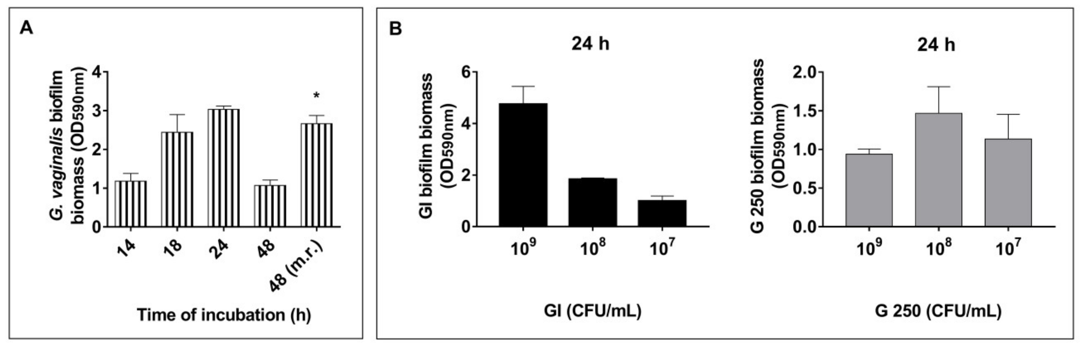
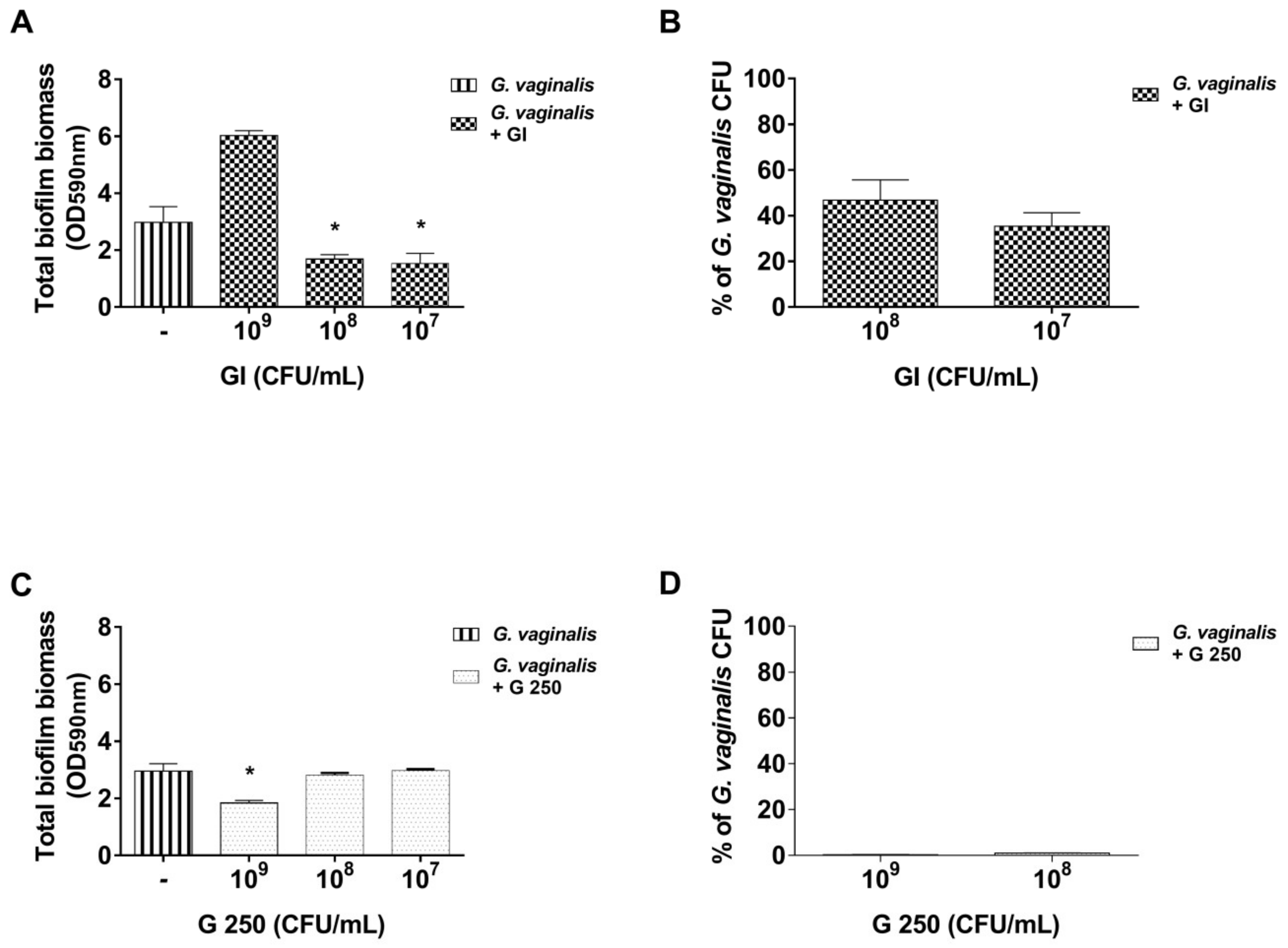
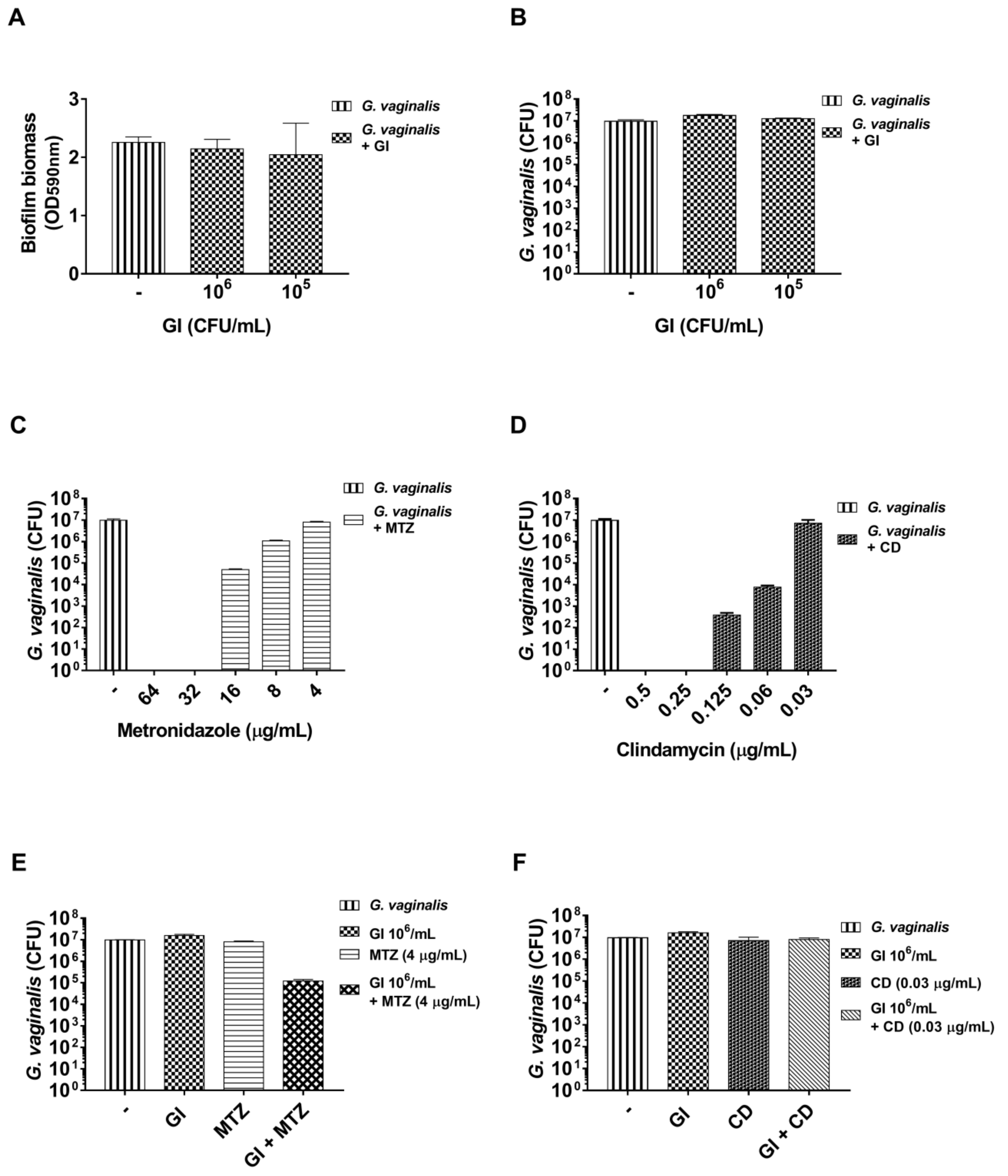
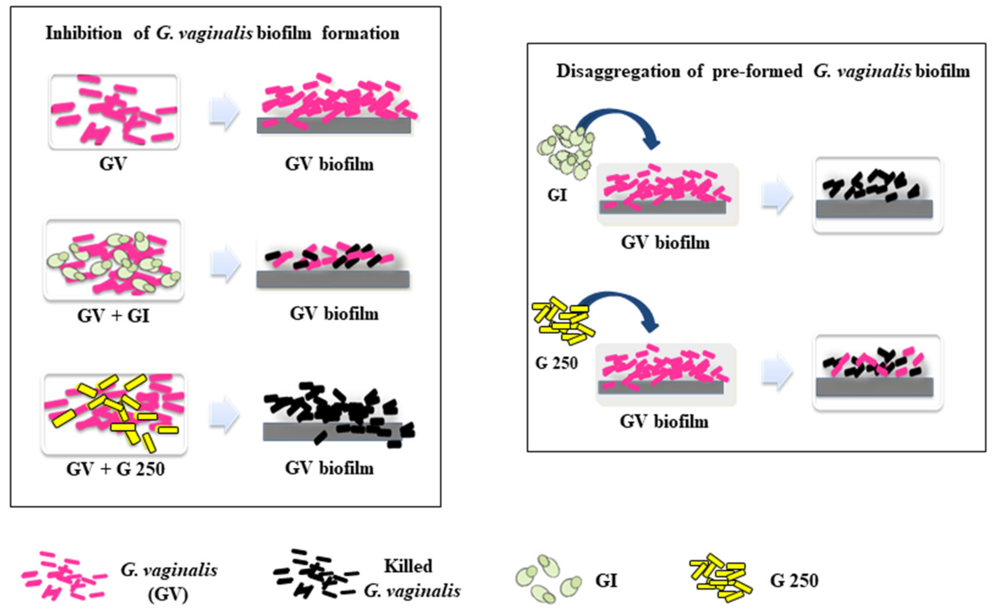
3.1. Effect of S. cerevisiae CNCM I-3856 (GI) and L. rhamnosus GG (G 250) on G. vaginalis Biofilm Formation

Firstly, the ability of a clinical isolate of G. vaginalis to develop a biofilm in our experimental conditions was evaluated. To this end, G. vaginalis (108 CFU/mL) was incubated for 14, 18, 24, and 48 h as described in the Materials and Methods section. Biofilm formation on an abiotic surface (polystyrene plates) was determined by CV biomass staining. The results, reported in Figure 1A, show that biofilm formation was observed up till 18–24 h of incubation and that the prolongation of incubation time resulted in a strong decrease of biofilm biomass, showing that G. vaginalis biofilm disintegration occurs from 24 h. This decrease was prevented by replacing the spent medium with fresh medium after 24 h (Figure 1A), as shown by the OD value obtained at 48 h medium renewal (m.r.).
It has been reported that biofilm formation by probiotic bacteria is considered a beneficial property because it could promote colonization and longer persistence on the mucosa of the host, thus inhibiting the growth of pathogenic bacteria [60]. As Saccharomyces spp. and Lactobacillus spp. have previously been shown to be able to form biofilm on abiotic surfaces [60,61], we tested the ability of GI and G 250 to form biofilm in our experimental system. The two probiotics were incubated for 24 h, as described in the Material and Methods section, and the CV staining method was used for biofilm biomass quantification. The results show that both probiotics were able to form biofilm on abiotic surfaces at all concentrations tested. In particular, GI produces biofilm in a dose-dependent manner whereas G 250 did not (Figure 1B).
The capacity of probiotics to inhibit G. vaginalis biofilm formation was then evaluated. To this end, GI, and G 250, at concentrations of 109, 108, and 107 CFU/mL, were mixed with G. vaginalis (108 CFU/mL) and biofilm development was evaluated after 24 h of incubation. The results show that, in the presence of GI at doses of 107 and 106 CFU/mL, the total biofilm biomass was significantly decreased compared with that of G. vaginalis mono-species biofilm (p < 0.05, Figure 2A), whereas at the dose of 109 CFU/mL, GI induced an increase in the overall mass of the biofilm, suggesting that the interaction between G. vaginalis and probiotics could have stimulated the growth of G. vaginalis, of the probiotic, or of both.
To determine the effect of GI on G. vaginalis viability in the dual-species biofilm, a CFU assay was performed by using the two concentrations that produced a significant reduction of total biofilm biomass compared to that of G. vaginalis alone. The results reported in Figure 2B show that both 108 and 107 CFU/mL of GI produced a strong reduction of G. vaginalis viability.
Regarding the effect of G 250 on G. vaginalis biofilm development, the results show that G 250 was able to cause a consistent inhibition of biofilm formation at a dose of 108 CFU/mL whereas no inhibition was observed at doses of 108 and 107 CFU/mL (Figure 2C).
To determine the effect of G 250 on G. vaginalis viability, a CFU assay was performed by using two doses: 109 that causes inhibition of total biofilm biomass formation and 108 that, like the dose of 107, did not. Of note, both the concentrations were able to achieve near-complete reduction of G. vaginalis viability (Figure 2D).

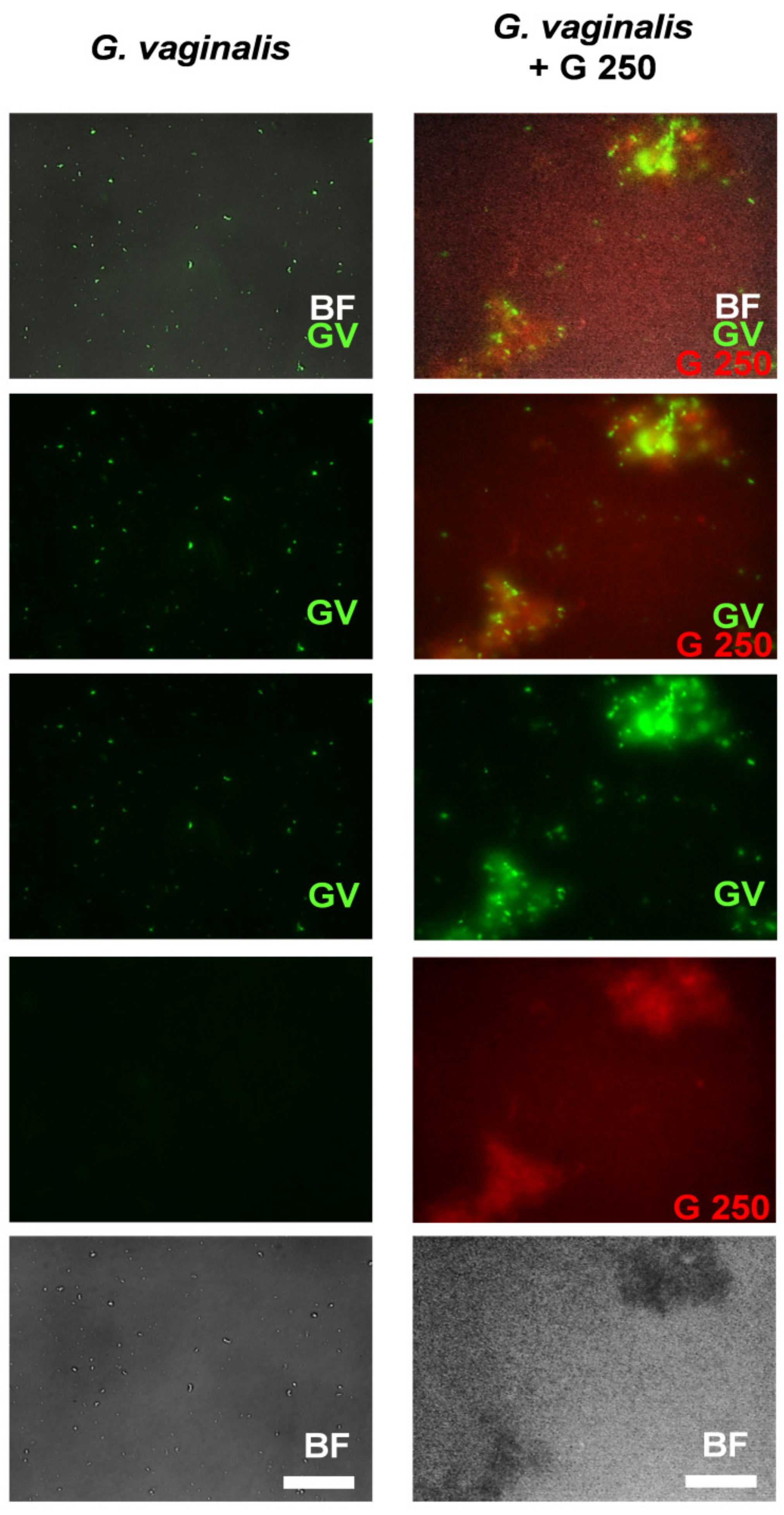
3.2. Co-Aggregation between L. rhamnosus GG (G250) and G. vaginalis

Co-aggregation is an important mechanism that influences development of complex multispecies biofilms [62] and can be involved in pathogens elimination by probiotics [63,64,65]. We recently demonstrated that GI does not induce G. vaginalis co-aggregation [49]. In this study, we analyzed if G 250 can co-aggregate with G. vaginalis. To this end, G 250 was incubated alone or mixed with G. vaginalis, as described in the Materials and Methods section. The results reported in Table 1 show that a low level of self-aggregation was manifested by G 250 while the strongest co-aggregation was observed with G. vaginalis.
A representative image taken using fluorescence microscopy demonstrating the capacity of probiotic to co-aggregate G. vaginalis is shown in Figure 3.

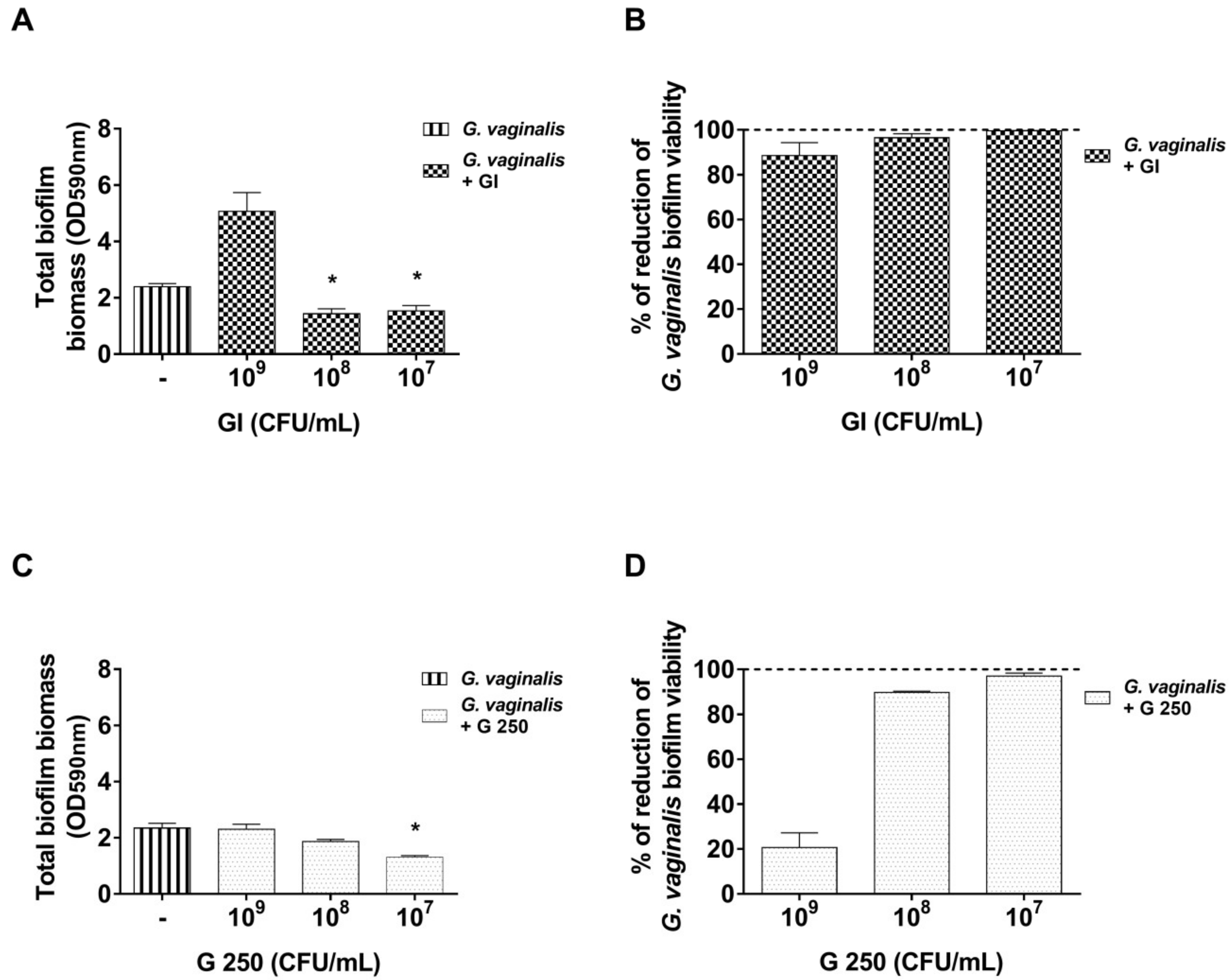
3.3. Effect of S. cerevisiae CNCM I-3856 (GI) and L. rhamnosus GG (G 250) on Preformed G. vaginalis Biofilm Disaggregation

To investigate the capacity of probiotics to disaggregate preformed biofilm, G. vaginalis (108 CFU/mL) was incubated for 24 h. Then, to avoid physiological disaggregation of a G. vaginalis biofilm (see Figure 1A), the medium was removed and fresh sBHI medium alone or containing GI or G 250 (109–107 CFU/mL) was added to the culture. After another 24 h of incubation, the biomass quantification and G. vaginalis biofilm viability were determined by the CV staining method and by a CFU assay, respectively. Our results show that GI at the doses of 108 and 107 CFU/mL significantly reduced biofilm biomass compared to the control (p < 0.05, Figure 4A).
Of note, all the doses tested (109–107 CFU/mL) killed almost 100% of G. vaginalis cells in the remaining biofilm (Figure 4B). Concerning G 250, our data show that it was able to induce a significant disaggregation of the preformed G. vaginalis biofilm only at 107 CFU/mL (p < 0.05, Figure 4C). The determination of G 250 effect on G. vaginalis biofilm viability showed that both 108 and 107 CFU/mL greatly reduced the viability of G. vaginalis (Figure 4D).
In subsequent experiments, we also evaluated if GI in association with suboptimal doses of metronidazole or clindamycin could be able to potentiate the antibiotic effect on mature G. vaginalis biofilm.

3.4. Effect of S. cerevisiae CNCM I-3856 (GI) Association with Metronidazole or Clindamycin on Preformed G. vaginalis Biofilm

To date, standard antibiotic therapies for G. vaginalis infection, metronidazole, and clindamycin, seem to be unable to fully eradicate BV vaginal biofilms [23].
In order to evaluate if GI can potentiate the antibiotic effect on preformed G. vaginalis biofilms, we first evaluated the suboptimal doses of the probiotic on biofilm biomass and viability. Our results show that GI at doses of 106 and 105 CFU/mL did not produce reduction both of total biofilm biomass and of G. vaginalis biofilm viability (Figure 5A,B).
Then, the MIC values for metronidazole and for clindamycin were evaluated. In our experimental conditions, metronidazole exhibited an MIC of 16 µg/mL and clindamycin exhibited an MIC of 0.0625 µg/mL. The minimal biofilm bactericidal concentration (BBC) for both antibiotics was then determined. To this end, 24 h preformed G. vaginalis biofilm was incubated for another 24 h with two-fold serial dilutions of metronidazole or clindamycin and G. vaginalis biofilm viability was determined by a CFU assay. As reported in Figure 5C,D, metronidazole and clindamycin exhibited BBC values of 32 µg/mL and 0.25 µg/mL, respectively, and the first dose that did not significantly reduce G. vaginalis viability was 4 µg/mL for metronidazole and 0.0312 µg/mL for clindamycin. These suboptimal concentrations have been selected for subsequent experiments.
The results reported in Figure 5E show that GI (106 CFU/mL) was able to potentiate the metronidazole activity and produced a very strong reduction of biofilm viability. Conversely, the results obtained from the association of GI with clindamycin showed that the combination of suboptimal doses of GI (106 CFU/mL) with suboptimal doses of clindamycin (0.0312 µg/mL) did not produce effects on G. vaginalis biofilm viability (Figure 5F).

4. Discussion

BV is a gynecological infection associated with the presence of a highly organized polymicrobial biofilm adhering to vaginal epithelial cells, with G. vaginalis having a key role in the constitution of this biofilm [28,66,67,68,69,70]. The establishment of this biofilm is a required step for BV initiation and development [16,71,72]. Conventional antibiotic therapy constitutes a first-line response but is ineffective in a high proportion of women, explaining the high recurrence rate [23,25,73]. Besides being associated with a number of side effects, including nausea, vomiting, and gastrointestinal complaints [23,74,75,76], antibiotic therapy, especially when prolonged or frequently repeated, can result in the development of resistant pathogens [18,23,25] without restoring a healthy vaginal microbiota [77]. Complementary and/or alternative therapeutic approaches for BV are therefore necessary. In the last years, several studies have reported the beneficial effect of potential anti-biofilm agents such as DNase [33]; retrocyclins [31]; antibacterial enzymes, peptides, antiseptics, and tensides [26,35]; quorum-sensing inhibitors subtilosin [32], and benzoyl peroxide [34]. Probiotics also seem to be a valid adjuvant or alternative option to the conventional therapeutic strategies both for prevention and for treatment of several human diseases such as bacterial vaginosis [37,38,43,78,79,80]. To date, there are limited studies on the effect of probiotics in inhibiting G. vaginalis biofilm formation or disaggregation [40,42,43] and none have been reported for S. cerevisiae. In this study, S. cerevisiae CNCM I-3856 and lactic acid bacteria L. rhamnosus GG (ATCC 53103) probiotics were tested for their ability to inhibit biofilm formation and to disaggregate mature G. vaginalis biofilms.
Recently, our research group demonstrated the beneficial effect of probiotics on vaginal candidiasis [46,47] and on BV [49,80]. In particular, in an experimental murine model of BV, we showed that S. cerevisiae CNCM I-3856 was able (i) to inhibit G. vaginalis adhesion to vaginal epithelium, (ii) to displace G. vaginalis adhered to epithelial cells, (iii) to reduce G. vaginalis vaginal load and sialidase activity, and iv) to decrease epithelial exfoliation [49].
Here, for the first time, we demonstrated that S. cerevisiae CNCM I-3856 as well as L. rhamnosus GG were able i) to significantly inhibit G. vaginalis biofilm formation, ii) to markedly reduce G. vaginalis viability among the biomass constituting the biofilm, iii) to induce disaggregation of preformed biofilms, and iv) to kill a consistent amount of bacterial cells in a G. vaginalis preformed biofilm (Figure 6).
Furthermore, S. cerevisiae strongly potentiates the antibiotic effects on G. vaginalis biofilm viability.
Several mechanisms [81] may interfere with biofilm formation and maintenance such as competition for nutrients and for binding sites, contact-dependent growth inhibition, secretion of inhibitory molecules, and inhibition of cell-to-cell communication. Given that we recently demonstrated that S. cerevisiae CNCM I-3856 (GI) is unable to co-aggregate with G. vaginalis [49], it is possible that S. cerevisiae CNCM I-3856 inhibition of G. vaginalis biofilm formation can be ascribed to biological instead of mechanical effects. Indeed, previous studies demonstrated that members of the Saccharomyces genus can produce extracellular protease and other compounds able to inhibit the growth of many pathogenic bacterial species [82,83]. In addition, given that the presence of the sialidase gene is associated with G. vaginalis biofilm formation [84], the capacity of S. cerevisiae CNCM I-3856 in inhibiting sialidase activity [49] could account for the observed effect.
Co-aggregation of pathogenic microorganisms is one of the mechanisms used by lactobacilli to maintain a healthy vaginal environment [65], and it is recognized as the one by which lactobacilli can exert their probiotic effects [65,85,86,87]. Indeed, differently from S. cerevisiae CNCM I-3856, L. rhamnosus GG was able to co-aggregate with G. vaginalis, suggesting that, by this interaction, L. rhamnosus GG produces around the G. vaginalis a microenvironment with an high concentration of antimicrobial substances [88] that not only may inhibit G. vaginalis growth but also may cause its death.
In the current study, we also demonstrated that S. cerevisiae CNCM I-3856 (108 and 107 CFU/mL) produced a significant disaggregation of the preformed G. vaginalis biofilm. This is consistent with our recent work showing that S. cerevisiae CNCM I-3856 is effective in displacing G. vaginalis adhered to vaginal and cervix epithelial cells [49]. Of note, S. cerevisiae CNCM I-3856 killed almost 100% of G. vaginalis cells in the biomass. This effect against G. vaginalis could be accounted for by two putative mechanisms. First, S. cerevisiae CNCM I-3856 can degrade the extracellular matrix by secreting enzymes [82] such as amylase, protease, and lipase that favor penetration of the yeast into the biofilm, allowing it to displace G. vaginalis from the abiotic surface. Second, the probiotic can have an antimicrobial activity by secreting specific compounds [82,83].
L. rhamnosus GG was also able to significantly disaggregate the G. vaginalis biofilm but only at 107 CFU/mL, probably because low doses facilitate penetration into the preformed biofilm. On the other hand, it was able to significantly reduce G. vaginalis viability in the biofilm in a dose-dependent manner. This is likely due to the production of antimicrobial compounds able to kill G. vaginalis cells [89,90,91].
In this study, we observed that the same doses of L. rhamnosus GG (107 CFU/mL) induced different effects in biofilm formation and biofilm disaggregation. This could be related to different mechanisms involved. Indeed, it is possible that low doses of L. rhamnosus GG are not sufficient to induce G. vaginalis coaggregation but could favor its penetration inside the biofilm, thus facilitating disruption.
Biofilms play an important role in conferring to microorganisms a marked increase in tolerance to antimicrobial agents [21]. The development of therapeutic combinations of antibiotics and probiotics may represent a promising alternative strategy to compromise biofilm maintenance, in particular, the use of yeast-based probiotics since yeast cannot promote the spread of antimicrobial resistance because exchange of antibiotic resistance genes with bacteria is unlikely. In our experimental system, the coadministration of S. cerevisiae CNCM I-3856 with metronidazole improved the efficacy of an otherwise suboptimal antibiotic dose. These results are encouraging as the use of low dosages of antibiotics could decrease side effects and the development of antibiotic resistant biofilms.
Our study presents, however, some limitations due to the fact that the biofilm obtained in this in vitro model is only constituted by G. vaginalis whereas the biofilm of bacterial vaginosis is a polymicrobial biofilm made up of G. vaginalis and other anaerobic bacteria, such as Prevotella bivia and Atopobium vaginae, which synergistically contribute to its formation [72,92].
Here, we demonstrate that both S. cerevisiae CNCM I-3856 and L. rhamnosus GG probiotics are able to inhibit G. vaginalis biofilm formation and to induce disaggregation of preformed biofilms. More importantly, we demonstrate that these probiotics can kill G. vaginalis during biofilm formation and can destroy almost completely G. vaginalis cells into mature biofilm. In addition, S. cerevisiae CNCM I-3856 strongly potentiate antibiotic efficacy. This should be considered important findings considering the difficulty to eradicate preformed biofilms from different bacteria [27,93,94,95].

5. Conclusions

The treatment of biofilm-involving infections is currently known to be very complicated partly due to the inefficiency of conventional antibiotic treatments in overcoming such infections.
Here, we proposed a novel and alternative approach for inhibition of G. vaginalis biofilm development and for disaggregation of the preformed one. Both probiotics evaluated were shown to be able to inhibit and to disrupt the G. vaginalis biofilm.
Furthermore, our results suggest that S. cerevisiae CNCM I-3856 is a good potential candidate to be used in combination with metronidazole for the treatment of bacterial vaginosis.

Author Contributions

Conceptualization, N.B. and A.V.; formal analysis, S.S., C.M., A.C.D., S.B., B.C., and S.P.; funding acquisition, N.B.; Investigation, S.S., and C.M.; methodology, S.S. and C.M.; project administration, C.M., N.B., and A.V.; resources, A.V.; supervision, A.V.; validation, A.V.; writing—original draft, C.M. and A.V.; writing—review and editing, C.M., N.B., A.C.D., and A.V. All authors contributed to the writing of the statement, agreed with its content and conclusions, and read and approved the final manuscript. All authors have read and agreed to the published version of the manuscript.

Funding

This work was performed in the frame of a service agreement supported by Lesaffre International.

Acknowledgments

The authors thank Sebastian Ku from Lesaffre Singapore for English language proofreading.

Conflicts of Interest

Nathalie Ballet and Amélie Cayzeele Decherf are full-time employed by the company Lesaffre International, Lesaffre Group, Marcq-en-Baroeul, France. All other authors declare no conflict of interest.

References

  1. Sobel, J.D. Bacterial Vaginosis. Annu. Rev. Med. 2000, 51, 349–356. [Google Scholar] [CrossRef]
  2. Guerra, B.; Ghi, T.; Quarta, S.; Morselli-Labate, A.M.; Lazzarotto, T.; Pilu, G.; Rizzo, N. Pregnancy outcome after early detection of bacterial vaginosis. Eur. J. Obstet. Gynecol. Reprod. Biol. 2006, 128, 40–45. [Google Scholar] [CrossRef]
  3. Leitich, H.; Bodner-Adler, B.; Brunbauer, M.; Kaider, A.; Egarter, C.; Husslein, P. Bacterial vaginosis as a risk factor for preterm delivery: A meta-analysis. Am. J. Obstet. Gynecol. 2003, 189, 139–147. [Google Scholar] [CrossRef] [PubMed]
  4. Acobsson, B.J.; Ernevi, P.P.; Hidekel, L.C.; Platz-Christensen, J.J. Bacterial vaginosis in early pregnancy may predispose for preterm birth and postpartum endometritis. Acta Obstet. Gynecol. Scand. 2002, 81, 1006–1010. [Google Scholar] [CrossRef] [PubMed]
  5. Rothman, K.J.; Funch, D.P.; Alfredson, T.; Brady, J.; Dreyer, N.A. Randomized field trial of vaginal douching, pelvic inflammatory disease and pregnancy. Epidemiology 2003, 14, 340–348. [Google Scholar] [CrossRef]
  6. Kavoussi, S.K.; Pearlman, M.D.; Burke, W.M.; Lebovic, D.I. Endometrioma complicated by tubo-ovarian abscess in a woman with bacterial vaginosis. Infect. Dis. Obstet. Gynecol. 2006, 2006, 84140. [Google Scholar] [CrossRef] [PubMed] [Green Version]
  7. Gallo, M.F.; Macaluso, M.; Warner, L.; Fleenor, M.E.; Hook, E.W.; Brill, I.; Weaver, M.A. Bacterial Vaginosis, Gonorrhea, and Chlamydial Infection Among Women Attending a Sexually Transmitted Disease Clinic: A Longitudinal Analysis of Possible Causal Links. Ann. Epidemiol. 2012, 22, 213–220. [Google Scholar] [CrossRef]
  8. Brotman, R.M. Vaginal microbiome and sexually transmitted infections: An epidemiologic perspective. J. Clin. Investig. 2011, 121, 4610–4617. [Google Scholar] [CrossRef]
  9. Koumans, E.H.; Sternberg, M.; Bruce, C.; McQuillan, G.; Kendrick, J.; Sutton, M.; Markowitz, L.E. The prevalence of bacterial vaginosis in the United States, 2001-2004; associations with symptoms, sexual behaviors, and reproductive health. Sex. Transm. Dis. 2007, 34, 864–869. [Google Scholar] [CrossRef]
  10. Ravel, J.; Gajer, P.; Abdo, Z.; Schneider, G.M.; Koenig, S.S.K.K.; McCulle, S.L.; Karlebach, S.; Gorle, R.; Russell, J.; Tacket, C.O.; et al. Vaginal microbiome of reproductive-age women. Proc. Natl. Acad. Sci. USA 2011, 108, 4680–4687. [Google Scholar] [CrossRef] [Green Version]
  11. Ceccarani, C.; Foschi, C.; Parolin, C.; D’Antuono, A.; Gaspari, V.; Consolandi, C.; Laghi, L.; Camboni, T.; Vitali, B.; Severgnini, M.; et al. Diversity of vaginal microbiome and metabolome during genital infections. Sci. Rep. 2019, 9, 14095. [Google Scholar] [CrossRef] [PubMed] [Green Version]
  12. Verhelst, R.; Verstraelen, H.; Claeys, G.; Verschraegen, G.; Delanghe, J.; Van Simaey, L.; De Ganck, C.; Temmerman, M.; Vaneechoutte, M. Cloning of 16S rRNA genes amplified from normal and disturbed vaginal microflora suggests a strong association between Atopobium vaginae, Gardnerella vaginalis and bacterial vaginosis. BMC Microbiol. 2004, 4, 16. [Google Scholar] [CrossRef] [Green Version]
  13. Muzny, C.A.; Taylor, C.M.; Swords, W.E.; Tamhane, A.; Chattopadhyay, D.; Cerca, N.; Schwebke, J.R. An Updated Conceptual Model on the Pathogenesis of Bacterial Vaginosis. J. Infect. Dis. 2019, 220, 1399–1405. [Google Scholar] [CrossRef] [PubMed]
  14. Amabebe, E.; Anumba, D.O.C. The vaginal microenvironment: The physiologic role of Lactobacilli. Front. Med. 2018, 5, 181. [Google Scholar] [CrossRef] [PubMed] [Green Version]
  15. Gilbert, N.M.; Lewis, W.G.; Lewis, A.L. Clinical Features of Bacterial Vaginosis in a Murine Model of Vaginal Infection with Gardnerella vaginalis. PLoS ONE 2013, 8, e59539. [Google Scholar] [CrossRef] [Green Version]
  16. Machado, A.; Cerca, N. Influence of biofilm formation by gardnerella vaginalis and other anaerobes on bacterial vaginosis. J. Infect. Dis. 2015, 212, 1856–1861. [Google Scholar] [CrossRef] [Green Version]
  17. Roselletti, E.; Sabbatini, S.; Perito, S.; Mencacci, A.; Vecchiarelli, A.; Monari, C. Apoptosis of vaginal epithelial cells in clinical samples from women with diagnosed bacterial vaginosis. Sci. Rep. 2020, 10, 1978. [Google Scholar] [CrossRef]
  18. Verstraelen, H.; Swidsinski, A. The biofilm in bacterial vaginosis: Implications for epidemiology, diagnosis and treatment: 2018 update. Curr. Opin. Infect. Dis. 2019, 32, 38–42. [Google Scholar] [CrossRef]
  19. Kostakioti, M.; Hadjifrangiskou, M.; Hultgren, S.J. Bacterial biofilms: Development, dispersal, and therapeutic strategies in the dawn of the postantibiotic era. Cold Spring Harb. Perspect. Med. 2013, 3, a010306. [Google Scholar] [CrossRef] [Green Version]
  20. Mah, T.F.C.; O’Toole, G.A. Mechanisms of biofilm resistance to antimicrobial agents. Trends Microbiol. 2001, 9, 34–39. [Google Scholar] [CrossRef]
  21. Macià, M.D.; Rojo-Molinero, E.; Oliver, A. Antimicrobial susceptibility testing in biofilm-growing bacteria. Clin. Microbiol. Infect. 2014, 20, 981–990. [Google Scholar] [CrossRef] [PubMed] [Green Version]
  22. Trampuz, A.; Widmer, A.F. Infections associated with orthopedic implants. Curr. Opin. Infect. Dis. 2006, 19, 349–356. [Google Scholar] [CrossRef] [PubMed]
  23. Machado, D.; Castro, J.; Palmeira-de-Oliveira, A.; Martinez-de-Oliveira, J.; Cerca, N. Bacterial vaginosis biofilms: Challenges to current therapies and emerging solutions. Front. Microbiol. 2016, 6, 1528. [Google Scholar] [CrossRef] [Green Version]
  24. Workowski, K.A.; Bolan, G.A. Sexually transmitted diseases treatment guidelines, 2015. MMWR Recomm. Reports 2015. [Google Scholar] [CrossRef]
  25. Bradshaw, C.S.S.; Morton, A.N.N.; Hocking, J.; Garland, S.M.M.; Morris, M.B.B.; Moss, L.M.M.; Horvath, L.B.B.; Kuzevska, I.; Fairley, C.K.K. High Recurrence Rates of Bacterial Vaginosis over the Course of 12 Months after Oral Metronidazole Therapy and Factors Associated with Recurrence. J. Infect. Dis. 2006, 193, 1478–1486. [Google Scholar] [CrossRef] [PubMed] [Green Version]
  26. Gottschick, C.; Szafranski, S.P.; Kunze, B.; Sztajer, H.; Masur, C.; Abels, C.; Wagner-Döbler, I. Screening of compounds against Gardnerella vaginalis biofilms. PLoS ONE 2016, 11, e0154086. [Google Scholar] [CrossRef] [PubMed]
  27. Swidsinski, A.; Mendling, W.; Loening-Baucke, V.; Swidsinski, S.; Dörffel, Y.; Scholze, J.; Lochs, H.; Verstraelen, H. An adherent Gardnerella vaginalis biofilm persists on the vaginal epithelium after standard therapy with oral metronidazole. Am. J. Obstet. Gynecol. 2008, 198, 97.e1–97.e6. [Google Scholar] [CrossRef]
  28. Swidsinski, A.; Guschin, A.; Tang, Q.; Dörffel, Y.; Verstraelen, H.; Tertychnyy, A.; Khayrullina, G.; Luo, X.; Sobel, J.D.; Jiang, X. Vulvovaginal candidiasis: Histologic lesions are primarily polymicrobial and invasive and do not contain biofilms. Am. J. Obstet. Gynecol. 2019, 220, 91.e1–91.e8. [Google Scholar] [CrossRef] [Green Version]
  29. Marrs, C.N.; Knobel, S.M.; Zhu, W.Q.; Sweet, S.D.; Chaudhry, A.R.; Alcendor, D.J. Evidence for Gardnerella vaginalis uptake and internalization by squamous vaginal epithelial cells: Implications for the pathogenesis of bacterial vaginosis. Microbes Infect. 2012, 14, 500–508. [Google Scholar] [CrossRef] [Green Version]
  30. Alves, P.; Castro, J.; Sousa, C.; Cereija, T.B.; Cerca, N. Gardnerella vaginalis Outcompetes 29 other bacterial species isolated from patients with bacterial vaginosis, using in an in vitro biofilm formation model. J. Infect. Dis. 2014, 210, 593–596. [Google Scholar] [CrossRef] [Green Version]
  31. Hooven, T.A.; Randis, T.M.; Hymes, S.R.; Rampersaud, R.; Ratner, A.J. Retrocyclin inhibits Gardnerella vaginalis biofilm formation and toxin activity. J. Antimicrob. Chemother. 2012, 67, 2870–2872. [Google Scholar] [CrossRef] [PubMed]
  32. Algburi, A.; Zehm, S.; Netrebov, V.; Bren, A.B.; Chistyakov, V.; Chikindas, M.L. Subtilosin Prevents Biofilm Formation by Inhibiting Bacterial Quorum Sensing. Probiotics Antimicrob. Proteins 2017, 9, 81–90. [Google Scholar] [CrossRef] [PubMed]
  33. Hymes, S.R.; Randis, T.M.; Sun, T.Y.; Ratner, A.J. DNase inhibits gardnerella vaginalis biofilms in vitro and in vivo. J. Infect. Dis. 2013, 207, 1491–1497. [Google Scholar] [CrossRef] [PubMed] [Green Version]
  34. Algburi, A.; Zehm, S.; Netrebov, V.; Weeks, R.; Zubovskiy, K.; Chikindas, M.L. Benzoyl Peroxide Inhibits Quorum Sensing and Biofilm Formation by Gardnerella vaginalis 14018. Infect. Dis. Obstet. Gynecol. 2018, 2018, 1426109. [Google Scholar] [CrossRef]
  35. Gottschick, C.; Deng, Z.L.; Vital, M.; Masur, C.; Abels, C.; Pieper, D.H.; Rohde, M.; Mendling, W.; Wagner-Döbler, I. Treatment of biofilms in bacterial vaginosis by an amphoteric tenside pessary-clinical study and microbiota analysis. Microbiome 2017, 5, 119. [Google Scholar] [CrossRef] [Green Version]
  36. McFarland, L.V. Use of probiotics to correct dysbiosis of normal microbiota following disease or disruptive events: A systematic review. BMJ Open 2014, 4, e005047. [Google Scholar] [CrossRef] [Green Version]
  37. Wang, Z.; He, Y.; Zheng, Y. Probiotics for the treatment of bacterial vaginosis: A meta-analysis. Int. J. Environ. Res. Public Health 2019, 16, 3859. [Google Scholar] [CrossRef] [Green Version]
  38. Li, C.; Wang, T.; Li, Y.; Zhang, T.; Wang, Q.; He, J.; Wang, L.; Li, L.; Yang, N.; Fang, Y. Probiotics for the treatment of women with bacterial vaginosis: A systematic review and meta-analysis of randomized clinical trials. Eur. J. Pharmacol. 2019, 864, 172660. [Google Scholar] [CrossRef]
  39. Superti, F.; De Seta, F. Warding off recurrent yeast and bacterial vaginal infections: Lactoferrin and lactobacilli. Microorganisms 2020, 8, 130. [Google Scholar] [CrossRef] [Green Version]
  40. Vuotto, C.; Longo, F.; Donelli, G. Probiotics to counteract biofilm-associated infections: Promising and conflicting data. Int. J. Oral Sci. 2014, 6, 189–194. [Google Scholar] [CrossRef] [Green Version]
  41. Lebeer, S.; Verhoeven, T.L.A.; Vélez, M.P.; Vanderleyden, J.; De Keersmaecker, S.C.J. Impact of environmental and genetic factors on biofilm formation by the probiotic strain Lactobacillus rhamnosus GG. Appl. Environ. Microbiol. 2007, 73, 6768–6775. [Google Scholar] [CrossRef] [PubMed] [Green Version]
  42. Saunders, S.; Bocking, A.; Challis, J.; Reid, G. Effect of Lactobacillus challenge on Gardnerella vaginalis biofilms. Colloids Surfaces B Biointerfaces 2007, 55, 138–142. [Google Scholar] [CrossRef] [PubMed]
  43. McMillan, A.; Dell, M.; Zellar, M.P.; Cribby, S.; Martz, S.; Hong, E.; Fu, J.; Abbas, A.; Dang, T.; Miller, W.; et al. Disruption of urogenital biofilms by lactobacilli. Colloids Surfaces B Biointerfaces 2011, 86, 58–64. [Google Scholar] [CrossRef] [PubMed]
  44. Segers, M.E.; Lebeer, S. Towards a better understanding of Lactobacillus rhamnosus GG - host interactions. Microb. Cell Fact. 2014, 13, S7. [Google Scholar] [CrossRef] [PubMed] [Green Version]
  45. Sivignon, A.; De Vallée, A.; Barnich, N.; Denizot, J.; Darcha, C.; Pignède, G.; Vandekerckove, P.; Darfeuille-Michaud, A. Saccharomyces cerevisiae CNCM I-3856 prevents colitis induced by AIEC bacteria in the transgenic mouse model mimicking Crohn’s disease. Inflamm. Bowel Dis. 2015, 21, 276–286. [Google Scholar] [CrossRef]
  46. Pericolini, E.; Gabrielli, E.; Ballet, N.; Sabbatini, S.; Roselletti, E.; Cayzeele Decherf, A.; Pélerin, F.; Luciano, E.; Perito, S.; Jüsten, P.; et al. Therapeutic activity of a Saccharomyces cerevisiae-based probiotic and inactivated whole yeast on vaginal candidiasis. Virulence 2017, 8, 74–90. [Google Scholar] [CrossRef] [Green Version]
  47. Gabrielli, E.; Pericolini, E.; Ballet, N.; Roselletti, E.; Sabbatini, S.; Mosci, P.; Decherf, A.C.; Pélerin, F.; Perito, S.; Jüsten, P.; et al. Saccharomyces cerevisiae-based probiotic as novel anti-fungal and anti-inflammatory agent for therapy of vaginal candidiasis. Benef. Microbes 2018, 9, 219–230. [Google Scholar] [CrossRef]
  48. Graf, K.; Last, A.; Gratz, R.; Allert, S.; Linde, S.; Westermann, M.; Gröger, M.; Mosig, A.S.; Gresnigt, M.S.; Hube, B. Keeping Candida commensal: How lactobacilli antagonize pathogenicity of Candida albicans in an in vitro gut model. DMM Dis. Model. Mech. 2019, 12, dmm039719. [Google Scholar] [CrossRef] [Green Version]
  49. Sabbatini, S.; Monari, C.; Ballet, N.; Mosci, P.; Decherf, A.C.; Pélerin, F.; Perito, S.; Scarpelli, P.; Vecchiarelli, A. Saccharomyces cerevisiae–based probiotic as novel anti-microbial agent for therapy of bacterial vaginosis. Virulence 2018, 9, 954–966. [Google Scholar] [CrossRef] [Green Version]
  50. Liévin-Le Moal, V.; Servin, A.L. Anti-infective activities of Lactobacillus strains in the human intestinal microbiota: From probiotics to gastrointestinal anti- infectious biotherapeutic agents. Clin. Microbiol. Rev. 2014, 27, 167–199. [Google Scholar] [CrossRef] [Green Version]
  51. Kurtzman, C.P.; Robnett, C.J. Identification of clinically important ascomycetous yeasts based on nucleotide divergence in the 5′ end of the large-subunit (26S) ribosomal DNA gene. J. Clin. Microbiol. 1997, 35, 1216–1223. [Google Scholar] [CrossRef] [PubMed] [Green Version]
  52. Kurtzman, C.P.; Robnett, C.J. Identification and phylogeny of ascomycetous yeasts from analysis of nuclear large subunit (26S) ribosomal DNA partial sequences. Antonie van Leeuwenhoek Int. J. Gen. Mol. Microbiol. 1998, 73, 331–371. [Google Scholar] [CrossRef] [PubMed]
  53. European Committee for Standardization. 15790:2008 PCR typing of probiotic strains of Saccharomyces cerevisiae (yeast); European Committee for Standardization: Brussels, Belgium, 2009; ISBN ISBN 978-0-580-61806-2. [Google Scholar]
  54. Westerik, N.; Kort, R.; Sybesma, W.; Reid, G. Lactobacillus rhamnosus probiotic food as a tool for empowerment across the value chain in Africa. Front. Microbiol. 2018, 9, 1501. [Google Scholar] [CrossRef] [PubMed] [Green Version]
  55. Zheng, J.; Wittouck, S.; Salvetti, E.; Franz, C.M.; Harris, H.M.; Mattarelli, P.; O’Toole, P.W.; Pot, B.; Vandamme, P.; Walter, J.; et al. A taxonomic note on the genus Lactobacillus: Description of 23 novel genera, emended description of the genus Lactobacillus Beijerinck 1901, and union of Lactobacillaceae and Leuconostocaceae. Int. J. Syst. Evol. Microbiol. 2020, 70, 2782–2858. [Google Scholar] [CrossRef] [PubMed]
  56. Machado, D.; Palmeira-de-Oliveira, A.; Cerca, N. Optimization of culture conditions for Gardnerella vaginalis biofilm formation. J. Microbiol. Methods 2015, 118, 143–146. [Google Scholar] [CrossRef] [PubMed] [Green Version]
  57. Jaffar, N.; Ishikawa, Y.; Mizuno, K.; Okinaga, T.; Maeda, T. Mature biofilm degradation by potential probiotics: Aggregatibacter actinomycetemcomitans versus Lactobacillus spp. PLoS ONE 2016, 11, e0159466. [Google Scholar] [CrossRef] [PubMed]
  58. Gabrielli, E.; Pericolini, E.; Cenci, E.; Monari, C.; Magliani, W.; Ciociola, T.; Conti, S.; Gatti, R.; Bistoni, F.; Polonelli, L.; et al. Antibody constant region peptides can display immunomodulatory activity through activation of the dectin-1 signalling pathway. PLoS ONE 2012, 7, e43972. [Google Scholar] [CrossRef] [Green Version]
  59. Noll, K.S.; Prichard, M.N.; Khaykin, A.; Sinko, P.J.; Chikindas, M.L. The natural antimicrobial peptide subtilosin acts synergistically with glycerol monolaurate, lauric arginate, and ε-poly-L-lysine against bacterial vaginosis-associated pathogens but not human lactobacilli. Antimicrob. Agents Chemother. 2012, 56, 1756–1761. [Google Scholar] [CrossRef] [Green Version]
  60. Salas-Jara, M.; Ilabaca, A.; Vega, M.; García, A. Biofilm Forming Lactobacillus: New Challenges for the Development of Probiotics. Microorganisms 2016, 4, 35. [Google Scholar] [CrossRef]
  61. Bojsen, R.K.; Andersen, K.S.; Regenberg, B. Saccharomyces cerevisiae—A model to uncover molecular mechanisms for yeast biofilm biology. FEMS Immunol. Med. Microbiol. 2012, 65, 169–182. [Google Scholar] [CrossRef] [Green Version]
  62. Rickard, A.H.; Gilbert, P.; High, N.J.; Kolenbrander, P.E.; Handley, P.S. Bacterial coaggregation: An integral process in the development of multi-species biofilms. Trends Microbiol. 2003, 11, 94–100. [Google Scholar] [CrossRef]
  63. Mastromarino, P.; Brigidi, P.; Macchia, S.; Maggi, L.; Pirovano, F.; Trinchieri, V.; Conte, U.; Matteuzzi, D. Characterization and selection of vaginal Lactobacillus strains for the preparation of vaginal tablets. J. Appl. Microbiol. 2002, 93, 884–893. [Google Scholar] [CrossRef] [PubMed]
  64. Kaewsrichan, J.; Peeyananjarassri, K.; Kongprasertkit, J. Selection and identification of anaerobic lactobacilli producing inhibitory compounds against vaginal pathogens. FEMS Immunol. Med. Microbiol. 2006, 48, 75–83. [Google Scholar] [CrossRef] [PubMed] [Green Version]
  65. Al Kassaa, I.; Hamze, M.; Hober, D.; Chihib, N.E.; Drider, D. Identification of vaginal lactobacilli with potential probiotic properties isolated from women in North Lebanon. Microb. Ecol. 2014, 67, 722–734. [Google Scholar] [CrossRef]
  66. Swidsinski, A.; Mendling, W.; Loening-Baucke, V.; Ladhoff, A.; Swidsinski, S.; Hale, L.P.; Lochs, H. Adherent biofilms in bacterial vaginosis. Obstet. Gynecol. 2005, 106, 1013–1023. [Google Scholar] [CrossRef] [Green Version]
  67. Menard, J.; Fenollar, F.; Henry, M.; Bretelle, F.; Raoult, D. Molecular Quantification of Gardnerella vaginalis and Atopobium vaginae Loads to Predict Bacterial Vaginosis. Clin. Infect. Dis. 2008, 47, 33–43. [Google Scholar] [CrossRef] [Green Version]
  68. Schilling, J.; Loening-Baucke, V.; Dörffel, Y. Increased Gardnerella vaginalis urogenital biofilm in inflammatory bowel disease. J. Crohn’s Colitis 2014, 8, 543–549. [Google Scholar] [CrossRef]
  69. Jung, H.S.; Ehlers, M.M.; Lombaard, H.; Redelinghuys, M.J.; Kock, M.M. Etiology of bacterial vaginosis and polymicrobial biofilm formation. Crit. Rev. Microbiol. 2017, 43, 651–667. [Google Scholar] [CrossRef] [Green Version]
  70. Hardy, L.; Cerca, N.; Jespers, V.; Vaneechoutte, M.; Crucitti, T. Bacterial biofilms in the vagina. Res. Microbiol. 2017, 168, 865–874. [Google Scholar] [CrossRef] [Green Version]
  71. Castro, J.; Alves, P.; Sousa, C.; Cereija, T.; França, Â.; Jefferson, K.K.; Cerca, N. Using an in-vitro biofilm model to assess the virulence potential of Bacterial Vaginosis or non-Bacterial Vaginosis Gardnerella vaginalis isolates. Sci. Rep. 2015, 5, 11640. [Google Scholar] [CrossRef] [Green Version]
  72. Muzny, C.A.; Schwebke, J.R. Biofilms: An Underappreciated Mechanism of Treatment Failure and Recurrence in Vaginal Infections. Clin. Infect. Dis. 2015, 61, 601–606. [Google Scholar] [CrossRef] [PubMed] [Green Version]
  73. Sanchez, S.; Garcia, P.J.; Thomas, K.K.; Catlin, M.; Holmes, K.K. Intravaginal metronidazole gel versus metronidazole plus nystatin ovules for bacterial vaginosis: A randomized controlled trial. Am. J. Obstet. Gynecol. 2004, 191, 1898–1906. [Google Scholar] [CrossRef] [PubMed]
  74. Schwebke, J.R.; Desmond, R.A. Tinidazole vs metronidazole for the treatment of bacterial vaginosis. Am. J. Obstet. Gynecol. 2011, 204, 211.e1–211.e6. [Google Scholar] [CrossRef] [PubMed] [Green Version]
  75. Abdali, K.; Jahed, L.; Amooee, S.; Zarshenas, M.; Tabatabaee, H.; Bekhradi, R. Comparison of the Effect of Vaginal Zataria multiflora Cream and Oral Metronidazole Pill on Results of Treatments for Vaginal Infections including Trichomoniasis and Bacterial Vaginosis in Women of Reproductive Age. Biomed. Res. Int. 2015, 2015, 683640. [Google Scholar] [CrossRef] [Green Version]
  76. Chavoustie, S.E.; Jacobs, M.; Reisman, H.A.; Waldbaum, A.S.; Levy, S.F.; Hillier, S.L.; Nyirjesy, P. Metronidazole vaginal gel 1.3% in the treatment of bacterial vaginosis: A dose-ranging study. J. Low. Genit. Tract Dis. 2015, 19, 129–134. [Google Scholar] [CrossRef] [Green Version]
  77. Mayer, B.T.; Srinivasan, S.; Fiedler, T.L.; Marrazzo, J.M.; Fredricks, D.N.; Schiffer, J.T. Rapid and profound shifts in the vaginal microbiota following antibiotic treatment for bacterial vaginosis. J. Infect. Dis. 2015, 212, 793–802. [Google Scholar] [CrossRef] [Green Version]
  78. Khani, S.; Hosseini, H.M.; Taheri, M.; Nourani, M.R.; Fooladi, A.A.I. Probiotics as an alternative strategy for prevention and treatment of human diseases: A review. Inflamm. Allergy - Drug Targets 2012, 11, 79–89. [Google Scholar] [CrossRef]
  79. Al-Ghazzewi, F.H.; Tester, R.F. Biotherapeutic agents and vaginal health. J. Appl. Microbiol. 2016, 121, 18–27. [Google Scholar] [CrossRef]
  80. Gaziano, R.; Sabbatini, S.; Roselletti, E.; Perito, S.; Monari, C. Saccharomyces cerevisiae-Based Probiotics as Novel Antimicrobial Agents to Prevent and Treat Vaginal Infections. Front. Microbiol. 2020, 11, 718. [Google Scholar] [CrossRef] [Green Version]
  81. Rendueles, O.; Ghigo, J.-M. Mechanisms of Competition in Biofilm Communities. Microbiol. Spectr. 2015, 3, 319–342. [Google Scholar] [CrossRef] [Green Version]
  82. Fakruddin, M.; Hossain, M.N.; Ahmed, M.M. Antimicrobial and antioxidant activities of Saccharomyces cerevisiae IFST062013, a potential probiotic. BMC Complement. Altern. Med. 2017, 17, 64. [Google Scholar] [CrossRef] [Green Version]
  83. Younis, G.; Awad, A.; Dawod, R.E.; Yousef, N.E. Antimicrobial activity of yeasts against some pathogenic bacteria. Vet. World 2017, 10, 979–983. [Google Scholar] [CrossRef] [PubMed] [Green Version]
  84. Hardy, L.; Jespers, V.; Van Bulck, M.D.; Buyze, J.; Mwambarangwe, L.; Musengamana, V.; Vaneechoutte, M.; Crucitti, T. The presence of the putative Gardnerella vaginalis sialidase A gene in vaginal specimens is associated with bacterial vaginosis biofilm. PLoS ONE 2017, 12, e0172522. [Google Scholar] [CrossRef] [PubMed] [Green Version]
  85. Osset, J.; Bartolomé, R.M.; García, E.; Andreu, A. Assessment of the Capacity of Lactobacillus to Inhibit the Growth of Uropathogens and Block Their Adhesion to Vaginal Epithelial Cells. J. Infect. Dis. 2001, 183, 485–491. [Google Scholar] [CrossRef] [PubMed] [Green Version]
  86. Aslim, B.; Onal, D.; Beyatli, Y. Factors influencing autoaggregation and aggregation of Lactobacillus delbrueckii subsp. bulgaricus isolated from handmade yogurt. J. Food Prot. 2007, 70, 223–227. [Google Scholar] [CrossRef] [PubMed]
  87. Younes, J.A.; van der Mei, H.C.; van den Heuvel, E.; Busscher, H.J.; Reid, G. Adhesion forces and coaggregation between vaginal staphylococci and lactobacilli. PLoS ONE 2012, 7, e36917. [Google Scholar] [CrossRef] [Green Version]
  88. Santos, C.M.A.; Pires, M.C.V.; Leão, T.L.; Hernández, Z.P.; Rodriguez, M.L.; Martins, A.K.S.; Miranda, L.S.; Martins, F.S.; Nicoli, J.R. Selection of Lactobacillus strains as potential probiotics for vaginitis treatment. Microbiology 2016, 162, 1195–1207. [Google Scholar] [CrossRef]
  89. Lu, R.; Fasano, S.; Madayiputhiya, N.; Morin, N.P.; Nataro, J.; Fasano, A. Isolation, identification, and characterization of small bioactive peptides from Lactobacillus GG conditional media that exert both anti-gram-negative and Gram-positive bactericidal activity. J. Pediatr. Gastroenterol. Nutr. 2009, 49, 23–30. [Google Scholar] [CrossRef]
  90. Coman, M.M.; Verdenelli, M.C.; Cecchini, C.; Silvi, S.; Orpianesi, C.; Boyko, N.; Cresci, A. In vitro evaluation of antimicrobial activity of Lactobacillus rhamnosus IMC 501®, Lactobacillus paracasei IMC 502® and SYNBIO® against pathogens. J. Appl. Microbiol. 2014, 117, 518–527. [Google Scholar] [CrossRef]
  91. Miquel, S.; Lagrafeuille, R.; Souweine, B.; Forestier, C. Anti-biofilm activity as a health issue. Front. Microbiol. 2016, 7, 592. [Google Scholar] [CrossRef]
  92. Castro, J.; Rosca, A.S.; Cools, P.; Vaneechoutte, M.; Cerca, N. Gardnerella vaginalis Enhances Atopobium vaginae Viability in an in vitro Model. Front. Cell. Infect. Microbiol. 2020, 10, 83. [Google Scholar] [CrossRef] [PubMed] [Green Version]
  93. Lewis, K. Persister Cells. Annu. Rev. Microbiol. 2010, 64, 357–372. [Google Scholar] [CrossRef] [PubMed]
  94. Conlon, B.P.; Rowe, S.E.; Lewis, K. Persister cells in biofilm associated infections. Adv. Exp. Med. Biol. 2015, 831, 1–9. [Google Scholar] [CrossRef] [PubMed]
  95. Figueiredo, A.M.S.; Ferreira, F.A.; Beltrame, C.O.; Côrtes, M.F. The role of biofilms in persistent infections and factors involved in ica-independent biofilm development and gene regulation in Staphylococcus aureus. Crit. Rev. Microbiol. 2017, 43, 602–620. [Google Scholar] [CrossRef]
Figure 1. Gardnerella vaginalis, Saccharomyces cerevisiae CNCM (French National Collection of Cultures of Microorganisms) I-3856 (GI), and Lacticaseibacillus rhamnosus GG (G 250) biofilm formation: (A) G. vaginalis (108 colony forming units (CFU)/mL) biofilm formation after 14, 18, 24, and 48 h of incubation. For the 48 h m.r. (medium renewal) value, the medium was replaced by a fresh medium after 24 h of incubation. (B) Biofilm formation by GI and G 250 (109, 108, and 107 CFU/mL) after 24 h. The biofilm biomass was measured by crystal violet staining (OD, optical density). Results are the mean ± SEM from 3 independent experiments (each with n = 3). * p < 0.05 48 h (m.r.) G. vaginalis biofilm vs. 48 h G. vaginalis biofilm. Data are the mean ± SEM from 4 independent experiments (each with n = 3).
Microorganisms 08 01294 g001
Figure 2. Effect of S. cerevisiae CNCM I-3856 (GI) and L. rhamnosus GG (G 250) on G. vaginalis biofilm formation: G. vaginalis (108 CFU/mL) was incubated in presence or absence of GI (A,B) or G 250 (C,D), both at 109–107 CFU/mL for 24 h as described in the Materials and Methods section. Then, the following were determined: (A,C) biofilm biomass development and (B,D) percentage of G. vaginalis CFU recovered from dual-species biofilm with respect to the CFU from a G. vaginalis single-species biofilm. Biofilm formation has been evaluated by crystal violet staining (OD, optical density). Data are the mean ± SEM from 4 independent experiments (each with n = 3) for biomass determination and one experiment (n = 3) for CFU evaluation. * p < 0.05 probiotic (GI or G 250)–G. vaginalis biofilm vs. G. vaginalis monomicrobial biofilm.
Microorganisms 08 01294 g002
Figure 3. Co-aggregation between L. rhamnosus GG (G 250) and G. vaginalis: G. vaginalis or fluorescein isothiocyanate-labeled (FITC)-G. vaginalis (109 CFU/mL) in 500 μL of phosphate-buffered saline (PBS) were mixed or not with equal volumes of G 250 or RHB-G 250 (G 250) (109 CFU/mL). The samples were vortexed for at least 10 s and incubated in a 24-well plate for 4 h at 37 °C under agitation. The suspensions were then photographed by fluorescence microscopy. Images are from one experiment (scale bar = 50 μm, magnification 20×). BF = bright field; G. vaginalis (GV) = green; and G 250 (G 250) = red.
Microorganisms 08 01294 g003
Figure 4. Effect of S. cerevisiae CNCM I-3856 (GI) and L. rhamnosus GG (G 250) on G. vaginalis biofilm disaggregation: G. vaginalis (108 CFU/mL) was incubated for 24 h to develop mature biofilm. After incubation, the medium was removed, GI (A,B) or G 250 (C,D) were added at different concentrations, and the co-cultures were incubated for a further 24 h as described in the Materials and Methods section. Then, the following were determined: (A,C) biomass disaggregation and (B,D) percentage of G. vaginalis biofilm viability reduction. Biofilm disaggregation has been evaluated by crystal violet staining (OD, optical density). The G. vaginalis biofilm viability has been determined by a colony forming units (CFU) assay. Percentages of biofilm viability reduction probiotics have been quantified with respect to untreated G. vaginalis biofilms which were taken as 100% (dotted line). Data are the mean ± SEM from 3 independent experiments (each with n = 3) for biomass determination and from 2 independent experiments (each with n = 2) for viability evaluation. * p < 0.05 GI or G 250-treated G. vaginalis biofilm vs. untreated G. vaginalis biofilm.
Microorganisms 08 01294 g004
Figure 5. Effect of S. cerevisiae CNCM I-3856 (GI)-antibiotic combination on preformed G. vaginalis biofilm: (A) G. vaginalis (108 CFU/mL) was incubated for 24 h to develop mature biofilm. After incubation, the medium was removed, GI was added at different concentrations, and the co-cultures were incubated for a further 24 h as described in the Materials and Methods section. Biofilm formation has been evaluated by crystal violet staining (OD, optical density). Data are the mean ± SEM from three experiments, each with n = 3. (B) G. vaginalis biofilm viability was determined by a colony forming units (CFU) assay. Data are the mean ± SEM from one experiment with n = 3. (C,D) G. vaginalis (108 CFU/mL) was incubated for 24 h to develop mature biofilm. After incubation, the medium was removed and metronidazole (MTZ) (C) or clindamycin (CD) (D) were added to the preformed biofilm and incubated for another 24 h to determine the minimal biofilm bactericidal concentration as described in the Materials and Methods section. Data are the mean ± SEM from one experiment with n = 3. (E) G. vaginalis (108 CFU/mL) preformed biofilm was incubated with GI (106 CFU/mL), metronidazole (4 µg/mL), or metronidazole (4 µg/mL) plus GI (106 CFU/mL) in the same experimental conditions as above described, and G. vaginalis biofilm viability was determined by a CFU assay. Data are the mean ± SEM from one experiment with n = 3. (F) G. vaginalis (108 CFU/mL) preformed biofilm was incubated with GI (106 CFU/mL), clindamycin (0.0312 µg/mL), or clindamycin (0.0312 µg/mL) plus GI (106 CFU/mL) in the same experimental conditions as above described. G. vaginalis biofilm viability has been determined by a colony forming unit (CFU) assay. Data are the mean ± SEM from one experiment with n = 3.
Microorganisms 08 01294 g005
Figure 6. Representation of anti-G. vaginalis biofilm activity of S. cerevisiae CNCM I-3856 (GI) and L. rhamnosus GG (G250).
Microorganisms 08 01294 g006
Table 1. Scores of G. vaginalis and L. rhamnosus GG (G 250) co-aggregation.
Co-Aggregation Scores *
G. vaginalisG 250G. vaginalis + G 250
EXP 1003
EXP 2013
EXP 3023.5
Mean score013.16
* Scores from 0 (no aggregation) to 4 (maximum aggregation).

