
Low-Dose Doxycycline Treatment Normalizes Levels of Some Salivary Metabolites Associated with Oral Microbiota in Patients with Primary Sjögren’s Syndrome

 , ,
, ,  , , and
Abstract
:1. Introduction
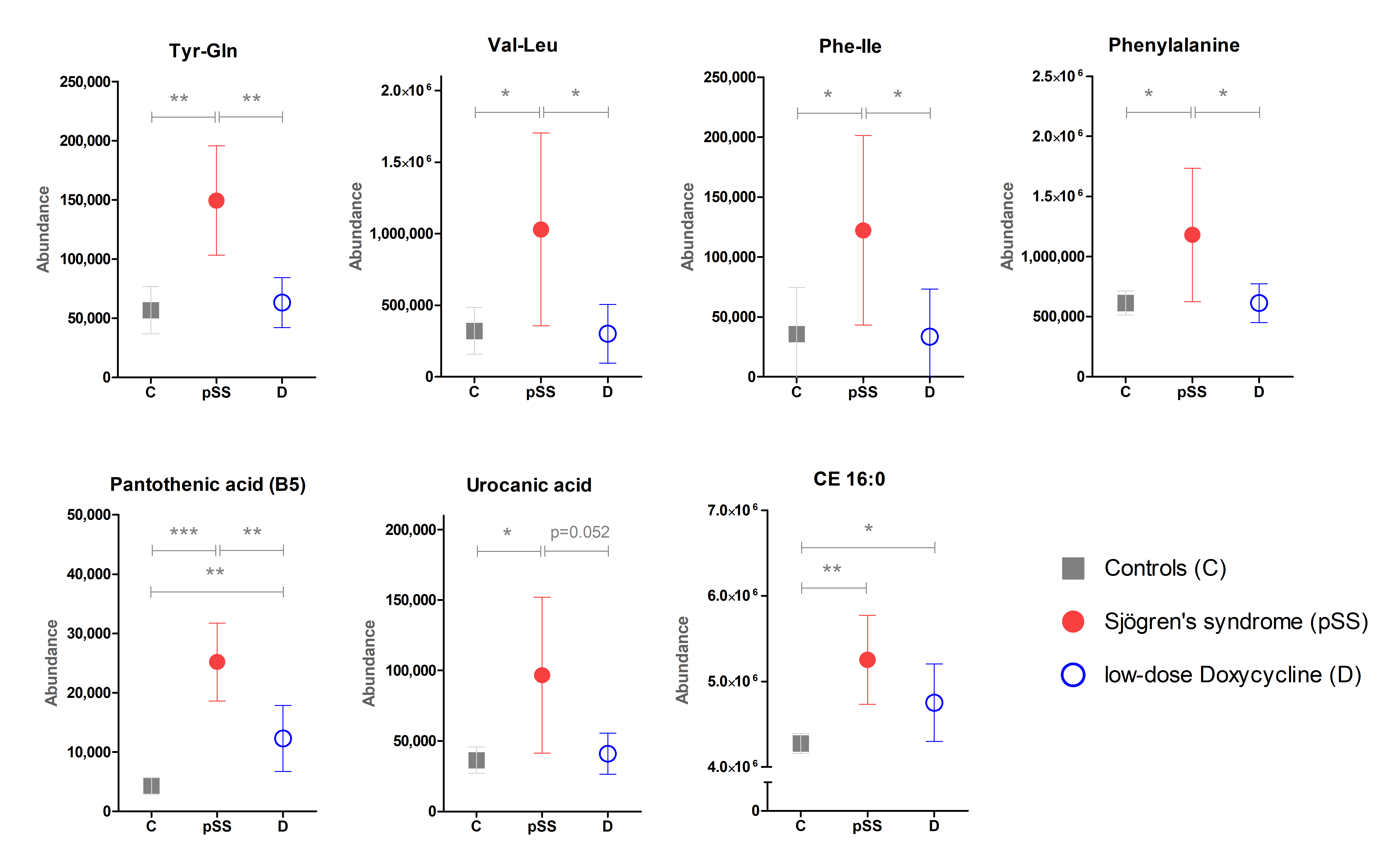
2. Results
3. Discussion
4. Materials and Methods
4.1. Ethical Statement
4.2. Participants
4.3. Collection of Salivary Samples
4.4. Metabolomics Analysis
Supplementary Materials
Author Contributions
Funding
Institutional Review Board Statement
Informed Consent Statement
Data Availability Statement
Acknowledgments
Conflicts of Interest
References
- Wong, D. Salivary Diagnostics; Wiley-Blackwell: Ames, IA, USA, 2008; pp. 27–29. [Google Scholar]
- Jazzar, A.A.; Shirlaw, P.J.; Carpenter, G.H.; Challacombe, S.J.; Proctor, G.B. Salivary S100A8/A9 in Sjögren’s syndrome accompanied by lymphoma. J. Oral Pathol. Med. 2018, 47, 900–906. [Google Scholar] [CrossRef] [Green Version]
- Cecchettini, A.; Finamore, F.; Ucciferri, N.; Donati, V.; Mattii, L.; Polizzi, E.; Ferro, F.; Sernissi, F.; Mosca, M.; Bombardieri, S.; et al. Phenotyping multiple subsets in Sjögren’s syndrome: A salivary proteomic SWATH-MS approach towards precision medicine. Clin. Proteom. 2019, 16, 26. [Google Scholar] [CrossRef]
- Zoukhri, D.; Rawe, I.; Singh, M.; Brown, A.; Kublin, C.L.; Dawson, K.; Haddon, W.F.; White, E.L.; Hanley, K.M.; Tusé, D.; et al. Discovery of putative salivary biomarkers for Sjögren’s syndrome using high resolution mass spectrometry and bioinformatics. J. Oral Sci. 2012, 54, 61–70. [Google Scholar] [CrossRef] [PubMed] [Green Version]
- Hauck, T.S.; Douglas, S.C.; Bookman, A. Sjogren’s syndrome in Canada: Diagnosis, treatment and patient perspectives. Connections 2013, 7. [Google Scholar]
- Herrala, M.; Mikkonen, J.J.; Pesonen, P.; Lappalainen, R.; Tjäderhane, L.; Niemelä, R.K.; Seitsalo, H.; Salo, T.; Myllymaa, S.; Kullaa, A.M. Variability of salivary metabolite levels in patients with Sjögren’s syndrome. J. Oral Sci. 2021, 63, 22–26. [Google Scholar] [CrossRef] [PubMed]
- Kageyama, G.; Saegusa, J.; Irino, Y.; Tanaka, S.; Tsuda, K.; Takahashi, S.; Sendo, S.; Morinobu, A. Metabolomics analysis of saliva from patients with primary Sjögren’s syndrome. Clin. Exp. Immunol. 2015, 182, 149–153. [Google Scholar] [CrossRef] [Green Version]
- Spratlin, J.L.; Serkova, N.J.; Eckhardt, S.G. Clinical applications of metabolomics in oncology: A review. Clin. Cancer Res. 2009, 15, 431–440. [Google Scholar] [CrossRef] [Green Version]
- Mikkonen, J.J.; Herrala, M.; Soininen, P.; Lappalainen, R.; Tjäderhane, L.; Seitsalo, H.; Niemelä, R.; Salo, T.; Kullaa, A.M.; Myllymaa, S. Metabolic profiling of saliva in patients with primary Sjögren’s syndrome. Metabolomics 2013, 3, 1. [Google Scholar]
- Gardner, A.; Carpenter, G.; So, P.W. Salivary Metabolomics: From Diagnostic Biomarker Discovery to Investigating Biological Function. Metabolites 2020, 10, 47. [Google Scholar] [CrossRef] [Green Version]
- Alam, J.; Lee, A.; Lee, J.; Kwon, D.I.; Park, H.K.; Park, J.H.; Jeon, S.; Baek, K.; Lee, J.; Park, S.H.; et al. Dysbiotic oral microbiota and infected salivary glands in Sjögren’s syndrome. PLoS ONE 2020, 15, e0230667. [Google Scholar] [CrossRef]
- Seitsalo, H.; Niemelä, R.K.; Marinescu-Gava, M.; Vuotila, T.; Tjäderhane, L.; Salo, T. Effectiveness of low-dose doxycycline (LDD) on clinical symptoms of Sjögren’s syndrome: A randomized, double-blind, placebo controlled cross-over study. J. Negat. Results Biomed. 2007, 6, 11. [Google Scholar] [CrossRef] [PubMed] [Green Version]
- Rusthen, S.; Kristoffersen, A.K.; Young, A.; Galtung, H.K.; Petrovski, B.É.; Palm, Ø.; Enersen, M.; Jensen, J.L. Dysbiotic salivary microbiota in dry mouth and primary Sjögren’s syndrome patients. PLoS ONE 2019, 14, e0218319. [Google Scholar] [CrossRef] [PubMed]
- Singh, M.; Teles, F.; Uzel, N.G.; Papas, A. Characterizing Microbiota from Sjögren’s Syndrome Patients. JDR Clin. Transl. Res. 2020. [Google Scholar] [CrossRef] [PubMed]
- Takahashi, N. Oral Microbiome Metabolism: From “Who Are They?” to “What Are They Doing?”. J. Dent. Res. 2015, 94, 1628–1637. [Google Scholar] [CrossRef]
- Van der Meulen, T.A.; Harmsen, H.J.; Bootsma, H.; Liefers, S.C.; Vich Vila, A.; Zhernakova, A.; Fu, J.; Wijmenga, C.; Spijkervet, F.K.; Kroese, F.G.; et al. Dysbiosis of the buccal mucosa microbiome in primary Sjögren’s syndrome patients. Rheumatology 2018, 57, 2225–2234. [Google Scholar] [CrossRef]
- van der Meulen, T.A.; Harmsen, H.J.; Vila, A.V.; Kurilshikov, A.; Liefers, S.C.; Zhernakova, A.; Fu, J.; Wijmenga, C.; Weersma, R.K.; de Leeuw, K.; et al. Shared gut, but distinct oral microbiota composition in primary Sjögren’s syndrome and systemic lupus erythematosus. J. Autoimmun. 2019, 97, 77–87. [Google Scholar] [CrossRef]
- Liebsch, C.; Pitchika, V.; Pink, C.; Samietz, S.; Kastenmüller, G.; Artati, A.; Suhre, K.; Adamski, J.; Nauck, M.; Völzke, H.; et al. The Saliva Metabolome in Association to Oral Health Status. J. Dent. Res. 2019, 98, 642–651. [Google Scholar] [CrossRef]
- Czumaj, A.; Szrok-Jurga, S.; Hebanowska, A.; Turyn, J.; Swierczynski, J.; Sledzinski, T.; Stelmanska, E. The Pathophysiological Role of CoA. Int. J. Mol. Sci. 2020, 21, 9057. [Google Scholar] [CrossRef]
- Hill, M.J. Intestinal flora and endogenous vitamin synthesis. Eur. J. Cancer Prev. Off. J. Eur. Cancer Prev. Organ. (ECP) 1997, 6 (Suppl. S1), S43–S45. [Google Scholar] [CrossRef]
- Lassalle, F.; Spagnoletti, M.; Fumagalli, M.; Shaw, L.; Dyble, M.; Walker, C.; Thomas, M.G.; Bamberg Migliano, A.; Balloux, F. Oral microbiomes from hunter-gatherers and traditional farmers reveal shifts in commensal balance and pathogen load linked to diet. Mol. Ecol. 2018, 27, 182–195. [Google Scholar] [CrossRef] [PubMed] [Green Version]
- Gheita, A.A.; Gheita, T.A.; Kenawy, S.A. The potential role of B5: A stitch in time and switch in cytokine. Phytother. Res. 2020, 34, 306–314. [Google Scholar] [CrossRef]
- He, W.; Hu, S.; Du, X.; Wen, Q.; Zhong, X.P.; Zhou, X.; Zhou, C.; Xiong, W.; Gao, Y.; Zhang, S.; et al. Vitamin B5 Reduces Bacterial Growth via Regulating Innate Immunity and Adaptive Immunity in Mice Infected with Mycobacterium tuberculosis. Front. Immunol. 2018, 9, 365. [Google Scholar] [CrossRef] [Green Version]
- Hug, D.H.; Dunkerson, D.D.; Hunter, J.K. The degradation of L-histidine and trans- and cis-urocanic acid by bacteria from skin and the role of bacterial cis-urocanic acid isomerase. J. Photochem. Photobiol. B 1999, 50, 66–73. [Google Scholar] [CrossRef]
- Belstrøm, D.; Holmstrup, P.; Bardow, A.; Kokaras, A.; Fiehn, N.E.; Paster, B.J. Comparative analysis of bacterial profiles in unstimulated and stimulated saliva samples. J. Oral Microbiol. 2016, 8, 30112. [Google Scholar] [CrossRef]
- Larsson, B.; Olivecrona, G.; Ericson, T. Lipids in human saliva. Arch. Oral Biol. 1996, 41, 105–110. [Google Scholar] [CrossRef]
- Matczuk, J.; Żendzian-Piotrowska, M.; Maciejczyk, M.; Kurek, K. Salivary lipids: A review. Adv. Clin. Exp. Med. Off. Organ Wroc. Med. Univ. 2016, 26, 1021–1029. [Google Scholar] [CrossRef]
- Slomiany, B.L.; Kosmala, M.; Nadziejko, C.; Murty, V.L.; Gwozdzinski, K.; Slomiany, A.; Mandel, I.D. Lipid composition and viscosity of parotid saliva in Sjögren syndrome in man. Arch. Oral Biol. 1986, 31, 699–702. [Google Scholar] [CrossRef]
- Vitali, C.; Bombardieri, S.; Jonsson, R.; Moutsopoulos, H.M.; Alexander, E.L.; Carsons, S.E.; Daniels, T.E.; Fox, P.C.; Fox, R.I.; Kassan, S.S.; et al. Classification criteria for Sjogren’s syndrome: A revised version of the European criteria proposed by the American-European consensus group. Ann. Rheum. Dis. 2002, 61, 554–558. [Google Scholar] [CrossRef] [Green Version]
- Niemela, R.K.; Takalo, R.; Paakko, E.; Suramo, I.; Paivansalo, M.; Salo, T.; Hakala, M. Ultrasonography of salivary glands in primary Sjogren’s syndrome. A comparison with magnetic resonance imaging and magnetic resonance sialography of parotid glands. Rheumatology 2004, 43, 875–879. [Google Scholar] [CrossRef] [Green Version]
- Navazesh, M. Methods for collecting saliva. Ann. N. Y. Acad. Sci. 1993, 694, 72–77. [Google Scholar] [CrossRef] [PubMed]
- Turunen, S.; Puurunen, J.; Auriola, S.; Kullaa, A.M.; Kärkkäinen, O.; Lohi, H.; Hanhineva, K. Metabolome of canine and human saliva: A non-targeted metabolomics study. Metabolomics 2020, 16, 90. [Google Scholar] [CrossRef] [PubMed]
- Klåvus, A.; Kokla, M.; Noerman, S.; Koistinen, V.M.; Tuomainen, M.; Zarei, I.; Meuronen, T.; Häkkinen, M.R.; Rummukainen, S.; Farizah Babu, A.; et al. “Notame”: Workflow for Non-Targeted LC-MS Metabolic Profiling. Metabolites 2020, 10, 135. [Google Scholar] [CrossRef] [PubMed] [Green Version]
- Sumner, L.W.; Amberg, A.; Barrett, D.; Beale, M.H.; Beger, R.; Daykin, C.A.; Fan, T.W.; Fiehn, O.; Goodacre, R.; Griffin, J.L.; et al. Proposed minimum reporting standards for chemical analysis Chemical Analysis Working Group (CAWG) Metabolomics Standards Initiative (MSI). Metabolomics 2007, 3, 211–221. [Google Scholar] [CrossRef] [PubMed] [Green Version]
- Pang, Z.; Chong, J.; Zhou, G.; Morais, D.; Chang, L.; Barrette, M.; Gauthier, C.; Jacques, P.E.; Li, S.; Xia, J. MetaboAnalyst 5.0: Narrowing the gap between raw spectra and functional insights. Nucl. Acids Res. 2021, 49, W388–W396. [Google Scholar] [CrossRef]


| Metabolite | ID | Healthy Controls, HC (C) | Sjögren’s Syndrome (pSS) without LDD | Sjögren’s Syndrome (pSS) with LDD (D) | pSS vs. C | D vs. C | D vs. pSS | |||||||||
|---|---|---|---|---|---|---|---|---|---|---|---|---|---|---|---|---|
| Mean | SD | Mean | SD | Mean | SD | VIP | p | d | VIP | p | d | VIP | p | d | ||
| Amino acids, peptides, and analogues | ||||||||||||||||
| 4-Guanidinobutyric acid | 2 | 16,864 | 4783 | 31,881 | 18,325 | 28,463 | 10,929 | 1.38 | 0.031 | 1.3 | 1.83 | 0.004 | 1.5 | 0.39 | 0.612 | −0.2 |
| Glutamic acid | 1 | 122,045 | 67,786 | 250,491 | 87,720 | 225,246 | 124,395 | 1.91 | 0.001 | 1.7 | 1.48 | 0.010 | 1.1 | 0.72 | 0.558 | −0.2 |
| Glycine betaine | 1 | 1241,617 | 504,031 | 1,999,692 | 964,523 | 1,849,530 | 974,498 | 1.43 | 0.041 | 1.0 | 1.31 | 0.045 | 0.8 | 0.33 | 0.708 | −0.2 |
| Isoleucine | 2 | 48,927 | 30,704 | 166,871 | 135,723 | 99,906 | 78,292 | 1.54 | 0.023 | 1.4 | 1.28 | 0.037 | 0.9 | 1.00 | 0.184 | −0.6 |
| Leucine | 2 | 112,620 | 46,369 | 448,733 | 419,771 | 190,974 | 115,700 | 1.56 | 0.032 | 1.4 | 1.37 | 0.026 | 1.0 | 1.54 | 0.088 | −1.0 |
| Phenylalanine | 1 | 614,751 | 174,399 | 1,180,634 | 775,776 | 612,765 | 291,252 | 1.41 | 0.048 | 1.2 | 0.53 | 0.982 | 0.0 | 1.58 | 0.050 | −1.1 |
| Tryptophan | 1 | 33,773 | 17,245 | 97,779 | 66,429 | 56,455 | 30,196 | 1.63 | 0.014 | 1.5 | 1.32 | 0.021 | 1.0 | 1.32 | 0.091 | −0.9 |
| Arg-Ser | 2 | 653,047 | 308,815 | 285,658 | 248,390 | 447,258 | 269,746 | 1.56 | 0.004 | −1.3 | 1.10 | 0.072 | −0.7 | 1.49 | 0.145 | 0.6 |
| Phe-Ile | 2 | 35,793 | 41,934 | 122,220 | 85,371 | 33,595 | 37,875 | 1.55 | 0.040 | 1.4 | 0.39 | 0.923 | −0.1 | 1.81 | 0.037 | −1.4 |
| Tyr-Gln | 2 | 56,858 | 34,559 | 149,463 | 64,758 | 63,153 | 33,358 | 1.96 | 0.001 | 1.9 | 0.48 | 0.641 | 0.2 | 2.23 | 0.002 | −1.8 |
| Val-Leu | 2 | 320,566 | 283,561 | 1,029,040 | 940,151 | 300,576 | 370,401 | 1.42 | 0.043 | 1.2 | 0.47 | 0.871 | −0.1 | 1.61 | 0.040 | −1.1 |
| Lipids and carnitines | ||||||||||||||||
| FA 16:0 | 2 | 19,113,838 | 2,988,053 | 23,755,538 | 5,398,164 | 21,707,731 | 6,550,119 | 1.40 | 0.029 | 1.1 | 1.01 | 0.181 | 0.5 | 0.58 | 0.403 | −0.3 |
| FA 16:1 | 2 | 291,295 | 177,097 | 601,015 | 353,046 | 406,038 | 534,487 | 1.46 | 0.025 | 1.2 | 0.64 | 0.442 | 0.3 | 0.91 | 0.283 | −0.4 |
| Azelaic acid | 1 | 46,004 | 5591 | 61,345 | 16,287 | 54,030 | 11,500 | 1.67 | 0.161 | 1.4 | 1.27 | 0.025 | 0.9 | 0.95 | 0.238 | −0.5 |
| Leucic acid | 2 | 61,064 | 25,243 | 42,491 | 15,691 | 45,390 | 20,708 | 1.25 | 0.037 | −0.9 | 1.00 | 0.089 | −0.7 | 1.23 | 0.706 | 0.2 |
| CE 16:0 | 1 | 4,277,005 | 204,081 | 5,253,366 | 726,120 | 4,751,349 | 817,147 | 2.03 | 0.002 | 2.1 | 1.23 | 0.045 | 0.9 | 1.02 | 0.122 | −0.7 |
| Cholesterol | 1 | 42,086 | 23,976 | 107,189 | 65,001 | 111,790 | 89,827 | 1.66 | 0.118 | 1.5 | 1.50 | 0.010 | 1.2 | 0.45 | 0.883 | 0.1 |
| Propionylcarnitine | 1 | 91,310 | 57,056 | 165,515 | 81,484 | 202,565 | 108,289 | 1.47 | 0.025 | 1.1 | 1.73 | 0.002 | 1.3 | 0.62 | 0.340 | 0.4 |
| Isobutyryl carnitine | 2 | 50,626 | 23,350 | 95,837 | 32,915 | 123,268 | 75,870 | 1.85 | 0.002 | 1.6 | 1.80 | 0.004 | 1.5 | 0.77 | 0.244 | 0.5 |
| LPC 18:0 | 2 | 0 | 0 | 24,441 | 13,129 | 71,932 | 99,287 | 0.01 | <0.001 | 0.68 | <0.001 | 0.84 | 0.170 | 0.8 | ||
| LPE 18:0 | 2 | 0 | 0 | 16,498 | 8405 | 17,072 | 8133 | 0.34 | <0.001 | 0.25 | <0.001 | 0.35 | 0.941 | 0.1 | ||
| Oleamide | 2 | 9,389,628 | 1,079,571 | 11,488,697 | 1,386,541 | 10,258,327 | 2,115,434 | 1.85 | <0.001 | 1.7 | 0.96 | 0.174 | 0.5 | 1.03 | 0.092 | −0.7 |
| Linoleamide | 2 | 30,287,081 | 4,290,249 | 37,683,667 | 8,799,870 | 35,644,986 | 12,192,656 | 1.41 | 0.030 | 1.1 | 1.04 | 0.140 | 0.7 | 0.44 | 0.639 | −0.2 |
| Palmitoleamide | 2 | 12,508,377 | 1,779,147 | 15,694,049 | 3,897,803 | 15,115,575 | 6,193,420 | 1.41 | 0.033 | 1.1 | 1.04 | 0.137 | 0.7 | 0.20 | 0.777 | −0.1 |
| Other | ||||||||||||||||
| Choline | 2 | 5,453,217 | 2,359,520 | 9,987,031 | 4,308,942 | 9,524,364 | 4,603,084 | 1.59 | 0.010 | 1.4 | 1.60 | 0.009 | 1.2 | 0.31 | 0.804 | −0.1 |
| Pantothenic acid (B5) | 1 | 4289 | 2431 | 25,186 | 9179 | 12,290 | 10,088 | 2.46 | <0.001 | 3.6 | 1.53 | 0.009 | 1.3 | 1.81 | 0.003 | −1.3 |
| MEHP | 1 | 101,276 | 15,673 | 58,944 | 15,552 | 88,751 | 27,838 | 2.35 | <0.001 | −2.7 | 0.87 | 0.146 | −0.6 | 1.77 | 0.002 | 1.4 |
| Xanthine | 1 | 173,813 | 123,732 | 548,441 | 260,922 | 601,704 | 302,916 | 2.05 | 0.001 | 1.9 | 2.15 | <0.001 | 2.0 | 0.41 | 0.644 | 0.2 |
| Urocanic acid | 1 | 36,386 | 16,273 | 96,702 | 77,360 | 40,992 | 25,246 | 1.47 | 0.037 | 1.3 | 0.41 | 0.572 | 0.2 | 1.55 | 0.052 | −1.1 |
| 1-Methylnicotinamide | 2 | 11,940 | 1594 | 28,433 | 16,638 | 33,923 | 33,704 | 1.68 | 0.039 | 1.8 | 1.27 | 0.069 | 1.2 | 0.42 | 0.664 | 0.2 |
| Biliverdin IX | 1 | 162,101 | 31,725 | 137,588 | 11,938 | 140,733 | 11,422 | 1.28 | 0.017 | −1.1 | 1.33 | 0.030 | −1.0 | 0.46 | 0.519 | 0.3 |
| N6-methyl-adenine | 2 | 55,816 | 27,354 | 119,280 | 86,943 | 102,200 | 74,734 | 1.35 | 0.049 | 1.1 | 1.35 | 0.037 | 0.9 | 0.39 | 0.618 | −0.2 |
| Nicotinic acid | 1 | 43,642 | 31,275 | 107,135 | 80,169 | 87,564 | 60,411 | 1.37 | 0.049 | 1.1 | 1.34 | 0.029 | 1.0 | 0.71 | 0.541 | −0.3 |
| Diethanolamine | 1 | 147,588 | 168,396 | 695,458 | 459,234 | 219,384 | 495,663 | 1.89 | 0.004 | 1.7 | 0.56 | 0.628 | 0.2 | 1.56 | 0.027 | −1.0 |
| Stearamide | 2 | 10,065,816 | 1,374,684 | 12,518,622 | 3,182,570 | 11,313,506 | 3,775,837 | 1.35 | 0.042 | 1.1 | 0.94 | 0.247 | 0.5 | 0.56 | 0.399 | −0.3 |
| Dibutyladipate | 2 | 73,783 | 33,110 | 43,796 | 6608 | 86,324 | 85,921 | 1.45 | 0.005 | −1.5 | 0.54 | 0.606 | 0.2 | 1.10 | 0.077 | 0.9 |
Publisher’s Note: MDPI stays neutral with regard to jurisdictional claims in published maps and institutional affiliations. |
© 2021 by the authors. Licensee MDPI, Basel, Switzerland. This article is an open access article distributed under the terms and conditions of the Creative Commons Attribution (CC BY) license (https://creativecommons.org/licenses/by/4.0/).
Share and Cite
Herrala, M.; Turunen, S.; Hanhineva, K.; Lehtonen, M.; Mikkonen, J.J.W.; Seitsalo, H.; Lappalainen, R.; Tjäderhane, L.; Niemelä, R.K.; Salo, T.; et al. Low-Dose Doxycycline Treatment Normalizes Levels of Some Salivary Metabolites Associated with Oral Microbiota in Patients with Primary Sjögren’s Syndrome. Metabolites 2021, 11, 595. https://doi.org/10.3390/metabo11090595
Herrala M, Turunen S, Hanhineva K, Lehtonen M, Mikkonen JJW, Seitsalo H, Lappalainen R, Tjäderhane L, Niemelä RK, Salo T, et al. Low-Dose Doxycycline Treatment Normalizes Levels of Some Salivary Metabolites Associated with Oral Microbiota in Patients with Primary Sjögren’s Syndrome. Metabolites. 2021; 11(9):595. https://doi.org/10.3390/metabo11090595
Chicago/Turabian StyleHerrala, Maria, Soile Turunen, Kati Hanhineva, Marko Lehtonen, Jopi J. W. Mikkonen, Hubertus Seitsalo, Reijo Lappalainen, Leo Tjäderhane, Raija K. Niemelä, Tuula Salo, and et al. 2021. "Low-Dose Doxycycline Treatment Normalizes Levels of Some Salivary Metabolites Associated with Oral Microbiota in Patients with Primary Sjögren’s Syndrome" Metabolites 11, no. 9: 595. https://doi.org/10.3390/metabo11090595