Article

Increase in Lower Limb Strength after Multimodal Pain Management in Patients with Low Back Pain

by
Moritz Kaiser
1,*,
Sara Brambrink
1,
Achim Benditz
1,*,
Leonard Achenbach
2,
Matthias Gehentges
1 and
Matthias Alexander König
1
1
Department of Orthopedic Surgery, Regensburg University Medical Center, 93077 Bad Abbach, Germany
2
Department of Orthopedic Surgery, König-Ludwig-Haus, Julius-Maximilians-University Würzburg, 97074 Würzburg, Germany
*
Authors to whom correspondence should be addressed.
Medicina 2022, 58(7), 837; https://doi.org/10.3390/medicina58070837
Submission received: 12 May 2022 / Revised: 7 June 2022 / Accepted: 19 June 2022 / Published: 22 June 2022
(This article belongs to the Section Neurology)

Abstract

:
Background and Objectives: The aim of the present study was to evaluate the efficacy of a multimodal pain therapy (MPM) regarding the objective parameter muscle strength of segment-dependent lower limb muscle groups before and after such a treatment. Materials and Methods: 52 patients with a history of low back pain and/or leg pain received standardized multimodal pain management. Strength of segment indicating lower limb muscles were assessed for each patient before and after ten days of treatment by handheld dynamometry. Results: Overall strength increased significantly from 23.6 kg ± 6.6 prior to treatment to 25.4 ± 7.3 after treatment, p ≤ 0.001. All muscle groups significantly increased in strength with exception of great toe extensors. Conclusions: Despite lower basic strength values at the beginning of treatment, all investigated muscle groups, except for the great toe extensors, showed a significant increase of overall strength after completion of the multimodal pain management concept. Increased overall strength could help with avoiding further need of medical care by supporting patients’ autonomy in daily life activities, as well as maintaining working abilities. Thus, our study is the first to show a significant positive influence on lower limb strength in patients with low back pain after a conservative MPM program.

1. Introduction

Chronic low back pain (LBP) is of increasing socio-economic importance [1,2]. In 2016, it was one of the five leading causes of years lived with a disability [3]. Furthermore, a prevalence of 1 in 5 in Germany, and 1 in 10 in the United States of America was reported [4,5]. Low back pain, and chronic back pain, is associated with a higher Body Mass Index (BMI) and old age [6,7]. In addition, pain in general is associated with higher immobilization and reduced strength to master daily life activities or the professional life [2]. To avoid persistent need of medical care and to maintain patients’ autonomy, the treatment of this disease is an important issue.
Chronic low back pain is often described in the literature as mixed pain. This means that most patients have both neuropathic and nociceptive pain components [8].
Unless there is an absolute surgical indication such as cauda equina syndrome (CES), conservative therapy should then be carried out first. Since, as mentioned before, chronic low back pain is normally a mixed pain, it should be addressed in a multidisciplinary approach. Different treatment options are described in the literature for the non-surgical treatment [9,10]. In this context, spinal injection therapy has an important role in the treatment of LBP. In addition, it has already been shown that lumbar injection therapy is an effective method for nerve root irritations [11,12].
In combination with accompanying treatments such as physical therapy and psychological counselling as a multimodal analgesia management, injection therapy can be used to control chronic pain symptoms and avoid surgery with accompanying treatments such as physical therapy and psychological counselling [13,14].
Up to now, the visual analogue scale (VAS) or the numeric rating scale (NRS) have mostly been used in the literature as indicators for successful therapy [14]. However, these scales are strictly subjective parameters that can be influenced by a variety of confounders. Patient dependent criteria have mostly been collected through a questionnaire [14,15,16,17]. Interestingly, up to now, no data has been available yet concerning the influence of multimodal pain management (MPM) on muscle strength.
The aim of the present study was to evaluate the efficacy of a multimodal pain therapy regarding the objective parameter muscle strength of segment-dependent lower limb muscle groups before and after such a treatment.

2. Material and Methods

2.1. Study Population

This prospective study was approved by the local ethics committee on 21 March 2018 (Nr. 18-931-101). Written informed consent was given by all patients participating in this study. Patients were included in the study if they were indicated for inpatient, multimodal pain management in a single center (Department of Orthopedic Surgery, Regensburg University Medical Center, Bad Abbach, Germany) from June 2018 to April 2019 and met the following inclusion criteria: age between 18 and 80 years, radicular pain originating from a specific nerve root or clearly attributable muscle strength impairment of the lower limb without surgical indication, and a pain level of at least 5 on the numeric rating scale.
Exclusion criteria were tumors with spinal implication, congenital spinal malformations, former spinal surgery, and rheumatic or inflammatory spinal diseases. Demographic data are shown in Table 1.

2.2. Treatment Algorithm
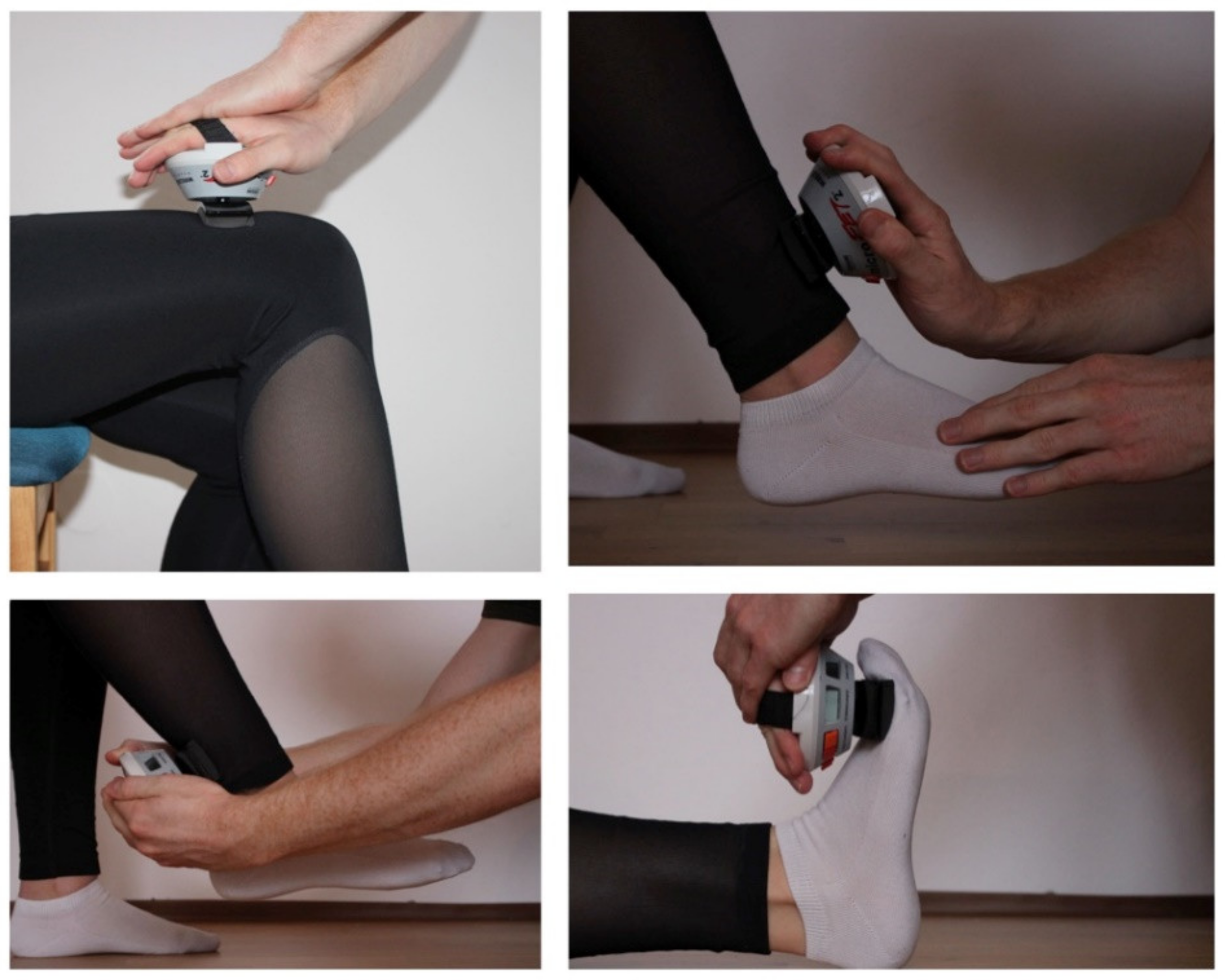
Inpatient care was provided for each patient for a total of 10 days. The concept consisted mainly of lumbar spinal nerve root analgesia (LSPA) in the affected region, using a freehand technique, which has already been described elsewhere [18]. The injections were administered twice a day in the morning and at noon. In addition, the patient received a bilateral facet joint infiltration at the level L4 to S1 and an epidural injection, as well a nerve root block. Undiluted Mepivacaine 1% was used and 10 mL were applied as LSPA. For the lumbar epidural injection, 8 mg Dexamethasone were combined with sterile saline solution. Again, 10 mL were applied. Facet joint injections contained 8 ml undiluted Mepivacaine 1% and 8 mg Dexamethasone. For nerve root blocks, 4 ml Mepivacaine 1% and 4 mg Dexamethasone were injected. The aforementioned injections were accompanied by physiotherapeutic and sports medical exercises, which included isometric strengthening of the back muscles on specific training equipment. A specific training program for lower limb muscles was not carried out. Additionally, the patients received proprioceptive training, electrotherapy, thermotherapy, instruction in progressive muscle relaxation, and if indicated, a psychotherapeutic program, which was embedded in a group therapy.

2.3. Measurement

A hand-held dynamometer (microFET2, Hoggan Health Industries, Salt Lake City, Utah, USA) was used to measure strength. All values were documented in kilogram. The measurements were performed on two different days. The first measurement took place on the day of admission to determine the basic strength values, and the second on the day of discharge. Each measurement was performed independently by two examiners for each assessment. Both examiners received instruction and training in the use of the measurement device prior to the study.
The contact surfaces were marked with a skin-friendly pencil to ensure equal measurement points. The marked areas were repainted by the patient himself, if necessary, (e.g., after personal hygiene acts). The contact surfaces were chosen in a manner that allowed for comfortable measurement with the highest achievable leverage. The best testing position for each muscle group was described by Mentiplay [19] While in a sitting position, the following movements were then carried out, one after the other, against the resistance of the dynamometer using the “break method” as described by Burns and Spanier [20]: hip flexion, knee extension, knee flexion. Then, the patient was moved into a lying position. Starting from the neutral joint position, the following movements were performed: foot extension, foot flexion, big toe extension. Each measurement was carried out bilaterally and all movements were measured in every patient. Three different types of transducer pads were available for the device in order to achieve a proper skin contact, a flat one, a curved one and a small flat one for Toes. Figure 1 illustrates exemplarily the testing positions for hip flexion, knee flexion, knee extension and ankle flexion.
To record the patients’ pain, a numerical rating scale (NRS) from 0 to 10 was used. The patients were interviewed before each measurement regarding the pain level. The strength values were documented immediately in standardized, electronic form. In addition, the descriptive data of every individual was collected (Table 1 and Table 2).

2.4. Statistical Analysis

For statistical analysis, continuous data are presented as mean values and standard deviation. Group comparisons were performed by two-sided t-tests for dependent variables. Absolute and relative frequencies were given for categorical data. Inter-observer agreement was assessed using the intraclass correlation coefficient (ICC). The following values were determined according to Koo et al. [21]: less than 0.5 poor, between 0.5 and 0.75 moderate, between 0.75 and 0.9 good, and greater than 0.90 excellent reliability. Differences of p < 0.05 were considered statistically significant. IBM SPSS Statistics 25 (SPSS Inc, Chicago, IL, USA) was used for analysis.

3. Results

In total, 52 Patients were included analyzed with a mean age of 63.2 years (±12.1), 25 male and 27 female. Patients were treated in-hospital for 9.19 days (±0.56) on average. The painful side was right in 12 patients, left in 15 patients, and both in 25 patients.
Overall Interrater Reliability was excellent with ICC = 0.94 for measurements at the day of admission and excellent with ICC = 0.96 for the second measurements at discharge.
NRS decreased significantly for both back and leg pain from 5.9 (±2.2) to 3.3 (±2.2) and 4.5 (±2.8) to 2.5 (±2.0), respectively, p < 0.001.
Infiltration of the facet joints were administered 45 times (86.5%), epidural injections 47 times (90.4%), and nerve root blocks 19 times (36.5%).

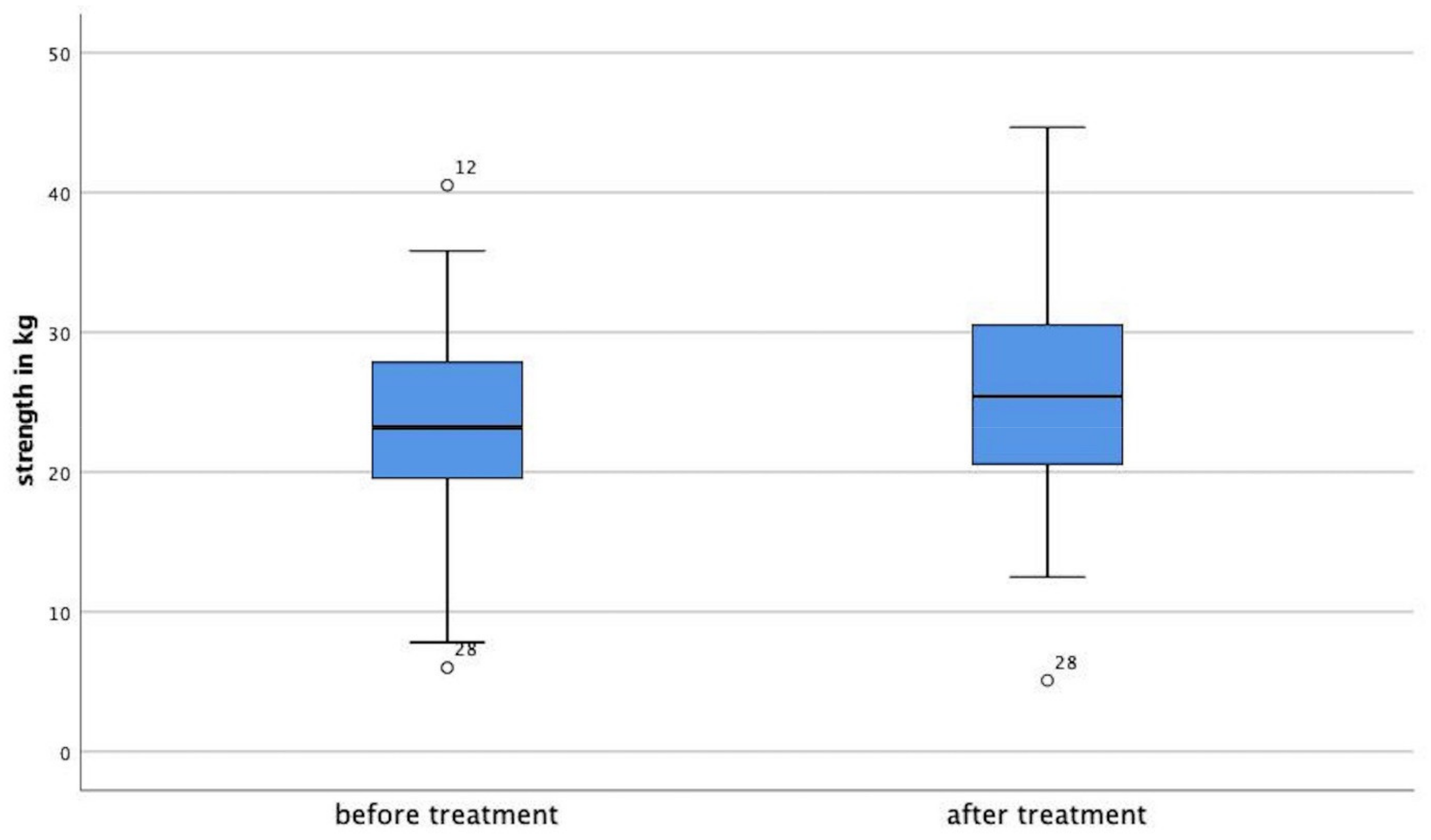
3.1. Strength Development

Hip flexors strength increased significantly (p = 0.013) by 7.2% from the first day to discharge.
For the knee extensors, the power of the muscle group increased significantly by 12.6% (p < 0.001).
Knee flexors showed an increased rate of 7.4%, (p < 0.001). Ankle extensor strength increased by 4.75% (p = 0.041). Ankle flexor strength increased by 9.5% (p < 0.001). For great toe extensors, a reduction of strength was noted by −4.0%, which was statistically not significant, p = 0.103. Mean values for strength development are shown in Table 2. Figure 2 shows Boxplots prior to treatment and after for mean overall strength.

3.2. Side-to-Side Differences

An overall significant side-to-side difference prior to treatment, considering all measurements on one side, was not noticed, p = 0.294. Overall strength on the right side was 23.96 kg ± 7.4, and on the left was 23.24 kg ± 6.7. After treatment, a significant side-to-side difference was recognized, p = 0.039. On the right side, overall strength was 25.87 kg ± 7.6, on the left side, it was 24.95 kg ± 7.3.
No adverse events were found in the study.

4. Discussion

The most important finding of this study was that lower limb strength increased significantly within a few days of non-operative treatment in patients with low back pain.
Up to now, objective data regarding the course of segment-dependent muscle strength in patients undergoing MPM are missing. Hence, the primary goal of this study was to objectify the influence of multimodal pain management with regard to the muscle strength development of lower limb muscles using a hand-held dynamometer.
There are several limitations of this study. First, the strength values were assessed only twice, at the start of the injection therapy and at its end. Therefore, our records do not contain any information about the course of strength development over the entire duration of therapy. Second, a handheld dynamometer has been used in the current study. However, a free hand technique cannot be as accurate as a fixed system due to imbalances in motion control of the raters and variances due to the raters themselves. Third, there is no comparison group, especially comparing injections to other treatment options like analgesia or physiotherapy alone in a multimodal analgesia concept. This might limit the interpretation of the data. In addition, the study group is very limited. Bigger study groups are needed to confirm the findings of this investigation. Since there were only two measurements in a very short period, it is not possible to predict a long-term result and how long the effect of MPM on muscle strength might last.
Researchers have to be aware when comparing different testers. As Wadsworth and Wikholm showed in their studies, intertester reliability is reduced when the strength of the subject is superior to the one of the examiner [22,23]. Third, the present study does not contain any information about the patient’s dominant side. Compared to the results in the literature, the presented measuring method could have an unintentional influence by the examiner himself, based on different force and gender as well as, of course, levers of the examiner [24,25].
In the present study, the interrater reliability results for both before treatment and after treatment proved to be excellent with values of 0.94 and 0.96, respectively. These findings are comparable to literature, where a total interrater reliability of 0.94 using the same device can be found [26]. Nonetheless, there are also inhomogeneous results, ranging from moderate to excellent interrater reliability between assessors testing muscle strength with a hand-held device [27,28,29].
In general, the comparison of the available data with the literature does not appear to be unambiguous due to inhomogeneous measurement methods. For hip flexion, the values of 24.8 kg ± 9.7 were found after treatment, which is a good average of the literature. A study conducted by Andrews et al., used a different testing position and device strength levels for hip flexion in asymptomatic, elderly adults, and found values of 18.8 kg, while Lasse Ishøi et al., reported a peak force of hip flexion in healthy young adults of up to 321 N (32.7 kg) with a similar testing setup [30,31].
In 2000, Stoll et al., published higher maximal isometric muscle strength in hip flexion values of 25.1 kp (1 kp = 9.81 N) to 38.0 kp in healthy adults [32]. The increased hip flexion muscle strength in healthy adults compared to patients with low back pain might be the result of pain-free range of motion. In the present study, knee extension and flexion force was 30.3 kg ± 12.09 and 21.75 kg ± 7.31, respectively, both after treatment. These findings are more or less in line with Stoll et al., which found slightly more muscle strength in these groups [32]. As they investigated healthy participants, the results in this study suggest a normalization of high pain levels resulting in low strength levels to the ones of asymptomatic adults due to MPM. Also, a significant rise in knee strength was found after treatment, suggesting patient relief from pain-related functional restrictions. Lee et al., reported that patients with a history of low back pain have significantly lower strength levels in total knee strength than a control group [28].
For ankle dorsal extension and plantar flexion, a significant increase was also found, showing a considerably higher base value for the plantar flexors of about 14% in comparison to the dorsal extensor, which increased even after treatment to about a 21% difference. Mentiplay et al., could show higher levels of baseline values, ranging from about 21 kg and 31 kg for dorsal extensors and about 48 kg and 51 kg for plantar flexors [19]. This indicates an even higher plantar flexion and dorsal extension strength ratio, which highlights the increasing strength ratio after treatment in the present study.
The results for great toe extensors in this study slightly decreased over the treatment period. However, clinical testing of the extension force of the big toe is difficult due to the small contact surface on the one hand and the rather low extension force of the big toe compared to the force of the examiner’s hands on the other. Interestingly, the extension force of the big toe has rarely been studied in literature, despite the fact that the extensor hallucis longus is the segment-dependent muscle of the L5 nerve root. In 1995, Jönsson et al., reported an improvement in the function of the extensor hallucis longus after surgical treatment of disc herniation and affection of the L5 nerve root, but the classification of the force ratios was only categorical and not absolute [33]. Further studies also did not present absolute data obtained by dynamometry, such as Hara et al., in 2011, who assessed extensor muscle strength by placing the middle finger of the investigator on the toe [34]. Riandini et al., used a Hoggan Microfet 3 but presented their data only relative [35]. The discrepancy in measurement should be the content of further research to compare these findings, since L5 nerve root irritations are common.
After treatment, the patients showed a significant side-to-side-difference, which was not the case prior to the multimodal pain management. According to Lanshammer et al., normally existing side-to-side differences reoccur, which emphasizes the positive effect of the multimodal pain therapy [36]. However, side differences are controversially discussed in literature. Andrews et al., did not see significant side-to-side differences in the lower limbs in asymptomatic patients [30]. The clinical relevance of the study is based on the fact that patient with low back Pain seem to highly benefit from a MPM. Handheld dynamometry is a useful instrument to monitor the success.

5. Conclusions

Despite lower basic strength values at the beginning of treatment, all investigated muscle groups, with the exception of the great toe extensors, showed a significant increase of overall strength after completion of the multimodal pain management concept. Increased overall strength could help with avoiding further need of medical care by supporting patients’ autonomy in daily life activities, as well as maintaining working abilities. Thus, to our best knowledge, the present study is the first to show a significant positive influence on lower limb strength in patients with low back pain after a conservative MPM program. Altough, our findings are of limited value (low level of evidence), and it is unknown whether these effects are sustained in the long term.

Author Contributions

M.K., M.A.K. and A.B. made substantial contributions to the conception and design of the study. M.K., M.A.K., M.G. and S.B. participated in the acquisition of data, analysis and statistics. All authors made contributions to the interpretation of data and have been involved in drafting the manuscript. All authors have read and agreed to the published version of the manuscript.

Funding

No funding was received for this study.

Institutional Review Board Statement

The study was approved by the local Ethics Committee (No. 18-943-101) prior to the initiation of this study.

Informed Consent Statement

Informed written consent was obtained from all study participants.

Data Availability Statement

The data that support the findings of this study are available from the corresponding author upon reasonable request.

Conflicts of Interest

The authors declare no conflict of interest.

References

  1. Katz, J.N. Lumbar disc disorders and low-back pain: Socioeconomic factors and consequences. JBJS 2006, 88, 21–24. [Google Scholar] [CrossRef]
  2. Benditz, A.; Loher, M.; Boluki, D.; Grifka, J.; Völlner, F.; Renkawitz, T.; Maderbacher, G.; Götz, J. Positive medium-term influence of multimodal pain management on socioeconomic factors and health care utilization in patients with lumbar radiculopathy: A prospective study. J. Pain Res. 2017, 10, 389. [Google Scholar] [CrossRef] [PubMed] [Green Version]
  3. Vos, T.; Abajobir, A.A.; Abate, K.H.; Abbafati, C.; Abbas, K.M.; Abd-Allah, F.; Abdulkader, R.S.; Abdulle, A.M.; Abebo, T.A.; Abera, S.F.; et al. Global, regional, and national incidence, prevalence, and years lived with disability for 328 diseases and injuries for 195 countries, 1990–2016: A systematic analysis for the Global Burden of Disease Study 2016. Lancet 2017, 390, 1211–1259. [Google Scholar] [CrossRef] [Green Version]
  4. Neuhauser, H.; Ellert, U.; Ziese, T. Chronic back pain in the general population in Germany 2002/2003: Prevalence and highly affected population groups. Gesundh. (Bundesverb. Arzte Offentlichen Gesundh.) 2005, 67, 685. [Google Scholar] [CrossRef]
  5. Freburger, J.K.; Holmes, G.M.; Agans, R.P.; Jackman, A.M.; Darter, J.D.; Wallace, A.S.; Castel, L.D.; Kalsbeek, W.D.; Carey, T.S. The rising prevalence of chronic low back pain. Arch. Intern. Med. 2009, 169, 251–258. [Google Scholar] [CrossRef] [Green Version]
  6. Heuch, I.; Heuch, I.; Hagen, K.; Zwart, J.A. Body mass index as a risk factor for developing chronic low back pain: A follow-up in the Nord-Trøndelag Health Study. Spine 2013, 38, 133–139. [Google Scholar] [CrossRef]
  7. Jacobs, J.M.; Hammerman-Rozenberg, R.; Cohen, A.; Stessman, J. Chronic back pain among the elderly: Prevalence, associations, and predictors. Spine 2006, 31, E203–E207. [Google Scholar] [CrossRef]
  8. Freynhagen, R.; Parada, H.A.; Calderon-Ospina, C.-A.; Chen, J.; Emril, D.R.; Fernandez-Villacorta, F.J.; Franco, H.; Ho, K.-Y.; Lara-Solares, A.; Li, C.C.-F.; et al. Current understanding of the mixed pain concept: A brief narrative review. Curr. Med. Res. Opin. 2019, 35, 1011–1018. [Google Scholar] [CrossRef] [Green Version]
  9. Knezevic, N.N.; Candido, K.D.; Vlaeyen, J.W.S.; Van Zundert, J.; Cohen, S.P. Low back pain. Lancet 2021, 398, 78–92. [Google Scholar] [CrossRef]
  10. Calderon-Ospina, C.A.; Nava-Mesa, M.O.; Arbeláez Ariza, C.E. Effect of Combined Diclofenac and B Vitamins (Thiamine, Pyridoxine, and Cyanocobalamin) for Low Back Pain Management: Systematic Review and Meta-analysis. Pain Med. 2020, 21, 766–781. [Google Scholar] [CrossRef] [Green Version]
  11. Buchner, M.; Zeifang, F.; Brocai, D.R.; Schiltenwolf, M. Epidural corticosteroid injection in the conservative management of sciatica. Clin. Orthop. Relat. Res. 2000, 375, 149–156. [Google Scholar] [CrossRef] [PubMed]
  12. Karppinen, J.; Malmivaara, A.; Kurunlahti, M.; Kyllönen, E.; Pienimaki, T.; Nieminen, P.; Ohinmaa, A.; Tervonen, O.; Vanharanta, H. Periradicular infiltration for sciatica: A randomized controlled trial. Spine 2001, 26, 1059–1067. [Google Scholar] [CrossRef] [PubMed]
  13. Faber, F.; Benditz, A.; Boluki, D.; Grifka, J. Application of image-guided injection for cervical and lumbar syndromes. Z. Rheumatol. 2020, 79, 367–378. [Google Scholar] [CrossRef]
  14. Benditz, A.; Madl, M.; Loher, M.; Grifka, J.; Boluki, D.; Linhardt, O. Prospective medium-term results of multimodal pain management in patients with lumbar radiculopathy. Sci. Rep. 2016, 6, 28187. [Google Scholar] [CrossRef] [PubMed] [Green Version]
  15. Guzmán, J.; Esmail, R.; Karjalainen, K.; Malmivaara, A.; Irvin, E.; Bombardier, C. Multidisciplinary rehabilitation for chronic low back pain: Systematic review. BMJ 2001, 322, 1511–1516. [Google Scholar] [CrossRef] [Green Version]
  16. Neubauer, E.; Zahlten-Hinguranage, A.; Schiltenwolf, M.; Buchner, M. Multimodale Therapie bei chronischem HWS- und LWS-Schmerz. Der Schmerz 2006, 20, 210–218. [Google Scholar] [CrossRef]
  17. Kamper, S.J.; Apeldoorn, A.T.; Chiarotto, A.; Smeets, R.; Ostelo, R.; Guzman, J.; van Tulder, M. Multidisciplinary biopsychosocial rehabilitation for chronic low back pain. Cochrane Database Syst. Rev. 2014, CD000963. [Google Scholar] [CrossRef]
  18. Kraemer, J.; Ludwig, J.; Bickert, U.; Owczarek, V.; Traupe, M. Lumbar epidural perineural injection: A new technique. Eur. Spine J. 1997, 6, 357–361. [Google Scholar] [CrossRef]
  19. Mentiplay, B.F.; Perraton, L.G.; Bower, K.J.; Adair, B.; Pua, Y.-H.; Williams, G.P.; McGaw, R.; Clark, R.A. Assessment of Lower Limb Muscle Strength and Power Using Hand-Held and Fixed Dynamometry: A Reliability and Validity Study. PLoS ONE 2015, 10, e0140822. [Google Scholar] [CrossRef] [PubMed] [Green Version]
  20. Burns, S.P.; Spanier, D.E. Break-technique handheld dynamometry: Relation between angular velocity and strength measurements. Arch. Phys. Med. Rehabil. 2005, 86, 1420–1426. [Google Scholar] [CrossRef]
  21. Koo, T.K.; Li, M.Y. A Guideline of Selecting and Reporting Intraclass Correlation Coefficients for Reliability Research. J. Chiropr. Med. 2016, 15, 155–163. [Google Scholar] [CrossRef] [PubMed] [Green Version]
  22. Wadsworth, C.T.; Nielsen, D.H.; Corcoran, D.S.; Phillips, C.E.; Sannes, T.L. Interrater reliability of hand-held dynamometry: Effects of rater gender, body weight, and grip strength. J. Orthop. Sports Phys. Ther. 1992, 16, 74–81. [Google Scholar] [CrossRef] [PubMed]
  23. Wikholm, J.B.; Bohannon, R.W. Hand-held dynamometer measurements: Tester strength makes a difference. J. Orthop. Sports Phys. Ther. 1991, 13, 191–198. [Google Scholar] [CrossRef] [PubMed] [Green Version]
  24. Krause, D.A.; Neuger, M.D.; Lambert, K.A.; Johnson, A.E.; DeVinny, H.A.; Hollman, J.H. Effects of examiner strength on reliability of hip-strength testing using a handheld dynamometer. J. Sport Rehabil. 2014, 23, 56–64. [Google Scholar] [CrossRef] [PubMed] [Green Version]
  25. Thorborg, K.; Bandholm, T.; Schick, M.; Jensen, J.; Hölmich, P. Hip strength assessment using handheld dynamometry is subject to intertester bias when testers are of different sex and strength. Scand. J. Med. Sci. Sports 2013, 23, 487–493. [Google Scholar] [CrossRef]
  26. Baschung Pfister, P.; de Bruin, E.D.; Sterkele, I.; Maurer, B.; de Bie, R.A.; Knols, R.H. Manual muscle testing and hand-held dynamometry in people with inflammatory myopathy: An intra- and interrater reliability and validity study. PLoS ONE 2018, 13, e0194531. [Google Scholar] [CrossRef]
  27. Bandinelli, S.; Benvenuti, E.; Del Lungo, I.; Baccini, M.; Benvenuti, F.; Di Iorio, A.; Ferrucci, L. Measuring muscular strength of the lower limbs by hand-held dynamometer: A standard protocol. Aging Clin. Exp. Res. 1999, 11, 287–293. [Google Scholar] [CrossRef]
  28. Poulsen, E.; Christensen, H.W.; Penny, J.Ø.; Overgaard, S.; Vach, W.; Hartvigsen, J. Reproducibility of range of motion and muscle strength measurements in patients with hip osteoarthritis–an inter-rater study. BMC Musculoskelet. Disord. 2012, 13, 242. [Google Scholar] [CrossRef] [Green Version]
  29. Malliaras, P.; Hogan, A.; Nawrocki, A.; Crossley, K.; Schache, A. Hip flexibility and strength measures: Reliability and association with athletic groin pain. Br. J. Sports Med. 2009, 43, 739–744. [Google Scholar] [CrossRef]
  30. Andrews, A.W.; Thomas, M.W.; Bohannon, R.W. Normative values for isometric muscle force measurements obtained with hand-held dynamometers. Phys Ther. 1996, 76, 248–259. [Google Scholar] [CrossRef] [Green Version]
  31. Ishøi, L.; Hölmich, P.; Thorborg, K. Measures of hip muscle strength and rate of force development using a fixated handheld dynamometer: Intra-tester intra-day reliability of a clinical set up. Int. J. Sports Phys. Ther. 2019, 14, 715–723. [Google Scholar] [CrossRef]
  32. Stoll, T.; Huber, E.; Seifert, B.; Michel, B.A.; Stucki, G. Maximal isometric muscle strength: Normative values and gender-specific relation to age. Clin. Rheumatol. 2000, 19, 105–113. [Google Scholar] [CrossRef] [PubMed]
  33. Jönsson, B.; Strömqvist, B. Motor Affliction of the L5 Nerve Root in Lumbar Nerve Root Compression Syndromes. Spine 1995, 20, 2012–2015. [Google Scholar] [CrossRef] [PubMed]
  34. Hara, Y.; Matsudaira, K.; Hara, N.; Oka, H. A comparison of muscle strength testing for great toe extension. J. Orthop. Sci. 2011, 16, 765–767. [Google Scholar] [CrossRef] [PubMed]
  35. Riandini, T.; Wee, H.L.; Khoo, E.Y.H.; Tai, B.C.; Wang, W.; Koh, G.C.H.; Tai, E.S.; Tavintharan, S.; Chandran, K.; Hwang, S.W.; et al. Functional status mediates the association between peripheral neuropathy and health-related quality of life in individuals with diabetes. Acta Diabetol. 2018, 55, 155–164. [Google Scholar] [CrossRef] [PubMed] [Green Version]
  36. Lanshammar, K.; Ribom, E.L. Differences in muscle strength in dominant and non-dominant leg in females aged 20–39 years—A population-based study. Phys. Ther. Sport 2011, 12, 76–79. [Google Scholar] [CrossRef]
Figure 1. Different testing positions.
Medicina 58 00837 g001
Figure 2. Overall mean strength in kg before and after treatment. Outliers are marked as circles.
Medicina 58 00837 g002
Table 1. Demographic data.
n52
Gender (male/female)25/27
Age (years ± SD)63.2 ± 12.1
BMI (kg/m2 ± SD)30.6 ± 6.3
Painful side (right/left/both)12/15/25
Treatment days (±SD)9.19 ± 0.56
BMI—Body Mass Index, SD—standard deviation.
Table 2. Strength development.
Muscle GroupM1 in kgM2 in kgp-Values
HF23.17 ± 8.6124.84 ± 9.70p = 0.013
KE26.92 +/− 10.2630.30 ± 12.09p ≤ 0.001
KF20.26 ± 7.0321.75 ± 7.31p ≤ 0.001
ADE29.75 ± 9.7630.90 ± 10.29p = 0.041
APF34.02 ± 7.1337.24 ± 7.3p ≤ 0.001
GTE7.71 ± 2.647.40 ± 2.31p = 0.103
M1—Measurement 1, M2—Measurement 2, HF—hip flexors, KE—knee extensors, KF—knee flexors, AE—ankle dorsal extensors, AF—ankle plantar flexors, GTE—great toe extensors.
Publisher’s Note: MDPI stays neutral with regard to jurisdictional claims in published maps and institutional affiliations.

