Identification of Prognostic Biomarker Signatures and Candidate Drugs in Colorectal Cancer: Insights from Systems Biology Analysis

 , , and
Abstract
:1. Introduction
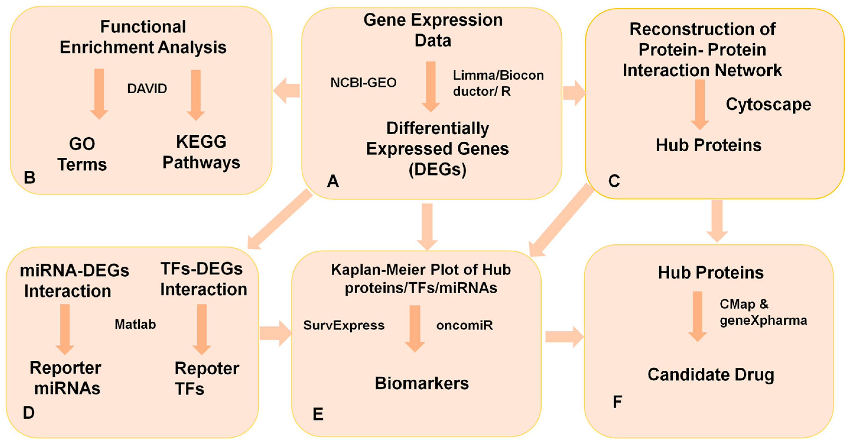
2. Materials and Methods
2.1. High-Throughput Microarray Gene Expression Datasets
2.2. Identification of Differentially Expressed Genes
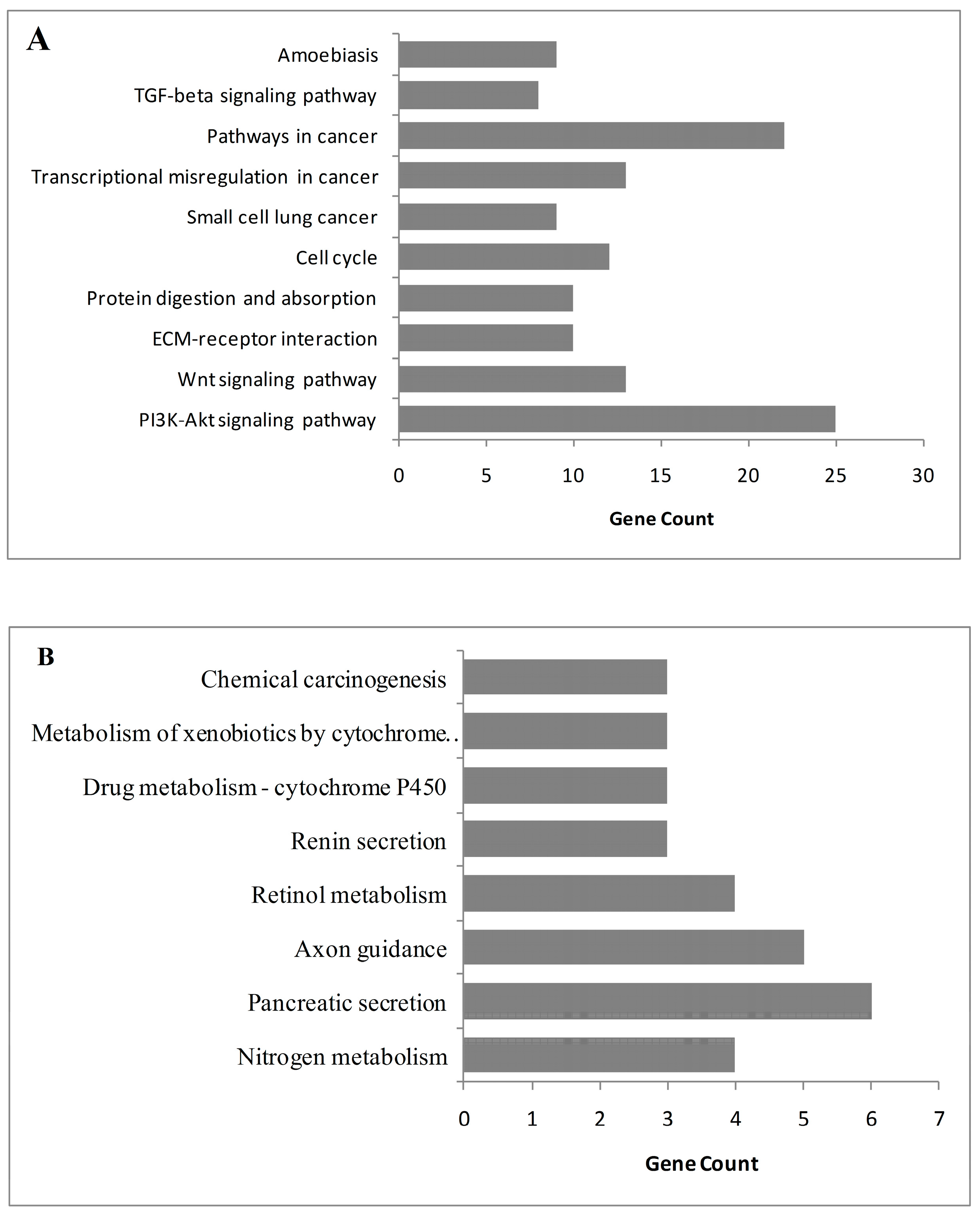
2.3. Gene Ontology and Pathway Analysis
2.4. Reconstruction and Analysis of Protein–Protein Interaction (PPI) Network in CRC
2.5. Identification of Reporter Biomolecules
2.6. Evaluation of the Prognostic Performance of Reporter Molecules
2.7. Identification of Candidate Drugs
3. Results
3.1. Identification of Differentially Expressed Genes
3.2. Analysis of Protein–Protein Interaction Network to Identify Hub Proteins
3.3. Identification of Regulatory Biomolecules
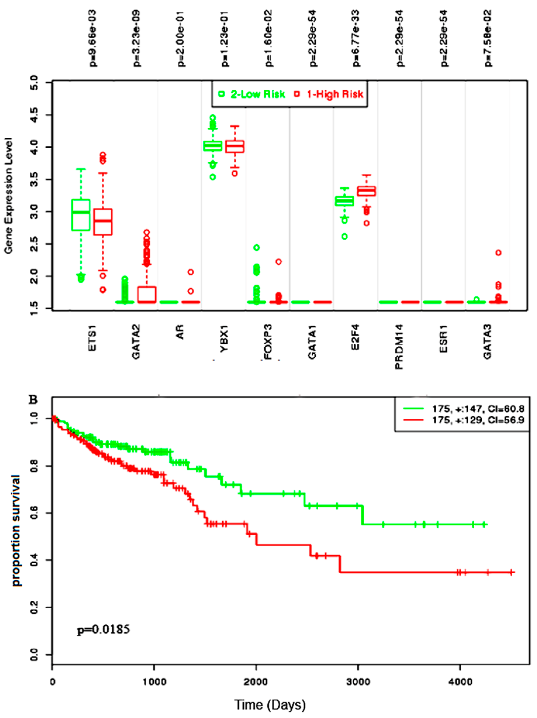
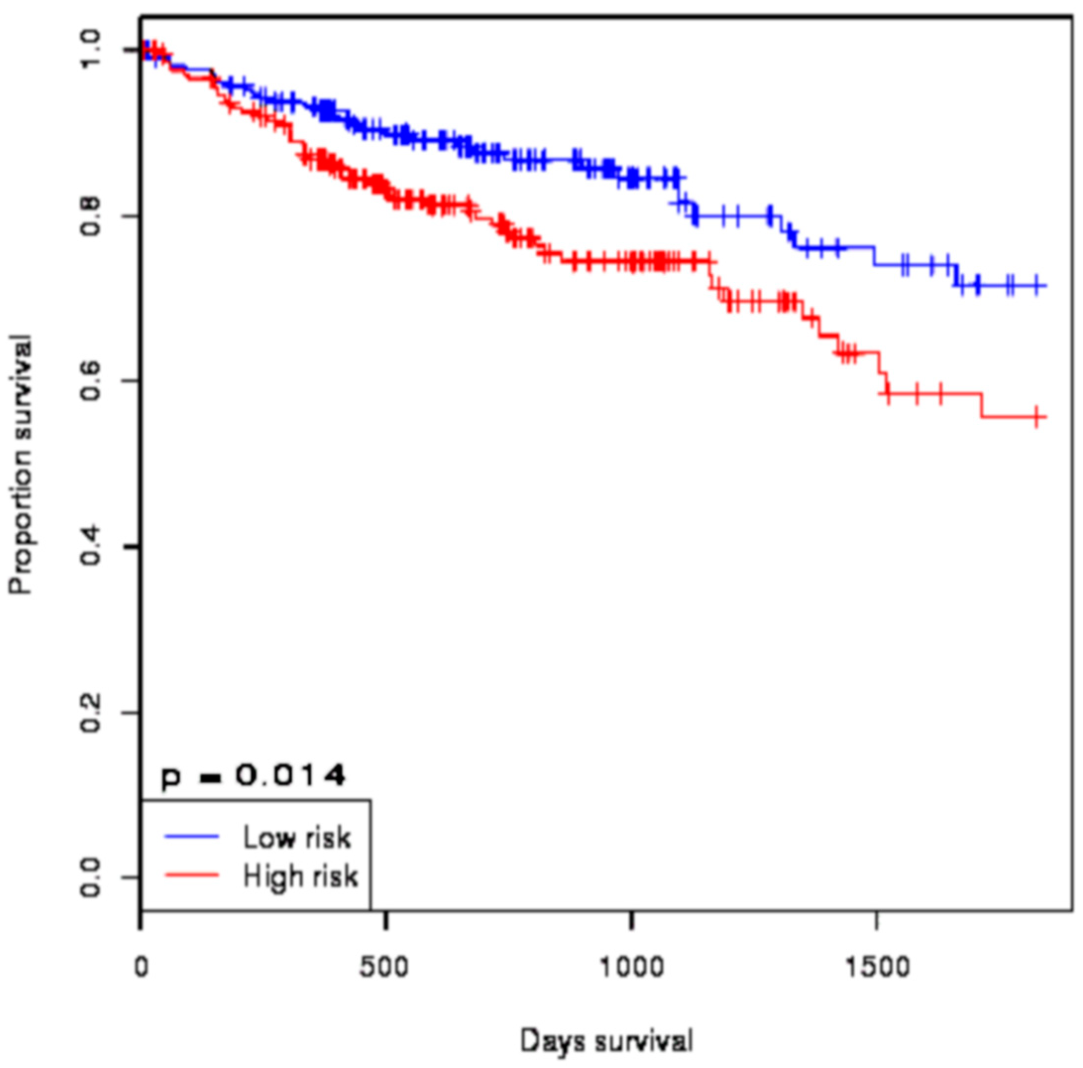
3.4. Survival Analysis of Biomolecules
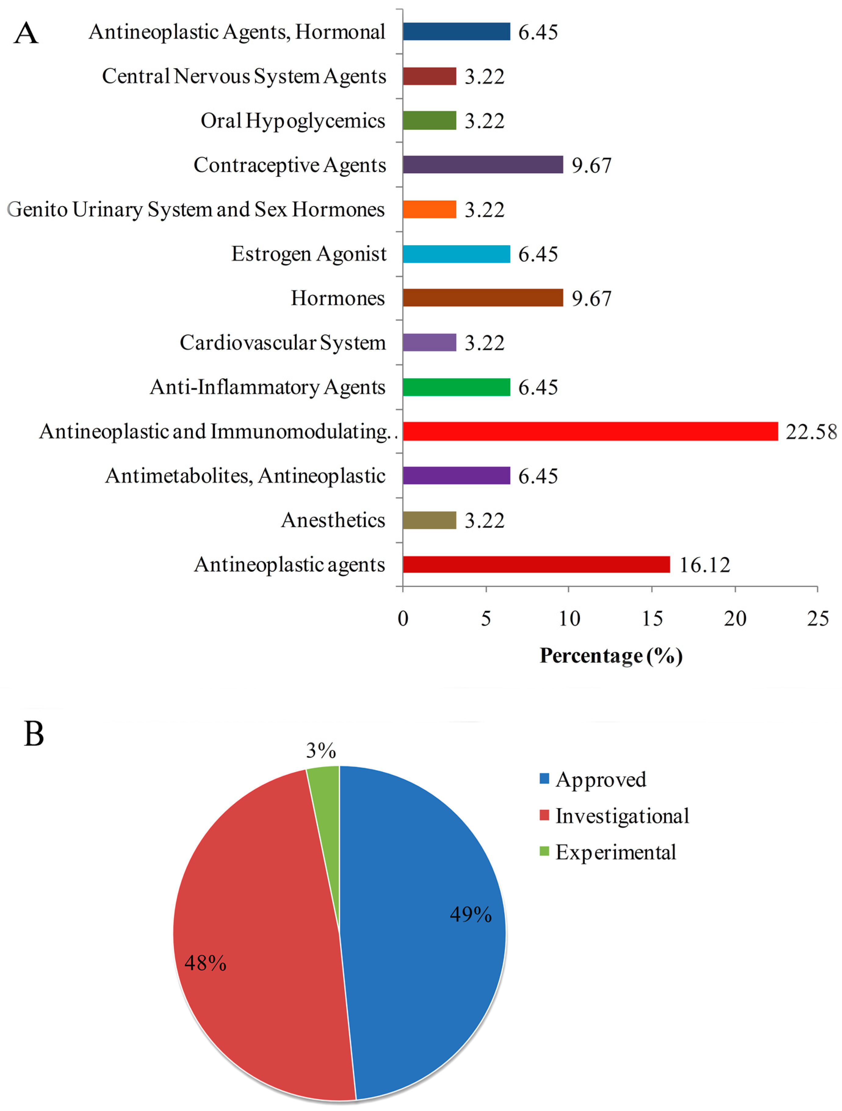
3.5. Identification of Candidate Drugs through In Silico Drug Repositioning
4. Discussion
5. Conclusions
Author Contributions
Funding
Acknowledgments
Conflicts of Interest
References
- Bray, F.; Ferlay, J.; Soerjomataram, I.; Siegel, R.L.; Torre, L.A.; Jemal, A. Global Cancer Statistics 2018: GLOBOCAN estimates of incidence and mortality worldwide for 36 cancers in 185 countries. CA Cancer J. Clin. 2018, 68, 394–424. [Google Scholar] [CrossRef] [PubMed]
- Arnold, M.; Sierra, M.S.; Laversanne, M.; Soerjomataram, I.; Jemal, A.; Bray, F. Global patterns and trends in colorectal cancer incidence and mortality. Gut 2017, 66, 683–691. [Google Scholar] [CrossRef] [PubMed]
- Markowitz, S.D.; Bertagnolli, M.M. Molecular Basis of Colorectal Cancer. N. Engl. J. Med. 2013, 361, 2449–2460. [Google Scholar] [CrossRef] [PubMed]
- Kheirelseid, E.A.H.; Miller, N.; Kerin, M.J. Molecular biology of colorectal cancer: Review of the literature. Am. J. Mol. Biol. 2013, 3, 72–80. [Google Scholar] [CrossRef]
- Grady, W.M.; Markowitz, S.D. The molecular pathogensis of colorectal cancer and its potential application to colorectal cancer screening. Dig. Dis. Sci. 2015, 60, 762–772. [Google Scholar] [CrossRef] [PubMed]
- Zarkavelis, G.; Boussios, S.; Papadaki, A.; Katsanos, K.H.; Christodoulou, D.K.; Pentheroudakis, G. Current and future biomarkers in colorectal cancer. Ann. Gastroenterol. 2017, 30, 613–621. [Google Scholar] [CrossRef] [PubMed]
- Krizkova, S.; Kepinska, M.; Emri, G.; Rodrigo, M.A.M.; Tmejova, K.; Nerudova, D.; Kizek, R.; Adam, V. Microarray analysis of metallothioneins in human diseases—A review. J. Pharm. Biomed. Anal. 2016, 117, 464–473. [Google Scholar] [CrossRef] [Green Version]
- Stewart, J.P.; Richman, S.; Maughan, T.; Lawler, M.; Dunne, P.D.; Salto-Tellez, M. Standardising RNA profiling based biomarker application in cancer-The need for robust control of technical variables. Biochim. Biophys. Acta 2017, 1868, 258–272. [Google Scholar] [CrossRef]
- Kamel, H.F.M.; Al-Amodi, H.S.A.B. Exploitation of Gene Expression and Cancer Biomarkers in Paving the Path to Era of Personalized Medicine. Genomics Proteomics Bioinformatics 2017, 15, 20–35. [Google Scholar] [CrossRef]
- Isella, C.; Terrasi, A.; Bellomo, S.E.; Petti, C.; Galatola, G.; Muratore, A.; Mellano, A.; Senetta, R.; Cassenti, A.; Sonetto, C.; et al. Stromal contribution to the colorectal cancer transcriptome. Nat. Genet. 2015, 47, 312–319. [Google Scholar] [CrossRef]
- Tripathi, M.K.; Deane, N.G.; Zhu, J.; An, H.; Mima, S.; Wang, X.; Padmanabhan, S.; Shi, Z.; Prodduturi, N.; Ciombor, K.K.; et al. Nuclear factor of activated T-cell activity is associated with metastatic capacity in colon cancer. Cancer Res. 2014, 74, 6947–6957. [Google Scholar] [CrossRef] [PubMed]
- Masuda, T.; Hayashi, N.; Kuroda, Y.; Ito, S.; Eguchi, H.; Mimori, K. MicroRNAs as biomarkers in colorectal cancer. Cancers (Basel) 2017, 9, 124. [Google Scholar] [CrossRef] [PubMed]
- Michael, M.Z.; O’ Connor, S.M.; van Holst Pellekaan, N.G.; Young, G.P.; James, R.J. Reduced accumulation of specific microRNAs in colorectal neoplasia. Mol. Cancer Res. 2003, 1, 882–891. [Google Scholar] [CrossRef] [PubMed]
- Barabási, A.L.; Gulbahce, N.; Loscalzo, J. Network medicine: A network-based approach to human disease. Nat. Rev. Genet. 2011, 12, 56–68. [Google Scholar] [CrossRef] [PubMed]
- Islam, T.; Rahman, M.R.; Gov, E.; Turanli, B.; Gulfidan, G.; Haque, M.A.; Arga, K.Y.; Mollah, M.N.H. Drug Targeting and Biomarkers in Head and Neck Cancers: Insights from Systems Biology Analyses. OMICS 2018, 22, 422–436. [Google Scholar] [CrossRef] [PubMed]
- Karagoz, K.; Lehman, H.; Stairs, D.; Sinha, R.; Arga, K.Y. Proteomic and metabolic signatures of esophageal squamous cell carcinoma. Curr. Cancer Drug Targets 2016, 16, 721–736. [Google Scholar] [CrossRef]
- Karagoz, K.; Sinha, R.; Arga, K.Y. Triple Negative Breast Cancer: A Multi-omics Network Discovery Strategy for Candidate Targets and Driving Pathways. OMICS 2015, 19, 115–130. [Google Scholar]
- Kori, M.; Arga, K.Y. Potential biomarkers and therapeutic targets in cervical cancer: Insights from meta-analysis of transcriptomics data within network biomedicine perspective. PLoS ONE 2018, 13, e0200717. [Google Scholar] [CrossRef]
- Gov, E.; Kori, M.; Arga, K.Y. Multiomics analysis of tumor microenvironment reveals Gata2 and miRNA-124-3p as potential novel biomarkers in ovarian cancer. OMICS 2017, 21, 603–615. [Google Scholar]
- Kori, M.; Gov, E.; Arga, K.Y. Molecular signatures of ovarian diseases: Insights from network medicine perspective. Syst. Biol. Reprod. Med. 2016, 62, 266–282. [Google Scholar] [CrossRef]
- Sevimoglu, T.; Turanli, B.; Bereketoglu, C.; Arga, K.Y.; Karadag, A.S. Systems biomarkers in psoriasis: Integrative evaluation of computational and experimental data at transcript and protein levels. Gene 2018, 647, 157–163. [Google Scholar] [CrossRef] [PubMed]
- Calimlioglu, B.; Karagoz, K.; Sevimoglu, T.; Kilic, E.; Gov, E.; Arga, K.Y. Tissue-Specific Molecular Biomarker Signatures of Type 2 Diabetes: An Integrative Analysis of Transcriptomics and Protein-Protein Interaction Data. OMICS 2015, 19, 563–573. [Google Scholar] [CrossRef]
- Lamb, J.; Crawford, E.D.; Peck, D.; Modell, J.W.; Blat, I.C.; Wrobel, M.J.; Lerner, J.; Brunet, J.P.; Subramanian, A.; Ross, K.N.; et al. The connectivity map: Using gene-expression signatures to connect small molecules, genes, and disease. Science 2006, 313, 1929–1935. [Google Scholar] [CrossRef] [PubMed]
- Turanli, B.; Gulfidan, G.; Arga, K.Y. Transcriptomic-Guided Drug Repositioning Supported by a New Bioinformatics Search Tool: geneXpharma. OMICS 2017, 21, 584–591. [Google Scholar] [CrossRef]
- Kagawa, Y.; Matsumoto, S.; Kamioka, Y.; Mimori, K.; Naito, Y.; Ishii, T.; Daisuke Okuzaki, D.; Nishida, N.; Maeda, S.; Naito, A.; et al. Cell cycle-dependent Rho GTPase activity dynamically regulates cancer cell motility and invasion in vivo. PLoS ONE 2013, 8, e83629. [Google Scholar] [CrossRef] [PubMed]
- Kogo, R.; Shimamura, T.; Mimori, K.; Kawahara, K.; Imoto, S.; Sudo, T.; Kogo, R.; Shimamura, T.; Mimori, K.; Kawahara, K.; et al. Long noncoding RNA HOTAIR regulates polycomb-dependent chromatin modification and is associated with poor prognosis in colorectal cancers. Cancer Res. 2011, 71, 6320–6326. [Google Scholar] [CrossRef] [PubMed]
- Barrett, T.; Wilhite, S.E.; Ledoux, P.; Evangelista, C.; Kim, I.F.; Tomashevsky, M.; Marshall, K.A.; Phillippy, K.H.; Sherman, P.M.; Holko, M.; et al. NCBI GEO: Archive for functional genomics data sets—Update. Nucleic Acids Res. 2013, 41, 991–995. [Google Scholar] [CrossRef]
- Bolstad, B.M.; Irizarry, R.A.; Astrand, M.; Speed, T.P. A comparison of normalization methods for high density oligonucleotide array data based on variance and bias. Bioinformatics 2003, 19, 185–193. [Google Scholar] [CrossRef] [Green Version]
- Smyth, G.K.; Ritchie, M.; Thorne, N. Linear Models for Microarray Data User’s Guide. Bioinformatics 2011, 20, 3705–3706. [Google Scholar] [CrossRef]
- Huang, D.W.; Sherman, B.T.; Lempicki, R.A. Systematic and integrative analysis of large gene lists using DAVID bioinformatics resources. Nat. Protoc. 2009, 4, 44–57. [Google Scholar] [CrossRef]
- Kanehisa, M.; Furumichi, M.; Tanabe, M.; Sato, Y.; Morishima, K. KEGG: New perspectives on genomes, pathways, diseases and drugs. Nucleic Acids Res. 2017, 45, D353–D361. [Google Scholar] [CrossRef] [PubMed]
- The Gene Ontology Consortium. Gene Ontology Consortium: Going forward. Nucleic Acids Res. 2015, 43, D1049–D1056. [Google Scholar] [CrossRef]
- Karagoz, K.; Sevimoglu, T.; Arga, K.Y. Integration of multiple biological features yields high confidence human protein interactome. J. Theor. Biol. 2016, 403, 85–96. [Google Scholar] [CrossRef]
- Smoot, M.E.; Ono, K.; Ruscheinski, J.; Wang, P.L.; Ideker, T. Cytoscape 2.8: New features for data integration and network visualization. Bioinformatics 2011, 27, 431–432. [Google Scholar] [CrossRef] [PubMed]
- Chin, C.H.; Chen, S.H.; Wu, H.H.; Ho, C.W.; Ko, M.T.; Lin, C.Y. cytoHubba: Identifying hub objects and sub-networks from complex interactome. BMC Syst. Biol. 2014, 8 (Suppl. 4). [Google Scholar] [CrossRef] [PubMed]
- Bader, G.D.; Hogue, C.W. An automated methods for finding molecular complexes in large protein interaction networks. BMC Bioinformatics 2003, 4, 2. [Google Scholar] [CrossRef]
- Gov, E.; Arga, K.Y.A. Interactive cooperation and hierarchical operation of microRNA and transcription factor crosstalk in human transcriptional regulatory network. IET Syst. Biol. 2016, 10, 219–228. [Google Scholar] [CrossRef]
- Bovolenta, L.A.; Acencio, M.L.; Lemke, N. HTRIdb: An open-access database for experimentally verified human transcriptional regulation interactions. BMC Genomics 2012, 13, 405. [Google Scholar] [CrossRef]
- Hsu, S.D.; Tseng, Y.T.; Shrestha, S.; Lin, Y.L.; Khaleel, A.; Chou, C.H.; Chu, C.F.; Huang, H.Y.; Lin, C.M.; Ho, S.Y.; et al. miRTarBase update 2014: An information resource for experimentally validated miRNA-target interactions. Nucleic Acids Res. 2014, 42, D78–D85; [Google Scholar] [CrossRef]
- Patil, K.R.; Nielsen, J. Uncovering transcriptional regulation of metabolism by using metabolic network topology. Proc. Natl. Acad. Sci. USA 2005, 102, 2685–2689. [Google Scholar] [CrossRef] [Green Version]
- Aguirre-Gamboa, R.; Gomez-Rueda, H.; Martínez-Ledesma, E.; Martínez-Torteya, A.; Chacolla-Huaringa, R.; Rodriguez-Barrientos, A.; Tamez-Peña, J.G.; Treviño, V. SurvExpress: An Online Biomarker Validation Tool and Database for Cancer Gene Expression Data Using Survival Analysis. PLoS ONE 2013, 8, e74250. [Google Scholar] [CrossRef] [PubMed]
- Wong, N.W.; Chen, Y.; Chen, S.; Wang, X. OncomiR: An online resource for exploring pan-cancer microRNA dysregulation. Bioinformatics 2018, 34, 713–715. [Google Scholar] [CrossRef] [PubMed]
- Guo, Y.; Bao, Y.; Ma, M.; Yang, W. Identification of key candidate genes and pathways in colorectal cancer by integrated bioinformatical analysis. Int. J. Mol. Sci. 2017, 18, 722. [Google Scholar] [CrossRef]
- Nagaraj, N.S.; Datta, P.K. Targetting the Transforming Growth factor-beta Signalling Pathway in Human Cancer. Expert Opin. Investig. Drugs 2010, 19, 77–91. [Google Scholar] [CrossRef] [PubMed]
- Sevimoglu, T.; Arga, K.Y. The role of protein interaction networks in systems biomedicine. Comput. Struct. Biotechnol. J. 2014, 11, 22–27. [Google Scholar] [CrossRef] [PubMed] [Green Version]
- Blaj, C.; Bringmann, A.; Urbischek, M.; Krebs, S.; Blum, H.; Fröhlich, T.; Arnold, G.J.; Krebs, S.; Blum, H.; Hermeking, H.; et al. ADNP is a repressor of WNT signaling in colon cancer that can be therapeutically induced. Eur. J. Cancer 2016, 61 (Suppl. 1). [Google Scholar] [CrossRef]
- Rask, K.; Thorn, M.; Ponten, F.; Kraaz, W.; Sundfeldt, K.; Hedin, L.; Enerbäck, S. Increased expression of the transcription factors CCAAT-enhancer binding protein-beta (C/EBBeta) and C/EBzeta (CHOP) correlate with invasiveness of human colorectal cancer. Int. J. Cancer 2000, 86, 337–343. [Google Scholar] [CrossRef]
- Yin, J.; Oh, Y.T.; Kim, J.Y.; Kim, S.S.; Choi, E.; Kim, T.H.; Hong, J.H.; Chang, N.; Cho, H.J.; Sa, J.K.; et al. Transglutaminase 2 inhibition reverses mesenchymal transdifferentiation of glioma stem cells by regulating C/EBPβ signaling. Cancer Res. 2017, 77, 4973–4984. [Google Scholar] [CrossRef] [PubMed]
- Balcerczak, E.; Pasz-Walczak, G.; Kumor, P.; Panczyk, M.; Kordek, R.; Wierzbicki, R.; Mirowski, M. Cyclin D1 protein and CCND1 gene expression in colorectal cancer. Eur. J. Surg. Oncol. 2005, 31, 721–726. [Google Scholar] [CrossRef]
- Porter, T.R.; Richards, F.M.; Houlston, R.S.; Evans, D.G.R.; Jankowski, J.A.; Macdonald, F.; Norbury, G.; Payne, S.J.; Fisher, S.A.; Tomlinson, I.; et al. Contribution of cyclin d1 (CCND1) and E-cadherin (CDH1) polymorphisms to familial and sporadic colorectal cancer. Oncogene 2002, 21, 1928–1933. [Google Scholar] [CrossRef] [Green Version]
- Wolter, F.; Akoglu, B.; Clausnitzer, A.; Stein, J. Downregulation of the cyclin D1/Cdk4 complex occurs during resveratrol-induced cell cycle arrest in colon cancer cell lines. J. Nutr. 2001, 131, 2197–2203. [Google Scholar] [CrossRef] [PubMed]
- Pek, M.; Yatim, S.M.J.M.; Chen, Y.; Li, J.; Gong, M.; Jiang, X.; Zhang, F.; Zheng, J.; Wu, X.; Yu, Q.; et al. Oncogenic KRAS-associated gene signature defines co-targeting of CDK4/6 and MEK as a viable therapeutic strategy in colorectal cancer. Oncogene 2017, 36, 4975–4986. [Google Scholar] [CrossRef]
- Tomonaga, T.; Matsushita, K.; Yamaguchi, S. Overexpression and Mistargeting of Centromere Protein-A in Human Primary Colorectal Cancer. Cancer Res. 2003, 63, 3511–3516. [Google Scholar] [PubMed]
- Tomonaga, T.; Matsushita, K.; Ishibashi, M.; Nezu, M.; Shimada, H.; Ochiai, T.; Yoda, K.; Nomura, F. Centromere protein H Is up-regulated in primary human colorectal cancer and its overexpression induces aneuploidy. Cancer Res. 2005, 65, 4683–4689. [Google Scholar] [CrossRef] [PubMed]
- Maruyama, T.; Farina, A.; Dey, A.; Cheong, J.; Bermudez, V.P.; Tamura, T.; Sciortino, S.; Shuman, J.; Hurwitz, J.; Ozato, K.; et al. A Mammalian Bromodomain Protein, Brd4, Interacts with Replication Factor C and Inhibits Progression to S Phase. Mol. Cell. Biol. 2002, 22, 6509–6520. [Google Scholar] [CrossRef] [PubMed] [Green Version]
- Da Costa, D.; Agathanggelou, A.; Perry, T.; Weston, V.; Petermann, E.; Zlatanou, A.; Oldreive, C.; Wei, W.; Stewart, G.; Longman, J.; et al. BET inhibition as a single or combined therapeutic approach in primary paediatric B-precursor acute lymphoblastic leukaemia. Blood Cancer J. 2013, 3, e126. [Google Scholar] [CrossRef]
- Dang, C.V.; Le, A.; Gao, P. MYC-induced cancer cell energy metabolism and therapeutic opportunities. Clin. Cancer Res. 2009, 15, 6479–6483. [Google Scholar] [CrossRef]
- Sikora, K.; Chan, S.; Evan, G.; Gabra, H.; Markham, N.; Stewart, J.; Watson, J. c-myc oncogene expression in colorectal cancer. Cancer 1987, 59, 1289–1295. [Google Scholar] [CrossRef] [Green Version]
- Castell, A.; Larsson, L.-G. Targeting MYC Translation in Colorectal Cancer. Cancer Discov. 2015, 5, 701–703. [Google Scholar] [CrossRef] [Green Version]
- Hellwig, D.; Emmerth, S.; Ulbricht, T.; Doring, V.; Hoischen, C.; Martin, R.; Samora, C.P.; McAinsh, A.D.; Carroll, C.W.; Straight, A.F.; et al. Dynamics of CENP-N kinetochore binding during the cell cycle. J. Cell Sci. 2011, 124, 3871–3883. [Google Scholar] [CrossRef] [Green Version]
- Jenster, G. The role of the androgen receptor in the development and progression of prostate cancer. Semin. Oncol. 1999, 26, 407–421. [Google Scholar] [PubMed]
- Seth, A.; Watson, D.K. ETS transcription factors and their emerging roles in human cancer. Eur. J. Cancer 2005, 41, 2462–2478. [Google Scholar] [CrossRef]
- Chen, L.; Jiang, B.; Wang, Z.; Liu, M.; Ma, Y.; Yang, H.; Xing, J.; Zhang, C.; Yao, Z.; Zhang, N.; et al. Expression and prognostic significance of GATA-binding protein 2 in colorectal cancer. Med. Oncol. 2013, 30, 498. [Google Scholar] [CrossRef] [PubMed]
- Zheng, R.; Blobel, G.A. Gata transcription factors and cancer. Genes Cancer 2010, 1, 1178–1188. [Google Scholar] [CrossRef] [PubMed]
- Prabhu, L.; Mundade, R.; Wang, B.; Wei, H.; Hartley, A.-V.; Martin, M.; McElyea, K.; Temm, C.J.; Sandusky, G.; Liu, Y.; et al. Critical role of phosphorylation of serine 165 of YBX1 on the activation of NF-κB in colon cancer. Oncotarget 2015, 6, 29396–29412. [Google Scholar] [CrossRef]
- Oda, Y.; Ohishi, Y.; Saito, T.; Hinoshita, E.; Uchiumi, T.; Kinukawa, N.; Iwamoto, Y.; Kohno, K.; Kuwano, M.; Tsuneyoshi, M.; et al. Nuclear expression of Y-box-binding protein-I correlates with P-glycoprotein and topoisomerase II alpha expression, and with poor prognosis in synovial sarcoma. J. Pathol. 2003, 199, 251–258. [Google Scholar] [CrossRef] [PubMed]
- Le Gouvello, S.; Bastuji-Garin, S.; Aloulou, N.; Mansour, H.; Chaumette, M.T.; Berrehar, F.; Seikour, A.; Charachon, A.; Karoui, M.; Leroy, K.; et al. High prevalence of Foxp3 and IL17 in MMR-proficient colorectal carcinomas. Gut 2008, 57, 772–779. [Google Scholar] [CrossRef] [Green Version]
- Nevins, J.R. The Rb/E2F pathway and cancer. Hum. Mol. Genet. 2001, 10, 699–703. [Google Scholar] [CrossRef] [Green Version]
- Garneau, H.; Paquin, M.C.; Carrier, J.C.; Rivard, N. E2F4 expression is required for cell cycle progression of normal intestinal crypt cells and colorectal cancer cells. J. Cell. Physiol. 2009, 221, 350–358. [Google Scholar] [CrossRef]
- Zhang, T.; Cui, G.; Bi, H.; Shi, H. PRDM14 Promotes the Migration of Human Non-small Cell Lung Cancer Through Extracellular Matrix Degradation in vitro. Chin. Med. J. 2015, 128, 373–377. [Google Scholar] [CrossRef]
- Nishikawa, N.; Toyota, M.; Suzuki, H.; Honma, T.; Fujikane, T.; Ohmura, T.; Ohe-Toyota, M.; Maruyama, R.; Sonoda, T.; Sasaki, Y.; et al. Gene amplification and overexpression of PRDM14 in breast cancers. Cancer Res. 2007, 67, 9649–9657. [Google Scholar] [CrossRef] [PubMed]
- Holst, F.; Stahl, P.R.; Ruiz, C.; Hellwinkel, O.; Jehan, Z.; Wendland, M.; Lebeau, A.; Terracciano, L.; Al-Kuraya, K.; Jänicke, F.; et al. Estrogen receptor alpha (ESR1) gene amplification is frequent in breast cancer. Nat. Genet. 2007, 39, 655–660. [Google Scholar] [CrossRef] [PubMed]
- Hrašovec, S.; Glavač, D. MicroRNAs as novel biomarkers in colorectal cancer. Front. Genet. 2012, 3, 180. [Google Scholar] [CrossRef] [PubMed]
- Mullany, L.E.; Herrick, J.S.; Sakoda, L.C.; Samowitz, W.; John, R.; Wolff, R.K.; Slattery, M.L. miRNA involvement in cell cycle regulation in colorectal cancer cases. Genes Cancer 2018, 9, 53–65. [Google Scholar] [CrossRef] [PubMed]
- Wu, K.; Zhao, Z.; Ma, J.; Chen, J.; Peng, J.; Yang, S.; He, Y. Deregulation of miR-193b affects the growth of colon cancer cells via transforming growth factor-β and regulation of the SMAD3 pathway. Oncol. Lett. 2017, 13, 2557–2562. [Google Scholar] [CrossRef] [PubMed] [Green Version]
- Trevisani, F.; Ghidini, M.; Larcher, A.; Lampis, A.; Lote, H.; Manunta, P.; Librandi, M.T.; Zagato, L.; Citterio, L.; Dell’Antonio, G.; et al. MicroRNA 193b-3p as a predictive biomarker of chronic kidney disease in patients undergoing radical nephrectomy for renal cell carcinoma. Br. J. Cancer 2016, 115, 1343–1350. [Google Scholar] [CrossRef] [PubMed] [Green Version]
- Schee, K.; Lorenz, S.; Worren, M.M.; Günther, C.C.; Holden, M.; Hovig, E.; Fodstad, O.; Meza-Zepeda, L.A.; Flatmark, K. Deep Sequencing the MicroRNA Transcriptome in Colorectal Cancer. PLoS ONE 2013, 8, e66165. [Google Scholar] [CrossRef]
- Wang, J.; Liu, L.; Sun, Y.; Xue, Y.; Qu, J.; Pan, S.; Li, H.; Qu, H.; Wang, J.; Zhang, J. miR-615-3p promotes proliferation and migration and inhibits apoptosis through its potential target CELF2 in gastric cancer. Biomed. Pharmacother. 2018, 101, 406–413. [Google Scholar] [CrossRef]


| Gene Ontology | Gene Ontology (GO) Term | # of Genes | Coverage (%) | p-Value |
|---|---|---|---|---|
| Upregulated genes | ||||
| Biological Process | Collagen fibril organization | 11 | 1.62 | 4.53 × 10⁻⁷ |
| Extracellular matrix organization | 22 | 3.24 | 2.94 × 10⁻⁶ | |
| Male gonad development | 14 | 2.06 | 1.53 × 10⁻⁵ | |
| Positive regulation of transcription from RNA polymerase II promoter | 58 | 8.56 | 3.90 × 10⁻⁵ | |
| Collagen catabolic process | 11 | 1.62 | 5.07 × 10⁻⁵ | |
| Cellular Component | Extracellular region | 84 | 12.4 | 2.40 × 10⁻⁵ |
| Cytoplasm | 216 | 31.9 | 5.80 × 10⁻⁵ | |
| Extracellular space | 70 | 10.3 | 1.50 × 10⁻⁴ | |
| Basement membrane | 11 | 1.62 | 2.56 × 10⁻⁴ | |
| Extracellular matrix | 23 | 3.39 | 3.34 × 10⁻⁴ | |
| Molecular Function | Protein binding | 354 | 52.3 | 8.10 × 10⁻⁸ |
| Protein homodimerization activity | 42 | 6.20 | 7.54 × 10⁻⁴ | |
| Growth factor activity | 15 | 2.21 | 1.04 × 10⁻³ | |
| Extracellular matrix binding | 6 | 0.88 | 1.47 × 10⁻³ | |
| Amino-acid transmembrane transporter activity | 7 | 1.03 | 4.43 × 10⁻³ | |
| Downregulated genes | ||||
| Biological Process | Bicarbonate transport | 5 | 4.90 | 5.89 × 10⁻⁵ |
| One-carbon metabolic process | 4 | 3.92 | 4.00 × 10⁻⁴ | |
| Chloride transmembrane transport | 5 | 4.90 | 1.06 × 10⁻³ | |
| Nervous system development | 7 | 6.86 | 2.63 × 10⁻³ | |
| Regulation of chloride transport | 2 | 1.96 | 9.62 × 10⁻³ | |
| Cellular Component | Plasma membrane | 31 | 30.4 | 0.0108 |
| Extracellular space | 14 | 13.7 | 0.0135 | |
| Integral component of membrane | 36 | 35.3 | 0.0163 | |
| Anchored component of membrane | 4 | 3.92 | 0.0179 | |
| Integral component of plasma membrane | 13 | 12.7 | 0.0421 | |
| Molecular Function | Carbonate dehydratase activity | 4 | 3.92 | 4.16×10⁻⁵ |
| Hormone activity | 5 | 4.90 | 0.0012 | |
| Zinc ion binding | 15 | 14.7 | 0.0018 | |
| UDP-galactose:β-N-acetylglucosamine β-1,3-galactosyltransferase activity | 3 | 2.94 | 0.0018 | |
| Chloride channel activity | 4 | 3.92 | 0.0025 | |
| Symbol | Description | Feature |
|---|---|---|
| Hub proteins | ||
| ADNP | Activity-dependent neuroprotector homeobox | Stimulatory and inhibitory effect on the growth of tumor cells |
| CEBPB | CCAAT/enhancer-binding protein beta | Involved in immune and inflammatory responses |
| CCND1 | Cyclin D1 (afflicted with cancers colonic adenocarcinomas, myeloma) | Cell-cycle regulatory protein |
| CD44 | CD44 molecule | Required in cell–cell interactions, migration |
| CDK4 | Cyclin-dependent kinase 4 | Cyclin D1 activates CDK4, which causes proliferation of cellular division. |
| CENPA | Centromere protein A (afflicted with colorectal cancer) | Central role in the assembly of kinetochore |
| CENPH | Centromere Protein H (afflicted with colorectal cancer) | Central role in the assembly of kinetochore proteins |
| RFC2 | Replication factor C subunit 2 | Encodes activator 1 small subunit family |
| MYC | Myc proto-oncogene | Regulator gene contributes to formation of many human cancers |
| CENPN | Centromere protein N | Involved in cell-cycle process |
| Symbol | Description | Feature |
|---|---|---|
| Reporter Transcription Factors | ||
| AR | Androgen receptor | Involved in prostate cancer |
| GATA1 | GATA binding protein 1 | Transcriptional activator or repressor |
| GATA2 | GATA binding protein 2 (afflicted with colorectal cancer) | Transcriptional activator |
| GATA3 | GATA binding protein 3 | Transcriptional activator |
| E2F4 | E2F transcription factor 4 | Controls of cell cycle |
| ETS1 | ETS proto-oncogene 1 | Involved in tumorigenesis |
| YBX1 | Y-box binding protein 1 | Aberrant expression is associated with cancer |
| PRADM14 | PR/SET domain 14 | Involved in breast cancer |
| ESR1 | Estrogen receptor 1 | Involved in breast cancer |
| FOXP3 | Forkhead box P3 (afflicted with colorectal cancer) | DNA binding |
| Reporter microRNAs | ||
| miR-193b-3p | MicroRNA 193 | Afflicted with CRC and epidermal squamous cell carcinoma |
| miR-615-3p | MicroRNA 615 | Afflicted with CRC |
| miR-16-5p | MicroRNA 16 | Potential biomarkers in gastric cancer |
| miR-26b-5p | MicroRNA 26 | Afflicted with CRC |
| let-7b-5p | MicroRNA 7 | Afflicted with CRC |
| miR-92a-3p | MicroRNA 92 | Afflicted with CRC |
| miR-124-3p | MicroRNA 124 | Afflicted with CRC, gastric and breast cancer |
| miR-484 | MicroRNA 484 | Afflicted with CRC |
| miR-192-5p | MicroRNA 192 | Afflicted with CRC |
| miR-93-5p | MicroRNA 93 | Afflicted with head and neck cancer |
| Target | Repositioned Drug | Drug Class/Status/Description |
|---|---|---|
| Hub protein | ||
| CCND1 | Gefitinib | Antineoplastic agent; approved; investigational/used in the treatment of cancer |
| Hydrocortisone | Anti-inflammatory agent; approved; used in the treatment of inflammation, allergy, collagen diseases, asthma, and some neoplastic conditions | |
| Irinotecan | Antineoplastic agent; approved; investigational/used in the treatment of colorectal cancer | |
| Letrozole | Antineoplastic agent; approved; investigational/introduced for treatment of breast cancer | |
| Lidocaine | Anesthetic; approved; local anesthetic and used as an antiarrhythmia agent | |
| Methotrexate | Antimetabolite, antineoplastic; approved; antineoplastic antimetabolite with immunosuppressant properties | |
| Sirolimus | Antineoplastic and immunomodulating agents; approved; investigational/a potent immunosuppressant which possesses both antifungal and antineoplastic properties | |
| Tamoxifen | Antineoplastic and immunomodulating agent; approved; for the treatment and prevention of breast cancer | |
| CDK4 | Gefitinib | Antineoplastic agent; approved; investigational/used in the treatment of cancer |
| Lidocaine | Anesthetic; approved; local anesthetic and used as an antiarrhythmia agent | |
| Sirolimus | Antineoplastic and immunomodulating agent; approved; investigational/a potent immunosuppressant which possesses both antifungal and antineoplastic properties | |
| MYC | Gefitinib | Antineoplastic agent; approved; investigational/used in the treatment of cancer |
| Tamoxifen | Antineoplastic and immunomodulating agent; approved; for the treatment and prevention of breast cancer | |
| Simvastatin | Cardiovascular system; approved; a lipid-lowering agent | |
| Reporter TFs | ||
| GATA3 | Azathioprine | Antineoplastic and immunomodulating agent; approved; immunosuppressive antimetabolite pro-drug |
| Daunorubicin | Antineoplastic and immunomodulating agent; approved; used in treatment of leukemia and other neoplasms | |
| Dexamethasone | Antineoplastic agent; approved, investigational, vet approved; for the treatment of endocrine disorders, rheumatic disorders, collagen diseases, dermatologic diseases | |
| Doxorubicin | Antineoplastic and immunomodulating agent; approved; investigational/used neoplastic conditions like acute lymphoblastic leukemia | |
| Mercaptopurine | Antimetabolite antineoplastic agent with immunosuppressant properties; approved; in the treatment of leukemia | |
| Methotrexate | Antimetabolite, antineoplastic; approved; antineoplastic antimetabolite with immunosuppressant properties | |
| ESR1 | Clomifene | Estrogen agonist, antagonist; approved; investigational/used mainly in female infertility due to anovulation to induce ovulation |
| Daunorubicin | Antineoplastic and immunomodulating agent; approved; used in treatment of leukemia and other neoplasms | |
| Dexamethasone | Antineoplastic agent; approved; investigational/for the treatment of endocrine disorders, rheumatic disorders, collagen diseases, dermatologic diseases | |
| Estriol | Estradiol congener; approved; investigational/used as a test to determine the general health of an unborn fetus | |
| Estrone | Hormone; approved; used for management of perimenopausal and postmenopausal symptoms | |
| Etoposide | Antineoplastic agent; approved; used in the treatment of refractory testicular tumors and in patients with small cell lung cancer | |
| Fulvestrant | Antineoplastic and immunomodulating agent; approved; investigational/a drug treatment of metastatic breast cancer | |
| Glibenclamide | Oral hypoglycemic; approved; used for the treatment of non-insulin-dependent diabetes mellitus | |
| Imipramine | Central nervous system agent; approved; antidepressant used for the relief of symptoms of depression | |
| Letrozole | Antineoplastic agent; approved; investigational/introduced for the treatment of breast cancer | |
| Megestrol | Antineoplastic and immunomodulating agent; approved; investigational/used in the palliative treatment of breast cancer | |
| Mifepristone | Abortifacient agent and blood-glucose-lowering agent; approved; investigational/for the medical termination of intrauterine pregnancy; also indicated to control hyperglycemia | |
| Progesterone | Contraceptive agent; approved, vet approved; progesterone acts on the uterus, the mammary glands, and the brain | |
| Raloxifene | Estrogen agonist, antagonist; approved; investigational/used to prevent osteoporosis in postmenopausal women | |
| Tamoxifen | Antineoplastic and immunomodulating agent; approved; for the treatment and prevention of breast cancer | |
| Testosterone | Androgen and estrogen; approved; investigational/in men, testosterone is produced primarily by the leydig cells of the testes; testerone in women functions to maintain libido and general wellbeing. | |
| AR | Cyproterone | Antineoplastic agent and hormone antagonist; approved; investigational/used in the treatment of hypersexuality in males, as a palliative in prostatic carcinoma |
| Flufenamic acid | Antiinflammatory and antirheumatic; experimental; analgesic, anti-inflammatory, and antipyretic properties | |
| Flutamide | Antineoplastic agent, hormonal; approved; investigational/for the management of metastatic carcinoma of the prostate | |
| Levonorgestrel | Contraceptive agent; approved; investigational/for the treatment of menopausal and postmenopausal disorders | |
| Mifepristone | Abortifacient agent and blood-glucose-lowering agent; approved; investigational/for the medical termination of intrauterine pregnancy; also indicated to control hyperglycemia | |
| Spironolactone | Agent causing hyperkalemia; approved; used primarily to treat low-renin hypertension, hypokalemia, and Conn’s syndrome | |
| Testosterone | Androgen and estrogen; approved; investigational/in men, testosterone is produced primarily by the interstitial cells of the testes; it functions to maintain libido and general wellbeing in women. | |
© 2019 by the authors. Licensee MDPI, Basel, Switzerland. This article is an open access article distributed under the terms and conditions of the Creative Commons Attribution (CC BY) license (http://creativecommons.org/licenses/by/4.0/).
Share and Cite
Rahman, M.R.; Islam, T.; Gov, E.; Turanli, B.; Gulfidan, G.; Shahjaman, M.; Akhter Banu, N.; Mollah, M.N.H.; Arga, K.Y.; Moni, M.A. Identification of Prognostic Biomarker Signatures and Candidate Drugs in Colorectal Cancer: Insights from Systems Biology Analysis. Medicina 2019, 55, 20. https://doi.org/10.3390/medicina55010020
Rahman MR, Islam T, Gov E, Turanli B, Gulfidan G, Shahjaman M, Akhter Banu N, Mollah MNH, Arga KY, Moni MA. Identification of Prognostic Biomarker Signatures and Candidate Drugs in Colorectal Cancer: Insights from Systems Biology Analysis. Medicina. 2019; 55(1):20. https://doi.org/10.3390/medicina55010020
Chicago/Turabian StyleRahman, Md. Rezanur, Tania Islam, Esra Gov, Beste Turanli, Gizem Gulfidan, Md. Shahjaman, Nilufa Akhter Banu, Md. Nurul Haque Mollah, Kazim Yalcin Arga, and Mohammad Ali Moni. 2019. "Identification of Prognostic Biomarker Signatures and Candidate Drugs in Colorectal Cancer: Insights from Systems Biology Analysis" Medicina 55, no. 1: 20. https://doi.org/10.3390/medicina55010020