Article

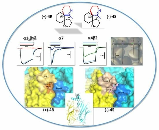
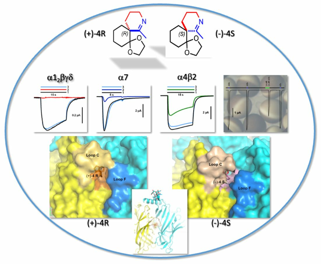
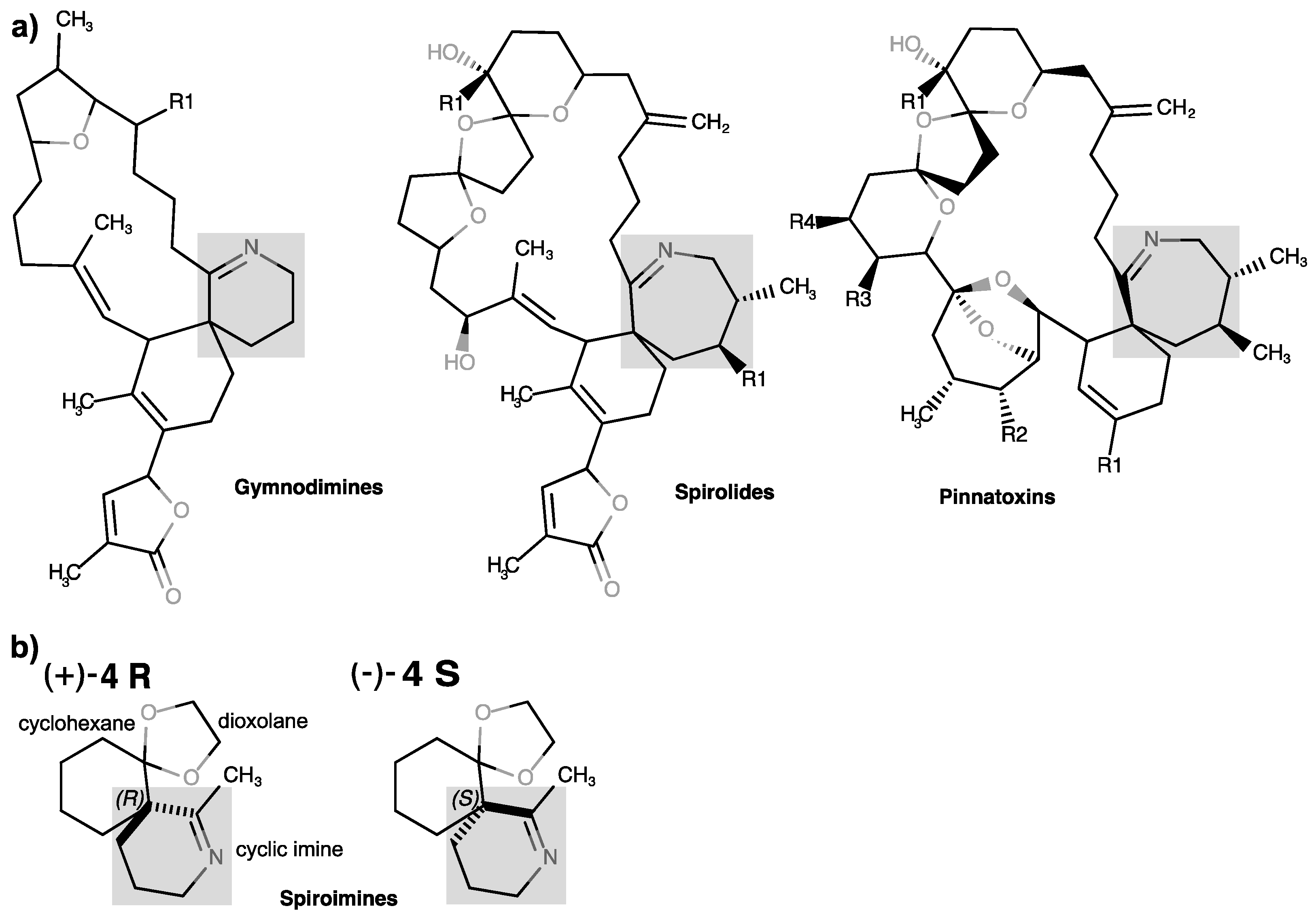
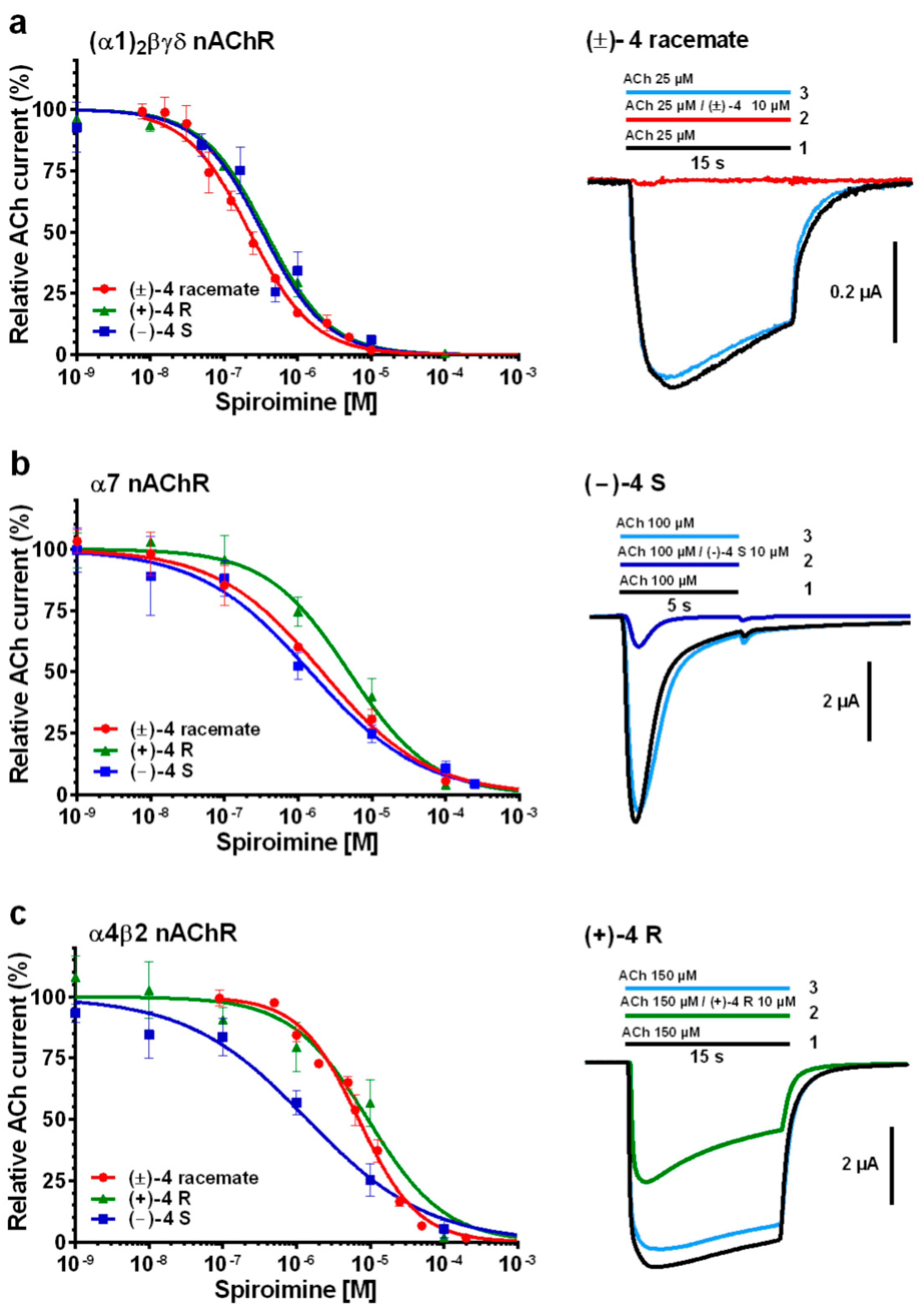
The Cyclic Imine Core Common to the Marine Macrocyclic Toxins Is Sufficient to Dictate Nicotinic Acetylcholine Receptor Antagonism

1
Lab “Architecture et Fonction des Macromolécules Biologiques” (AFMB), Aix-Marseille Univ, CNRS, Faculté des Sciences Campus Luminy, 13288 Marseille cedex 09, France
2
Institut de Chimie des Substances Naturelles (ICSN), Univ Paris-Saclay, CNRS, 91198 Gif-sur-Yvette, France
3
Service d’Ingénierie Moléculaire pour la Santé (SIMoS) EMR CNRS 9004, Département Médicaments et Technologies pour la Santé, Institut des Sciences du Vivant Frédéric Joliot, CEA, INRAE, Université Paris-Saclay, 91191 Gif-sur-Yvette, France
4
Skaggs School of Pharmacy and Pharmaceutical Sciences (SSPPS), University of California San Diego, La Jolla, CA 92093-0751, USA
5
Centre de Recherche en Neurobiologie et Neurophysiologie de Marseille (CRN2M), Aix Marseille Univ, CNRS, 13344 Marseille, France
*
Author to whom correspondence should be addressed.
Current address: Institut des Sciences Moléculaires (ISM), Univ Bordeaux, CNRS, Bordeaux INP, 33400 Talence, France
Current address: CEREGE, Aix-Marseille Univ, CNRS, IRD, Collège de France, INRAE, 13545 Aix-en-Provence, France
Mar. Drugs 2024, 22(4), 149; https://doi.org/10.3390/md22040149
Submission received: 13 February 2024 / Revised: 22 March 2024 / Accepted: 25 March 2024 / Published: 27 March 2024
(This article belongs to the Special Issue Marine Biotoxins 2.0)

Abstract

:
Macrocyclic imine phycotoxins are an emerging class of chemical compounds associated with harmful algal blooms and shellfish toxicity. Earlier binding and electrophysiology experiments on nAChR subtypes and their soluble AChBP surrogates evidenced common trends for substantial antagonism, binding affinities, and receptor-subtype selectivity. Earlier, complementary crystal structures of AChBP complexes showed that common determinants within the binding nest at each subunit interface confer high-affinity toxin binding, while distinctive determinants from the flexible loop C, and either capping the nest or extending toward peripheral subsites, dictate broad versus narrow receptor subtype selectivity. From these data, small spiroimine enantiomers mimicking the functional core motif of phycotoxins were chemically synthesized and characterized. Voltage-clamp analyses involving three nAChR subtypes revealed preserved antagonism for both enantiomers, despite lower subtype specificity and binding affinities associated with faster reversibility compared with their macrocyclic relatives. Binding and structural analyses involving two AChBPs pointed to modest affinities and positional variability of the spiroimines, along with a range of AChBP loop-C conformations denoting a prevalence of antagonistic properties. These data highlight the major contribution of the spiroimine core to binding within the nAChR nest and confirm the need for an extended interaction network as established by the macrocyclic toxins to define high affinities and marked subtype specificity. This study identifies a minimal set of functional pharmacophores and binding determinants as templates for designing new antagonists targeting disease-associated nAChR subtypes.

Graphical Abstract

1. Introduction

Cyclic imine toxins are a globally distributed and emerging family of marine macrocyclic biotoxins comprising seven different main groups of low-molecular-weight organic compounds: the gymnodimines, spirolides, pinnatoxins, pteriatoxins, portimines, prorocentrolides, and spiroprorocentrimine (for reviews, see [1,2,3,4,5]). Even though most of the discovered toxins with a characteristic cyclic imine function were found primarily in shellfish, most of the available evidence strongly indicates that marine dinoflagellates are responsible for the production of cyclic imine toxins. These lipophilic toxins as well as a large number of acyl derivative products of shellfish metabolism [6,7,8,9,10,11] can bioaccumulate and contaminate filter-feeding (bivalves) edible mollusks and other marine invertebrate species [12]. Therefore, they represent a risk for shellfish consumers (for reviews, see [13,14]), especially since some emerging cyclic imine toxins have the ability to cross the intestinal, blood–brain, and placental barriers [15].
The Karenia selliformis dinoflagellate produces gymnodimines A–C [16,17,18,19,20], while 12-methyl gymnodimine-A, 12-methyl gymnodimine-B, gymnodimine-D, and 16-desmethyl gymnodimine-D are produced by the dinoflagellate Alexandrium ostenfeldii [21,22,23,24,25,26]. The use of liquid chromatography-high resolution mass spectrometry has considerably expanded the number of known gymnodimine congeners (gymnodimines F–J) [11]. The dinoflagellate A. ostenfeldii has primarily been associated with the production of spirolides [27,28], which constitute the largest and most highly diverse group among the cyclic imine toxins [29,30,31,32]. The cosmopolitan benthic dinoflagellate Vulcanodinium rugosum [33,34,35] is the producer of pinnatoxins [36,37,38], portimines A and B [39,40], and kabirimine [41]. Pteriatoxins A–C, first isolated from the Okinawan oyster Pteria penguin [42], are supposed to be shellfish metabolites of a cyclic imine toxin precursor [43]. The dinoflagellate origin of pteriatoxins remains to be determined. Finally, Prorocentrum lima and P. maculosum have been related to the biosynthesis of prorocentrolides A and B, respectively [44,45,46], of which three analogs, prorocentrolide C and 4-hydroxyprorocentrolide from the benthic dinoflagellate P. lima [47], and spiro-prorocentrimine from a benthic Prorocentrum sp. of Taiwan [30,48], were also isolated.
The chemical structures of the cyclic imine toxins exhibit a rich diversity involving a macrocyclic ring (14 to 27 carbon atoms) and two constant moieties: the cyclic imine (mostly found as a spiroimine) and the spiroketal ring system. In prorocentrolides, the 26-membered carbo-macrocycle or 28-membered macrocyclic lactone is arranged around a hexahydroisoquinoline that incorporates the cyclic imine group [43,44]. In turn, the cyclic imines are composed of 5-membered (portimines), 6-membered (gymnodimines, spiroprorocentrimine, prorocentrolides, and kabirimine), or 7-membered rings (spirolides, pinnatoxins, pteriatoxins). The other structural constituents of the ring system are one or two tetrahydrofurans (in portimine and gymnodimine A, and in gymnodimine D, respectively), or a tetrahydropyran (in prorocentrolides and spiroprorocentrimine), or more complex 6,5-(spirolides H and I), 6,6,5-(spirolide G), 6,5,5-(spirolides A–F), or 6,5,6-spiroketal moieties (in pinnatoxins and pteriatoxins) (for reviews, see [3,30,49]).
Most, if not all, of these toxins have been identified as competitive antagonists of the nicotinic acetylcholine receptors (nAChRs), initially from the central neurological symptoms and rapid lethality that they induced in mice, and then through the recording of nicotinic currents from various nAChR subtypes (for reviews, see [3,50,51]). The nAChRs are prototypical cation-selective, ligand-gated ion channels (LGIC) that mediate fast neurotransmission in the central and peripheral nervous systems [52,53]. They belong to the Cys-loop subfamily of LGICs and are formed by distinct combinations of five subunits that confer selectivity in pharmacological properties and regional tissue locations. In mammals, the diversity in the nAChR subunit subtypes and assemblies is most evident in the central nervous system, where up to nine α and three β subunits have been described, of which some can arrange as homopentamers with five acetylcholine (ACh)-binding sites (e.g., α7 and α9 subtypes) or as heteropentamers of two different subunits with either two or three ACh-binding sites (e.g., α3β2 and α4β2 subtypes) [54,55,56]. An additional level of complexity arises from the assembly of four distinct subunits in the muscle-type nAChR, α12βγδ [54,56]. The various nAChRs used in this study are further described in Appendix A, and their subunit sequences are displayed in Figure A1.
During the last decade, an impressive number of medium- to high-resolution structures of several full-length LGICs in the absence or presence of bound ligands have been solved by X-ray crystallography or cryo-electron microscopy (EM) (for reviews, see [55,56,57,58]) including those of the α7 [59], α4β2 [60], and α12βγδ nAChR subtypes [61]. These structures provided fundamental insights into the gating mechanisms of LGICs. However, despite the remarkable technological and experimental progress, using full-length receptors for identifying the molecular determinants involved in agonist and competitive antagonist binding remains both time and cost consuming, and risky in terms of success.
The soluble ACh-binding proteins (AChBP) from mollusks form homopentameric assemblies of subunits homologous to the N-terminal, extracellular ligand binding domain (LBD) of the nAChR [62,63,64] (Figure A1). In addition to the overall structural features of the subunits, the aromatic side chains that form the ligand-binding pocket at the subunit interfaces are well conserved in the nAChR family, with greater variability for residues at the complementary or (−) face than the principal or (+) face of each interface. The binding pocket of AChBP possesses all of the functional residues identified in the nAChR LBD, and its extension toward various directions of the interface provides multiple means for selective accommodation of the nicotinic ligands [63,64,65,66,67,68]. Overall, nicotinic full and partial agonists recognize the “core agonist signature motif” central to the binding pocket and capped by loop C, located on the (+) face, whereas the larger competitive antagonists also extend toward peripheral directions along the interface, resulting in the opening of loop C and often in greater subtype selectivity than seen for agonists (for recent reviews, see [69,70]). However, AChBPs are devoid of the transmembrane domains and intracellular loops typical for nAChRs, and hence, of the capacity of conducting ions and mediating neurotransmission.
Previously, we documented the parameters and mode of binding of two pairs of closely related macrocyclic imine toxins, 13-desmethyl spirolide C (SPX) and (−)-gymnodimine-A (GYM) [71], and pinnatoxins A (PnTxA) and G (PnTxG) [72] (Figure 1) to several representative nAChR subtypes and two AChBP subtypes. Our data identified the molecular determinants on both the toxins and receptors dictating potent nicotinic antagonism. They also identified those responsible for the broad selectivity of SPX and GYM toward the various nAChR subtypes, and for the narrow selectivity of the pinnatoxins toward the muscle-type α12βγδ or neuronal α7 nAChR subtypes versus the neuronal α3β2 and α4β2 nAChRs. In particular, we showed that affinity is dictated by the protonated imine nitrogen common to the macrocyclic toxins, while specificity is imposed by toxin determinants extending out of the agonist-binding nest toward apical, radial, or ‘membrane’ sides of the LBD. Since peptidic neurotoxins acting as subtype-selective nAChR antagonists (e.g., curarimimetic α-neurotoxin, waglerins, α-conotoxins) are polar molecules unable to cross the blood–brain barrier, we propose that the lipophilic macrocyclic imine framework might offer new avenues for distinguishing nAChR subtype functions in the brain.
A synthetic tetrahydrofuran fragment mimicking the C10–C20 skeleton of GYM was found not to bind the α12βγδ nAChR [73]. In contrast, a series of small synthetic analogs of its 6,6-spiroimine core were found to inhibit ACh-evoked nicotinic currents on the α4β2 and α12βγδ nAChR subtypes, although they were much less active than the parental phycotoxin. These data revealed that the spiroimine moiety is critical for the blockade of nAChRs and pointed to it as a possible pharmacophore of this group of toxins [74]. To confirm the identity of the minimal core motif dictating nAChR antagonism, we then synthesized a new analog of 6,6-spiroimine, differing from the previous ones through incorporation of a dioxolane moiety. This molecule was first generated as a (±) racemate and named “spiroimine” [75].
Here, from this racemic spiroimine, we purified and characterized the two enantiomers, (+) R and (−) S, and carried out a comprehensive study of their mode of action by recording voltage-clamp currents from the muscle-type α12βγδ and neuronal α7 and α4β2 nAChRs along with binding parameters on AChBPs from Aplysia californica (A-AChBP) and Lymnaea stagnalis (L-AChBP), and by solving X-ray structures of their A-AChBP complexes. This study identified a minimal set of functional determinants and binding sites as a framework for the design of new effectors targeting disease-associated nAChR subtypes.

2. Results and Discussion

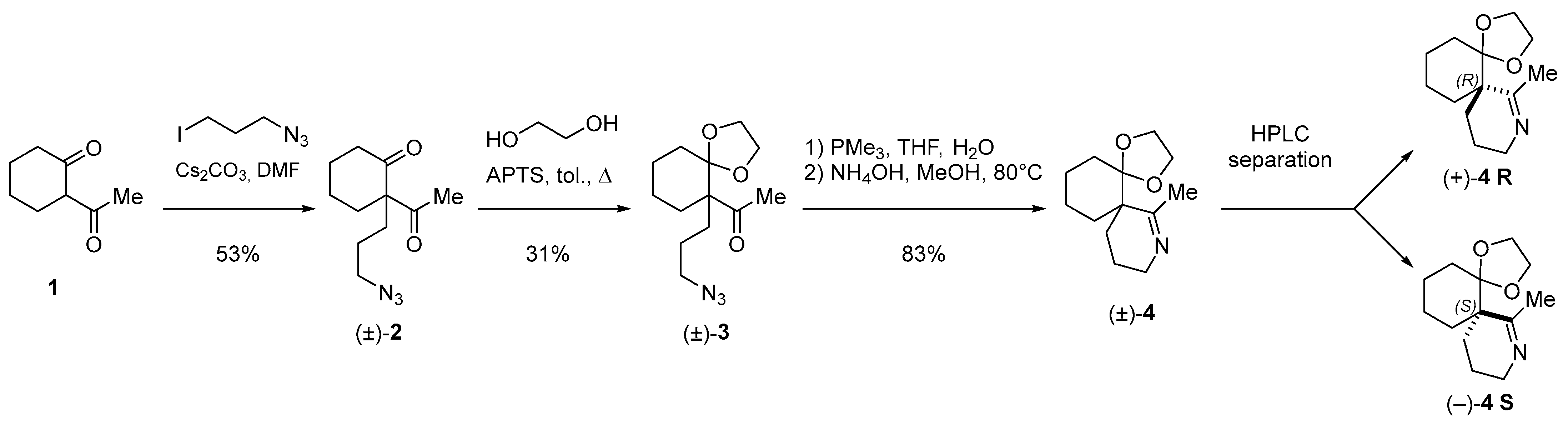
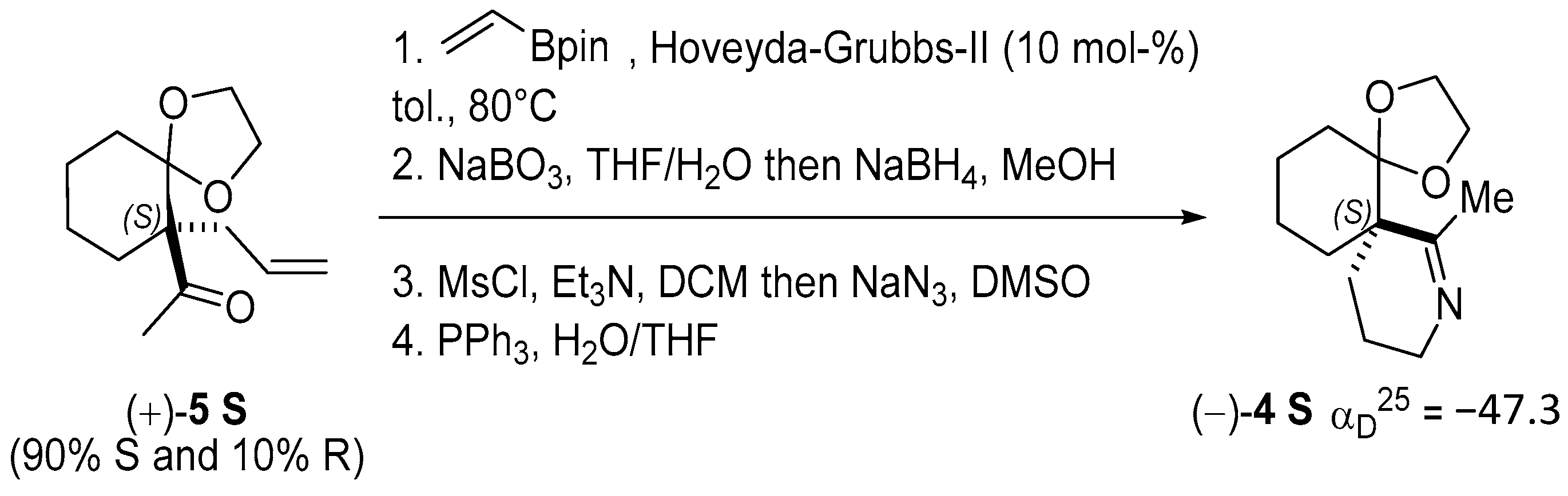
2.1. Chemical Synthesis and Characterization of the Spiroimine Enantiomers

The spiroimine (±)-4 racemate was synthesized in three steps from diketone 1 using standard procedures (Scheme 1; Appendix B). Separation by chiral HPLC of the two enantiomers present in (±)-4 yielded virtually pure spiroimines (+)-4 and (−)-4.
To assign the absolute configuration of the enantiopure spiroimines, (−)-4 was also synthesized from ketone (+)-5 S, prepared according to [76]. Spiroimine (−)-4 was found to have configuration S at the quaternary carbon (Scheme 2; Appendix B), and hence, spiroimine (+)-4 was considered to be of the opposite configuration R. The two enantiomers were therefore designated as (+)-4 R and (−)-4 S.
Initial electrophysiology experiments on nAChRs and binding and crystallography experiments on AChBPs were carried out using the racemic spiroimine (±)-4. These were then complemented by more comprehensive experiments performed with the enantiopure spiroimines (+)-4 R and (−)-4 S.

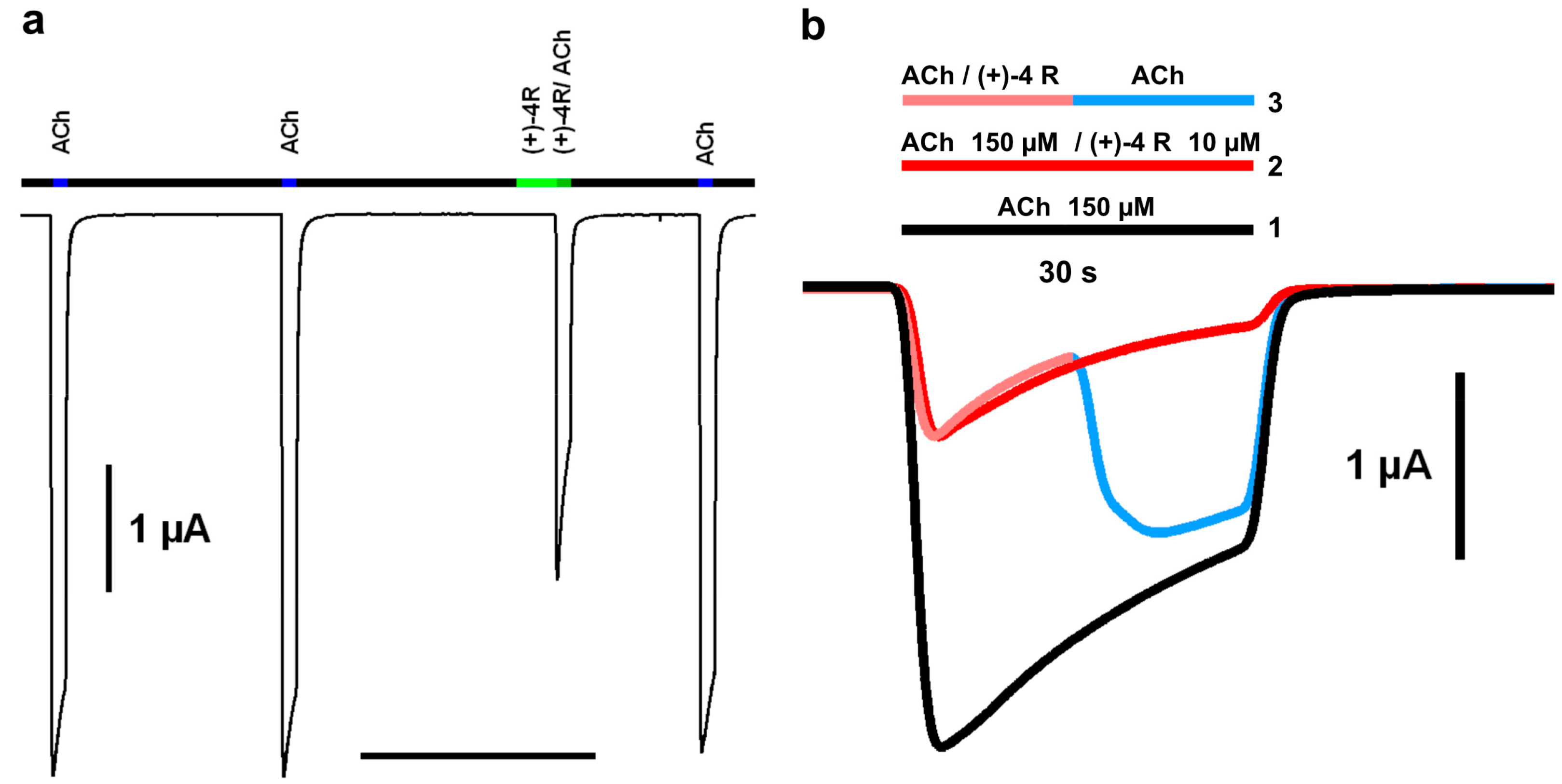
2.2. Functional Characteristics and nAChR Subtype Selectivity of the Spiroimine Racemate and Enantiomers

Functional analysis of the (±)-4 racemate and (+)-4 R and (−)-4 S enantiomers used either manual or automated two-electrode voltage-clamp (TEVC) recordings on Xenopus laevis oocytes at −60 mV holding membrane potential. The oocytes were either micro-transplanted with Torpedo α12βγδ-enriched electrocyte membranes or transfected with human α7 or α4β2-encoding cDNAs. When applied alone (see Experimental Procedures), none of the three spiroimines induced nAChR channel opening, thereby discarding any agonistic activity toward these nAChR subtypes. Instead, when applied together with ACh at its EC50 concentration for each nAChR subtype, all three spiroimines clearly behaved as nicotinic antagonists, whose potency depended on the receptor subtype examined (Figure 2; Table 1).
Indeed, the concentration–response curves recorded on the α12βγδ nAChR showed a full antagonistic effect at high spiroimine concentration, with IC50 values in the low µM range for the three spiroimines, with no stereospecificity and no apparent cooperativity or difference in the binding affinities to the αγ and αδ interfaces (Figure 2a left; Table 1). Perfusion of a mixed ACh/spiroimine solution markedly inhibited the ACh-evoked current (IACh), as here exemplified with the (±)-4 racemate (Figure 2a right, tracing 2). When the α12βγδ nAChR was in the desensitized state, the washout of the bound spiroimine with ACh triggered the immediate reopening of the receptor channel and full recovery of the nicotinic current up to the amplitude level of the control desensitized state, a feature denoting fast reversibility of channel blockade by the spiroimine (Figure 2a right, tracing 3).
Concentration–response curves recorded on the α7 nAChR also showed a full antagonistic effect at high spiroimine concentration, with IC50 values in the medium µM range, and slightly higher, albeit not statistically different, for the (+)-4 R enantiomer compared to its two relatives (Figure 2b left; Table 1). Unexpectedly, negative cooperativity was observed, higher for the (−)-4 S enantiomer than the (+)-4 R enantiomer, and averaged for the (±)-4 racemate, suggesting the contribution of an allosteric component to α7 antagonism by the spiroimines along with stereospecific modes of binding. Here again, recovery of the ACh-evoked current following the washout of bound spiroimine was fast and complete, as here exemplified with enantiomer (−)-4 S (Figure 2b right, tracing 3).
Similar antagonistic potency trends were again obtained for the α4β2 nAChR, with IC50 values in the medium-to-high µM range, but now significantly higher, by ca. one order of magnitude, for both the (±)-4 racemate and (+)-4 R enantiomer compared to their (−)-4 S relative (Figure 2c left; Table 1). Here again, negative cooperativity was observed, higher for the more potent (−)-4 S enantiomer than for the (+)-4 R enantiomer and (±)-4 racemate. Recovery of the ACh-evoked current was fast and complete, as here exemplified with enantiomer (+)-4 R (Figure 2c right, tracing 3; Figure 3a) and with the (±)-4 racemate using a slightly different protocol (Figure 3b).
Overall, these data point to similar antagonistic potencies of the three spiroimines for the muscle-type α12βγδ nAChR and a slightly higher potency of the (−)-4 S enantiomer for the neuronal α7 and α4β2 nAChR subtypes compared to its two relatives, with a one order of magnitude stereospecificity for α4β2. However, they also point to a higher selectivity, by 13- and 25-fold, of the (+)-4 R enantiomer for the α12βγδ nAChR compared with the α7 and α4β2 subtypes. The IC50 values recorded for the (±)-4 racemate were overall consistent with the presence of the two enantiomers in a 1:1 ratio (see Experimental Procedures).
These data also support the role of the spiroimine moiety in the cyclic imine toxins as a main pharmacophore, as previously approached using 6,6-spiroimine analogs of GYM [74], whereas a synthetic tetrahydrofuran fragment mimicking the C10–C20 skeleton of GYM was found not to bind the α12βγδ nAChR [73]. Hence, developing spiroimine-like bioactive molecules may be a promising strategy for designing new effectors targeting disease-associated nAChR subtypes. In turn, the weak antagonistic potency of the spiroimines, lower than that of GYM by two (α12βγδ) to three (α7) to four (α4β2) orders of magnitude [71,77], likely proceeds from a much shorter residence time (i.e., greater koff value) within the orthosteric binding site of the nAChRs due to the limited number of possible interactions with the subunit interface enabled by their small molecular size. Competition binding studies on nAChRs expressed at the surface of mammalian cells were not performed; however, these parameters were explored through binding studies on A- and L-AChBP and the structural analysis of spiroimine-A-AChBP complexes (see below).

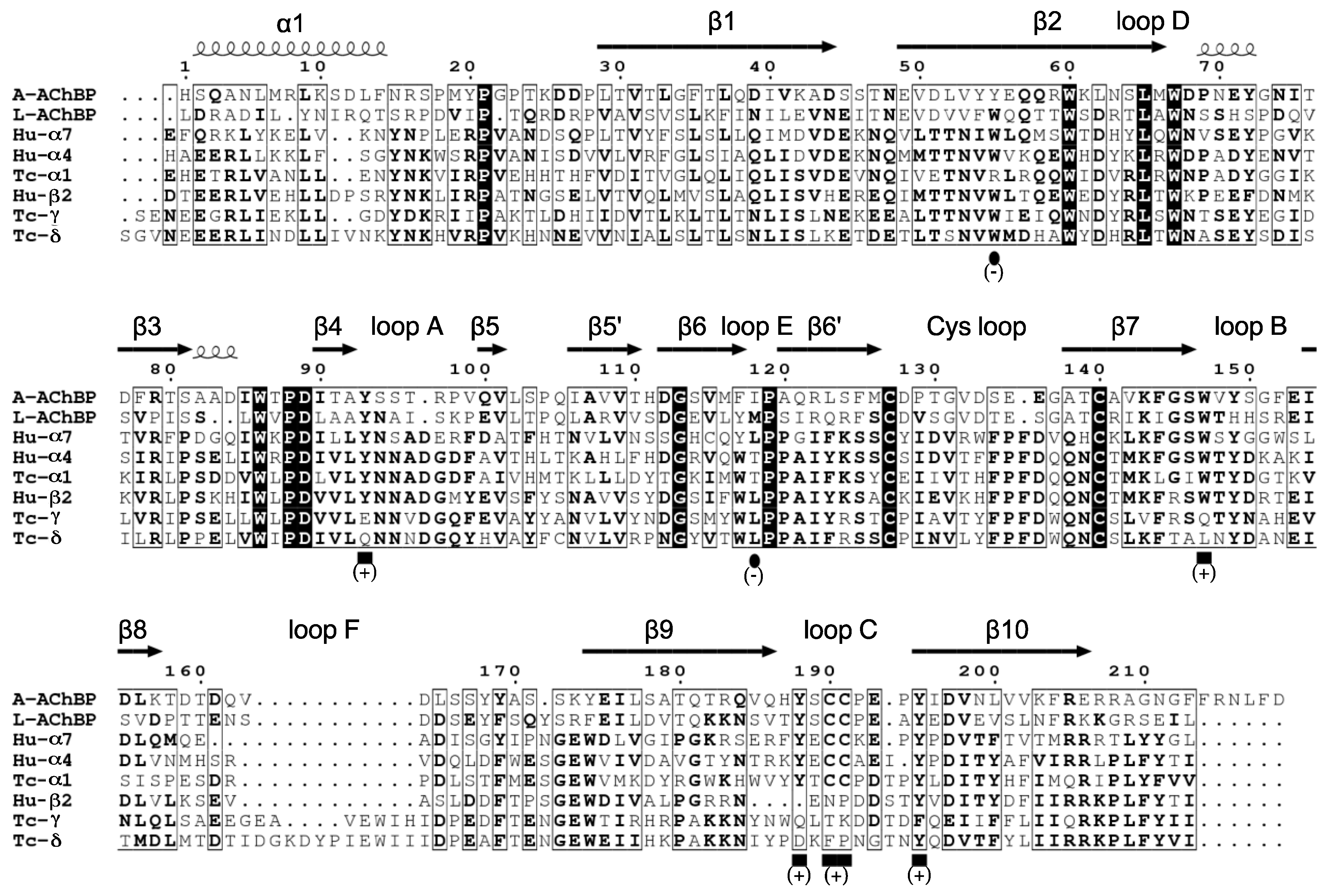
2.3. Binding Characteristics of the Spiroimine Enantiomers toward A- and L-AChBPs

The sequence differences and distinctive binding affinities of A- and L-AChBP for nicotinic ligands render them useful templates for approaching ligand binding to various nAChR subtypes. These differences are exemplified by the A-AChBP lower affinity for ACh, but higher affinity for some α7-specific peptidic antagonists, a feature that renders it more “α7-like” than L-AChBP [63,64,69,70].
Equilibrium dissociation constants were determined either from ratios (Kd/(koff/kon)) of rate constants determined using multiple kinetic means (kon, koff, koff/GAL) or directly from stopped-flow measurements (Kd/SFeq, Kd/SPAeq) (see Experimental Procedures). Overall, interaction of the spiroimines with each AChBP was found to be clearly stereospecific (Table 2). Specificity was even more pronounced with L-AChBP with a one-to-two orders of magnitude difference between the equilibrium constants (Kd values) of the (+)-4 R and (−)-4 S enantiomers. This difference most likely arises from the slower association (lower kon values) and faster dissociation (higher koff value) of enantiomer (+)-4 R, as also suggested by the kinetic values obtained for racemic (±)-4 and reflected in the fraction of the complex too low for reliable detection under the experimental conditions of the stopped-flow measurement. Enantiomer (+)-4 R was a tighter binder to A-AChBP, albeit with a modest stereospecificity of up to two-fold arising largely from slower complex dissociation (lower koff values). In turn, enantiomer (−)-4 S bound tighter to L-AChBP with Kd values in the 10−7 M range, resulting in the tightest complex of all combinations, while enantiomer (+)-4 R bound with Kd values in the 10−5 M range, not permitting the separation of rate constants for complex formation and dissociation under pre-equilibrium conditions.
The interaction constant values determined by the different experimental approaches generally agreed well (Table 2). Both the koff and koff/GAL values and the Kd/(koff/kon), Kd/SFeq and Kd/SPAeq values determined in differently designed assays were comparable. Only the Kd/SPAeq value determined for enantiomer (−)-4 S binding to L-AChBP was ca. one order of magnitude higher than the Kd/(koff/kon) and Kd/SFeq values determined in two different stopped-flow based assays (performed under pre-equilibrium and equilibrium conditions of spiroimine interaction to AChBP). Hence, this divergent value may have to be considered with caution, although in terms of the associated ΔΔG value, the difference is not significant. Quite surprisingly however, the higher enantioselectivity for L-AChBP was also reflected in the biphasic curve obtained upon stop-flow recording of the binding rates (Kd/SFeq values). The lower enantioselectivity (i.e., comparable affinities) of enantiomers (+)-4 R and (−)-4 S for A-AChBP (Kd values in the 2.2–5.4 µM range) compared to L-AChBP (12–0.32 µM range) justified the use of A-AChBP for the structural analyses.

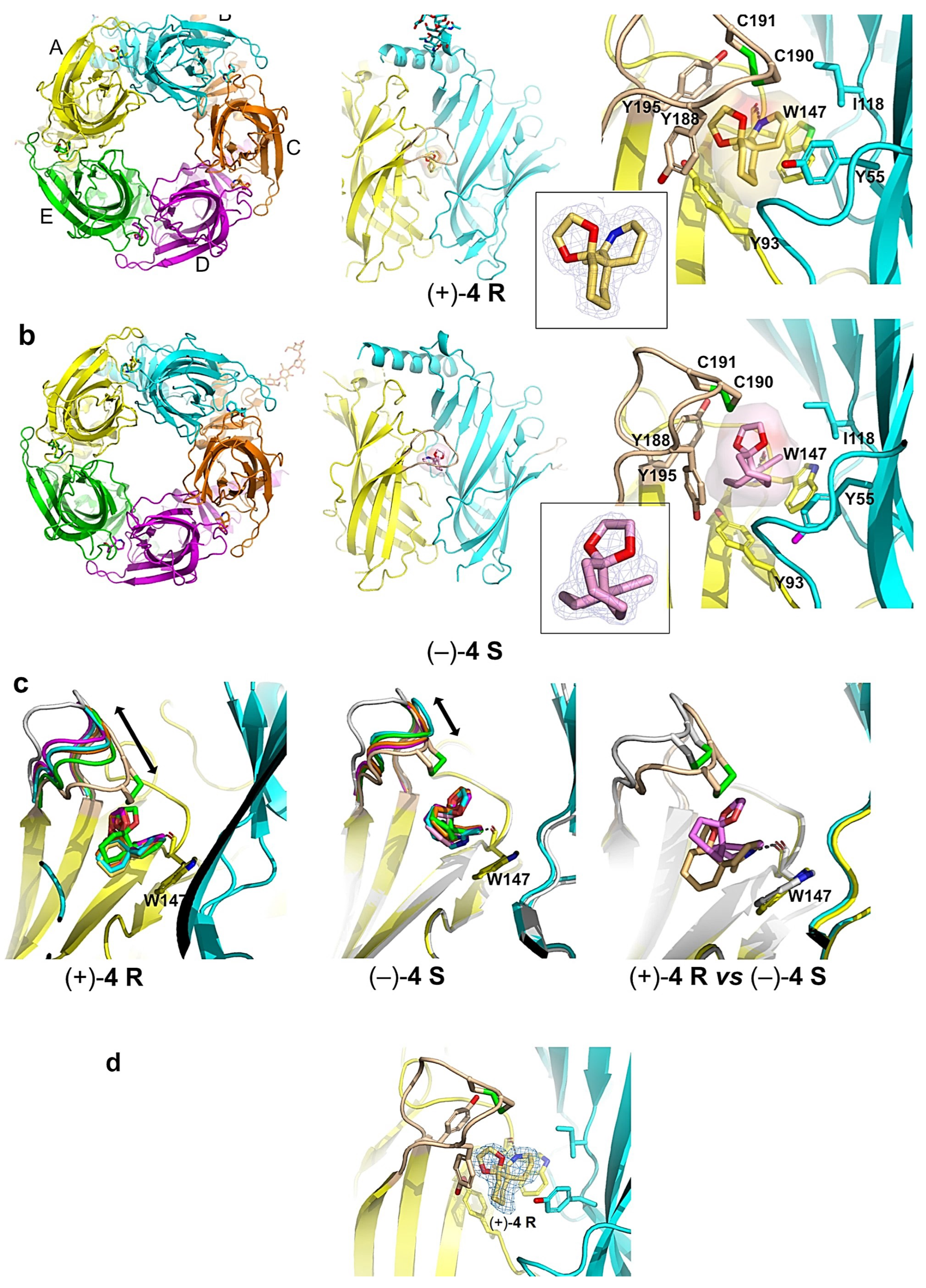
2.4. Overall View of the Crystalline Spiroimine-AChBP Complexes

The structures of A-AChBP bound with spiroimines (+)-4 R and (−)-4 S were solved in the 1.85–2.00 Å resolution range from preformed stoichiometric complexes (Table S1; Figure 4). Both showed the same tight homopentameric ring assembly of subunits as found in all previous AChBP structures (for reviews, see [69,70]). The ligand binding pocket encompasses a nest of five electron-rich aromatic side chains provided by residues Tyr93, Trp147, Tyr188 and Tyr195 on the principal (+) face of the subunit interface and residue Tyr55 on the complementary (−) face. This pocket is partially sheltered from the solvent by loop C, which is found at the outer perimeter of the pentamer and harbors at its tip a disulfide bridge linking the vicinal Cys190 and Cys191 residues, linked into a Cys-trans-Cys disulfide bridge (aka oxidized cysteinyl-cysteinyl ring), a signature determinant for nAChR α subunits.
Both structures showed very similar positions of the bound spiroimines at all five subunit interfaces within a pentamer (see. r.m.s.d. values in Experimental Procedures). Despite the moderate binding affinities (Table 2), the well-defined electron densities revealed full ligand occupancy at all five binding sites (Figure 4) due to the high protein concentration (ca. 100-fold the Kd values) and slight molar excess of spiroimine over A-AChBP used for complex formation and crystallization (see Experimental procedures).

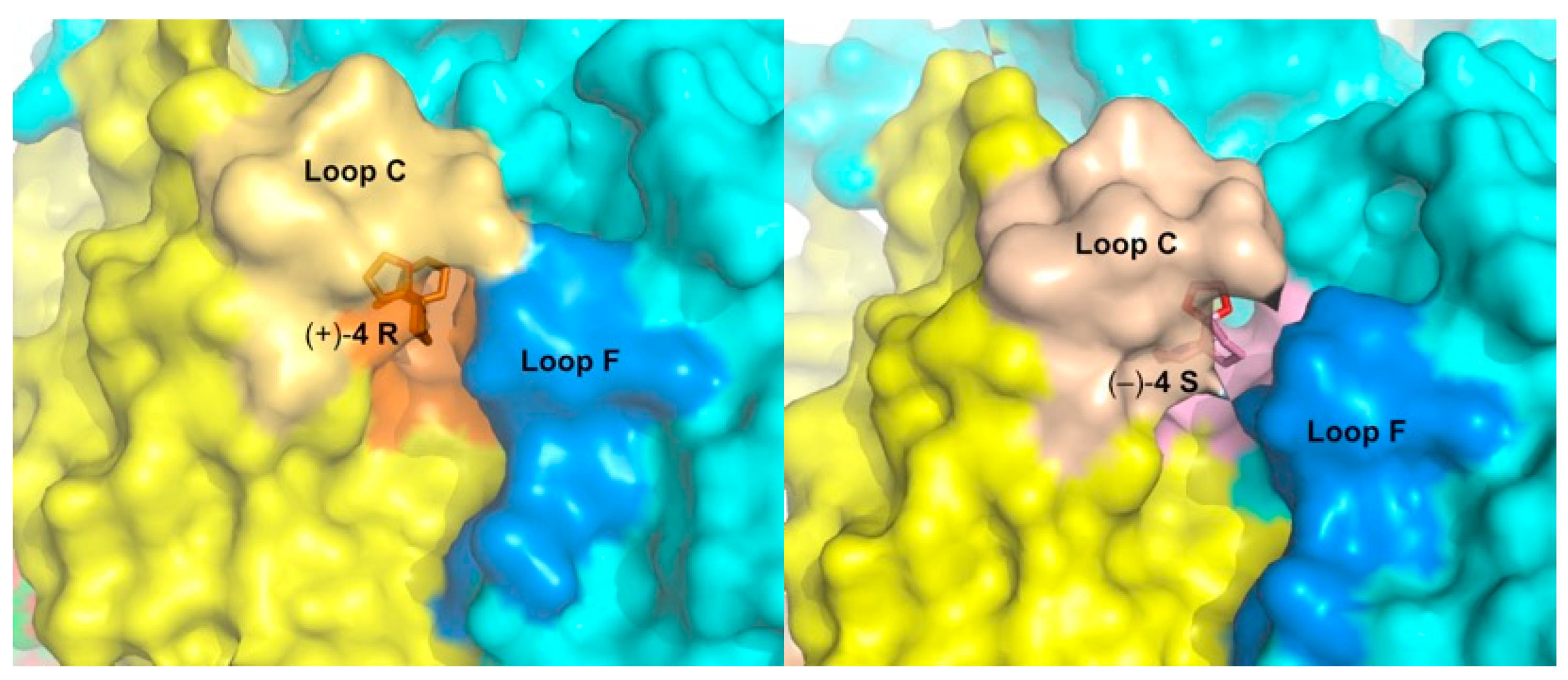
2.5. Detailed Description of the Crystalline Spiroimine-AChBP Complexes

In the A-AChBP complex with enantiomer (+)-4 R, the compact 3-ring skeleton of the spiroimine comfortably accommodates the core agonist signature motif central to the binding pocket (Figure 4 and Figure 5) and ideally positions the 6-membered cyclic imine ring nearly parallel to the indole ring of Trp147 in loop B (π-π stacking), and within H bond distances (2.6–2.7 Å) of the Trp147 carbonyl oxygen, with a typical Ĥ angle value of ~125° between the hydrogen bond donor, acceptor, and acceptor antecedent [78] (Table S2), as previously observed for the parental macrocyclic imine toxins (Figure 6). Such conservation of a protonated imine nitrogen (see discussion in [74] and reference 18 in it, along with the crystallization conditions in the Experimental procedures) tethers the spiroimine core centered within the binding pocket and greatly contributes to the binding affinity. The dioxolane ring makes nearly edge-to-face stacking interactions with Tyr188 and Tyr195 (loop C) from the (+) face, while the cyclohexane ring makes nearly face-to-face stacking interactions with Tyr93 from the (+) face and Tyr55 (loop D) from the (−) face (Figure 4 and Figure A1). However, spiroimine binding involves virtually no interaction with residues from loop F at the complementary (−) face of the subunit interface (Figure 4, Figure 5 and Figure A1). This observation is consistent with the limited contribution of loop F to the binding of SPX and GYM versus its significant contribution for laterally accommodating the bridged ketal group specific to the pinnatoxins [71,72] (Figure 6).
In the A-AChBP complex with enantiomer (−)-4 S, the cyclic imine ring, nearly perpendicular to the indole ring of Trp147 (T-shaped interaction), only partially overlaps that of the (+)-4 R enantiomer, with long-range H bond distances (2.8–3.5 Å) to the carbonyl oxygen of Trp147 and an Ĥ angle value of ~142° (Figure 4 and Figure 5; Table S2). As a result, the position of the cyclohexane ring is displaced toward the tip of loop C, and the dioxolane ring projects in the opposite direction at the entrance of the binding pocket, thus making limited interactions with the A-AChBP residues. Such a drastic shift in the position of the 3-ring system for the two enantiomers is associated with differences in the loop-C position (Figure 4, Figure 5 and Figure 6, and see below) and may account for the slightly (ca. 2-fold) higher affinity of enantiomer (+)-4 R for the two AChBPs compared to enantiomer (−)-4 S (Table 2). In turn, the one order of magnitude higher affinity of enantiomer (−)-4 S for L-AChBP compared to A-AChBP (Table 2; Figure 6b and Figure A1) may correlate with the presence of substitutions Met114 (for Ile118) and Trp53 (for Tyr55) in the vicinity of the cyclic imine and cyclohexane rings. This is consistent with the ca. 5-fold higher affinity of enantiomer (+)-4 R compared with the (−)-4 S enantiomer for the α7 and α4β2 nAChRs (Table 1; Figure A1). Here again, there is almost no interaction between the bound spiroimine and loop-F residues (Figure 4, Figure 5, and Figure A1).
Co-crystallization of A-AChBP with the (±)-4 racemate unambiguously led to a bound (+)-4 R enantiomer at all five subunit interfaces in a virtually identical position to that seen in the (+)-4 R complex, with only a minor trace (i.e., occupancy below ~10%) of a bound (−)-4 S (Figure 4d). Per se, this observation would suggest a much lower dissociation rate for enantiomer (+)-4 R relative to its (−)-4 S congener than the one recorded through the binding studies (Table 2). Whether this is due to the more acidic and hydrophobic composition of the crystallization liquor used for the (±)-4 complex compared with the other two complexes (see Appendix D) or to conformational remodeling of the complex during crystal nucleation and growth [79] is unknown.
In all three complexes, the loop-C position shows a high degree of variability with respect to the different orientation of the 3-ring system in each enantiomer associated with slightly different orientations of the bound enantiomers at each binding interface (Figure 4c). In the (+)-4 R complex, loop C adopts a closed, agonist-bound position in one subunit along with a range of positions intermediate between those found in apo (2BYN) and agonist-bound A-AChBP (2BYQ, epibatidine) in the other four subunits. In the (±)-4 and (−)-4 S complexes, loop C clusters around intermediate positions in all five subunits. Moreover, at one interface of the (−)-4 S complex, the tip of loop C (Tyr188-Glu193) adopts two alternate conformations with up to 3 Å distance at position Cys190, an observation emphasizing the high conformational dynamics of loop C, even in the crystalline state. The average loop-C conformation in the spiroimine-AChBP complexes contrasts with the wide-open, “antagonist” position observed in the complexes with the parental, bulky macrocyclic spirolides/phycotoxins [71,72] or other large antagonists (Figure 6). However, the loop-C “intermediate” positions associated with its loose interactions with the spiroimines resemble more the position observed in HEPES-bound or apo AChBPs [63,67,80] than that in complexes with small agonists [65,67,68] (Figure 6). A reverse situation was reported for 2-aminopyrimidine agonists inducing an unusually large opening of loop C [81]. This comparative analysis further reflects the antagonist properties of these small spiroimines and illustrates, here within a single AChBP pentamer, how loop C can behave as a highly flexible sensor to adapt its configuration to the chemical features and position of the ligand within the binding pocket, as previously documented for many structurally-unrelated nicotinic ligands.
To explore a possible correlation between the spiroimine binding poses and interactions at the A-AChBP subunit interfaces and their binding affinities in each complex, from the structure coordinates, we calculated the DrugScore eXtended (DSX) scores, which combine distance-dependent atom–atom potentials, torsion angle potentials, and solvent-accessible surface-dependent potentials [82] (Table 3).
The smaller mean score for the interaction of enantiomer (+)-4 R at all five A-AChBP subunit interfaces compared to that for enantiomer (−)-4 S suggests tighter binding to A-AChBP, independently of the loop-C conformation. The ratio of the (+)-4 R/(−)-4 S mean scores, equal to 1.22, also indicates computational prediction from the structures for a tighter binding of enantiomer (+)-4 R, which should be reflected by a ca. one order of magnitude difference in the Kd values for the two complexes [82]. The fact that our experimentally determined values do not differ that much from each other (Table 2) may arise either from differences in the “experimental” conditions or from five interfaces per complex making too small a statistical sample, or from the limited significance of comparing differences in affinities in the micromolar range, or, here again, from time-dependent conformational remodeling of the complexes in the crystal state but not during the binding experiments in solution [79]. Overall, the DX scoring approach appears consistent with the prevailing presence of enantiomer (+)-4 R in the structure of the complex prepared from the (±)-4 racemate (Figure 4).

2.6. Structural Comparisons

Certain features of the A-AChBP complexes with the (+)-4 R and (−)-4 S enantiomers closely resemble those found in the complexes with the bulkier macrocyclic imine compounds GYM and SPX (see r.m.s.d. values in Experimental Procedures), with in particular, the positions of the cyclic imine and cyclohexene rings (Figure 6). In fact, the orientation of the cyclic imine ring in the (−)-4 S complex virtually overlaps that of GYM. Structural overlay of these two complexes shows that the lack of the bis-spiroacetal/tetrahydrofuran and butyrolactone ring systems of GYM in the apical and membrane directions of the subunit interface, respectively, is partially compensated by the new dioxolane ring in the spiroimines that abuts against the Tyr188 and Tyr195 phenol rings in loop C. The loop-C conformational, large-amplitude motions observed across the five binding interfaces in the (+)-4 R and (−)-4 S complexes, coupled with alternate conformations of its tip (a behavior neither observed in complexes with macrocyclic phycotoxins nor in those with small agonists), appear to be a signature of the minimal spiroimine core motif. This comparison also suggests substituent modification at key positions around the 3-ring framework of spiroimines, notably in the cyclohexane ring, to confer greater binding selectivity and specificity on them. For example, a polar group attached to the cyclohexene ring would be expected to target residues in or near loop F at the complementary face of the subunit interface.
Comparisons with humanized A-AChBP-α7-chimeras in the apo conformation [80] and the extracellular LBDs of the α4β2, α7, and α12βγδ nAChRs [59,60,83] showed a conserved aromatic nest with the exception of a Trp (in α4 and α7) or Arg (α1) residue in place of A-AChBP Tyr55 at the (−) face of the interface, and the positions of Tyr188 and Tyr195 within loop C at the (+) face (Figure 6 and Figure A1; Table S2). Moreover, conservation of the “interacting” residues at the αγ and αδ interfaces of the α12βγδ nAChR is consistent with the Hill slope of ~1 observed in the functional assays (Figure 2, Figure 6 and Figure A1; Table S2). Concerning the α3β2 nAChR, which was not included in the present study, a similar binding mode and antagonistic potency as for the α4β2 subtype can be predicted due to their high sequence homology in the spiroimine binding site (see Figure S1 in [70]).

3. Experimental Procedures

3.1. Chemical Synthesis, Separation, and Enantiomeric Characterization of the Spiroimines

Racemic spiroimine was synthesized from diketone 1 by alkylation with 1-azido-3-iodopropane in the presence of anhydrous cesium carbonate to afford the azido-ß-keto ketone (±)-2 in 53% yield (Scheme 1; Appendix B). The cyclohexanone function of (±)-2 was then protected with ethylene glycol to generate the corresponding dioxolane (±)-3 in 31% yield. Then, the azide function of (±)-3 was reduced with triphenylphosphine, followed by cyclization under basic conditions to generate spiroimine (±)-4 in 83% yield. Each compound was characterized by 1H and 13C NMR spectroscopy in CDCl3 (Figures S1–S6). The two enantiomers present in a 1:1 ratio in (±)-4 were then separated by semi-preparative chiral HPLC in Hept/EtOH 80:20 (v/v) to afford (+)-4 (t = 4.54 min, α D 24 = +144.5) (c = 0.25, CHCl3) and (−)-4 (t = 6.21 min, α D 24 = −163.5) (c = 0.25, CHCl3) with a purity greater than 99% (Figure S7). The racemate and pure enantiomers were lyophilized and stored at −20 °C. Stock solutions at 10 mM (racemate) and 20 mM (enantiomers) in MeOH prepared from the lyophilized compounds were stored as aliquots at −80 °C.
The synthesis of enantiomer (−)-4 from ketone 5 (a mixture of (+)-5 S and (−)-5 R in a 90:10 molar ratio, prepared according to [76]), was adapted from [74]. In brief, Ru-catalyzed cross-metathesis with vinyl pinacol boronate (step 1), followed by oxidation, then reduction (step 2), afforded the primary alcohol (not shown). The latter was converted into the azide (step 3), which was reduced with triphenylphosphine (step 4) to give spiroimine (−)-4 ( α D 24 = −47.3 (c = 0.25, CHCl3) (Scheme 2), of absolute configuration (S) at the quaternary carbon, as indicated by the negative sign of the optical rotation.

3.2. Analysis of ACh-Evoked Currents

The sources for live animals and biological materials, the procedures for nAChR microtransplantation and expression in oocytes, and the procedures for manual (α12βγδ nAChR) and automated (α7 and α4β2 nAChRs) TEVC recording are detailed in Appendix C. In brief, our TEVC protocol comprised 2 to 3 pulses of ACh at a concentration equivalent to its EC50 value, such as: 15 s at 25 µM for α12βγδ nAChR, 5 s at 100 µM for α7 nAChR, 15 or 30 s at 150 µM for α4β2 nAChR (perfusion flow, 8 mL/min). Then, the oocyte was perfused for 45 s with the tested spiroimine at a given concentration, immediately followed by the co-application of ACh and the spiroimine at the same concentrations and times as indicated above. The oocyte was washed with Ringer’s solution, and then recovery of the ACh-evoked current was recorded.
The concentration–inhibition curves were analyzed as previously detailed [71,72] using the equation I = Imax [L]nH/(EC50nH + [L]nH) (Equation (1)), where I is the measured agonist-evoked current, [L] is the agonist concentration, EC50 is the agonist concentration that evoked half the maximal current (Imax), and nH is the Hill coefficient. For antagonist inhibition, current (I) values were normalized to the Imax value recorded from the same oocyte to yield the fractional (%) response data. IC50 values were established from the concentration–response curves by fitting to the equation F = 1/[1 + ([X]/IC50)nH] (Equation (2)), where F is the fractional response obtained in the presence of the inhibitor at concentration [X], and IC50 is the inhibitor concentration reducing the ACh-evoked amplitude by half. Data analysis was performed without constraints as Log[spiroimine] versus response (three parameters), with a Hill slope ~1. Statistical significance of differences between the control and test values was assessed using either the two-tailed Student’s “t” test or the Kolmogorov–Smirnov two-sample test and p < 0.05.

3.3. Ligand Binding to the AChBPs

The procedures for stable expression and purification of A- and L-AChBPs are described in Appendix D. Rate constants for association (kon) and dissociation (koff) were determined by multiple kinetic means, largely as previously described [64,67,71,72]. Measurement of kon and koff values entailed the direct admixture of reactants and monitoring the quenching of AChBP native Trp fluorescence. Individual rate constants were determined from linear regression of the experimental mono-exponential decay of Trp fluorescence intensity (kobs) using the linear “approach-to-equilibrium” relationship kobs = kon[spiroimine] + koff (Equation (3)). Additional measurements of koff (koff/GAL) employed stopped-flow measurement of the rate of occupation of free AChBP binding sites by a competing ligand (gallamine, used in large excess relative to its Kd) to form a non-quenching gallamine-AChBP complex. Equilibrium dissociation constants were determined either from the ratios of rate constants (Kd/(koff/kon)), or from stopped-flow measurements of the rates of gallamine or epibatidine binding to pre-equilibrated spiroimine-AChBP complexes formed using increasing spiroimine concentrations (Kd/SFeq), or by the scintillation proximity assay (SPA) where spiroimine competition against the binding of [3H]epibatidine (to L-AChBP) or [3H]methyllycaconitine (to A-AChBP) was monitored at equilibrium (Kd/SPAeq). In brief, in the SPA assay, to titrate those binding sites made available upon toxin dissociation, each equilibrated spiroimine-AChBP complex, at 250 pM in binding sites and a slight molar excess of toxin, was mixed with the [3H]ligand at a concentration well above its Kd value. The time course of [3H]ligand binding was monitored over several hours. All experiments were performed in triplicate, in which individual data differed by less than 20%.

3.4. Structure Determination and Refinement

The procedures for the formation and crystallization of the spiroimine-A-AChBP complexes and for data collection are described in Appendix D. The structure of the A-AChBP complex with spiroimine (+)-4 R was solved by molecular replacement with PHASER [84] using the apo A-AChBP structure (PDB: 2BYN [67]) as a search model, and that of the complex with spiroimine (−)-4 S by difference Fourier synthesis with REFMAC [85]. The models of the spiroimine-A-AChBP complexes were improved by manual adjustment with COOT [86] and refined with REFMAC including TLS refinement with each subunit defining a TLS group. In each case, a random set of reflections was set aside for cross-validation purposes. The molecular structures of spiroimines (+)-4 R and (−)-4 S and the associated library files containing the stereochemical and parametric data were generated with SKETCHER [87]. Ligands were fitted into unbiased Fo–Fc difference electron density maps calculated after 10 cycles of rigid-body refinement. Automated solvent building used COOT. The A-AChBP-spiroimine interfaces were analyzed with the PISA server [88]. Data collection and refinement statistics are reported in Table S1.
The final structures comprise A-AChBP residues His1-Arg207/Arg208 for each subunit in the single homopentamer present in the asymmetric unit, and a fully resolved spiroimine in each of the five binding sites. For each complex, the N-terminal FLAG sequence could be fully resolved in one of the five subunits, and a tetrasaccharide moiety linked to Asn74 was visible in another subunit. High temperature factors and weak electron densities were associated with the other four FLAG sequences and with the five surface loops Asn15–Met19. Five chlorine ions in the (+)-4 R complex and one isopropanol molecule and six chlorine ions in the (−)-4 S complex (all arising from the crystallization liquor or the purification buffer) could also be modeled. Quality of the models was validated using COOT and MOLPROBITY [89], with ~97% of residues in favored regions of the Ramachandran plot and no outliers. Data collection and refinement statistics are reported in Table S1.

3.5. Structural Analyses and Comparisons

Comparison of the spiroimine-A-AChBP complexes with other AChBP structures included those of A-AChBP in the apo form (PDB: 2BYN [67]) and bound with phycotoxins SPX and GYM (2WZY and 2X00 [71]), pinnatoxins PnTxA and PnTxG (4XHE and 4XK9 [72]), and the small agonist representative, EPI (2BYQ [67]); of L-AChBP in the “apo” (HEPES-bound) form (1I9B [63]); and of humanized A-AChBP-α7-chimeras (mutants I and II; 3T4M and 3SIO [80]. The average root-mean-square deviation (r.m.s.d) between A-AChBP subunits bound with spiroimine (+)-4 R with closed and intermediate states of loop C is 0.6 Å; between the five A-AChBP subunits bound with spiroimine (−)-4 S with intermediate states of loop C, it is 0.27 Å; between the A-AChBP subunits bound to spiroimine (+)-4 R with a closed loop C versus bound to spiroimine (−)-4 S with an intermediate state of loop C, it is 0.43 Å; and between the nine A-AChBP subunits with intermediate states of loop C, it is in the 0.27–0.43 Å range (all for 204–210 Cα atoms).
Comparisons with structures of full-length nAChRs included the human α7 bound to EPI (cryo-EM structure 7KOQ [59]), the human α4β2 in the apo form (X-ray structure 5KXI [60]), and the Torpedo α12βγδ receptor in the apo form (cryo-EM structure 7SMM [83]). The average r.m.s.d. between A-AChBP subunits bound with spiroimine (+)-4 R with a closed loop C or with spiroimine (−)-4 S with an intermediate loop C and the extracellular ligand-binding domains of the above-mentioned nAChRs is in the 1.3–1.7 Å range for 181–194 Cα atoms.

3.6. Figures

Figure 1 was generated with Marvin (ChemAxon); Scheme 1 and Scheme 2 and those in Appendix B with ChemDraw; Figure 2 and Figure 3 with GraphPad Prism 9.0 (GraphPad Software, San Diego, CA, USA) and pCLAMP 9.0 (Molecular Devices, LLC., San Jose, CA, USA); Figure 4, Figure 5 and Figure 6 with PyMOL [90]; Figure A1 with ESPript [91]; Figures S1–S6 with TopSpin; and Figure S7 with MassLynx.

4. Concluding Remarks

These data emphasize the intrinsic capacity of the spiroimine enantiomers synthesized in this study to block selected nAChR subtypes in a competitive manner, highlight the major contribution of the spiroimine core of macrocyclic imine toxins to binding within the nAChR aromatic nest (aka “agonist-competitive antagonist site” [92]), and confirm the need for extended interaction networks as established by the macrocyclic toxins to define high affinities and the variable levels of subtype specificity dictated by their capacity to extend in all directions of the subunit interface. Hence, this study identifies a minimal set of functional pharmacophores and binding determinants (Table S2) as templates for designing second-generation spiroimine-like bioactive molecules with full antagonistic properties targeting disease-associated nAChR subtypes. From that perspective, enantiomer (+)-4 R, with its higher specificity for the α12βγδ nAChR compared with the α7 and α4β2 subtypes, and its tighter binding to key residue Trp147 in the A-AChBP binding pocket compared with enantiomer (−)-4 S, appears as a suitable starting point in a context of muscle-linked diseases.
These data also contribute to challenge a conclusion built over the years that small molecules containing a quaternary nitrogen and strictly occupying the agonist binding pocket at the subunit interface, without extending substituents outside of this core, cannot act as nicotinic antagonists. Initial insights against this “dogma” include the small cyclic, disulfide-containing marine molecule, nereistoxin (aka N,N-dimethyl-1,2-dithiolan-4-amine), shown to act as a nicotinic antagonist with sub-millimolar IC50 values [93]. Availability of a crystal structure of a nereistoxin-AChBP complex would be of interest to complement the current study.

Supplementary Materials

The following supporting information can be downloaded from https://www.mdpi.com/article/10.3390/md22040149/s1. Figure S1: 1H NMR spectrum (CDCl3) of 2-acetyl-2-(3-azidopropyl)cyclohexanone (±)-2. Figure S2: 13C NMR spectrum (CDCl3) of 2-acetyl-2-(3-azidopropyl)cyclohexanone (±)-2. Figure S3: 1H NMR spectrum (CDCl3) of 1-[6-(3-azidopropyl)-1,4-dioxaspiro[4.5]dec-6-yl]ethanone (±)-3. Figure S4: 13C NMR spectrum (CDCl3) of 1-[6-(3-azidopropyl)-1,4-dioxaspiro[4.5]dec-6-yl]ethanone (±)-3. Figure S5: 1H NMR spectrum (CDCl3) of 7-methyl-1,4-dioxa-8-azadispiro[4.0.5.4]pentadec-7-ene (±)-4. Figure S6: 13C NMR spectrum (CDCl3) of 7-methyl-1,4-dioxa-8-azadispiro[4.0.5.4]pentadec-7-ene (±)-4. Figure S7: Separation of enantiomers (+)-4 and (−)-4 from the (±)-4 racemate by chiral HPLC. Table S1: X-ray crystallography—Data collection and refinement statistics. Table S2: Geometric parameters of key interactions between the (+)-4 R and (−)-4 S enantiomers and side chains in the A-AChBP binding pocket.

Author Contributions

Conceptualization, R.A., Z.R., C.G., J.M. and P.M.; Methodology, Y.B., G.S., L.C., R.A., Z.R., C.G., J.M. and P.M.; Validation, Y.B., G.S., L.C., R.A., Z.R., P.T., C.G., J.M. and P.M.; Formal analysis, Y.B., G.S., L.C., R.A., Z.R., S.C., C.G., J.M. and P.M.; Investigation, Y.B., G.S., L.C., R.A., Z.R. and S.C.; Resources, Y.B., G.S., R.A., Z.R., P.T., C.G., J.M. and P.M.; Data curation, Y.B., G.S., R.A., Z.R. and P.M.; writing—original draft preparation, Y.B., G.S., L.C., R.A., Z.R., C.G., J.M. and P.M.; Writing—review and editing, Y.B., G.S., L.C., R.A., Z.R., C.G., J.M. and P.M.; Visualization, Y.B., G.S., L.C., R.A., Z.R., J.M. and P.M.; Supervision, P.T., C.G., J.M. and P.M.; Project administration, C.G., J.M. and P.M.; Funding acquisition, Y.B., L.C., R.A., P.T., C.G., J.M. and P.M. All authors have read and agreed to the published version of the manuscript.

Funding

This work was supported by an ICSN-CNRS grant (to C.G. and L.C.); grants NEUROSPIROIMINE ANR-PCV-07-194417 (to J.M.), ICH-NEURO-MET ANR-21-CE34-0024 (to R.A.), and FRISBI ANR-10-INSB-0005 (to the AFMB laboratory); USPHS grants R37-GM18360 and P42-ES-010337 (to P.T.); and the CNRS/AMU/UCSD Associated International Laboratory “Structure-Guided Investigations into Disease States and Therapeutic Strategies” (LIA SGIDSTS) (to P.M. and P.T.).

Institutional Review Board Statement

The Torpedo marmorata fish and Xenopus laevis frogs used for this study were maintained and treated in strict adherence with the European Community guidelines for laboratory animal handling, and according to a protocol approved by the Ethical Committee for Animal Experimentation (C2EA-59 of Paris Center-South, to R.A.). All efforts were made to minimize animal suffering and to reduce the number of animals used. All animal experimental procedures were approved by the Animal Ethics Committee of the CEA, by the French General Directorate for Research and Innovation (project APAFIS#5310-2016042016067330 v3 protocol authorized to R.A.).

Data Availability Statement

The data presented in this study are included in the main text, Appendix A, Appendix B, Appendix C and Appendix D, and the Supplementary Materials. The atomic coordinates and structure factors of the A-AChBP complexes with enantiopure spiroimines (+)-4 R and (−)-4 S and the (±)-4 racemate have been deposited at the RCSB-PDB (www.rcsb.org) with accession codes 8Q1M, 8QTL, and 8QX2, respectively.

Acknowledgments

We gratefully acknowledge the expert assistance of Kwok-Yiu Ho (UCSD, La Jolla, CA) for the AChBP binding assays and Karine Barral, Philippe Leone, Julien Lescar, Miguel Ortiz-Lombardia, Florence Vincent (AFMB lab), Sébastien Santini (IGS, Marseille), the ID14-EH1 and ID23-1 staff of the European Synchrotron Radiation Facility (ESRF, Grenoble), and the Proxima-1 staff of the SOLEIL synchrotron (Gif-sur-Yvette) during data collection. We thank Isabel Bermudez (Oxford Brookes University, Oxford, UK) and Pierre-Jean Corringer (Institut Pasteur, Paris, France) for the kind gift of the nAChR subunit cDNAs.

Conflicts of Interest

The authors declare no conflicts of interest. The funders had no role in the design of the study; in the collection, analysis, or interpretation of data; in the writing of the manuscript; nor in the decision to publish the results.

Appendix A

Receptors Used or Mentioned in this Study

The prototype of vertebrate skeletal muscle nAChR, found in large amounts in the Torpedo electric organ, is a transmembrane heteropentameric assembly of four homologous subunits with a α12βγδ stoichiometry (Figure A1). Nicotinic agonists and competitive antagonists bind with distinctive affinities at the two, α/γ, and α/δ subunit interfaces. In the peripheral and central nervous systems, the human α4β2 subtype, which plays predominant roles in both pre- and postsynaptic functions, has variable stoichiometries involving two or three α/β subunit interfaces and ligand binding sites. Typically, agonist binding to the two or three sites of the α12βγδ or α4β2 nAChR in the closed channel state selects conformational transitions leading to channel opening, and then to nAChR desensitization. Competitive antagonist binding to at least one site blocks channel function. When the affinities of the two sites differ, the antagonism of function correlates with the occupation of the higher affinity site [94]. In turn, the homopentameric α7 subtype, also found at pre- and postsynaptic neuronal locations, encompasses five interfacial α/α binding sites with identical affinity [95].
Figure A1. Sequence alignment for the extracellular ligand-binding domains of the AChBP and nAChR subunits related to this study. The A-AChBP secondary structure elements, along with loops A (or β4-β5), B (β7-β8), C (β9-β10), D (β2), E (β6-β6′), F (β8-β9), and the Cys-loop (β6′-β7), are indicated and labeled above the alignment. Below the alignment, A-AChBP residues, whose side chains interact with the bound spiroimines and are conserved in L-AChBP and nAChR subunits α1, α4, α7, are indicated by squares, while interacting non-conserved residues are indicated by ovals. Assignment of these residues to the principal (+) and complementary (−) faces of the subunit interface is indicated by the corresponding signs.
Marinedrugs 22 00149 g0a1

Appendix B

Appendix B.1. Chemical Synthesis of 2-acetyl-2-(3-azidopropyl)cyclohexanone (±)-2 (C11H17N3O2, 223.2760) (Figure A2)

To a solution of diketone 1 (0.49 mL, 3.79 mmol) in dry DMF (6 mL) was added anhydrous cesium carbonate (102.3 mg, 4.17 mmol) at RT. The mixture was stirred for 10 min, then 1-azido-3-iodopropane (1.6 g, 7.58 mmol) was added and the mixture stirred for 20 h at RT. The reaction was quenched with a saturated NaCl solution and the aqueous layer extracted with ethyl acetate. The combined organic layers were washed with a saturated NaCl solution and dried over MgSO4. The solvent was evaporated under vacuum and the residue purified over silica gel (Hept to Hept/EtOAc: 8/2) to afford a colorless oil (446 mg, 53%).
Figure A2. Chemical structure of the 2-acetyl-2-(3-azidopropyl)cyclohexanone racemate.
Marinedrugs 22 00149 g0a2
IR: υ (film) 2941–2867, 2090, 1713, 1693 cm−1. MS: (ESI) m/z: 246 (100) [M + Na]. HRMS: calculated for C11H17O2Na: 246.1218; found: 246.1211. 1H: (300 MHz; CDCl3) δ 3.28 (t, J = 6.5 Hz, 2H), 2.46 (m, 2H), 2.29 (m, 1H), 2.10 (s, 3H), 2.04–1.84 (m, 2H), 1.80–1.59 (m, 4H), 1.52–1.30 (m, 3H). 13C: (75 MHz; CDCl3) δ 209.8 (Cq), 206.6 (Cq), 67.2 (Cq), 51.5 (CH2), 41.6 (CH2), 34.3 (CH2), 31.1 (CH2), 27.1 (CH2), 26.1 (CH3), 23.6 (CH2), 22.1 (CH2).

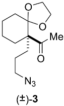
Appendix B.2. Chemical Synthesis of 1-[6-(3-azidopropyl)-1,4-dioxaspiro[4.5]dec-6-yl]ethanone (±)-3 (C13H21N3O3, 267.3290) (Figure A3)

To a solution of diketone (±)-2 (180 mg, 0.807 mmol) in toluene (2 mL) was added ethylene glycol (0.09 mL, 1.61 mmol). A catalytic amount of para-toluene sulfonic acid was then introduced. The flask was fitted with a Dean–Stark and the solvent heated under reflux overnight. The reaction was quenched with a saturated Na2CO3 solution and the aqueous phase extracted twice with ethyl acetate. The combined organic layers were washed with brine dried over Na2SO4. The solvent was evaporated under vacuum and the crude was purified over silica gel (Hept to Hept/EtOAc: 8/2) to afford a colorless oil (m = 66.1 mg, 31%).
Figure A3. Chemical structure of the 1-[6-(3-azidopropyl)-1,4-dioxaspiro[4.5]dec-6-yl]ethanone racemate.
Marinedrugs 22 00149 g0a3
IR: υ (film) 2936–2866, 2091, 1697, 1177 cm−1. MS: (ESI) m/z: 290 (100) [M + Na]. HRMS: calculated for C13H21N3O3Na: 290.1481; found: 290.1483. 1H: (500 MHz; CDCl3) δ 3.98–3.85 (m, 4H), 3.26 (dd, J = 6.7, 6.7 Hz, 2H), 2.22 (s, 3H), 2.06 (m, 2H), 1.69 (m, 1H), 1.64–1.56 (m, 3H), 1.54–1.40 (m, 4H), 1.35 (m, 1H), 1.16 (m, 1H). 13C: (75 MHz; CDCl3) δ 210.9 (Cq), 111.1 (Cq), 64.4 (CH2), 64.0 (CH2), 58.4 (Cq), 51.8 (CH2), 31.6 (CH2), 28.9 (CH3), 28.8 (CH2), 27.6 (CH2), 23.5 (CH2), 22.9 (CH2), 20.7 (CH2).

Appendix B.3. Chemical Synthesis of 7-methyl-1,4-dioxa-8-azadispiro[4.0.5.4]pentadec-7-ene (±)-4 (C13H21NO2, 223.3160) (Figure A4)

To a solution of azide (±)-3 (22.3 mg, 83 μmol) in THF:H2O (9:1, 1.2 mL) was added trimethylphosphine (1 M in toluene, 87 μL, 87 μmol) under argon. The mixture was stirred overnight at RT. The solvent was evaporated under vacuum and the crude mixture dissolved in methanol (3 mL), then ammonia (28%, 1 mL) was added. The mixture was heated to 80 °C for 2 h. The solvent was evaporated and the residue purified over silica gel (DCM with 1% NH4OH to DCM/MeOH/95/5 with 1% NH4OH) to give a colorless oil (m = 15.4 mg, 83%).
Figure A4. Chemical structure of the 7-methyl-1,4-dioxa-8-azadispiro[4.0.5.4]pentadec-7-ene racemate.
Marinedrugs 22 00149 g0a4
IR: υ (film) 3364, 2927–2865, 1638, 1089 cm−1. MS: (ESI) m/z: 224 (100) [M + H]. HRMS: calculated for C13H22NO2: 224.1651; found: 224.1659. 1H: (500 MHz; CDCl3) δ 3.90–3.77 (m, 4H), 3.56 (m, 2H), 3.48 (m, 2H), 2.20 (ddd, J = 13.1, 5.2, 4.8 Hz, 1H), 2.15 (s, 3H), 2.07 (m, 1H), 1.92 (m, 1H), 1.71 (ddd, J = 13.1, 4.8, 4.8 Hz, 1H), 1.66–1.57 (m, 2H), 1.56–1.33 (m, 6H). 13C: (75 MHz; CDCl3) δ 172.8 (Cq), 113.2 (Cq), 64.6 (CH2), 63.8 (CH2), 48.8 (CH2), 46.1 (Cq), 33.9 (CH2), 31.3 (CH2), 28.5 (CH2), 26.5 (CH3), 23.1 (CH2), 19.7 (CH2), 19.6 (CH2).

Appendix C

Appendix C.1. Live Animals and Biological Materials

Torpedo marmorata fish and adult female Xenopus laevis frogs were purchased live from the Station Biologique de Roscoff (CNRS-Sorbonne Université, Roscoff, France) and the TEFOR Paris-Saclay (Gif sur Yvette, France). The cDNAs encoding the subunits forming the human α7 neuronal nAChR and α4β2 subtypes were kindly provided by Isabel Bermudez (Oxford Brookes University, Oxford, UK) and Pierre-Jean Corringer (Institut Pasteur, Paris, France), respectively.

Appendix C.2. Microtransplantation and Expression of the nAChRs in Xenopus Oocytes

Stage V–VI Xenopus laevis oocytes [96] were surgically removed from mature female frogs by laparotomy under tricaine (ethyl 3-aminobenzoate methanesulfonate salt, Sigma Aldrich, Saint Quentin, France) anesthesia, and placed in a Ca2+-free medium made of HEPES 5 mM pH 7.6, NaCl 88 mM, KCl 2.5 mM, and MgCl2 1 mM [97]. Oocytes were defolliculated manually and maintained at 18 °C in a Barth’s solution made of HEPES 7.5 mM pH 7.6, NaCl 88 mM, KCl 1 mM, CaCl2 0.41 mM, MgSO4 0.82 mM, Ca(NO3)2 0.33 mM, NaHCO3 2.5 mM, and supplemented with kanamycin 0.1 mg/mL [98,99,100].
Torpedo membranes containing α12βγδ nAChR were microtransplanted into the oocyte cytoplasm by microinjecting a membrane suspension (50 nL at 2.5 µg/µL, i.e., 125 ng total protein, in 5 mM glycine) [101] with an electronic micro-injector (Nanoliter2000 Micro4 Controller, World Precision Instruments Inc., Hertfordshire, UK) mounted on a microscope [99]. Human α7 and α4β2 nAChRs were expressed through microinjection, into the oocyte nucleus, of α7 mRNA and of a 1:1 (w/w) mixture of α4 and β2 cDNAs (a ratio favoring expression of the α43β22 population over the α42β23 population) (50 nL at 1 µg/µL) in a pRc/CMV expression vector (Invitrogen) [102]. Oocytes were maintained at 18 °C and TEVC recordings were performed 1–4 days later in a room with controlled temperature (20 °C).

Appendix C.3. Voltage-Clamp Recordings in Oocytes

Manual TEVC on α12βγδ nAChR oocytes was carried out using an OC-725B amplifier (Warner Instrument Corp., Hamden, CT, USA). Voltage and current borosilicate glass microelectrodes were filled with KCl 3 M and had 0.5–1.5 MΩ tip resistances. For data acquisition and recording, a pCLAMP-9/Digidata-1322A system (Molecular Devices, San Jose, CA, USA) was used. The oocyte recording chamber of 300 μL was superfused at a high rate (8–12 mL.min−1) with a modified oocyte Ringer’s solution made of HEPES 5 mM pH 7.4, NaCl 100 mM, KCl 2.8 mM, and BaCl2 0.3 mM, where BaCl2 substitution to the original CaCl2 prevents secondary activation of a Ca2+-dependent Cl current [103]. Oocytes were first incubated for 45 s with the spiromine to be assayed, and then ACh was applied for 5, 15, or 30 s using a computer-controlled multi-valve perfusion system (VC-6, Warner Instruments). To enable full receptor recovery from desensitization between successive ACh applications, 3-min perfusion intervals with the modified Ringer’s solution were used.
Automated TEVC on α7 and α4β2 nAChR oocytes was performed using a HiClamp system (Multi Channel Systems, GmbH., Reutlingen, Germany) [104]. Oocytes free of follicular cells were transferred from the 96-wells of the oocyte microplate to a recording basket carrying the reference bath electrode. Oocytes were automatically impaled and clamped at a holding potential of −60 mV using borosilicate glass microelectrodes of 0.5–5 MΩ tip resistances, filled with a 1:1 (v/v) mixture of KCl 1 M and KAc 1 M. The clamped oocytes were exposed twice or thrice to ACh by incubation into a well containing ACh (at the EC50 for the assayed nAChR subtype) to obtain a control IACh peak amplitude. Then, the oocytes were incubated with a given spiroimine alone for 45 s to test for a direct agonist effect, followed by immediate incubation with an ACh/spiroimine mixture. Finally, the clamped oocyte was moved to a washing well containing oocyte Ringer’s solution for 30 s prior to its transfer to the washing station for 3 min.

Appendix D

Appendix D.1. Stable Expression and Purification of the AChBPs

A- and L-AChBP, flanked with an N-terminal FLAG epitope with residues numbered (-8)DYKDDDDKL(0), were expressed as soluble, exported proteins using synthetic cDNA and stably-transfected HEK293S-GnTI cells selected for G418 resistance [67], and cultured in Dulbecco’s modified Eagle’s medium (MediaTech CellGro, Albany, NY, USA) containing fetal bovine serum (FBS) 2% (v/v). The conditioned medium was collected every 1–3 days for up to 4 weeks, supplemented with NaN3 0.02% (w/v) and stored at 4 °C. AChBPs were purified on immobilized anti-FLAG M2 antibody (Sigma-Aldrich) with elution using FLAG peptide 100 μg/mL in TRIS-HCl 50 mM pH 7.4, NaCl 150 mM, NaN3 0.02% (w/v) [64]. Purified proteins were dialyzed against the same buffer, concentrated by ultrafiltration, filtered on a 0.22 µm membrane, and stored on ice at 4 °C.

Appendix D.2. Formation and Crystallization of the Spiroimine-AChBP Complexes and Data Collection

The complexes were formed using ca. 240–320 μM in A-AChBP subunit (ca. 6–8 mg/mL) and a 3-fold molar excess of the (±)-4 racemate and purified (−)-4 S enantiomer or a 1.5-fold molar excess of the purified (+)-4 R enantiomer (incubation: 1 h at room temperature, then overnight at 4 °C). Crystallization was achieved manually by vapor diffusion at 18 °C using 1 μL hanging drops and a 1:1 (v/v) protein-to-well solution ratio with the following well solutions: for the (±)-4 racemate, PEG-1500 10% (w/v), sodium citrate 100 mM pH 5.5, NaCl 100 mM, isopropanol 24% (v/v); for spiroimine (+)-4 R, PEG-4K 17% (w/v), TRIS-HCl 100 mM pH 7.5, sodium citrate 200 mM pH 6.0, glycerol 10% (v/v); and for spiroimine (−)-4 S, PEG-4K 15% (w/v), HEPES 100 mM pH 7.5, isopropanol 10% (v/v), glycerol 10% (v/v). Crystals were rapidly soaked in the well solution complemented with 20% glycerol and flash-cooled in liquid nitrogen. Diffraction data for the racemic (±)-4 and spiroimine (+)-4 R complexes were collected at beamlines ID23-1 and ID14-EH1 of the European Synchrotron Radiation Facility (Grenoble, France), respectively, while those for the spiroimine (−)-4 S complex were collected at beamline Proxima1 of the SOLEIL synchrotron (Gif-sur-Yvette, France). Data were integrated with XDS [105] and scaled and merged using the CCP4 suite [106] of programs POINTLESS [107], AIMLESS [108], and TRUNCATE [109]. Random sets of 5% of reflections were set aside for Rfree cross-validation purposes.

References

  1. Davidson, K.; Baker, C.; Higgins, C.; Higman, W.; Swan, S.; Veszelovszki, A.; Turner, A.D. Potential threats posed by new or emerging marine biotoxins in UK waters and examination of detection methodologies used for their control: Cyclic imines. Mar. Drugs 2015, 13, 7087–7112. [Google Scholar] [CrossRef] [PubMed]
  2. Stivala, C.E.; Benoit, E.; Aráoz, R.; Servent, D.; Novikov, A.; Molgó, J.; Zakarian, A. Synthesis and biology of cyclic imine toxins, an emerging class of potent, globally distributed marine toxins. Nat. Prod. Rep. 2015, 32, 411–435. [Google Scholar] [CrossRef] [PubMed]
  3. Molgó, J.; Marchot, P.; Aráoz, R.; Benoit, E.; Iorga, B.I.; Zakarian, A.; Taylor, P.; Bourne, Y.; Servent, D. Cyclic imine toxins from dinoflagellates: A growing family of potent antagonists of the nicotinic acetylcholine receptors. J. Neurochem. 2017, 142 (Suppl. S2), 41–51. [Google Scholar] [CrossRef] [PubMed]
  4. Farabegoli, F.; Blanco, L.; Rodríguez, L.P.; Vieites, J.M.; Cabado, A.G. Phycotoxins in marine shellfish: Origin, occurrence and effects on humans. Mar. Drugs 2018, 16, 188. [Google Scholar] [CrossRef] [PubMed]
  5. Kim, Y.S.; An, H.J.; Kim, J.; Jeon, Y.J. Current situation of palytoxins and cyclic imines in Asia-pacific countries: Causative phytoplankton species and seafood poisoning. Int. J. Environ. Res. Public Health 2022, 19, 4921. [Google Scholar] [CrossRef] [PubMed]
  6. Aasen, J.A.B.; Hardstaff, W.; Aune, T.; Quilliam, M.A. Discovery of fatty acid ester metabolites of spirolide toxins in mussels from Norway using liquid chromatography/tandem mass spectrometry. Rapid Commun. Mass Spectrom. 2006, 20, 1531–1537. [Google Scholar] [CrossRef] [PubMed]
  7. McCarron, P.; Rourke, W.A.; Hardstaff, W.; Pooley, B.; Quilliam, M.A. Identification of pinnatoxins and discovery of their fatty acid ester metabolites in mussels (Mytilus edulis) from eastern Canada. J. Agric. Food Chem. 2012, 60, 1437–1746. [Google Scholar] [CrossRef] [PubMed]
  8. De la Iglesia, P.; McCarron, P.; Diogène, J.; Quilliam, M.A. Discovery of gymnodimine fatty acid ester metabolites in shellfish using liquid chromatography/mass spectrometry. Rapid Commun. Mass Spectrom. 2013, 27, 643–653. [Google Scholar] [CrossRef]
  9. Aráoz, R.; Barnes, P.; Séchet, V.; Delepierre, M.; Zinn-Justin, S.; Molgó, J.; Zakarian, A.; Hess, P.; Servent, D. Cyclic imine toxins survey in coastal european shellfish samples: Bioaccumulation and mode of action of 28-O-palmitoyl ester of pinnatoxin-G—First report of portimine-A bioaccumulation. Harmful Algae 2020, 98, 101887. [Google Scholar] [CrossRef]
  10. Ji, Y.; Che, Y.; Wright, E.J.; McCarron, P.; Hess, P.; Li, A. Fatty acid ester metabolites of gymnodimine in shellfish collected from China and in mussels (Mytilus galloprovincialis) exposed to Karenia selliformis. Harmful Algae 2020, 92, 101774. [Google Scholar] [CrossRef]
  11. Varriale, F.; Tartaglione, L.; Cinti, S.; Milandri, A.; Dall’Ara, S.; Calfapietra, A.; Dell’Aversano, C. Development of a data dependent acquisition-based approach for the identification of unknown fast-acting toxins and their ester metabolites. Talanta 2021, 224, 121842. [Google Scholar] [CrossRef] [PubMed]
  12. Kvrgic, K.; Lesic, T.; Aysal, A.I.; Dzafic, N.; Pleadin, J. Cyclic imines in shellfish and ascidians in the northern Adriatic Sea. Food Addit. Contam. Part B-Surveill. 2020, 14, 12–22. [Google Scholar] [CrossRef]
  13. Otero, P.; Silva, M. Emerging marine biotoxins in European waters: Potential risks and analytical challenges. Mar. Drugs 2022, 20, 199. [Google Scholar] [CrossRef] [PubMed]
  14. Finch, S.C.; Harwood, D.T.; Boundy, M.J.; Selwood, A.I. A review of cyclic imines in shellfish: Worldwide occurrence, toxicity and assessment of the risk to consumers. Mar. Drugs 2024, 22, 129. [Google Scholar] [CrossRef] [PubMed]
  15. Servent, D.; Malgorn, C.; Bernes, M.; Gil, S.; Simasotchi, C.; Hérard, A.S.; Delzescaux, T.; Thai, R.; Barbe, P.; Keck, M.; et al. First evidence that emerging pinnatoxin-G, a contaminant of shellfish, reaches the brain and crosses the placental barrier. Sci. Total Environ. 2021, 790, 148125. [Google Scholar] [CrossRef] [PubMed]
  16. Seki, T.; Satake, M.; MacKenzie, L.; Kaspar, H.F.; Yasumoto, T. Gymnodimine, a new marine toxin of unprecedented structure isolated from New Zealand oysters and the dinoflagellate, Gymnodinium sp. Tetrahedron Lett. 1995, 36, 7093–7096. [Google Scholar] [CrossRef]
  17. Miles, C.O.; Wilkins, A.L.; Stirling, D.J.; MacKenzie, A.L. New analogue of gymnodimine from a gymnodinium species. J. Agric. Food Chem. 2000, 48, 1373–1376. [Google Scholar] [CrossRef]
  18. Miles, C.O.; Wilkins, A.L.; Stirling, D.J.; MacKenzie, A.L. Gymnodimine C, an isomer of gymnodimine B, from Karenia selliformis. J. Agric. Food Chem. 2003, 51, 4838–4840. [Google Scholar] [CrossRef]
  19. Haywood, A.J.; Steidinger, K.A.; Truby, E.W.; Bergquist, P.R.; Bergquist, P.L.; Adamson, J.; MacKenzie, L. Comparative morphology and molecular phylogenetic analysis of three new species of the genus Karenia (Dinophyceae) from New Zealand. J. Phycol. 2004, 40, 165–179. [Google Scholar] [CrossRef]
  20. Tang, Z.; Qiu, J.; Wang, G.; Ji, Y.; Hess, P.; Li, A. Development of an efficient extraction method for harvesting gymnodimine-A from large-scale cultures of Karenia selliformis. Toxins 2021, 13, 793. [Google Scholar] [CrossRef]
  21. Van Wagoner, R.M.; Misner, I.; Tomas, C.R.; Wright, J.L.C. Occurrence of 12-methylgymnodimine in a spirolide-producing dinoflagellate Alexandrium peruvianum and the biogenetic implications. Tetrahedron Lett. 2011, 52, 4243–4246. [Google Scholar] [CrossRef]
  22. Van de Waal, D.B.; Tillmann, U.; Martens, H.; Krock, B.; van Scheppingen, Y.; John, U. Characterization of multiple isolates from an Alexandrium ostenfeldii bloom in The Netherlands. Harmful Algae 2015, 49, 94–104. [Google Scholar] [CrossRef]
  23. Anttila, M.; Strangman, W.; York, R.; Tomas, C.; Wright, J.L. Biosynthetic studies of 13-desmethylspirolide C produced by Alexandrium ostenfeldii (= A. peruvianum): Rationalization of the biosynthetic pathway following incorporation of (13)C-labeled methionine and application of the odd-even rule of methylation. J. Nat. Prod. 2016, 79, 484–489. [Google Scholar] [CrossRef] [PubMed]
  24. Harju, K.; Koskela, H.; Kremp, A.; Suikkanen, S.; de la Iglesia, P.; Miles, C.O.; Krock, B.; Vanninen, P. Identification of gymnodimine D and presence of gymnodimine variants in the dinoflagellate Alexandrium ostenfeldii from the Baltic Sea. Toxicon 2016, 112, 68–76. [Google Scholar] [CrossRef] [PubMed]
  25. Martens, H.; Tillmann, U.; Harju, K.; Dell’Aversano, C.; Tartaglione, L.; Krock, B. Toxin variability estimations of 68 Alexandrium ostenfeldii (Dinophyceae) strains from The Netherlands reveal a novel abundant gymnodimine. Microorganisms 2017, 5, 29. [Google Scholar] [CrossRef]
  26. Zurhelle, C.; Nieva, J.; Tillmann, U.; Harder, T.; Krock, B.; Tebben, J. Identification of novel gymnodimines and spirolides from the marine dinoflagellate Alexandrium ostenfeldii. Mar. Drugs 2018, 16, 446. [Google Scholar] [CrossRef]
  27. Cembella, A.D.; Lewis, N.I.; Quilliam, M.A. The marine dinoflagellate Alexandrium ostenfeldii [Dinophyceae] as the causative organism of spirolide shellfish toxins. Phycologia 2000, 39, 67–74. [Google Scholar] [CrossRef]
  28. Hu, T.; Burton, I.W.; Cembella, A.D.; Curtis, J.M.; Quilliam, M.A.; Walter, J.A.; Wright, J.L. Characterization of spirolides a, c, and 13-desmethyl c, new marine toxins isolated from toxic plankton and contaminated shellfish. J. Nat. Prod. 2001, 64, 308–312. [Google Scholar] [CrossRef]
  29. Guéret, S.M.; Brimble, M.A. Spiroimine shellfish poisoning (SSP) and the spirolide family of shellfish toxins: Isolation, structure, biological activity and synthesis. Nat. Prod. Rep. 2010, 27, 1350–1366. [Google Scholar] [CrossRef]
  30. Molgó, J.; Benoit, E.; Aráoz, R.; Zakarian, A.; Iorga, B.I. Spirolides and cyclic imines: Toxicological profile. In Marine and Freshwater Toxins; Gopalakrishnakone, P., Haddad, V., Jr., Tubaro, A., Kim, E., Kem, W.R., Eds.; Springer: Dordrecht, The Netherlands, 2015; pp. 193–217. [Google Scholar] [CrossRef]
  31. Nieva, J.A.; Tebben, J.; Tillmann, U.; Wohlrab, S.; Krock, B. Mass spectrometry-based characterization of new spirolides from Alexandrium ostenfeldii (Dinophyceae). Mar. Drugs 2020, 18, 505. [Google Scholar] [CrossRef]
  32. Long, M.; Krock, B.; Castrec, J.; Tillmann, U. Unknown extracellular and bioactive metabolites of the genus Alexandrium: A review of overlooked toxins. Toxins 2021, 13, 905. [Google Scholar] [CrossRef] [PubMed]
  33. Rhodes, L.; Smith, K.; Selwood, A.; McNabb, P.; van Ginkel, R.; Holland, P.; Munday, R. Production of pinnatoxins by a peridinoid dinoflagellate isolated from Northland, New Zealand. Harmful Algae 2010, 9, 384–389. [Google Scholar] [CrossRef]
  34. Rhodes, L.; Smith, K.; Selwood, A.; McNabb, P.; Munday, R.; Suda, S.; Molenaar, S.; Hallegraeff, G. Dinoflagellate Vulcanodinium rugosum identified as the causative organism of pinnatoxins in Australia, New Zealand and Japan. Phycologia 2011, 50, 624–628. [Google Scholar] [CrossRef] [PubMed]
  35. Nézan, E.; Chomérat, N. Vulcanodinium rugosum gen. nov., sp. nov. (dinophyceae): A new marine dinoflagellate from the French Mediterranean coast. Cryptogam. Algol. 2011, 32, 3–18. [Google Scholar] [CrossRef]
  36. Uemura, D.; Chou, T.; Haino, T.; Nagatsu, A.; Fukuzawa, S.; Zheng, S.Z.; Chen, H.S. Pinnatoxin-a—A toxic amphoteric macrocycle from the okinawan bivalve Pinna muricata. J. Am. Chem. Soc. 1995, 117, 1155–1156. [Google Scholar] [CrossRef]
  37. Selwood, A.I.; Miles, C.O.; Wilkins, A.L.; van Ginkel, R.; Munday, R.; Rise, F.; McNabb, P. Isolation, structural determination and acute toxicity of pinnatoxins E, F and G. J. Agric. Food Chem. 2010, 58, 6532–6542. [Google Scholar] [CrossRef] [PubMed]
  38. Selwood, A.I.; Wilkins, A.L.; Munday, R.; Gu, H.; Smith, K.F.; Rhodes, L.L.; Rise, F. Pinnatoxin H: A new pinnatoxin analogue from a South China Sea Vulcanodinium rugosum isolate. Tetrahedron Lett. 2014, 55, 5508–5510. [Google Scholar] [CrossRef]
  39. Fribley, A.M.; Xi, Y.; Makris, C.; Alves-de-Souza, C.; York, R.; Tomas, C.; Wright, J.L.C.; Strangman, W.K. Identification of portimine B, a new cell permeable spiroimine that induces apoptosis in oral squamous cell carcinoma. ACS Med. Chem. Lett. 2018, 10, 175–179. [Google Scholar] [CrossRef] [PubMed]
  40. Selwood, A.I.; Wilkins, A.L.; Munday, R.; Shi, F.; Rhodes, L.L.; Holland, P.T. Portimine: A bioactive metabolite from the benthic dinoflagellate Vulcanodinium rugosum. Tetrahedron Lett. 2013, 4, 4705–4707. [Google Scholar] [CrossRef]
  41. Hermawan, I.; Higa, M.; Hutabarat, P.U.B.; Fujiwara, T.; Akiyama, K.; Kanamoto, A.; Haruyama, T.; Kobayashi, N.; Higashi, M.; Suda, S.; et al. Kabirimine, a new cyclic imine from an Okinawan dinoflagellate. Mar. Drugs 2019, 17, 353. [Google Scholar] [CrossRef]
  42. Takada, N.; Umemura, N.; Suenaga, K.; Uemura, D. Structural determination of pteriatoxins A, B and C, extremely potent toxins from the bivalve Pteria penguin. Tetrahedron Lett. 2001, 42, 3495–3497. [Google Scholar] [CrossRef]
  43. Hao, J.; Matsuura, F.; Kishi, Y.; Kita, M.; Uemura, D.; Asai, N.; Iwashita, T. Stereochemistry of pteriatoxins A, B, and C. J. Am. Chem. Soc. 2006, 128, 7742–7743. [Google Scholar] [CrossRef] [PubMed]
  44. Torigoe, K.; Murata, M.; Yasumoto, T.; Iwashita, T. Prorocentrolide, a toxic nitrogenous macrocycle from a marine dinoflagellate, Prorocentrum lima. J. Am. Chem. Soc. 1988, 110, 7876–7877. [Google Scholar] [CrossRef]
  45. Hu, T.; deFreitas, S.W.; Curtis, J.M.; Oshima, Y.; Walter, J.A.; Wright, J.L.C. Isolation and structure of prorocentrolide B, a fast-acting toxin from Prorocentrum maculosum. J. Nat. Prod. 1996, 59, 1010–1014. [Google Scholar] [CrossRef] [PubMed]
  46. Amar, M.; Aráoz, R.; Iorga, B.I.; Yasumoto, T.; Servent, D.; Molgó, J. Prorocentrolide-A from cultured Prorocentrum lima dinoflagellates collected in Japan blocks subtypes of nicotinic acetylcholine receptors. Toxins 2018, 10, 97. [Google Scholar] [CrossRef] [PubMed]
  47. Lee, S.; Yang, A.R.; Yoo, Y.D.; Jeong, E.J.; Rho, J.R. Relative configurational assignment of 4-hydroxyprorocentrolide and prorocentrolide C isolated from a benthic dinoflagellate (Prorocentrum lima). J. Nat. Prod. 2019, 82, 1034–1039. [Google Scholar] [CrossRef] [PubMed]
  48. Lu, C.-K.; Lee, G.-H.; Huang, R.; Chou, H.-N. Spiro-prorocentrimine, a novel macrocyclic lactone from a benthic Prorocentrum sp. of Taiwan. Tetrahedron Lett. 2001, 42, 1713–1716. [Google Scholar] [CrossRef]
  49. Molgó, J.; Aráoz, R.; Benoit, E.; Iorga, B. Cyclic imine toxins: Chemistry, origin, metabolism, pharmacology, toxicology and detection. In Seafood and Freshwater Toxins: Pharmacology, Physiology and Detection; Botana, L.M., Ed.; CRC Press: Boca Raton, FL, USA, 2014; pp. 951–989. [Google Scholar] [CrossRef]
  50. Otero, A.; Chapela, M.J.; Atanassova, M.; Vieites, J.M.; Cabado, A.G. Cyclic imines: Chemistry and mechanism of action: A review. Chem. Res. Toxicol. 2011, 24, 1817–1829. [Google Scholar] [CrossRef] [PubMed]
  51. Kasheverov, I.; Kudryavtsev, D.; Shelukhina, I.; Nikolaev, G.; Utkin, Y.; Tsetlin, V. Marine origin ligands of nicotinic receptors: Low molecular compounds, peptides and proteins for fundamental research and practical applications. Biomolecules 2022, 12, 189. [Google Scholar] [CrossRef]
  52. Taylor, P. Agents acting at the neuromuscular junction and autonomic ganglia. In Goodman and Gilman’s The Pharmacological Basis of Therapeutics; Brunton, L.L., Lazo, J.S., Parker, K.L., Eds.; McGraw–Hill: New York, NY, USA, 2006; pp. 217–236. [Google Scholar]
  53. Changeux, J.P. The nicotinic acetylcholine receptor: The founding father of the pentameric ligand-gated ion channel superfamily. J. Biol. Chem. 2012, 287, 40207–40215. [Google Scholar] [CrossRef]
  54. Le Novère, N.; Corringer, P.J.; Changeux, J.P. The diversity of subunit composition in nAChRs: Evolutionary origins, physiologic and pharmacologic consequences. J. Neurobiol. 2002, 53, 447–456. [Google Scholar] [CrossRef] [PubMed]
  55. Gharpure, A.; Noviello, C.M.; Hibbs, R.E. Progress in nicotinic receptor structural biology. Neuropharmacology 2020, 171, 108086. [Google Scholar] [CrossRef]
  56. Delgado-Vélez, M.; Quesada, O.; Villalobos-Santos, J.C.; Maldonado-Hernández, R.; Asmar-Rovira, G.; Stevens, R.C.; Lasalde-Dominicci, J.A. Pursuing high-resolution structures of nicotinic acetylcholine receptors: Lessons learned from five decades. Molecules 2021, 26, 5753. [Google Scholar] [CrossRef] [PubMed]
  57. Unwin, N. Nicotinic acetylcholine receptor and the structural basis of neuromuscular transmission: Insights from Torpedo postsynaptic membranes. Q. Rev. Biophys. 2013, 46, 283–322. [Google Scholar] [CrossRef] [PubMed]
  58. Changeux, J.P. The nicotinic acetylcholine receptor: A typical ‘allosteric machine’. Philos. Trans. R. Soc. Lond. B Biol. Sci. 2018, 373, 20170174. [Google Scholar] [CrossRef] [PubMed]
  59. Noviello, C.M.; Gharpure, A.; Mukhtasimova, N.; Cabuco, R.; Baxter, L.; Borek, D.; Sine, S.M.; Hibbs, R.E. Structure and gating mechanism of the α7 nicotinic acetylcholine receptor. Cell 2021, 184, 2121–2134.e13. [Google Scholar] [CrossRef] [PubMed]
  60. Morales-Perez, C.L.; Noviello, C.M.; Hibbs, R.E. X-ray structure of the human α4β2 nicotinic receptor. Nature 2016, 538, 411–415. [Google Scholar] [CrossRef]
  61. Rahman, M.M.; Teng, J.; Worrell, B.T.; Noviello, C.M.; Lee, M.; Karlin, A.; Stowell, M.H.B.; Hibbs, R.E. Structure of the native muscle-type nicotinic receptor and inhibition by snake venom toxins. Neuron 2020, 106, 952–962.e5. [Google Scholar] [CrossRef]
  62. Smit, A.B.; Syed, N.I.; Schaap, D.; van Minnen, J.; Klumperman, J.; Kits, K.S.; Lodder, H.; van der Schors, R.C.; van Elk, R.; Sorgedrager, B.; et al. A glia-derived acetylcholine-binding protein that modulates synaptic transmission. Nature 2001, 411, 261–268. [Google Scholar] [CrossRef]
  63. Brejc, K.; van Dijk, W.J.; Klaassen, R.V.; Schuurmans, M.; van Der Oost, J.; Smit, A.B.; Sixma, T.K. Crystal structure of an ACh-binding protein reveals the ligand-binding domain of nicotinic receptors. Nature 2001, 411, 269–276. [Google Scholar] [CrossRef]
  64. Hansen, S.B.; Talley, T.T.; Radić, Z.; Taylor, P. Structural and ligand recognition characteristics of an acetylcholine-binding protein from Aplysia californica. J. Biol. Chem. 2004, 279, 24197–24202. [Google Scholar] [CrossRef] [PubMed]
  65. Celie, P.H.; van Rossum-Fikkert, S.E.; van Dijk, W.J.; Brejc, K.; Smit, A.B.; Sixma, T.K. Nicotine and carbamylcholine binding to nicotinic acetylcholine receptors as studied in AChBP crystal structures. Neuron 2004, 41, 907–914. [Google Scholar] [CrossRef] [PubMed]
  66. Bourne, Y.; Talley, T.T.; Hansen, S.B.; Taylor, P.; Marchot, P. Crystal structure of a Cbtx-AChBP complex reveals essential interactions between snake alpha-neurotoxins and nicotinic receptors. EMBO J. 2005, 24, 1512–1522. [Google Scholar] [CrossRef] [PubMed]
  67. Hansen, S.B.; Sulzenbacher, G.; Huxford, T.; Marchot, P.; Taylor, P.; Bourne, Y. Structures of Aplysia AChBP complexes with nicotinic agonists and antagonists reveal distinctive binding interfaces and conformations. EMBO J. 2005, 24, 3635–3646. [Google Scholar] [CrossRef] [PubMed]
  68. Hibbs, R.E.; Sulzenbacher, G.; Shi, J.; Talley, T.T.; Conrod, S.; Kem, W.R.; Taylor, P.; Marchot, P.; Bourne, Y. Structural determinants for interaction of partial agonists with acetylcholine binding protein and neuronal alpha7 nicotinic acetylcholine receptor. EMBO J. 2009, 28, 3040–3051. [Google Scholar] [CrossRef] [PubMed]
  69. Shahsavar, A.; Gajhede, M.; Kastrup, J.S.; Balle, T. Structural studies of nicotinic acetylcholine receptors: Using acetylcholine-binding protein as a structural surrogate. Basic Clin. Pharmacol. Toxicol. 2016, 118, 399–407. [Google Scholar] [CrossRef] [PubMed]
  70. Camacho-Hernandez, G.A.; Taylor, P. Lessons from nature: Structural studies and drug design driven by a homologous surrogate from invertebrates, AChBP. Neuropharmacology 2020, 179, 108108. [Google Scholar] [CrossRef] [PubMed]
  71. Bourne, Y.; Radić, Z.; Aráoz, R.; Talley, T.T.; Benoit, E.; Servent, D.; Taylor, P.; Molgó, J.; Marchot, P. Structural determinants in phycotoxins and AChBP conferring high affinity binding and nicotinic AChR antagonism. Proc. Natl. Acad. Sci. USA 2010, 107, 6076–6081. [Google Scholar] [CrossRef] [PubMed]
  72. Bourne, Y.; Sulzenbacher, G.; Radić, Z.; Aráoz, R.; Reynaud, M.; Benoit, E.; Zakarian, A.; Servent, D.; Molgó, J.; Taylor, P.; et al. Marine macrocyclic imines, pinnatoxins A and G: Structural determinants and functional properties to distinguish neuronal α7 from muscle α1(2)βγδ nAChRs. Structure 2015, 23, 1106–1115. [Google Scholar] [CrossRef]
  73. Toumieux, S.; Beniazza, R.; Desvergnes, V.; Aráoz, R.; Molgó, J.; Landais, Y. Synthesis of the gymnodimine tetrahydrofuran core through a Ueno-Stork radical cyclization. Org. Biomol. Chem. 2011, 9, 3726–3732. [Google Scholar] [CrossRef]
  74. Duroure, L.; Jousseaume, T.; Aráoz, R.; Barré, E.; Retailleau, P.; Chabaud, L.; Molgó, J.; Guillou, C. 6,6-Spiroimine analogs of (−)-gymnodimine A: Synthesis and biological evaluation on nicotinic acetylcholine receptors. Org. Biomol. Chem. 2011, 9, 8112–8118. [Google Scholar] [CrossRef] [PubMed]
  75. Alonso, E.; Otero, P.; Vale, C.; Alfonso, A.; Antelo, A.; Giménez-Llort, L.; Chabaud, L.; Guillou, C.; Botana, L.M. Benefit of 13-desmethyl spirolide C treatment in triple transgenic mouse model of Alzheimer disease: Beta-amyloid and neuronal markers improvement. Curr. Alzheimer Res. 2013, 10, 279–289. [Google Scholar] [CrossRef] [PubMed]
  76. Hayashi, T.; Kanehira, K.; Hagihara, T.; Kumada, M.J. Asymmetric synthesis catalyzed by chiral ferrocenylphosphine-transition metal complexes. 5. Palladium-catalyzed asymmetric allylation of active methine compounds. J. Org. Chem. 1998, 53, 113–120. [Google Scholar] [CrossRef]
  77. Hauser, T.A.; Hepler, C.D.; Kombo, D.C.; Grinevich, V.P.; Kiser, M.N.; Hooker, D.N.; Zhang, J.; Mountfort, D.; Selwood, A.; Akireddy, S.R.; et al. Comparison of acetylcholine receptor interactions of the marine toxins, 13-desmethylspirolide C and gymnodimine. Neuropharmacology 2012, 62, 2239–2250. [Google Scholar] [CrossRef]
  78. Tan, K.P.; Singh, K.; Hazra, A.; Madhusudhan, M.S. Peptide bond planarity constrains hydrogen bond geometry and influences secondary structure conformations. Curr. Res. Struct. Biol. 2020, 3, 1–8. [Google Scholar] [CrossRef] [PubMed]
  79. Bourne, Y.; Radić, Z.; Taylor, P.; Marchot, P. Conformational remodeling of femtomolar inhibitor-acetylcholinesterase complexes in the crystalline state. J. Am. Chem. Soc. 2010, 132, 18292–18300. [Google Scholar] [CrossRef] [PubMed]
  80. Nemecz, Á.; Taylor, P. Creating an α7 nicotinic acetylcholine recognition domain from the acetylcholine-binding protein: Crystallographic and ligand selectivity analyses. J. Biol. Chem. 2011, 286, 42555–42565. [Google Scholar] [CrossRef] [PubMed]
  81. Kaczanowska, K.; Camacho Hernandez, G.A.; Bendiks, L.; Kohs, L.; Cornejo-Bravo, J.M.; Harel, M.; Finn, M.G.; Taylor, P. Substituted 2-aminopyrimidines selective for α7-nicotinic acetylcholine receptor activation and association with acetylcholine binding proteins. J. Am. Chem. Soc. 2017, 139, 3676–3684. [Google Scholar] [CrossRef] [PubMed]
  82. Neudert, G.; Klebe, G. DSX: A knowledge-based scoring function for the assessment of protein-ligand complexes. J. Chem. Inf. Model. 2011, 51, 2731–2745. [Google Scholar] [CrossRef]
  83. Rahman, M.M.; Basta, T.; Teng, J.; Lee, M.; Worrell, B.T.; Stowell, M.H.B.; Hibbs, R.E. Structural mechanism of muscle nicotinic receptor desensitization and block by curare. Nat. Struct. Mol. Biol. 2022, 29, 386–394. [Google Scholar] [CrossRef]
  84. McCoy, A.J. Solving structures of protein complexes by molecular replacement with Phaser. Acta Crystallogr. D Biol. Crystallogr. 2007, 63, 32–41. [Google Scholar] [CrossRef] [PubMed]
  85. Murshudov, G.N.; Vagin, A.A.; Dodson, E.J. Refinement of macromolecular structures by the maximum-likelihood method. Acta Crystallogr. D Biol. Crystallogr. 1997, 53, 240–255. [Google Scholar] [CrossRef] [PubMed]
  86. Emsley, P.; Cowtan, K. Coot: Model-building tools for molecular graphics. Acta Crystallogr. D Biol. Crystallogr. 2004, 60, 2126–2132. [Google Scholar] [CrossRef] [PubMed]
  87. CCP4. The CCP4 suite: Programs for protein crystallography. Acta Crystallogr. D. Biol. Crystallogr. 1994, 50, 760–763. [Google Scholar] [CrossRef] [PubMed]
  88. Krissinel, E.; Henrick, K. Inference of macromolecular assemblies from crystalline state. J. Mol. Biol. 2007, 372, 774–797. [Google Scholar] [CrossRef] [PubMed]
  89. Davis, I.W.; Leaver-Fay, A.; Chen, V.B.; Block, J.N.; Kapral, G.J.; Wang, X.; Murray, L.W.; Arendall, W.B.; Snoeyink, J.; Richardson, J.S.; et al. MolProbity: All-atom contacts and structure validation for proteins and nucleic acids. Nucleic Acids Res. 2007, 35, W375–W383. [Google Scholar] [CrossRef] [PubMed]
  90. Schrödinger, LLC. The PyMOL Molecular Graphics System, Version 1.5.0.4; Schrödinger, LLC: New York, NY, USA, 2010. [Google Scholar]
  91. Robert, X.; Gouet, P. Deciphering key features in protein structures with the new ENDscript server. Nucleic Acids Res. 2014, 42, W320–W324. [Google Scholar] [CrossRef] [PubMed]
  92. Taylor, P.; Shyong, Y.J.; Samskey, N.; Ho, K.Y.; Radić, Z.; Fenical, W.; Sharpless, K.B.; Kovarik, Z.; Camacho-Hernandez, G.A. Ligand design for human acetylcholinesterase and nicotinic acetylcholine receptors, extending beyond the conventional and canonical. J. Neurochem. 2021, 158, 1217–1222. [Google Scholar] [CrossRef] [PubMed]
  93. Kem, W.R.; Andrud, K.; Bruno, G.; Xing, H.; Soti, F.; Talley, T.T.; Taylor, P. Interactions of nereistoxin and its analogs with vertebrate nicotinic acetylcholine receptors and molluscan ACh binding proteins. Mar. Drugs 2022, 20, 49. [Google Scholar] [CrossRef]
  94. Sine, S.M.; Taylor, P. Relationship between reversible antagonist occupancy and the functional capacity of the acetylcholine receptor. J. Biol. Chem. 1981, 56, 6692–6699. [Google Scholar] [CrossRef]
  95. Bouzat, C. New insights into the structural bases of activation of Cys-loop receptors. J. Physiol. Paris 2012, 106, 23–33. [Google Scholar] [CrossRef] [PubMed]
  96. Dumont, J.N. Oogenesis in Xenopus laevis (Daudin). I. Stages of oocyte development in laboratory maintained animals. J. Morphol. 1972, 136, 153–179. [Google Scholar] [CrossRef] [PubMed]
  97. Villeneuve, P.; Esnault, G.; Benoit, E.; Molgó, J.; Aráoz, R. A monitoring study of repetitive surgical oocyte harvest in Xenopus laevis. In Toxins and Ion Transfers; Collection Meetings in Toxinology; Barbier, J., Benoit, E., Gilles, N., Ladant, D., Martin-Eauclaire, M.F., Mattei, C., Molgó, J., Popoff, M.R., Servent, D., Eds.; SFET Publication: Gif-sur-Yvette, France, 2011; pp. 173–178. ISSN 1760-6004. Available online: http://sfet.asso.fr/international/e-book-rt/e-book-rt.html (accessed on 27 November 2011).
  98. Kharrat, R.; Servent, D.; Girard, E.; Ouanounou, G.; Amar, M.; Marrouchi, R.; Benoit, E.; Molgó, J. The marine phycotoxin gymnodimine targets muscular and neuronal nicotinic acetylcholine receptor subtypes with high affinity. J. Neurochem. 2008, 107, 952–963. [Google Scholar] [CrossRef] [PubMed]
  99. Krieger, F.; Mourot, A.; Aráoz, R.; Kotzyba-Hibert, F.; Molgó, J.; Bamberg, E.; Goeldner, M. Fluorescent agonists for the Torpedo nicotinic acetylcholine receptor. ChemBioChem 2008, 9, 1146–1153. [Google Scholar] [CrossRef] [PubMed]
  100. Aráoz, R.; Ouanounou, G.; Iorga, B.I.; Goudet, A.; Alili, D.; Amar, M.; Benoit, E.; Molgó, J.; Servent, D. The neurotoxic effect of 13,19-didesmethyl and 13-desmethyl spirolide C phycotoxins is mainly mediated by nicotinic rather than muscarinic acetylcholine receptors. Toxicol. Sci. 2015, 147, 156–167. [Google Scholar] [CrossRef]
  101. Miledi, R.; Palma, E.; Eusebi, F. Microtransplantation of neurotransmitter receptors from cells to Xenopus oocyte membranes: New procedure for ion channel studies. Methods Mol. Biol. 2006, 322, 347–355. [Google Scholar] [CrossRef]
  102. Aráoz, R.; Servent, D.; Molgó, J.; Iorga, B.I.; Fruchart-Gaillard, C.; Benoit, E.; Gu, Z.; Stivala, C.; Zakarian, A. Total synthesis of pinnatoxins A and G, and revision of the mode of action of pinnatoxin A. J. Am. Chem. Soc. 2011, 133, 10499–10511. [Google Scholar] [CrossRef]
  103. Sands, S.B.; Costa, A.C.; Patrick, J.W. Barium permeability of neuronal nicotinic receptor alpha 7 expressed in Xenopus oocytes. Biophys. J. 1993, 65, 2614–2621. [Google Scholar] [CrossRef] [PubMed]
  104. Cieslikiewicz-Bouet, M.; Naldi, M.; Bartolini, M.; Pérez, B.; Servent, D.; Jean, L.; Aráoz, R.; Renard, P.-Y. Functional characterization of multifunctional ligands targeting acetylcholinesterase and alpha 7 nicotinic acetylcholine receptor. Biochem. Pharmacol. 2020, 177, 114010. [Google Scholar] [CrossRef]
  105. Kabsch, W. XDS. Acta Crystallogr. D Biol. Crystallogr. 2010, 66, 125–132. [Google Scholar] [CrossRef]
  106. Winn, M.D.; Ballard, C.C.; Cowtan, K.D.; Dodson, E.J.; Emsley, P.; Evans, P.R.; Keegan, R.M.; Krissinel, E.B.; Leslie, A.G.; McCoy, A.; et al. Overview of the CCP4 suite and current developments. Acta Crystallogr. D Biol. Crystallogr. 2011, 67, 235–242. [Google Scholar] [CrossRef] [PubMed]
  107. Evans, P. Scaling and assessment of data quality. Acta Crystallogr. D Biol. Crystallogr. 2006, 62, 72–82. [Google Scholar] [CrossRef] [PubMed]
  108. Evans, P.R.; Murshudov, G.N. How good are my data and what is the resolution? Acta Crystallogr. D Biol. Crystallogr. 2013, 69, 1204–1214. [Google Scholar] [CrossRef] [PubMed]
  109. French, S.; Wilson, K. On the treatment of negative intensity observations. Acta Crystallogr. 1978, 34, 517–525. [Google Scholar] [CrossRef]
Figure 1. Chemical structures of the gymnodymine, spirolide, and pinnatoxin variants sharing a spiroimine core, and of the spiroimine enantiomers synthesized in this study. (a) The 6- or 7-membered cyclic imines are highlighted with a gray background. The spiroketal ring system is at the top of the macrocyclic ring. Locations of group substitutions (R1 to R4) that differ among the variants are indicated. (b) Spiroimines (+)-4 R and (−)-4 S with their chiral center and ring systems labeled.
Marinedrugs 22 00149 g001
Scheme 1. Racemic synthesis of spiroimine (±)-4 and separation of the (+)-4 and (−)-4 enantiomers. The yield at each step is indicated.
Marinedrugs 22 00149 sch001
Scheme 2. Determination of the absolute configuration of (−)-4 S.
Marinedrugs 22 00149 sch002
Figure 2. Competitive antagonism of Torpedo muscle-type and human neuronal nAChR subtypes by the spiroimines. (a) Left: Concentration-dependent inhibition of IACh peak amplitude in α12βγδ nAChR by the (±)-4 racemate (red curve) and the (+)-4 R (green curve) and (−)-4 S enantiomers (blue curve). Right: Superimposed nicotinic currents evoked by perfusing an oocyte with ACh alone (25 µM, 15 s, control black tracing 1), then with a mix of ACh and (±)-4 racemate (25 µM and 10 µM [i.e., 45-fold the IC50 of the racemate for α12βγδ, see Table 1] respectively, 15 s, red tracing 2), and after a 210 s wash with Ringer’s solution, again with ACh (25 µM, 15 s, light blue tracing 3) [representative data]. (b) Left: Concentration-dependent inhibition of IACh peak amplitude in α7 nAChR by the three spiroimines [same color codes as in left panel (a)]. Right: Superimposed inward currents elicited by perfusing an oocyte with ACh alone (100 µM, 5 s, control black tracing 1), then with a mix of ACh and enantiomer (−)-4 S (100 µM and 10 µM [i.e., 7-fold the IC50 of (−)-4 S for α7] respectively, 5 s, dark blue tracing 2), and after a 210 s wash with Ringer’s solution, again with ACh (100 µM, 5 s, light blue tracing 3). (c) Left: Concentration-dependent inhibition of IACh peak amplitude in α4β2 nAChR by the three spiroimines [same color codes as in left panels (a,b)]. Right: Superimposed ACh-currents evoked by perfusing an oocyte with ACh alone (150 µM, 30 s, control black tracing 1), then with a mix of ACh and enantiomer (+)-4 R (150 µM and 10 µM [i.e., 3-fold the IC50 of (+)-4 R for α4β2], respectively, 15 s, green tracing 2), and after a 210 s wash with Ringer’s solution, again with ACh (150 µM, 15 s, light blue tracing 3). The IC50 values and confidence intervals calculated from these data are reported in Table 1.
Marinedrugs 22 00149 g002
Figure 3. Typical nicotinic current recordings and effect of the (+)-4 R enantiomer and (±)-4 racemate on the α4β2 nAChR. (a) Two control inward currents were evoked by successive ACh pulses (150 µM, 15 s, blue lines) with an intermediate 120 s wash using standard medium. After another 120 s wash, the oocyte was perfused with enantiomer (+)-4 R alone (10 µM, 30 s, light green line), which elicited no inward current per se. Subsequent perfusion with a mix of ACh and (+)-4 R (150 µM and 10 µM, respectively, 15 s, dark green line) elicited a reduced nicotinic inward current compared to the controls. After a 90 s wash, recovery of the initial current amplitude was ascertained by repeating the control ACh perfusion (blue line). Horizontal calibration: 120 s. (b) Superimposed inward currents evoked by perfusing an oocyte with ACh (150 µM, 30 s, control black tracing 1), then with a mix of ACh and (±)-4 racemate (150 µM and 10 µM, respectively, 30 s, red tracing 2), and then with the same ACh/racemate mix (15 s, pink tracing 3) immediately followed by ACh alone (150 µM, 15 s, light blue tracing 3). Note the fast reversibility of the (±)-4 racemate.
Marinedrugs 22 00149 g003
Figure 4. Overall and close-up views of the pentameric spiroimine-A-AChBP complexes. The A-AChBP pentamer in the (a) spiroimine (+)-4 R and (b) spiroimine (−)-4 S complexes is viewed from the “membrane” side (left), and in a radial perspective toward one subunit interface with the apical side at the top and the “membrane” side at the bottom (center). The clockwise labeling order of the subunits (A–E) denotes their contribution to the principal (+) and complementary (−) faces of each interface (A–B, B–C, etc.). Bound spiroimines (+)-4 R (light orange bonds and molecular surface, red oxygens, blue nitrogen) and (−)-4 S (pink bonds and molecular surface, red oxygens, blue nitrogen) are perfectly ordered, as assessed by the quality of the 1.8 Å- and 2.0 Å-resolution weighted 2Fo–Fc electron density maps contoured at 1.2σ (in blue) shown as insets. Close-up views (right) of the bound spiroimines in the aromatic nest at the subunit interface, showing details of their binding environment (radial perspective). The locations of the bound enantiomers are similar, but their main orientations differ. The side chains at the principal (+) and complementary (−) faces of the displayed interface that interact specifically with the spiroimines are displayed in yellow and cyan, respectively. The Cys190-Cys191 disulfide bridge embedded at the tip of loop C is displayed in brown with green sulfur atoms. Dashed lines denote hydrogen bonds. The geometric parameters of key interactions between the spiroimine enantiomers and the side chains in the A-AChBP binding pocket are reported in Table S2. (c) Overlays of the five A-AChBP subunits in each of the (+)-4 R (left) and (−)-4 S (center) complex showing variations/fluctuations (arrow) in the loop C conformation, and of the (+)-4 R and (−)-4 S complexes (right). Loop C in apo A-AChBP is displayed in grey. (d) Close-up view of bound spiroimine (+)-4 R in the aromatic nest found at the subunit interface after A-AChBP co-crystallization with the (±)-4 racemate. The (+)-4 R identity of the bound enantiomer selected by A-AChBP from the racemate is evident. Color codes for the spiroimine enantiomers and A-AChBP loops and determinants are the same in all panels.
Marinedrugs 22 00149 g004
Figure 5. Molecular surfaces buried by the bound (+)-4 R (left, orange enantiomer and surface, gold capping loop C, blue loop F) and (−)-4 S (right, pink enantiomer and surface, beige capping loop C, blue loop F) at the A-AChBP subunit interface (yellow principal (+) face, cyan complementary (−) face), viewed radially from the pentamer outer periphery (same orientation as in Figure 4). The interfacial surface areas buried by the bound spiroimines of ~350 Å2 are two-fold smaller than those buried by the macrocyclic toxins (~600–750 Å2 [71,72]).
Marinedrugs 22 00149 g005
Figure 6. Structural comparison of the spiroimine-A-AChBP complexes with other AChBP and full-length nAChR complexes. Overlays of A-AChBP bound with enantiomers (+)-4 R and (−)-4 S with: A-AChBP bound with (a) the macrocyclic toxins GYM (green, PDB 2X00), SPX (orange, 2WZY), PnTxA (pink, 4XHE), PnTxG (purple, 4XK9); (b) L-AChBP in “apo” form (1I9B), A-AChBP bound with EPI (blue, 2BYQ), and A-AChBP-α7 chimera mutant II (white, 3SIO); (c) the extracellular LBDs of human α7 nAChR bound to EPI (cryo-EM, 7KOQ), human α4β2 nAChR in the apo form (X-rays, 5KXI), and Torpedo α12βγδ nAChR in the apo form (cryo-EM, 7SMM). Residues in the ligand-binding pockets of L-AChBP and the three nAChRs that differ from those in A-AChBP are highlighted in grey and labeled in italics.
Marinedrugs 22 00149 g006
Table 1. Inhibition constants (IC50 values) for the effect of the spiroimines on ACh-evoked nicotinic currents recorded from oocytes either transplanted with the Torpedo muscle-type α12βγδ nAChR or expressing the human neuronal α7 or α4β2 nAChRs.
nAChR SubtypeIC50 (µM) a and Hill Slope b
Racemic (±)-4Spiromine (+)-4 RSpiroimine (−)-4 S
Torpedo α12βγδ0.24 (0.19–0.26) a0.37 (0.29–0.47) a0.33 (0.18–0.61) a
~1 b~1 b~1 b
Human α72.11 (1.39–3.21) a4.88 (3.16–7.51) a1.45 (0.89–2.36) a
0.61 (0.47–0.75) b0.78 (0.54–1.01) b0.58 (0.44–0.72) b
Human α4β26.96 (5.77–8.39) a9.31 (3.26–26.59) a1.41 (0.63–3.15) a
1.08 (0.86–1.29) b0.83 (0.15–1.52) b0.53 (0.32–0.74) b
a,b Values from individual experiments performed in triplicate or quadruplicate with the 95% confidence intervals provided in parentheses. The experimental curves associated with these data are reported in Figure 2.
Table 2. Kinetic constants for association (kon) and dissociation (koff and koff/GAL) and equilibrium dissociation constants (Kd/(koff/kon), Kd/SFeq, Kd/SPAeq) for spiroimine binding to purified AChBPs.
AChBP SubtypeKinetic/Equilibrium ParametersRacemic (±)-4 Spiroimine (+)-4 RSpiroimine (−)-4 S
L-AChBPkon (109 M−1min−1) a3.6 ± 0.6n.d.7.8 ± 0.1
koff (103 min−1) a6.2 ± 1.6n.d.3.2 ± 1.1
koff/GAL (103 min−1) a6.7 ± 0.6n.d.6.2 ± 0.1
Kd/(koff/kon) (μM) b1.7n.a.0.41
Kd/SFeq (μM) c0.32/15 d120.32
Kd/SPAeq (μM) a2.6 ± 1.912 ± 3(3.4 ± 0.3) e
A-AChBPkon (109 M−1 min−1) a6.3 ± 1.27.4 ± 0.46.1 ± 0
koff (103 min−1) a18 ± 216 ± 126 ± 1
koff/GAL (103 min−1) a15 ± 014 ± 124 ± 1
Kd/(koff/kon) (μM) b2.92.24.3
Kd/SFeq (μM) c4.23.15.4
Kd/SPAeq (μM) a2.4 ± 1.52.3 ± 0.42.4 ± 0.5
a Average ± the variation (n = 2) or mean ± SD (n > 2) of distinct experiments performed in triplicate. b Calculated from the rate constants. c Single value from a single experiment performed in triplicate. d Two constants issued from a biphasic curve. e Divergent value relative to other approaches. n.d., not detectable. n.a., not applicable. For definitions of koff/GAL, Kd/(koff/kon), Kd/SFeq, Kd/SPAeq, please refer to Section 3.3. in the Experimental Procedures.
Table 3. DrugScore eXtended (DSX) scores for spiroimine binding at each A-AChBP subunit interface.
InterfaceDSX Score (kJ/mol)
(+)-4 R Complex(−)-4 S Complex(±)-4 Complex
(A*-B)−94.628 a−77.480 b−76.901 b
(B*-C)−77.500 b−69.469 b−70.878 b
(C*-D)−82.139 b−67.328 b−74.733 b
(D*-E)−83.353 b−64.331 b−76.814 b
(E*-A)−86.909 b−68.729 b−78.242 b
Mean score ± SD (n = 5)−84.906 ± 6.392−69.467 ± 4.892−75.514 ± 2.879
The subunit contributing the principal (+) face of each interface is indicated by an asterisk (see Figure 4). a Closed loop C. b Intermediate loop-C conformation. Smaller DSX score values reflect tighter binding. However, DSX scores do not represent the Gibbs free energy of interaction between the enantiomers and their binding surfaces and cannot be used to determine the respective Kd values. Scores were calculated in Nanome (Nanome Inc., San Diego, CA, USA).
Disclaimer/Publisher’s Note: The statements, opinions and data contained in all publications are solely those of the individual author(s) and contributor(s) and not of MDPI and/or the editor(s). MDPI and/or the editor(s) disclaim responsibility for any injury to people or property resulting from any ideas, methods, instructions or products referred to in the content.

