
Cytotoxic Minor Piericidin Derivatives from the Actinomycete Strain Streptomyces psammoticus SCSIO NS126

 , ,
, ,
Abstract
:1. Introduction
2. Results
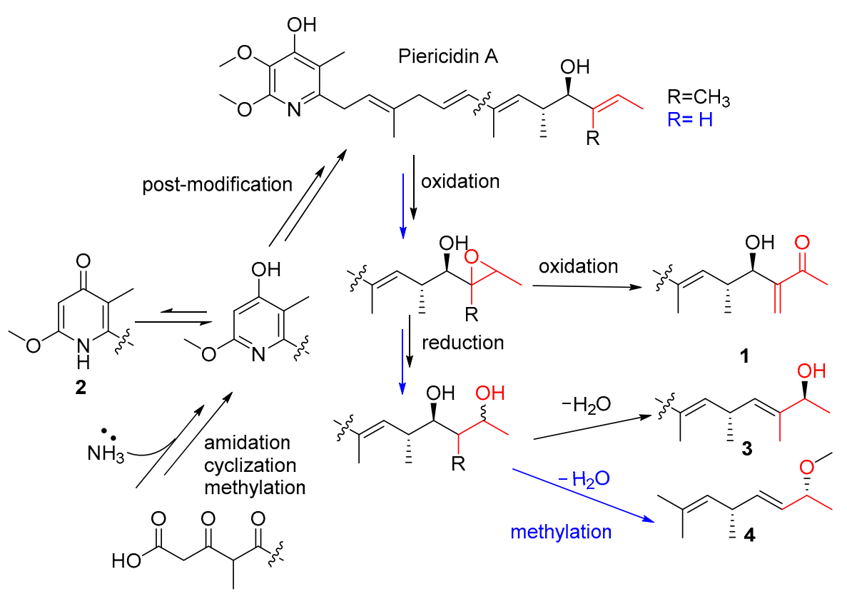
3. Discussion
4. Materials and Methods
4.1. General Experimental Procedures
4.2. Collection of THE Information on, and Cultivation of, the NS126 Strain
4.3. Isolation and Purification
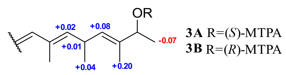
4.4. Mono-MTPA Esters of Piericidin Q (3)
4.5. Acid Hydrolysis of 11-Demethyl-Glucopiericidin A (8)
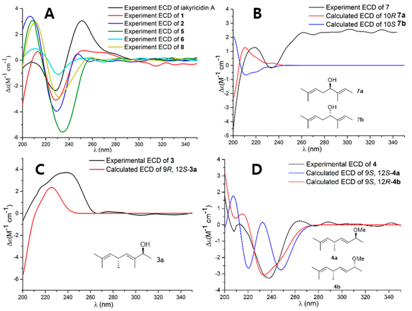
4.6. ECD Calculation
4.7. Cell Culture and Cytotoxic Bioassay
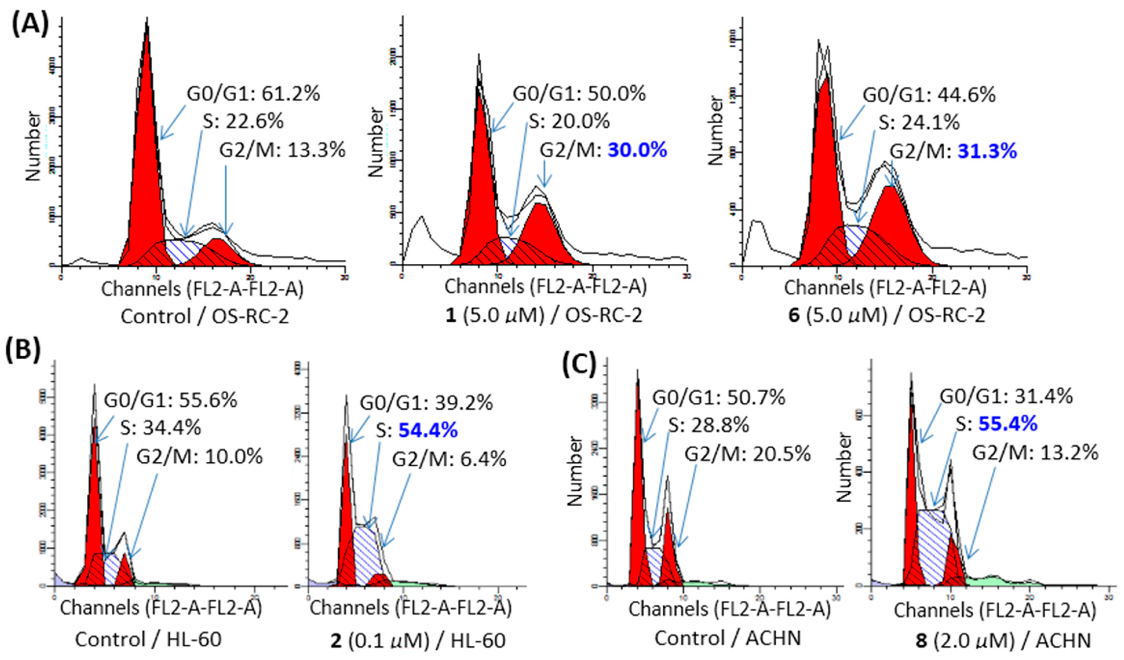
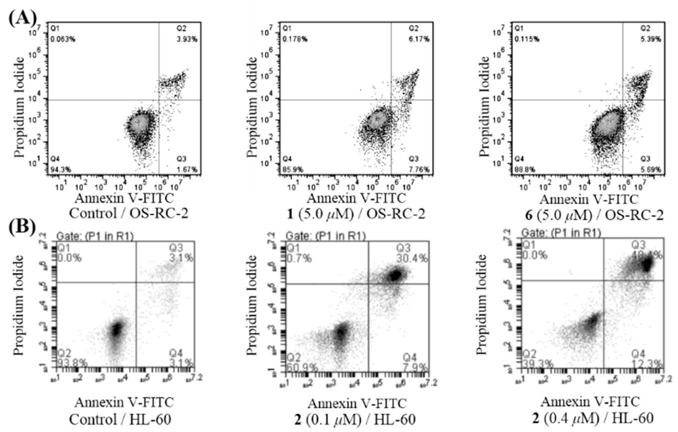
4.8. Cell Cycle and Apoptosis Assay
5. Conclusions
Supplementary Materials
Author Contributions
Funding
Institutional Review Board Statement
Informed Consent Statement
Conflicts of Interest
References
- Carroll, A.R.; Copp, B.R.; Davis, R.A.; Keyzers, R.A.; Prinsep, M.R. Marine natural products. Nat. Prod. Rep. 2020, 37, 175–223. [Google Scholar] [CrossRef] [PubMed]
- Jagannathan, S.V.; Manemann, E.M.; Rowe, S.E.; Callender, M.C.; Soto, W. Marine actinomycetes, new sources of biotechnological products. Mar. Drugs 2021, 19, 365. [Google Scholar] [CrossRef] [PubMed]
- Wang, Y.-N.; Meng, L.-H.; Wang, B.-G. Progress in research on bioactive secondary metabolites from deep-sea derived microorganisms. Mar. Drugs 2020, 18, 614. [Google Scholar] [CrossRef] [PubMed]
- Zhou, X.; Fenical, W. The unique chemistry and biology of the piericidins. J. Antibiot. 2016, 69, 582–593. [Google Scholar] [CrossRef] [PubMed]
- Liang, Z.; Chen, Y.; Gu, T.; She, J.; Dai, F.; Jiang, H.; Zhan, Z.; Li, K.; Liu, Y.; Zhou, X.; et al. LXR-mediated regulation of marine-derived piericidins aggravates high cholesterol diet-induced cholesterol metabolism disorder in mice. J. Med. Chem. 2021, 64, 9943–9959. [Google Scholar] [CrossRef]
- Zhou, X.; Liang, Z.; Li, K.; Fang, W.; Tian, Y.; Luo, X.; Chen, Y.; Zhan, Z.; Zhang, T.; Liao, S.; et al. Exploring the natural piericidins as anti-renal cell carcinoma agents targeting Peroxiredoxin 1. J. Med. Chem. 2019, 62, 7058–7069. [Google Scholar] [CrossRef]
- Li, K.; Liang, Z.; Chen, W.; Luo, X.; Fang, W.; Liao, S.; Lin, X.; Yang, B.; Wang, J.; Tang, L.; et al. Iakyricidins A-D, antiproliferative piericidin analogues bearing a carbonyl group or cyclic skeleton from Streptomyces iakyrus SCSIO NS104. J. Org. Chem. 2019, 84, 12626–12631. [Google Scholar] [CrossRef]
- Espeso, E.A.; Tilburn, J.; Arst, H.N.; Penalva, M.A. pH regulation is a major determinant in expression of a fungal penicillin biosynthetic gene. Embo. J. 1993, 12, 3947–3956. [Google Scholar] [CrossRef]
- Haneburger, I.; Eichinger, A.; Skerra, A.; Jung, K. New insights into the signaling mechanism of the pH-responsive, membrane-integrated transcriptional activator CadC of Escherichia coli. J. Biol. Chem. 2011, 286, 10681–10689. [Google Scholar] [CrossRef] [Green Version]
- Wilson, Z.E.; Brimble, M.A. Molecules derived from the extremes of life. Nat. Prod. Rep. 2009, 26, 44–71. [Google Scholar] [CrossRef]
- Lin, X.; Hetharua, B.; Lin, L.; Xu, H.; Zheng, T.; He, Z.; Tian, Y. Mangrove sediment microbiome: Adaptive microbial assemblages and their routed biogeochemical processes in Yunxiao mangrove national nature reserve, China. Microb. Ecol. 2019, 78, 57–69. [Google Scholar] [CrossRef]
- Matsumoto, M.; Mogi, K.I.; Nagaoka, K.; Ishizeki, S.; Kawahara, R.; Nakashima, T. New piericidin glucosides, glucopiericidin A and glucopiericidin B. J. Antibiot. 1987, 40, 149–156. [Google Scholar] [CrossRef]
- Lipshutz, B.H.; Amorelli, B. Total synthesis of piericidin A1. Application of a modified negishi carboalumination-nickel-catalyzed cross-coupling. J. Am. Chem. Soc. 2009, 131, 1396–1397. [Google Scholar] [CrossRef] [Green Version]
- Kominato, K.; Watanabe, Y.; Hirano, S.I.; Kioka, T.; Tone, H. Mer-A2026A and B, novel piericidins with vasodilating effect. II. Physicochemical properties and chemical structures. J. Antibiot. 1995, 48, 103–105. [Google Scholar] [CrossRef] [Green Version]
- Ueda, J.; Togashi, T.; Matukura, S.; Nagai, A.; Nakashima, T.; Komaki, H.; Anzai, K.; Harayama, S.; Doi, T.; Takahashi, T. A novel nuclear export inhibitor JBIR-02, a new piericidin discovered from Streptomyces sp. ML55. J. Antibiot. 2007, 60, 459–462. [Google Scholar] [CrossRef] [Green Version]
- Chen, Y.L.; Zhang, W.J.; Zhu, Y.G.; Zhang, Q.B.; Tian, X.P.; Zhang, S.; Zhang, C.S. Elucidating hydroxylation and methylation steps tailoring piericidin A1 biosynthesis. Org. Lett. 2014, 16, 736–739. [Google Scholar] [CrossRef]
- Ye, X.S.; He, J.; Cheng, Y.C.; Zhang, L.; Qiao, H.Y.; Pan, X.G.; Zhang, J.; Liu, S.N.; Zhang, W.K.; Xu, J.K. Cornusides A-O, bioactive iridoid glucoside dimers from the fruit of Cornus officinalis. J. Nat. Prod. 2017, 80, 3103–3111. [Google Scholar] [CrossRef]
- Han, X.N.; Liu, Z.Z.; Zhang, Z.Z.; Zhang, X.M.; Zhu, T.T.; Gu, Q.Q.; Li, W.L.; Che, Q.; Li, D.H. Geranylpyrrol A and piericidin F from Streptomyces sp CHQ-64 Delta rdmF. J. Nat. Prod. 2017, 80, 1684–1687. [Google Scholar] [CrossRef]
- Liu, Q.; Yao, F.; Chooi, Y.H.; Kang, Q.J.; Xu, W.; Li, Y.R.; Shao, Y.C.; Shi, Y.F.; Deng, Z.X.; Tang, Y.; et al. Elucidation of piericidin A1 biosynthetic locus revealed a thioesterase-dependent mechanism of α-pyridone ring formation. Chem. Biol. 2012, 19, 243–253. [Google Scholar] [CrossRef] [Green Version]
- Liu, Z.; Xiao, F.; Cai, S.; Liu, C.; Li, H.; Wu, T.; Jiang, Y.; Wang, X.; Che, Q.; Zhu, T.; et al. Effective generation of glucosylpiericidins with selective cytotoxicities and insights into their biosynthesis. Appl. Environ. Microbiol. 2021, 87, e00294. [Google Scholar] [CrossRef]
- Yang, B.; Tao, H.; Lin, X.; Wang, J.; Liao, S.; Dong, J.; Zhou, X.; Liu, Y. Prenylated indole alkaloids and chromone derivatives from the fungus Penicillium sp SCSI0041218. Tetrahedron 2018, 74, 77–82. [Google Scholar] [CrossRef]
- Frisch, M.J.; Trucks, G.W.; Schlegel, H.B.; Scuseria, G.E.; Robb, M.A.; Cheeseman, J.R.; Scalmani, G.; Barone, V.; Mennucci, B.; Petersson, G.A.; et al. Gaussian 09, Revision x.x; Gaussian Inc.: Wallingford, CT, USA, 2009. [Google Scholar]
- Tan, Y.; Yang, B.; Lin, X.; Luo, X.; Pang, X.; Tang, L.; Liu, Y.; Li, X.; Zhou, X. Nitrobenzoyl sesquiterpenoids with cytotoxic activities from a marine-derived Aspergillus ochraceus fungus. J. Nat. Prod. 2018, 81, 92–97. [Google Scholar] [CrossRef]
- Luo, X.; Lin, X.; Tao, H.; Wang, J.; Li, J.; Yang, B.; Zhou, X.; Liu, Y. Isochromophilones A–F, cytotoxic chloroazaphilones from the marine mangrove endophytic fungus diaporthe sp. SCSIO 41011. J. Nat. Prod. 2018, 81, 934–941. [Google Scholar] [CrossRef]


| Pos. | 1 a | 2 a | 3 a | 4 b | 5 b | 6 c | 7 a | 8 a |
|---|---|---|---|---|---|---|---|---|
| 1 | 3.39 (d, 6.9) | 3.36 (d, 6.9) | 3.42 (d, 6.9) | 3.37 (d, 6.9) | 3.37 (d, 6.9) | 3.25 (d, 6.9) | 3.35 (d, 6.9) | 3.48 (d, 6.9) |
| 2 | 5.28 (t, 6.9) | 5.18 (t, 6.9) | 5.27 (t, 6.9) | 5.41 (t, 6.9) | 5.40 (d, 6.9) | 5.30 (t, 6.9) | 5.31 (t, 6.9) | 5.26 (t, 6.9) |
| 4 | 2.76 (d, 7.1) | 2.79 (d, 6.9) | 2.77 (d, 7.0) | 2.78 (d, 6.9) | 2.78 (d, 6.7) | 2.72 (d, 6.8) | 2.76 (d, 6.9) | 2.80 (d, 7.1) |
| 5 | 5.45 (m) | 5.50 (m) | 5.53(m) | 5.57 (m) | 5.52 (m) | 5.48 (m) | 5.53 (m) | 5.55 (m) |
| 6 | 6.02 (d,15.8) | 6.08 (d, 5.5) | 6.02 (d, 6.2) | 6.05 (d, 15.8) | 6.07 (d, 6.0) | 5.86 (d, 5.7) | 6.03(d,15.5) | 6.08 (d, 6.1) |
| 8 | 5.26 (d, 10.4) | 5.30 (d, 9.2) | 5.22 (d, 9.2) | 5.20 (d, 9.1) | 5.26 (d, 9.4) | 5.19 (d, 9.7) | 5.30 (t, 6.7) | 5.30 (d, 9.5) |
| 9 | 2.75 (m) | 2.64 (m) | 3.42 (m) | 3.18 (m) | 2.63 (m) | 2.50 (m) | 2.35 (m) | 2.83 (m) |
| 10 | 4.52 (d, 4.0) | 3.69 (d, 8.2) | 5.26 (d, 9.0) | 5.53 (m) | 3.31 (m) | 3.59 (d, 7.1) | 3.96 (t, 7.0) | 4.00 (d, 8.0) |
| 11 | 5.27(m) | 5.26 (m) | 5.52 (m) | |||||
| 12 | 5.44 (q, 6.8) | 4.11 (q, 6.9) | 3.65 (m) | 5.60 (m) | 5.32 (q, 6.4) | 5.44 (q, 5.9) | 5.65 (m) | |
| 13 | 2.30 (s) | 1.62 (d, 6.8) | 1.20 (d, 6.9) | 1.21 (dd, 2.1, 6.3) | 1.73 (d, 6.7) | 1.54 (d, 6.6) | 1.60 (d, 5.4) | 1.71 (d, 6.4) |
| 14 | 6.21 (s); 6.01 (s) | 1.60 (s) | 1.65 (d, 1.3) | 3.23 (s) | 3.22 (s) | 1.50 (s) | 1.60 (s) | |
| 15 | 1.03 (d, 6.9) | 0.81 (d, 6.9) | 1.01 (d, 6.8) | 1.08 (t, 6.3) | 0.93 (d, 6.9) | 0.77 (d, 6.8) | 1.02 (d, 6.9) | |
| 16 | 1.57 (s) | 1.74 (s) | 1.74 (s) | 1.74 (s) | 1.72 (s) | 2.17(m); 2.12 (m) | 1.71 (s) | 1.75 (s) |
| 17 | 1.74 (s) | 1.74 (s) | 1.74 (s) | 1.75 (s) | 1.74 (s) | 1.70 (s) | 1.73 (s) | 1.78 (s) |
| 18 | 0.93 (t, 7.4) | |||||||
| 3′ | 5.88 (s) | |||||||
| 7′ | 3.96 (s) | 3.86 (s) | 4.00 (s) | 3.95 (s) | 3.95 (s) | 3.77 (s) | 3.90 (s) | 4.09 (s) |
| 8′ | 3.76 (s) | 3.77 (s) | 3.86 (s) | 3.86 (s) | 3.61 (s) | 3.74 (s) | 3.81 (s) | |
| 9′ | 2.06 (s) | 1.96 (s) | 2.07 (s) | 2.09 (s) | 2.09 (s) | 1.95 (s) | 2.04 (s) | 2.11 (s) |
| 1″ | 4.33 (d, 7.8) | |||||||
| 2″ | 3.20 (m) | |||||||
| 3″ | 3.36(m) | |||||||
| 4″ | 3.66(m) | |||||||
| 5″ | 3.20 (m) | |||||||
| 6″ | 3.65 (m); 3.78 (m) |
| Pos. | 1 a | 2 a | 3 a | 4 b | 5 b | 6 c | 7 a | 8 a |
|---|---|---|---|---|---|---|---|---|
| 1 | 34.8, CH2 | 32.3, CH2 | 34.1, CH2 | 34.6, CH2 | 34.6, CH2 | 34.2, CH2 | 34.9, CH2 | 33.4, CH2 |
| 2 | 122.9, CH | 121.0, CH | 122.2, CH | 122.2, CH | 122.1, CH | 122.2, CH | 123.4, CH | 121.5, CH |
| 3 | 136.7, C | 138.4, C | 137.1, C | 135.1, C | 135.2, C | 133.9, C | 135.9, C | 137.9, CH |
| 4 | 43.9, CH2 | 43.9, CH2 | 43.9, CH2 | 43.3, CH2 | 43.3, CH2 | 42.4, CH2 | 44.0, CH2 | 43.9, CH2 |
| 5 | 126.4, CH | 125.7, CH | 126.3, CH | 126.2, CH | 125.6, CH | 124.2, CH | 126.5, CH | 126.2, CH |
| 6 | 137.6, CH | 138.1, CH | 137.6, CH | 136.0, CH | 136.4, CH | 134.5, CH | 137.4, CH | 137.8, CH |
| 7 | 135.2, C | 134.8, C | 132.7, C | 133.0, C | 133.5, C | 138.1, C | 135.9, C | 134.6, C |
| 8 | 132.9, CH | 135.8, CH | 136.6, CH | 134.4, CH | 133.6, CH | 134.6, CH | 127.8, CH | 134.4, CH |
| 9 | 38.4, CH | 37.6, CH | 32.6, CH | 35.2, CH | 37.6, CH | 36.1, CH | 34.9, CH2 | 38.1, CH |
| 10 | 73.7, CH | 83.7, CH | 130.3, CH | 137.1, CH | 86.8, CH | 80.4, CH | 78.5, CH | 86.1, CH |
| 11 | 152.0, C | 136.6, C | 138.0, CH | 129.8, CH | 129.9, CH | 137.8, C | 138.7, C | 131.5, CH |
| 12 | 201.8, C | 122.8, CH | 73.9, CH | 78.2, CH | 129.9, CH | 119.4, CH | 121.5, CH | 129.4, CH |
| 13 | 26.5, CH3 | 13.1, CH3 | 22.0, CH3 | 21.6, CH3 | 17.9, CH3 | 12.8, CH3 | 13.1, CH3 | 18.0, CH3 |
| 14 | 126.8, CH2 | 11.1, CH3 | 11.8, CH3 | 55.8, CH3 | 56.3, CH3 | 11.4, CH3 | 11.1, CH3 | - |
| 15 | 18.5, CH3 | 18.1, CH3 | 21.8, CH3 | 21.0, CH3 | 16.8, CH3 | 18.3, CH3 | - | 16.4, CH3 |
| 16 | 13.0, CH3 | 13.1, CH3 | 12.9, CH3 | 12.8, CH3 | 13.0, CH3 | 19.7, CH2· | 12.9, CH3 | 13.0, CH3 |
| 17 | 16.7, CH3 | 16.7, CH3 | 16.7, CH3 | 16.8, CH3 | 16.8, CH3 | 16.5, CH3 | 16.6, CH3 | 16.7, CH3 |
| 18 | - | - | - | - | - | 13.8, CH3 | - | - |
| 2′ | 155.9, C | 161.8, C | 155.8, C | 153.7, C | 153.6, C | 154.3, C | 156.1, C | 155.7, C |
| 3′ | 130.6, C | 92.8, CH | 131.2, C | 127.9, C | 127.9, C | 128.5, C | 130.1, C | 131.5, C |
| 4′ | 158.5, C | 179.1, C | 159.8, C | 154.1, C | 154.1, C | 157.1, C | 157.2, C | 161.6, C |
| 5′ | 115.2, C | 117.6, C | 115.9, C | 112.1, C | 112.1, C | 112.9, C | 114.8, C | 116.8, C |
| 6′ | 151.2, C | 149.8, C | 150.5, C | 151.0, C | 151.1, C | 148.9, C | 151.7, C | 149.9, C |
| 7′ | 54.4, CH3 | 56.1, CH3 | 55.7, CH3 | 53.2, CH3 | 53.2, CH3 | 52.3, CH3 | 53.7, CH3 | 57.3, CH3 |
| 8′ | 61.0, CH3 | - | 61.2, CH3 | 60.8, CH3 | 60.8, CH3 | 59.7, CH3 | 60.8, CH3 | 61.4, CH3 |
| 9′ | 10.8, CH3 | 10.3, CH3 | 10.7, CH3 | 10.6, CH3 | 10.6, CH3 | 10.6, CH3 | 10.8, CH3 | 10.7, CH3 |
| 1″ | - | - | - | - | - | - | - | 103.3, CH |
| 2″ | - | - | - | - | - | - | - | 75.5, CH |
| 3″ | - | - | - | - | - | - | - | 78.3, CH |
| 4″ | - | - | - | - | - | - | - | 71.6, CH |
| 5″ | - | - | - | - | - | - | - | 77.9, CH |
| 6″ | - | - | - | - | - | - | - | 62.7, CH2 |
| No. | ACHN | 786-O | OS-RC-2 | HL-60 | K562 | MOLT-4 |
|---|---|---|---|---|---|---|
| 1 | >50 | >50 | 2.2 ± 0.21 | 12 ± 1.02 | >50 | >50 |
| 2 | >50 | >50 | 42.9 ± 5.3 | 0.08 ± 0.01 | >50 | / |
| 3 | >50 | >50 | >50 | 0.08 ± 0.01 | >50 | / |
| 4 | >50 | >50 | >50 | 0.08 ± 0.01 | >50 | / |
| 5 | >50 | >50 | >50 | 0.1 ± 0.01 | >50 | / |
| 6 | >50 | >50 | 4.5 ± 0.37 | 9.8 ± 0.52 | >50 | >50 |
| 7 | >50 | >50 | 15 ± 1.3 | >50 | >50 | >50 |
| 8 | 2.3 ± 0.32 | 12.0 ± 1.2 | 28.7 ± 3.2 | 1.3 ± 0.14 | 5.5 ± 0.69 | / |
| PA b | 0.40 ± 0.02 | 30 ± 3.2 | 5.2 ± 0.67 | 8.5 ± 0.86 | 2.4 ± 0.28 | 25 ± 2.6 |
Publisher’s Note: MDPI stays neutral with regard to jurisdictional claims in published maps and institutional affiliations. |
© 2021 by the authors. Licensee MDPI, Basel, Switzerland. This article is an open access article distributed under the terms and conditions of the Creative Commons Attribution (CC BY) license (https://creativecommons.org/licenses/by/4.0/).
Share and Cite
Li, K.; Su, Z.; Gao, Y.; Lin, X.; Pang, X.; Yang, B.; Tao, H.; Luo, X.; Liu, Y.; Zhou, X. Cytotoxic Minor Piericidin Derivatives from the Actinomycete Strain Streptomyces psammoticus SCSIO NS126. Mar. Drugs 2021, 19, 428. https://doi.org/10.3390/md19080428
Li K, Su Z, Gao Y, Lin X, Pang X, Yang B, Tao H, Luo X, Liu Y, Zhou X. Cytotoxic Minor Piericidin Derivatives from the Actinomycete Strain Streptomyces psammoticus SCSIO NS126. Marine Drugs. 2021; 19(8):428. https://doi.org/10.3390/md19080428
Chicago/Turabian StyleLi, Kunlong, Ziqi Su, Yongli Gao, Xiuping Lin, Xiaoyan Pang, Bin Yang, Huaming Tao, Xiaowei Luo, Yonghong Liu, and Xuefeng Zhou. 2021. "Cytotoxic Minor Piericidin Derivatives from the Actinomycete Strain Streptomyces psammoticus SCSIO NS126" Marine Drugs 19, no. 8: 428. https://doi.org/10.3390/md19080428