Article

Cold-Adapted Glutathione S-Transferases from Antarctic Psychrophilic Bacterium Halomonas sp. ANT108: Heterologous Expression, Characterization, and Oxidative Resistance

School of Marine Science and Technology, Harbin Institute of Technology, Weihai 264209, China
*
Author to whom correspondence should be addressed.
Mar. Drugs 2019, 17(3), 147; https://doi.org/10.3390/md17030147
Submission received: 22 January 2019 / Revised: 20 February 2019 / Accepted: 25 February 2019 / Published: 1 March 2019
(This article belongs to the Special Issue Marine Antioxidant)

Abstract

:
Glutathione S-transferases are one of the most important antioxidant enzymes to protect against oxidative damage induced by reactive oxygen species. In this study, a novel gst gene, designated as hsgst, was derived from Antarctic sea ice bacterium Halomonas sp. ANT108 and expressed in Escherichia coli (E. coli) BL21. The hsgst gene was 603 bp in length and encoded a protein of 200 amino acids. Compared with the mesophilic EcGST, homology modeling indicated HsGST had some structural characteristics of cold-adapted enzymes, such as higher frequency of glycine residues, lower frequency of proline and arginine residues, and reduced electrostatic interactions, which might be in relation to the high catalytic efficiency at low temperature. The recombinant HsGST (rHsGST) was purified to apparent homogeneity with Ni-affinity chromatography and its biochemical properties were investigated. The specific activity of the purified rHsGST was 254.20 nmol/min/mg. The optimum temperature and pH of enzyme were 25 °C and 7.5, respectively. Most importantly, rHsGST retained 41.67% of its maximal activity at 0 °C. 2.0 M NaCl and 0.2% H2O2 had no effect on the enzyme activity. Moreover, rHsGST exhibited its protective effects against oxidative stresses in E. coli cells. Due to its high catalytic efficiency and oxidative resistance at low temperature, rHsGST may be a potential candidate as antioxidant in low temperature health foods.

1. Introduction

Antarctica is isolated geographically from other continents and presents an extremely harsh environment on the earth. Antarctic environment has the characteristics of low temperature, high salinity, low nutrient availability, and strong ultraviolet (UV) radiation [1]. To adapt to the extreme conditions, microorganisms can produce a series of enzymes and display unique metabolic properties. Therefore, Antarctic microorganisms play a crucial part in the field of biomass conversion and have great potential applications, which have aroused more attention [1]. Sea ice is an important component of Antarctic climate and ecosystems. The increased oxygen concentration associated with the high salt concentration and low temperature of sea ice can induce the generation of reactive oxygen species (ROS) [2]. To remove the influence of ROS, sea ice microorganisms have especial antioxidant defense systems to protect against oxidative damage [2]. In the antioxidant defense systems, Glutathione S-transferases (GSTs) play a vital role in the regulation of the detoxification and redox balance of ROS [3]. Additionally, GSTs can catalyze the combination of glutathione (GSH) and various active electrophiles through affinity attacks and couplings [4]. GSTs have been widely described from animals [5], plants [6], and microorganisms [7]. Recently, a GST with an optimum temperature of 40 °C has been identified in the Antarctic psychrophilic bacteria Pseudoalteromonas sp. [8]. The crystallization and X-ray crystallographic of another GST from Antarctic clam Laternula elliptica were also studied [9]. To date, little information is available concerning the structural characteristics and biochemical properties of GSTs from Antarctic organisms.
Soluble GSTs can be divided into three categories: cytosolic, microsomal, and mitochondrial GSTs [10]. GSTs are composed of an amino acid chain, which contains the C-terminal domains and N-terminal domains (like-thioredoxin) [11]. The N-terminal domains with α-helixes and β-strands can contribute to GSH binding sites, and the C-terminal domains can provide more amino acid residues that interact with a variety of hydrophobic xenobiotics substrates. Besides, compared with N-terminal domains, the C-terminals of GSTs exhibit more structural changes, which can recognize and bind to a variety of known electrophilic compounds as GST substrates [12]. Currently, GSTs have been widely used as biomarkers of toxicity [13] and in pharmaceutical industries to scavenge organic hydroperoxides and ROS [14]. Furthermore, GSTs have been used as an indicator of certain diseases, especially hepatic infection [15]. Therefore, GSTs have different biological activities due to the differences of the conserved sequence regions, substrate specificity, and biochemical characterizations.
Antarctic sea ice microorganisms would be a novel source of antioxidant enzymes. Based on our recent studies, several cold-active antioxidant proteins were separated from Antarctic sea ice microorganisms and widely used in industrial sectors due to their excellent biochemical properties [16,17]. Halomonas is the type genus of the family Halomonadaceae belonging to the class Gammaproteobacteria. The genome sequence of Halomonas sp. strain KO116 isolated from sea surface [18] and the physiological features of Halomonas lionensis from sea sediment have also been reported [19]. In this study, a novel gst gene (hsgst) was cloned from Antarctic sea ice bacterium Halomonas sp. ANT108 and heterologous expressed in E. coli BL21. Furthermore, its biochemical properties and oxidative resistance were investigated.

2. Results and Discussion

2.1. Identification of the hsgst Gene

Sequence analysis of the full-length hsgst gene revealed an open reading frame (ORF) of 603 bp, encoding a protein of 200 amino acids with a predicted molecular weight of 21.76 kDa and the theoretical isoelectric point (pI) of 5.79. The length of Taenia multiceps gst gene was 606 bp encoding 201 amino acids, which was close to the length of hsgst gene [20]. Besides, the length of gst gene from Cydia pomonella and Sus scrofa were 648 bp encoding 215 amino acids (24.2 kDa) and 669 bp encoding 222 amino acids (25.3 kDa), respectively, which were both slightly longer than the hsgst gene [6]. The nucleotide sequence of the hsgst dene was deposited into GenBank database (accession numbers MH719093).
Based on sequence alignments with the related GSTs, HsGST shared the highest identity (90.0%) with HcGST from Halomonas campaniensis. The dimer interface of HsGST was comprised of L91, A92, G95, L96, G99 and R133 (Figure 1). In general, soluble GSTs have two binding domains with the GSH binding site (G-site) and the substrate binding site (H-site) [21]. The H-site (Y8, F9, V11, R14, V111, Y115, N211 and G212) and G-site (Y8, R14, W42, K50, Q57, L58, P59, Q71, S72, E104 and D105) were identified in the gst gene from Laternula elliptica [22]. Similarly, VpGSTp from Venerupis philippinarum also possessed the H-site (Y8, F9, V11, R14, V103, Y107, N200 and G201) and G-site (Y8, R14, W39, K45, Q52, L53, P54, Q65, S66, E96 and D97) [23]. However, in this study, only one H-site (Q98, M101, D102, A161 and Y164) was found in the sequence of HsGST. Additionally, the N-terminal domain interface (L91, Q98, L153, I156, T157, V160 and Y164) of HsGST was also identified in this sequence (Figure 1).

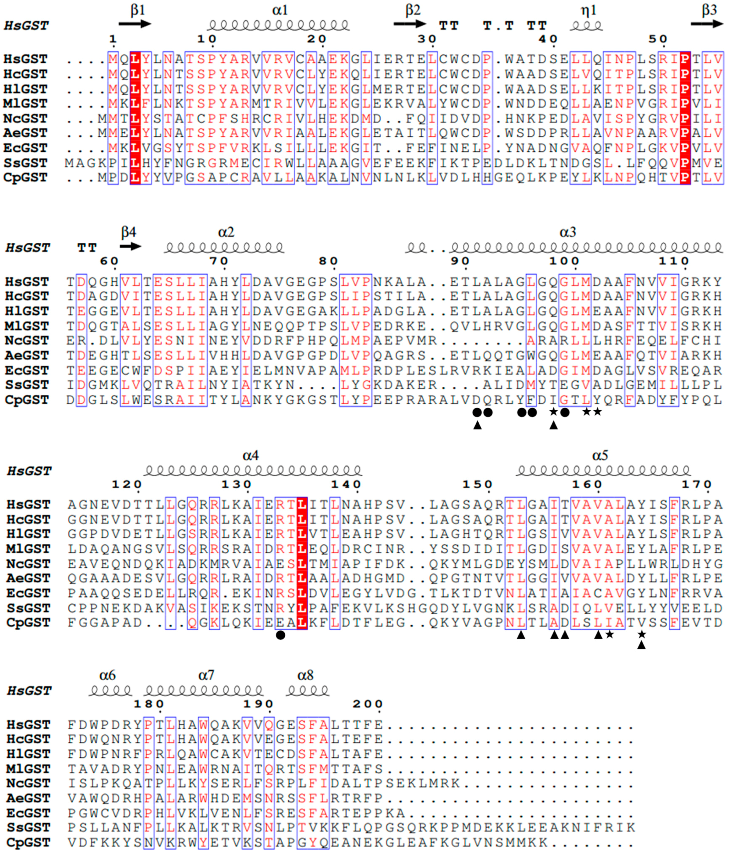
2.2. Homology Modeling Analysis

The 3D structure of HsGST showed that it consisted of 8 α-helixes and 4 β-strands (Figure 2A). For the validation of the structural model, 82.00% of the residues had an averaged 3D-1D score ≥ 0.2. The ERRAT program also showed an overall quality value of 94.27% for the 3D structure. These parameters demonstrated that the structural model of HsGST was well qualified. As can be seen in Figure 2B, the structural model of HsGST superimposed well with EcGST. The H-sites of HsGST and EcGST were also effectively located in close proximity (Figure 2B). EcGST (PDB ID: 3R2Q) was a mesophilic GST from E. coli K12 encoded 202 amino acids, and the sequence identity of EcGST to HsGST was 30% using BLASTp. In fact, there were differences in the frequencies of the amino acid residues between the cold-adapted enzymes and the homologous mesophilic enzymes [24]. As shown in Table 1, in the comparison with EcGST, HsGST possessed of a higher frequency of G (Gly), lower frequency of P (Pro) and R (Arg), regarded as a structural characteristic of cold-adapted enzymes, which endowed enzymes with increased conformational flexibility and low-temperature catalytic competence [24].
Furthermore, electrostatic interactions were an important factor for maintaining the secondary and tertiary structure in cold-adapted enzymes [25]. Compared to EcGST, HsGST exhibited reduced electrostatic interactions, especially hydrogen bonds and salt bridges, resulting in a decrease in the rigidity of the proteins, which were also obviously observed in other cold-adapted enzymes such as dienelactone hydrolase [26] and glycosylase [27]. In addition, in the comparison with EcGST, HsGST had less hydrophobic interactions, which might contribute to the increased structural flexibility and thermolability of cold-adapted enzymes [26,28]. The increased structural flexibility could improve the efficiency of substrates binding to the catalytic sites with high possibility, thereby reducing activation energy and increasing substrates turnover rates [26].

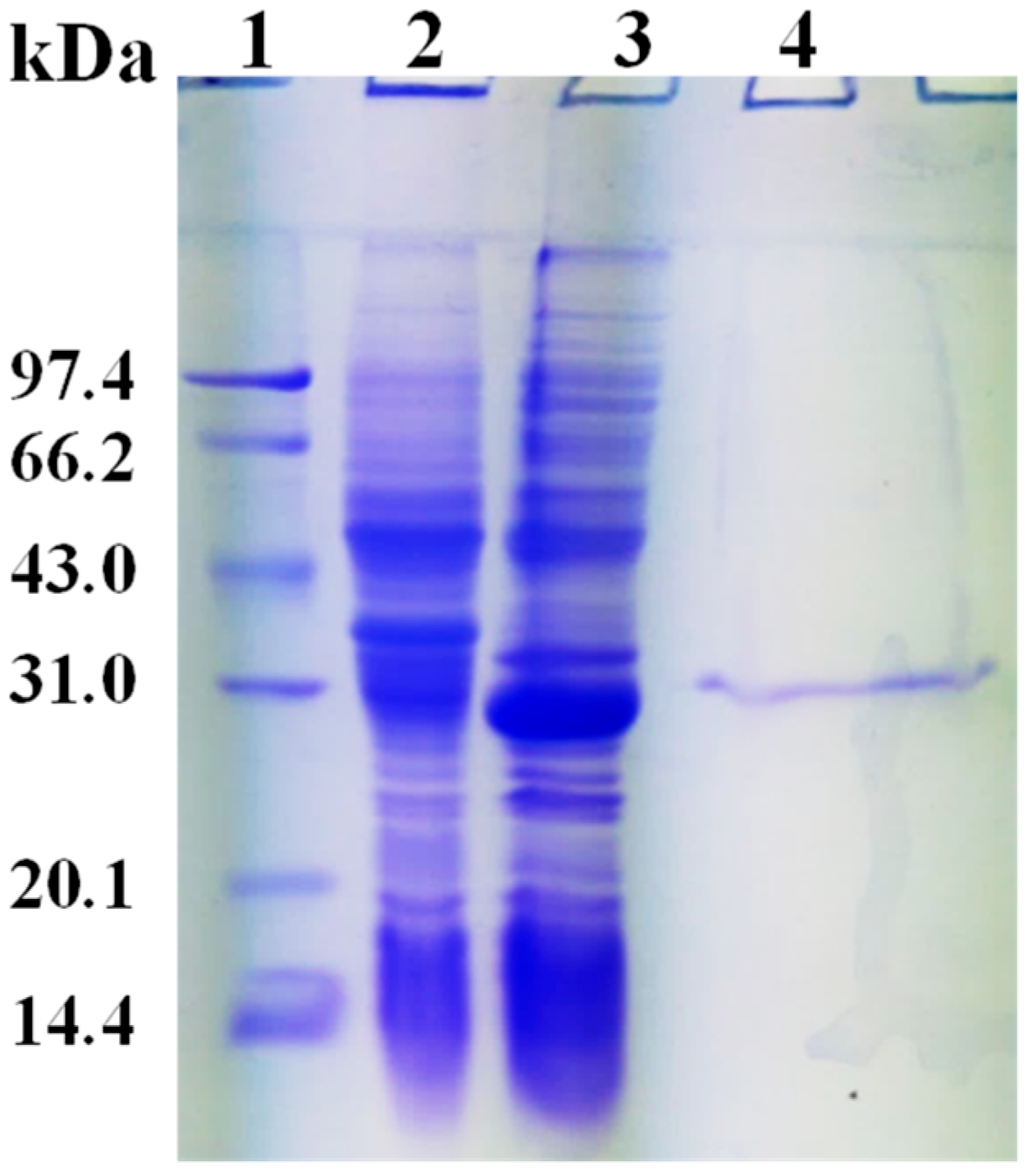
2.3. Expression and Purification of rHsGST

As shown in Figure 3 lane 3, the recombinant protein with an estimated molecular mass of 28 kDa was expressed in E. coli BL21. The purified protein using nickel nitrilotriacetic acid (Ni-NTA) resins exhibited a single band in terms of sodium dodecyl sulfate polyacrylamide gel electrophoresis (SDS-PAGE) (Figure 3, lane 4). In addition, 2.18 mg protein of rHsGST was purified from the crude extract. The purification fold and yields of rHsGST were 3.62 and 27.75%, respectively. The specific activity of rHsGST was 254.20 nmol/min/mg, which was higher than the GST isolated from Liposcelis entomophila (183 nmol/min/mg) and lower than that isolated from L. bostrychophila (412 nmol/min/mg) [29].

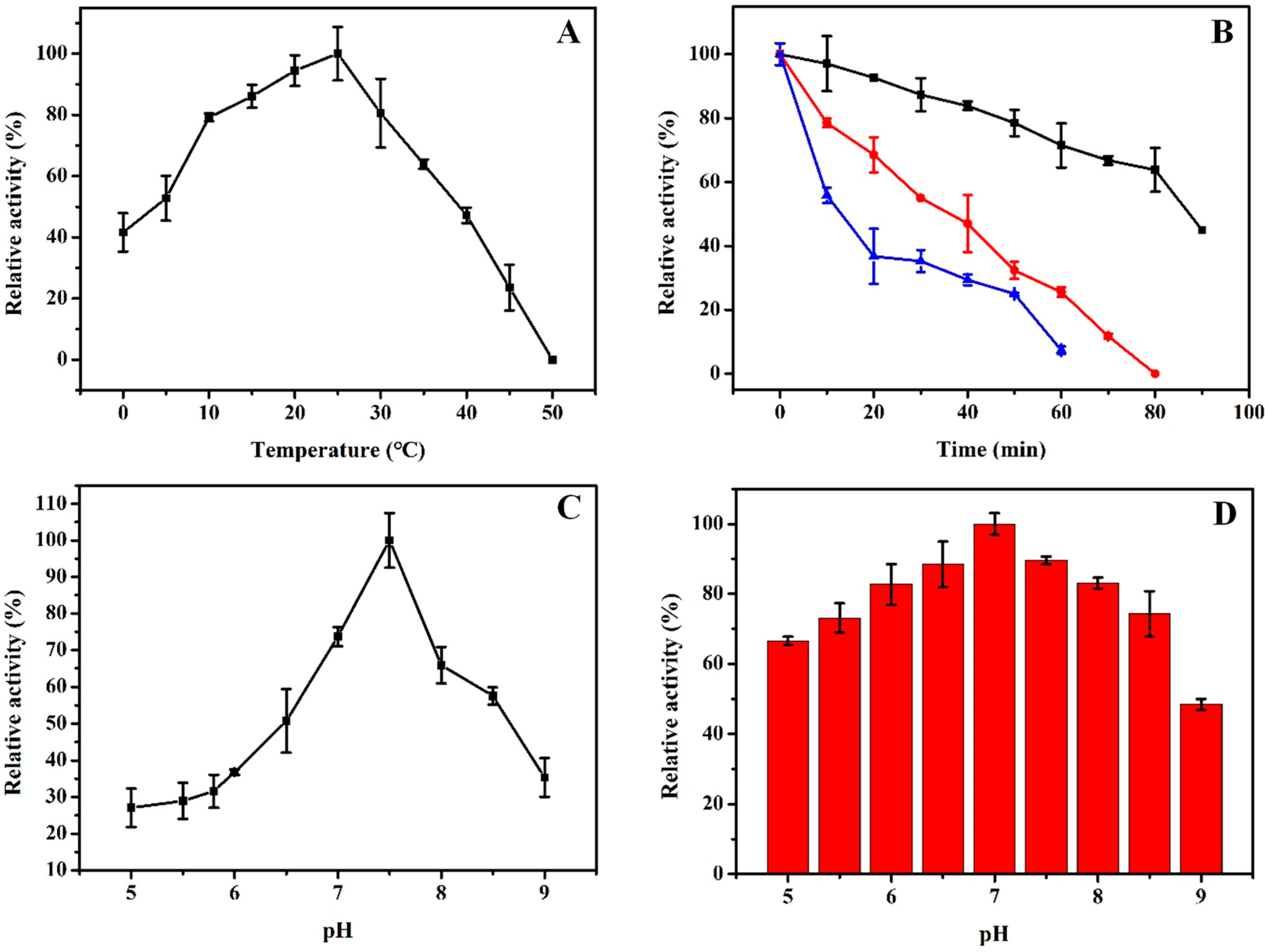
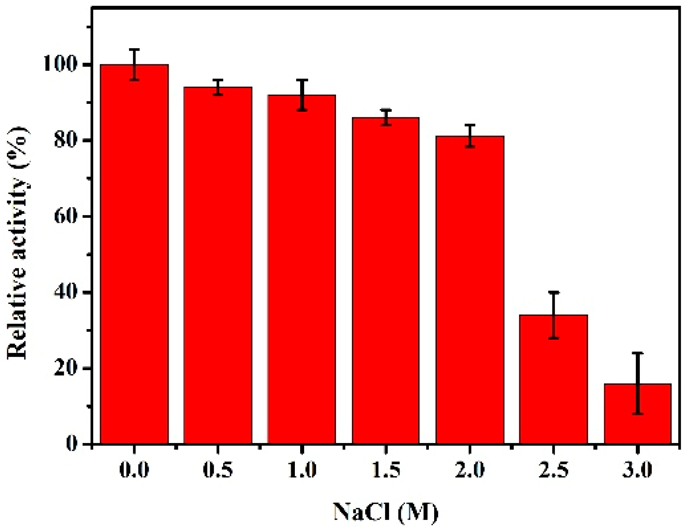
2.4. Biochemical Characterizations of rHsGST

The optimum temperature of rHsGST was measured over a temperature range of 0–50 °C (Figure 4A). rHsGST had the maximal activity at 25 °C and remained 41.67% of the maximal activity even at 0 °C, while the enzyme was inactivated at 50 °C. However, the optimum temperature of the GST from the Laternula elliptica [22] and GST from codling moth (Cydia pomonella) [30] were 35 °C and 50 °C, respectively. For the thermostability of rHsGST, the half-life of the enzyme was at 35 °C for 90 min, and rHsGST was completely inactivated after 1 h incubation at 45 °C (Figure 4B). According to other reports, GSTs from Pseudoalteromonas sp. [8] and Monopterus albus [31] retained 50% and 90% of the maximal activity at 40 °C for 15 min, respectively. These results indicated that rHsGST belonged to the cold-adapted enzyme, which was consistent with the results of homology modeling. rHsGST showed the maximal activity at pH 7.5 (Figure 4C), while the optimum pH of GST from Spodoptera exigua [32] and Locusta migratoria manilensis [33] was 6.0 and 8.0, respectively. Moreover, rHsGST retained about 50% and 65% of its maximal activity at pH 6.5 and pH 8.5, respectively. Similar results were also obtained in rRfGSTθ from black rockfish [34]. Consequently, it possessed good activity in neutral and weakly alkaline conditions. The stability of rHsGST was studied at various pH values from 5.0 to 9.0 (Figure 4D). The enzyme retained more than 60% of the maximal activity at pH 5.0–8.5. It illustrated that rHsGST had better stability over a wide pH range, which was similar with the GST from the locust [33].
As shown in Figure 5, rHsGST exhibited more than 80% of its initial activity at 0.5–2 M NaCl. However, the activity declined sharply at 2.5 M. These results illustrated that the rHsGST was a salt-tolerant enzyme that could adapt to the Antarctic sea ice environment with a high salt concentration. The activity of GST isolated from Pseudoalteromonas sp. ANT506 was lower than rHsGST, which remained about 50% of the initial activity in the existence of 2.0 M NaCl [8].
The effects of different reagents on rHsGST activity were also investigated (Table 2). The activity of rHsGST was not detected in the presence of metal ions Ca2+, Ni2+, Cu2+ and Sn2+. On the contrary, the activity of GST from Trichinella spiralis was promoted by Ni2+ [35]. Interestingly, Fe2+ could improve the rHsGST activity, and H2O2 also kept the enzyme activity with 97.09%. rHsGST that was treated with ethanol showed the strongest inhibitory effect, and the activity was only 29.03% of control. Besides, the activity of rHsGST treated with SDS retained 54.84%, a similar result was also obtained in the recent study of the TsGST from Trichinella spiralis [35].

2.5. Kinetics and Thermodynamics Parameters

Kinetic parameters were determined by linweaver-burk plot, Vmax of substrate chlorodinitrobenzene (CDNB) and GSH were 714.29 and 243.90 nmol/min/mg, respectively (Table 3). Compared with the results of LmGSTu1, the Vmax of CDNB in this study was much higher than 250 nmol/min/mg [36]. Furthermore, the Km of GSH was 0.27 mM, which was lower than the value of CDNB (2.86 mM). These consequences illustrated that rHsGST had a higher substrate affinity for GSH than for CDNB.
Thermodynamic parameters were investigated using the CDNB as the substrate (Table 4). Obviously, as the temperature increased from 10 °C to 30 °C, the values of ΔG appeared a growing tendency. The values of ΔH were changed from 34.11 to 33.65 KJ/mol at temperatures from 10 to 30 °C. Besides, the values of ΔS were −101.48, −101.63, −101.77, −101.91, and −102.05 J/mol, followed by the temperature 10, 15, 20, 25, and 30 °C, respectively. The negative value of ΔS could increase the order of the activated transition state of catalysis [37].

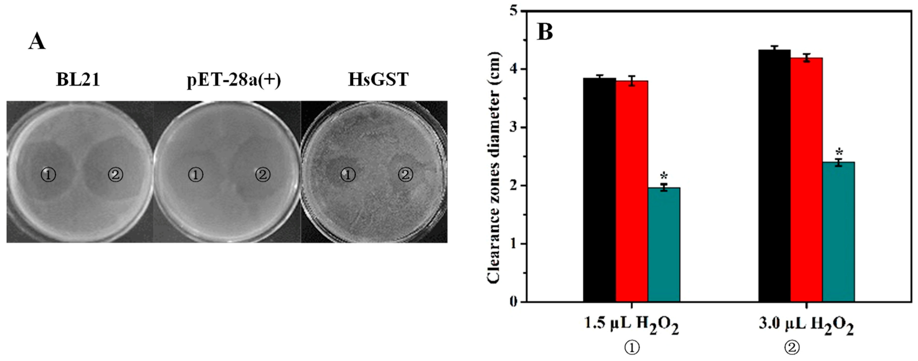
2.6. Disk Diffusion Assay

It is known that H2O2 can cause oxidative stress that may induce cell dysfunction [38]. As can be seen from Figure 6, the clearance zones in the recombinant bacteria plate were significantly smaller than those of controls (p < 0.05). This result indicated that rHsGST had an important antioxidant effect on helping living tissues to overcome the oxidative stress caused by H2O2. Similarly, GSTs from Haliotis discus discus and Apis cerana cerana could also protect cells against oxidative stress produced by H2O2 [39,40].

3. Materials and Methods

3.1. Strains and Materials

Antarctic sea ice bacterial samples (68° 30′ E, 65° 00′ S) were cultured in 2216E medium at the logarithmic growth phase, then the culture solution was extracted and streaked into a solid medium by inoculating loop. The medium was then cultured at 12 °C for 72 h to pick up a single colony, which was identified as Halomonas sp. according to the sequence analysis of 16S rRNA genes (accession number: MK494178). The isolated strain had an optimum growth temperature of 10–12 °C. Plasmid vector pET-28a(+) and receptor E. coli BL21 were maintained by our laboratory. T4 DNA ligase, BamHI and HindIII and SanPrep column DNA gel recovery kit were purchased from Takara Biotechnology (Dalian) Co., Ltd (Beijing, China).

3.2. Identification of hsgst Gene

Using high-throughput technologies to sequence and annotate the genome of Halomonas sp. ANT108 (data not shown). The sequencing of full-length hsgst gene was amplified by PCR applying by the forward primers 5′-ATAGGATCCATGCAGC TCTATTTAA-3′ and the reverse primer 5′-AGTAAGCTTGTTCAAACGTGG TAAG-3′ (the BamHI and HindIII sites were underlined, respectively) based on its genome sequence. The complete amino acid sequences of the HsGST were obtained through an ORF finder (http://www.ncbi.nlm.nih.gov/gorf/gorf.html). Multiple sequence alignments were performed using the Bioedit program.

3.3. Analysis of HsGST

The 3D structure model of HsGST was established using the SWISS-MODEL server and verified using SAVES v5.0 (http://servicesn.mbi.ucla.edu/SAVES/) (University of California, Los Angeles, CA, USA). The visualization of homology modeling was performed using PyMOL software (Version No.2.2.0, DeLano Scientific LLC, CA, USA). Meanwhile, salt bridges were predicted to use ESBRI (Evaluating the Salt Bridges in Proteins). Besides, protein intramolecular interactions were predicted by the Protein Interactions Calculator (PIC) Online website (http://pic.mbu.iisc. ernet.in/job.html).

3.4. Expression and Purification of hsgst Gene in E. coli

The hsgst gene was inserted into the plasmid of the pET-28a(+) vector. Then, the recombinant plasmid was transferred into the receptor E. coli BL21. The positive strain was verified by PCR and inoculated in LB medium supplemented with 0.1 mg/mL kanamycin (Kana). Afterwards, 1 mM isopropyl-β-d-thiogalactoside (IPTG) was added to induce the hsgst gene expression, and the 1 L cultures were pro-longed for 8 h at 25 °C. The induced cells were centrifuged and disrupted by sonication (JY96-IIN, Shanghai, China) to obtain inclusion bodies. Then the inclusion bodies were washed with PBS (pH 8.0) and treated with 8 M urea at 25 °C for 1 h. The mixture was centrifuged at 7500 rpm for 15 min. The supernatant was diluted for 30 times by adding PBS (pH 8.0) at 25 °C for 2 h, which was the procedure of the rHsGST refolding. The protein solution was centrifuged at 12,000 rpm for 15 min. The supernatant was the crude extract of rHsGST. The protein was purified using Ni-NTA resins affinity chromatography (GE Healthcare, Uppsala, Sweden). The protein molecular weight and purity of the rHsGST were determined by SDS-PAGE with 12.0% polyacrylamide gels.

3.5. Assay of rHsGST Activity and Protein Concentration

GST activity was determined according to the conjugation of CDNB and GSH in the presence of the enzyme [12]. One unit of enzyme activity was defined as the quantity of CDNB conjugated product catalyzed synthetically per milligram per minute at 25 °C. Protein concentrations were measured by the method of Bradford [41].

3.6. Biochemical Characteristics of rHsGST

The optimum temperature of the rHsGST was determined at different temperatures (0–50 °C) by the standard enzyme assay. To evaluate the thermostability of rHsGST, the enzyme was incubated at 35, 40, and 45 °C for 90 min, respectively. The optimum pH the rHsGST was measured at 25 °C and the following buffers were 0.1 mol/L acetate buffer (pH 5.0–5.8), 0.1 mol/L Na2HPO4/Na2HPO4 (pH 5.8–8.0), 0.1 mol/L Tris-HCl (pH 8.0–9.0). The stability of rHsGST at different pH was determined by incubating the enzyme in the buffers mentioned above at 25 °C for 1 h and the remaining activity was measured. Furthermore, the effect of NaCl on the rHsGST was evaluated by adding NaCl (0–3.0 M) and incubated at 25 °C for 1 h. The remaining activity was measured by means of the standard enzyme assay. In order to study the effects of various reagents on the rHsGST activity, the protein solution and different reagents solution were firstly mixed for 1 h at 25 °C, and then the remaining activity was measured by means of the standard enzyme assays.

3.7. Kinetics and Thermodynamics Parameters

Kinetic parameters were determined using five different concentrations of CDNB (0.5, 1.0, 1.5, 2.0, 2.5, 3.0, 3.5, 4.0 and 4.5 mM) and GSH (0.1, 0.2, 0.3, 0.4, 0.5, 0.6, 0.7, 0.8 and 0.9 mM) at 25 °C, respectively. Km and Vmax were measured using the Micchaelis-Menten equation [42]. Through the determination of kinetic, the kcat was obtained. The kcat was determined at 10–30 °C and the line was defined with lnkcat and 1/T. Then, the ΔG, ΔH, and ΔS were calculated, respectively [42].

3.8. Disk Diffusion Assay

To compare the survival efficiency of untransformed BL21, BL21/pET-28a(+) and recombinant BL21/pET-28a(+)-HsGST, a disk diffusion assay was performed as described previously [43]. The IPTG-induced bacterial cultures were uniformly spread on LB agar plates, and two filter paper disks (diameter 3 mm) were placed equidistant on each agar plate. Then, 1.5 and 3 μL of 30% H2O2 were added to the filter paper disks, respectively. The treated plates were incubated at 37 °C for 12 h, and the diameters of the clearance zone were measured.

4. Conclusions

A novel hsgst gene from Halomonas sp. ANT108 was cloned, expressed, and characterized in the present study. Furthermore, HsGST had the structural characteristics of cold-adapted enzymes by homology modeling. After purification, rHsGST exhibited different catalytic capabilities compared with other GSTs, such as the optimum temperature, thermolability, and high tolerance in the presence of H2O2 and high salt concentration. Moreover, E. coli cells overexpressing hsgst displayed the protective effects against oxidative stress. Although there are already numerous GSTs available in the market, the high catalytic efficiency and oxidative resistance at low temperature of rHsGST may make it a potential candidate as an antioxidant in low temperature health foods such as marine surimi, protamine, and low temperature meat products.

Author Contributions

Y.H., C.Q. and Q.W. (Quanfu Wang) took charge of the research and designed the experiments; C.Q., Y.W. (Yifan Wang), Y.W. (Yatong Wang), X.R. and Q.W. (Qifeng Wei) performed the experiments and analyzed the data; Y.H., C.Q. and Q.W. (Quanfu Wang) wrote the paper. All authors reviewed the manuscript and approved the final version.

Fundings

This study was funded by the National Natural Science Foundation of China (41876149), the Natural Science Foundation of Shandong Province (ZR2017MC046) and the Key Research and Development Plan of Shandong Province (2018GHY115021).

Conflicts of Interest

The authors declare no conflict interest.

References

  1. Kostadinova, N.; Tosi, S.; Spassova, B.; Angelova, M. Comparison of the oxidative stress response of two Antarctic fungi to different growth temperatures. Pol. Polar Res. 2017, 38, 393–408. [Google Scholar] [CrossRef] [Green Version]
  2. Chattopadhyay, M.K.; Raghu, G.; Sharma, Y.V.R.K.; Biju, A.R.; Rajasekharan, M.V.; Shivaji, S. Increase in oxidative stress at low temperature in an Antarctic bacterium. Curr. Microbiol. 2011, 62, 544–546. [Google Scholar] [CrossRef] [PubMed]
  3. Vijayakumar, H.; Thamilarasan, S.K.; Shanmugam, A.; Natarajan, S.; Jung, H.J.; Park, J.I.; Kim, H.; Chung, M.Y.; Nou, I.S. Glutathione transferases superfamily: Cold-inducible expression of distinct GST genes in Brassica oleracea. Int. J. Mol. Sci. 2016, 17, 1211. [Google Scholar] [CrossRef] [PubMed]
  4. Dhar, K.; Dhar, A.; Rosazza, J.P.N. Glutathione S-transferase isoenzymes from Streptomyces griseus. Appl. Environ. Microbiol. 2003, 69, 707–710. [Google Scholar] [CrossRef] [PubMed]
  5. Abunnaja, M.S.; Kurogi, K.; Mohammed, Y.I.; Sakakibara, Y.; Suiko, M.; Hassoun, E.A.; Liu, M.C. Identification and characterization of the zebrafish glutathione S-transferase Pi-1. J. Biochem. Mol. Toxicol. 2017, 31, e21948. [Google Scholar] [CrossRef] [PubMed]
  6. Yang, X.Q.; Zhang, Y.L. Characterization of glutathione S-transferases from Sus scrofa, Cydia pomonella and Triticum aestivum: Their responses to cantharidin. Enzyme Microb. Technol. 2015, 69, 1–9. [Google Scholar] [CrossRef] [PubMed]
  7. Jung, U.; Cho, Y.S.; Seong, H.M.; Kim, S.J.; Kim, Y.C.; Chung, A.S. Characterization of a novel glutathione S-Transferase from Pseudomonas sp. DJ77. J. Biochem. Mol. Biol. 1996, 29, 111–115. [Google Scholar]
  8. Shi, Y.L.; Wang, Q.F.; Hou, Y.H.; Hong, Y.Y.; Han, X.; Yi, J.L.; Qu, J.J.; Lu, Y. Molecular cloning, expression and enzymatic characterization of glutathione S-transferase from Antarctic sea-ice bacteria Pseudoalteromonas sp. ANT506. Microbiol. Res. 2014, 169, 179–184. [Google Scholar] [CrossRef] [PubMed]
  9. Jang, E.H.; Park, H.; Park, A.K.; Moon, J.H.; Chi, Y.M.; Ahn, I.Y. Crystallization and preliminary X-ray crystallographic studies of the q-class glutathione S-transferase from the Antarctic clam Laternula elliptica. Acta Crystallogr. Sect. F Struct. Biol. Cryst. Commun. 2008, 64, 1132–1134. [Google Scholar] [CrossRef] [PubMed]
  10. Liu, J.Y.; Yang, X.Q.; Zhang, Y.L. Characterization of a lambda-cyhalothrin metabolizing glutathione S-transferase CpGSTd1 from Cydia pomonella (L.). Appl. Microbiol. Biotechnol. 2014, 98, 8947–8962. [Google Scholar] [CrossRef] [PubMed]
  11. Kayum, M.A.; Nath, U.K.; Park, J.I.; Biswas, M.K.; Choi, E.K.; Song, J.Y.; Kim, H.T.; Nou, I.S. Genome-Wide identification, characterization, and expression profiling of glutathione S-transferase (GST) family in pumpkin reveals likely role in cold-stress tolerance. Genes 2018, 9, 84. [Google Scholar] [CrossRef] [PubMed]
  12. Pandey, T.; Singh, S.K.; Chhetri, G.; Tripathi, T.; Singh, A.K. Characterization of a highly pH stable chi-class glutathione S-transferase from Synechocystis PCC 6803. PLoS ONE 2015, 10, e0126811. [Google Scholar] [CrossRef] [PubMed]
  13. Hoarau, P.; Gnassia-Barelli, M.; Romeo, M.; Girard, J.P. Differential induction of glutathione S-transferases in the clam Ruditapes decussatus exposed to organic compounds. Environ. Toxicol. Chem. 2001, 20, 523–529. [Google Scholar] [CrossRef] [PubMed]
  14. Gajewska, B.; Kazmierczak, B.; Kuzma-Kozakiewicz, M.; Jamrozik, Z.; Baranczyk-Kuzma, A. GSTP1 polymorphisms and their association with glutathione transferase and peroxidase activities in patients with motor neuron disease. CNS Neurol. Disord.-Drug Targets 2015, 14, 1328–1333. [Google Scholar] [CrossRef] [PubMed]
  15. Aguayo, V.; Valdes, B.; Espino, A.M. Assessment of Fasciola hepatica glutathione S-transferase as an antigen for serodiagnosis of human chronic fascioliasis. Acta Trop. 2018, 186, 41–49. [Google Scholar] [CrossRef] [PubMed]
  16. Wang, Q.F.; Wang, Y.F.; Hou, Y.H.; Shi, Y.L.; Han, H.; Miao, M.; Wu, Y.Y.; Liu, Y.P.; Yue, X.N.; Li, Y.J. Cloning, expression and biochemical characterization of recombinant superoxide dismutase from Antarctic psychrophilic bacterium Pseudoalteromonas sp. ANT506. J. Basic Microbiol. 2016, 56, 753–761. [Google Scholar] [CrossRef] [PubMed]
  17. Wang, Y.T.; Han, H.; Cui, B.Q.; Hou, Y.H.; Wang, Y.F.; Wang, Q.F. A glutathione peroxidase from Antarctic psychrotrophic bacterium Pseudoalteromonas sp ANT506: Cloning and heterologous expression of the gene and characterization of recombinant enzyme. Bioengineered 2017, 8, 742–749. [Google Scholar] [CrossRef] [PubMed]
  18. O’Dell, K.B.; Woo, H.L.; Utturkar, S.; Klingeman, D.; Brown, S.D.; Hazen, T.C. Genome sequence of Halomonas sp. Strain KO116, an ionic liquid tolerant marine bacterium isolated from a Lignin-Enriched Seawater Microcosm. Genome Announc. 2015, 3, e00402. [Google Scholar] [CrossRef]
  19. Gaboyer, F.; Vandenabeele-Trambouze, O.; Cao, J.; Ciobanu, M.C.; Jebbar, M.; Le Romancer, M.; Alain, K. Physiological features of Halomonas lionensis sp. nov., a novel bacterium isolated from a Mediterranean Sea sediment. Res. Microbiol. 2014, 165, 490–500. [Google Scholar] [CrossRef] [PubMed]
  20. Sun, Y.; Wang, Y.; Huang, X.; Gu, X.; Lai, W.; Peng, X.; Yang, G. Characterization of glutathione S-transferase and its immunodiagnostic potential for detecting. Taenia multiceps. Vet. Parasitol. 2017, 242, 31–37. [Google Scholar] [CrossRef] [PubMed]
  21. Mannervik, B.; Danielson, U.H. Glutathione transferases—Structure and catalytic activity. CRC Crit. Rev. Biochem. 1988, 23, 283–337. [Google Scholar] [CrossRef] [PubMed]
  22. Kim, M.; Ahn, I.Y.; Cheon, J.; Park, H. Molecular cloning and thermal stress-induced expression of a pi-class glutathione S-transferase (GST) in the Antarctic bivalve Laternula elliptica. Comp. Biochem. Phys. A. Mol. Integr. Physiol. 2009, 152, 207–213. [Google Scholar] [CrossRef] [PubMed]
  23. Xu, C.Q.; Pan, L.Q.; Liu, N.; Wang, L.; Miao, J.J. Cloning, characterization and tissue distribution of a pi-class glutathione S-transferase from clam (Venerupis philippinarum): Response to benzo [alpha] pyrene exposure. Comp. Biochem. Phys. C 2010, 152, 160–166. [Google Scholar] [CrossRef] [PubMed]
  24. Simpson, P.J.L.; Codd, R. Cold adaptation of the mononuclear molybdoenzyme periplasmic nitrate reductase from the Antarctic bacterium Shewanella gelidimarina. Biochem. Biophys. Res. Commun. 2011, 414, 783–788. [Google Scholar] [CrossRef] [PubMed]
  25. Siddiqui, K.S.; Cavicchioli, R. Cold-adapted enzymes. Annu. Rev. Biochem. 2006, 75, 403–433. [Google Scholar] [CrossRef] [PubMed]
  26. Hashim, N.H.F.; Mahadi, N.M.; Illias, R.M.; Feroz, S.R.; Abu Bakar, F.D.; Murad, A.M.A. Biochemical and structural characterization of a novel cold-active esterase-like protein from the psychrophilic yeast Glaciozyma antarctica. Extremophiles 2018, 22, 607–616. [Google Scholar] [CrossRef] [PubMed]
  27. Leiros, I.; Moe, E.; Lanes, O.; Smalas, A.O.; Willassen, N.P. The structure of uracil-DNA glycosylase from Atlantic cod (Gadus morhua) reveals cold-adaptation features. Acta. Crystallogr. D 2003, 59, 1357–1365. [Google Scholar] [CrossRef] [PubMed]
  28. Ramli, A.N.M.; Mahadi, N.M.; Shamsir, M.S.; Rabu, A.; Joyce-Tan, K.H.; Murad, A.M.A.; Illias, R.M. Structural prediction of a novel chitinase from the psychrophilic Glaciozyma antarctica PI12 and an analysis of its structural properties and function. J. Comput. Aided Mol. Des. 2012, 26, 947–961. [Google Scholar] [CrossRef] [PubMed]
  29. Dou, W.; Xiao, L.S.; Niu, J.Z.; Jiang, H.B.; Wang, J.J. Characterization of the purified glutathione S-transferases from two Psocids Liposcelis bostrychophila and L. entomophila. Agric. Sci. China 2010, 9, 1008–1016. [Google Scholar] [CrossRef]
  30. Huang, X.L.; Fan, D.S.; Liu, L.; Feng, J.N. Identification and characterization of glutathione S-transferase genes in the Antennae of Codling Moth (Lepidoptera: Tortricidae). Ann. Entomol. Soc. Am. 2017, 110, 409–416. [Google Scholar] [CrossRef]
  31. Huang, Q.; Liang, L.; Wei, T.; Zhang, D.M.; Zeng, Q.Y. Purification and partial characterization of glutathione transferase from the teleost Monopterus albus. Comp. Biochem. Phys. C 2008, 147, 96–100. [Google Scholar] [CrossRef] [PubMed]
  32. Wan, H.; Zhan, S.; Xia, X.D.; Xu, P.F.; You, H.; Jin, B.R.; Li, J.H. Identification and functional characterization of an epsilon glutathione S-transferase from the beet armyworm (Spodoptera exigua). Pestic. Biochem. Phys. 2016, 132, 81–88. [Google Scholar] [CrossRef] [PubMed]
  33. Qin, G.H.; Jia, M.; Liu, T.; Zhang, X.Y.; Guo, Y.P.; Zhu, K.Y.; Ma, E.B.; Zhang, J.Z. Heterologous expression and characterization of a sigma glutathione S-transferase involved in carbaryl detoxification from oriental migratory locust, Locusta migratoria manilensis (Meyen). J. Insect Physiol. 2012, 58, 220–227. [Google Scholar] [CrossRef] [PubMed]
  34. Jayasinghe, J.D.H.E.; Bathige, S.D.N.K.; Nam, B.H.; Noh, J.K.; Lee, J. Comprehensive characterization of three glutathione S-transferase family proteins from black rockfish (Sebastes schlegelii). Comp. Biochem. Physiol. C Toxicol. Pharmacol. 2016, 189, 31–43. [Google Scholar] [CrossRef] [PubMed]
  35. Cui, J.; Li, L.G.; Jiang, P.; Liu, R.D.; Yang, X.; Liu, L.N.; Liu, P.; Zhang, S.B.; Wang, Z.Q. Biochemical and functional characterization of the glutathione S-transferase from Trichinella spiralis. Parasitol. Res. 2015, 114, 2007–2013. [Google Scholar] [CrossRef] [PubMed]
  36. Qin, G.H.; Jia, M.; Liu, T.; Zhang, X.Y.; Guo, Y.P.; Zhu, K.Y.; Ma, E.B.; Zhang, J.Z. Characterization and functional analysis of four glutathione S-transferases from the Migratory Locust, Locusta migratoria. PLoS ONE 2013, 8, e58410. [Google Scholar] [CrossRef] [PubMed]
  37. Siddiqui, K.S.; Cavicchioli, R.; Thomas, T. Thermodynamic activation properties of elongation factor 2 (EF-2) proteins from psychrotolerant and thermophilic Archaea. Extremophiles 2002, 6, 143–150. [Google Scholar] [CrossRef] [PubMed]
  38. Coyle, C.H.; Martinez, L.J.; Coleman, M.C.; Spitz, D.R.; Weintraub, N.L.; Kader, K.N. Mechanisms of H2O2-induced oxidative stress in endothelial cells. Free Radic. Biol. Med. 2006, 4, 2206–2213. [Google Scholar] [CrossRef] [PubMed]
  39. Sandamalika, W.M.G.; Priyathilaka, T.T.; Liyanage, D.S.; Lee, S.; Lim, H.K.; Lee, J. Molecular characterization of kappa class glutathione S-transferase from the disk abalone (Haliotis discus discus) and changes in expression following immune and stress challenges. Fish Shellfish Immunol. 2018, 77, 252–263. [Google Scholar] [CrossRef] [PubMed]
  40. Zhang, Y.Y.; Yan, H.R.; Lu, W.J.; Li, Y.Z.; Guo, X.Q.; Xu, B.H. A novel Omega-class glutathione S-transferase gene in Apis cerana cerana: Molecular characterisation of GSTO2 and its protective effects in oxidative stress. Cell Stress Chaperones 2013, 18, 503–551. [Google Scholar] [CrossRef] [PubMed]
  41. Bradford, M.M. A rapid and sensitive method for the quantitation of microgram quantities of protein utilizing the principle of protein-dye binding. Anal. Biochem. 1976, 72, 248–254. [Google Scholar] [CrossRef]
  42. Lonhienne, T.; Gerday, C.; Feller, G. Psychrophilic enzymes: Revisiting the thermodynamic parameters of activation may explain local flexibility. BBA-Protein Struct. Mol. Enzymol. 2000, 1543, 1–10. [Google Scholar] [CrossRef]
  43. Lee, Y.M.; Lee, K.W.; Park, H.; Park, H.G.; Raisuddin, S.; Ahn, I.Y.; Lee, J.S. Sequence, biochemical characteristics and expression of a novel Sigma-class of glutathione S-transferase from the intertidal copepod, Tigriopus japonicus with a possible role in antioxidant defense. Chemosphere 2007, 69, 893–902. [Google Scholar] [CrossRef] [PubMed]
Figure 1. Alignment of deduced amino acid sequences of HsGST with other GSTs. HsGST, Halomonas sp. ANT108 GST (GI: MH719093); HcGST, Halomonas campaniensis GST (GI: WP_088700156); HlGST, Halomonas lionensis GST (GI: WP_083025363); MlGST, Marinobacterium litorale GST (GI: WP_027854519); AeGST, Alkalilimnicola ehrlichii GST (GI: WP_011629671); NcGST, Nitrosomonas communis GST (GI: AKH37377); EcGST, E. coli K12 GST (PDB ID: 3R2Q); SsGST, Sus scrofa GST (GI: NM214389) and CpGST, Cydia pomonella GST (GI: EU887533). Symbols: ●, dimer interface; ★, substrate binding pocket (H-site); ▲, N-terminal domain interface.
Marinedrugs 17 00147 g001
Figure 2. 3D structure model of HsGST and structural superimposition with EcGST. (A) Cartoon representation of HsGST. The α-helices and β-strands are colored in blue and magenta, respectively. The catalytic triad residues are indicated as stick models colored in red. (B) The structural superposition of HsGST (cyan), EcGST (blue). The catalytic triad residues are indicated as stick models colored in yellow.
Marinedrugs 17 00147 g002
Figure 3. Expression and purification analysis of HsGST. Lane 1: protein molecular weight marker; lane 2: crude extract from the BL21/pET-28a(+); lane 3: crude extract from the BL21/pET-28a(+)-HsGST with IPTG induction; lane 4: purified rHsGST with Ni-NTA column.
Marinedrugs 17 00147 g003
Figure 4. Effects of temperatures and pH on the purified rHsGST activity. (A) The optimal temperature was determined by measuring the activity at temperatures from 0 to 50 °C. (B) Effect of temperatures on the stability of the purified rHsGST. The enzyme was incubated at 35 °C (■, black), 40 °C (●, red), and 45 °C (▲, blue) for 90 min. (C) The optimal pH was determined by measuring the activity at pH from 5.0 to 9.0. (D) Effect of pH on the stability of the purified rHsGST. The enzyme was incubated at 25 °C for 1 h. The maximal activity was taken as 100%.
Marinedrugs 17 00147 g004
Figure 5. Effect of NaCl on the purified rHsGST activity.
Marinedrugs 17 00147 g005
Figure 6. Disk diffusion assay against E. coli BL21, BL21/pET-28a(+) and BL21/pET-28a(+)-HsGST. (A) The clearance zone diameters (cm) were measured in the plates with E. coli BL21, BL21/pET-28a(+) and BL21/pET-28a(+)-HsGST after oxidative stress with H2O2. (B) The clearance zone diameters of BL21 (black), BL21/pET-28a(+) (red) and BL21/pET-28a(+)-HsGST (blue) were displayed in a bar graph form. Data are presented as mean (n = 3) ± SD. * p < 0.05, representing a significant difference from the control.
Marinedrugs 17 00147 g006
Table 1. Comparison of structural adaptation features between HsGST and EcGST.
HsGSTEcGSTExpected Effect on HsGST
Electrostatic interactions Protein stability
Salt bridges17
Hydrogen bonds165272
Aromatic interactions76
Cation-Pi interactions54
Hydrophobic interactions159180Thermolability
G (Gly)1311Flexibility
P (Pro)1013
R (Arg)1114
G substitution
(HsGST→EcGST)
G76→N74, G78→A76, G95→A95,
G97→A97, G110→Q111, G115→A116,
G124→R125, G147→K147,
G154→A154, G191→R191
P substitution
(EcGST→HsGST)
P64→L66, P84→A86, P114→Y113,
P172→F172, P199→F199, P200→E200
P substitution
(HsGST→EcGST)
P142→V142, P170→V170, P175→C175Stability
Table 2. Effects of different reagents on the rHsGST activity.
ReagentConcRelative Activity (%)ReagentConcRelative Activity (%)
None 100.00Ba2+5 mM93.54 ± 2.90
K+5 mM38.71 ± 3.22Ca2+5 mMND
Ni2+5 mMNDMn2+5 mM70.97 ± 3.54
Fe2+5 mM148.38 ± 6.45Ethanol25%29.03 ± 7.41
Zn2+5 mM41.94 ± 1.61H2O20.2%97.09 ± 2.90
Mg2+5 mM35.48 ± 7.41SDS5 mM54.84 ± 6.45
Cu2+5 mMNDEDTA5 mM56.45 ± 4.83
Sn2+5 mMNDDTT5 mMND
Conc: Concentration; ND: activity was not detected.
Table 3. Kinetic constants of the rHsGST.
SubstrateCDNBGSH
Vmax (nmol/min/mg)714.29243.90
Km (mM)2.860.27
kcat (1/s)53.6220.14
kcat/Km (1/s/mM)18.7574.59
Table 4. Thermodynamic constants of the rHsGST.
Temperature10 °C15 °C20 °C25 °C30 °C
ΔG (KJ/mol)62.8563.3563.8264.1564.59
ΔH (KJ/mol)34.1134.0533.9933.7733.65
ΔS (J/mol)−101.48−101.63−101.77−101.91−102.05
T × ΔS (KJ/mol)−28.73−29.29−29.83−30.38−30.94

