Article

Penicibrocazines A–E, Five New Sulfide Diketopiperazines from the Marine-Derived Endophytic Fungus Penicillium brocae

1
Key Laboratory of Experimental Marine Biology, Institute of Oceanology, Chinese Academy of Sciences, Nanhai Road 7, Qingdao 266071, China
2
College of Earth Science, University of Chinese Academy of Sciences, Yuquan Road 19A, Beijing 100049, China
*
Author to whom correspondence should be addressed.
These authors contributed equally to this work.
Mar. Drugs 2015, 13(1), 276-287; https://doi.org/10.3390/md13010276
Submission received: 6 December 2014 / Accepted: 25 December 2014 / Published: 7 January 2015

Abstract

:
Five new sulfide diketopiperazine derivatives, namely, penicibrocazines A–E (15), along with a known congener (6), were isolated and identified from the culture extract of Penicillium brocae MA-231, an endophytic fungus obtained from the fresh tissue of the marine mangrove plant Avicennia marina. The structures of these compounds were elucidated by detailed interpretation of NMR and mass spectroscopic data and the structures of compounds 1 and 3 were confirmed by single-crystal X-ray diffraction analysis. All these compounds were examined for cytotoxic and antimicrobial activities. Compounds 26 exhibited antimicrobial activity against some of the tested strains with MIC values ranging from 0.25 to 64 μg/mL.

1. Introduction

Thiodiketopiperazine derivatives, which generally possess a 6-5-6-5-6 diketopiperazine skeleton with a disulfide bridge or S-methyl group(s), have attracted considerable attention due to their diversified structures and potent biological activities [1,2,3]. As part of our recently initiated program aimed at the discovery of bioactive secondary metabolites from marine-derived fungi, a series of structurally interesting and biologically active natural products has been described [4,5,6,7,8,9]. Very recently, six new cytotoxic bisthiodiketopiperazine derivatives, brocazines A–F, have been isolated from the culture extract of Penicillium brocae MA-231, an endophytic fungus obtained from the fresh tissue of the marine mangrove plant Avicennia marina [10]. Further work on the remaining fractions of the fungus resulted in the isolation of five new sulfide diketopiperazine derivatives, namely, penicibrocazines A–E (15) and one known analog (6) (Figure 1). The structures of these compounds were determined by detailed analysis of the NMR and mass spectrometric data and compounds 1 and 3 were confirmed by single-crystal X-ray diffraction analysis. All these compounds were examined for cytotoxic and antimicrobial activities. Details of the isolation, structure elucidation, and biological activity of compounds 16 are reported herein.
Figure 1. Structures of the isolated compounds 16 and reference compound epicoccin G.
Marinedrugs 13 00276 g001

2. Results and Discussion

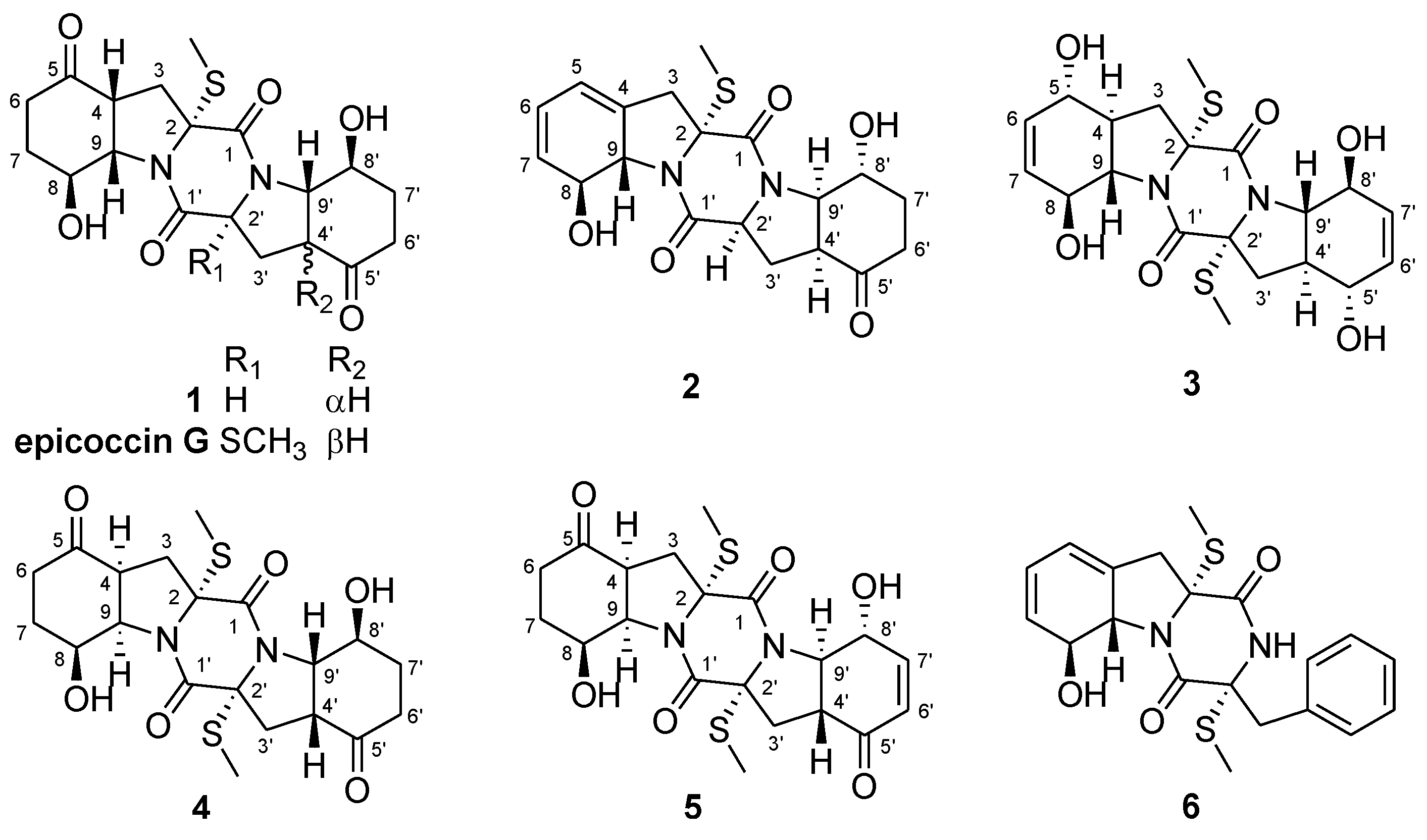
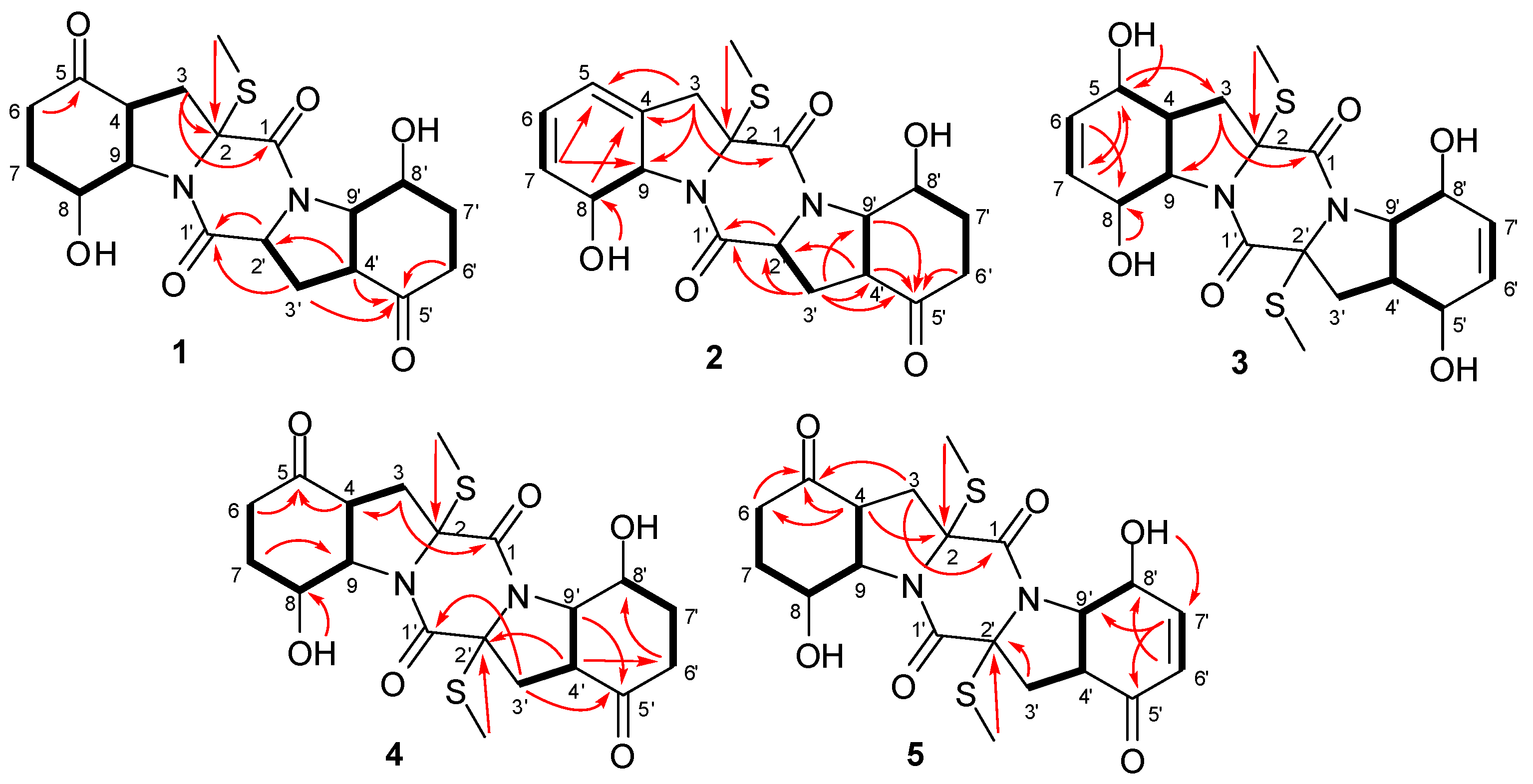
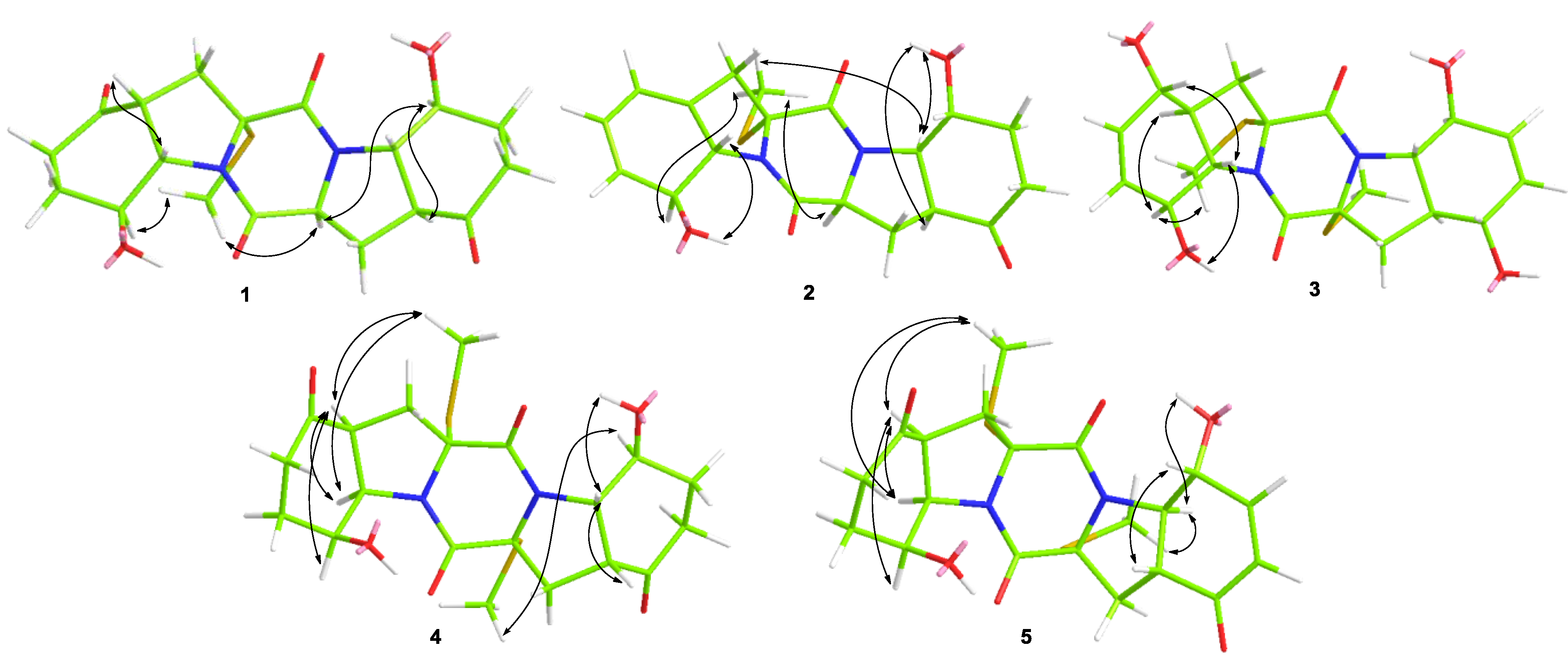
2.1. Structure Elucidation of the New Compounds

Compound 1 was obtained as colorless crystals. Its molecular formula was determined as C19H24N2O6S on the basis of HRESIMS, implying nine degrees of unsaturation. Detailed analyses of the 1H and 13C NMR data (Table 1 and Table 2, Supplementary Figures S1 and S2) indicated the presence of one methyl, six methylenes, seven sp3 methines, and five quaternary carbons (including two ketones and two ester/amide carbonyl carbons). The NMR data of 1 revealed some structural similarities to epicoccin G, a disulfide diketopiperazine isolated from the fungus Epicoccum nigrum [2,11]. However, the signal for a quaternary carbon resonating at δC 71.6 (C-2') in epicoccin G was absent in the 13C NMR spectrum of 1. Instead, resonances for a methine group at δH 4.57 (H-2') and δC 59.6 (C-2') were observed in the NMR spectra of 1 (Table 1 and Table 2). Moreover, signals for one of the two S-methyl groups (δH 1.90/δC 14.2, CH3) in epicoccin G [2] was absent in the NMR spectra of 1, indicating the lack of a S-methyl group in 1. This deduction was confirmed by the 1H-1H COSY correlations from H2-3' to H-2' and H-4' as well as by the observed HMBC correlations from H-2' and H-3' to C-1' and from H-4' to C-2', as shown in Figure 2.
Table 1. 1H NMR (500 MHz) data of compounds 15 (δ in ppm, J in Hz).
Position1 a2 b3 b4 b5 c
2.38, m (overlap)2.89, d (16.0)2.48, m (overlap)2.28, m (overlap)2.63, m (overlap)
2.32, m (overlap)3.08, d (16.0)2.08, t (12.7)2.22, m (overlap)3.07, d (13.8)
43.30, dd (8.7, 8.0) 2.30, m2.96, t (8.0)3.14, t (8.6)
5 5.93, d (2.3)4.08, d (8.8)
62.42, m (overlap)5.89, dd (9.6, 2.3)5.55,d (10.0)α 2.77, dd (13.3, 7.3) β 2.27, m (overlap)2.33, m (overlap)
7α 2.40, m (overlap) β 1.84, td (12.2, 6.2)5.59, d (9.6)5.69, d (10.0)1.90, m2.00, m
84.15, dd (13.1, 6.2)4.53, d (13.4)4.16, d (8.2)4.33, brs4.27, brs
93.57, dd (13.1, 8.0)4.69, d (13.4)3.43, dd (8.2, 11.9)4.29, d (8.0)4.51, br m
2'4.57, dd (10.4, 7.1)4.45, dd (10.3, 7.0)
3'α2.02, m (overlap)2.56, m (overlap)2.48, m (overlap)2.21, m (overlap)2.33, m (overlap)
3'β2.92, dd (13.1, 7.1)2.04, ddd (12.4, 10.3, 8.3)2.08, t (12.7)2.28, m (overlap)2.53, dd (12.9, 4.5)
4'3.24, dd (12.8, 6.7)3.12, m (overlap)2.30, m3.40, m3.47, td (13.0, 4.5)
5' 4.08, d (8.8)
6'α 2.36, m (overlap) β 2.56, ddd (17.5, 6.3, 1.8)α 2.60, m (overlap) β 2.28, dt (16.4, 5.6)5.55, d (10.0)α 2.59, ddd (18.2, 12.4, 5.2) β 2.27, m (overlap)6.01, d (10.0)
7'α 2.12, m β 2.04, m (overlap)α 1.74, m β 1.95, dt (12.5, 5.6)5.69, d (10.0)α 1.58, m β 2.27, m (overlap)6.86, d (10.0)
8'3.66, m4.36, m (overlap)4.16, d (8.2)4.00, dd (13.2, 7.4)4.60, d (8.2)
9'4.45, dd (12.8, 8.2)4.36, m (overlap)3.43, dd (8.2, 11.9)3.65,dd (13.2, 8.8)3.86, dd (13.0, 8.2)
2-SMe2.09, s2.10, s2.15, s1.93, s2.09, s
2'-SMe 2.15, s2.07, s2.22, s
5-OH 5.31, brs
5'-OH 5.31, brs
8-OH 5.68, s5.87, s5.36, brs4.83, brs
8'-OH 5.41, brs5.87, s5.89, s6.21, s
a Measured in CDCl3, b Measured in DMSO-d6, c Measured in acetone-d6.
Figure 2. Key 1H-1H COSY (bold lines) and HMBC (red arrows) correlations of compounds 15.
Marinedrugs 13 00276 g002
Table 2. 13C NMR (125 MHz) data of compounds 15 (δ in ppm).
Position1 a2 b3 b4 b5 c
1165.9, C163.7, C168.5, C169.0, C166.9, C
274.0, C74.7, C72.4, C71.1, C72.4, C
331.6, CH238.4, CH235.0, CH231.2, CH235.5, CH2
448.1, CH133.8, CH43.3, CH43.8, CH45.5, CH
5206.5, C118.7, CH68.8, CH207.4, C207.4, C
637.1, CH2123.3, CH133.3, CH34.2, CH234.8, CH2
730.8, CH2130.0, CH129.9, CH25.8, CH227.4, CH2
871.0, CH73.8, CH71.3, CH64.7, CH70.2, CH
969.9, CH68.0, CH67.8, CH63.3, CH67.2, CH
1'169.6, C169.0, C168.5, C165.2, C170.4, C
2'59.6, CH58.8, CH72.4, C72.7, C73.9, C
3'27.1, CH229.2, CH235.0, CH231.0, CH232.5, CH2
4'46.9, CH46.0, CH43.3, CH47.0, CH47.4, CH
5'207.9, C209.6, C68.8, CH206.8, C196.4, C
6'36.2, CH234.1, CH2133.3, CH36.7, CH2129.1, CH
7'26.7, CH226.6, CH2129.9, CH33.7, CH2152.6, CH
8'72.2, CH64.0, CH71.3, CH71.1, CH73.8, CH
9'68.1, CH65.0, CH67.8, CH69.0, CH69.8, CH
2-SMe14.5, CH313.2, CH314.3, CH314.1, CH314.9, CH3
2'-SMe 14.3, CH314.3, CH314.9, CH3
a Measured in CDCl3; b Measured in DMSO-d6; c Measured in acetone-d6.
The relative configuration of compound 1 was determined by analysis of its 1H-1H coupling constants and NOESY experiment. The coupling constants for H-4 and H-9 (JH-4/H-9 = 8.0 Hz) as well as for H-4' and H-9' (JH-4'/H-9' = 12.8 Hz), revealed the cis-relationship between H-4 and H-9 and trans-relationship of H-4' and H-9'. The NOE correlations from SCH3-2 to H-8, H-2', and H-8' as well as from H-8' to H-2' and H-4' indicated the same orientation of these groups (Figure 3). An X-ray crystallographic experiment (Figure 4) confirmed the structure and relative configuration of 1. The Cu/Kα radiation allowed the assignment of the absolute configuration of all the stereogenic centers in 1 as 2R, 4R, 8S, 9S, 2'S, 4'S, 8'S, and 9'S. The Electronic Circular Dichroism (ECD) spectrum of 1 showed negative Cotton Effect (CE) at 254 nm and positive CE at 218 nm (Supplementary Information). The negative CE around 254 nm is indicative for the 2R configuration of TDKPs [12]. Based on the above data, the structure of 1 was determined and it was named as penicibrocazine A.
Compound 2 was obtained as yellowish solid. The HRESIMS experiment established its molecular formula C19H22N2O5S (10 degrees of unsaturation). The 1H and 13C NMR chemical shifts for the left portion of 2 were identical to those of 6 (phomazine B) [13], whereas the right portion closely matched to that of 1. Detailed analysis of the 1H-1H COSY and HMBC correlations (Figure 2) confirmed the planar structure of 2. The relative configuration of compound 2 was determined by analysis of the NOESY spectrum. The NOE correlation from 8-OH to H-9 indicated the opposite orientation of H-8 and H-9, whereas correlations from 8'-OH to H-4' and H-9' and from 2-SMe to H-8, H-2' and H-9' indicated the same face of these groups (Figure 3). The absolute configuration of 2 was determined by comparison of its CD spectrum with that of phomazine B [13]. Three chromophoric groups including the S-methyl group, skewed diene, and an allylic hydroxy group contributed to the long-wavelength band around 270 nm [12]. Compound 2 showed similar CEs to those of phomazine B, indicating that the absolute configuration of the left portion at C-2, C-8, and C-9 of 2 are the same as those of phomazine B. These data in conjunction with chemical shifts and NOESY correlations, as compared with those of 1 from C-1' to C-9', allowed the assignment of the absolute configuration of all the stereogenic centers in 2 as 2R, 8S, 9S, 2'S, 4'S, 8'R, and 9'R. The structure of 2 was thus elucidated and it was named as penicibrocazine B.
Figure 3. NOESY correlations of compounds 15.
Marinedrugs 13 00276 g003
Figure 4. X-ray structure of compounds 1 and 3.
Marinedrugs 13 00276 g004
Penicibrocazine C (3) was obtained as colorless crystals and its molecular formula was determined as C20H26N2O6S2 (nine degrees of unsaturation) on the basis of HRESIMS data. Its 1H and 13C NMR spectroscopic data (Table 1 and Table 2) revealed the presence of one methyl, one methylene, six methines (with two olefinic and three oxygenated/nitrogenated) and two quaternary carbons (with one amide carbonyl). These data only accounted for half of the elemental composition, implying a symmetrical feature for 3. The structure of 3 was deduced from exhaustive analyses of the 1H-1H COSY and HMBC spectra. In the 1H-1H COSY spectrum, correlations from H-3 to H-4, from H-4 to H-5 and H-9, from H-8 to H-9, and from H-6 to H-7 were observed, while in the HMBC spectrum, a full set of all possible 2J and 3J correlations, such as from H-3 to C-1, C-2, C-5, and C-9, from H-6 to C-4 and C-8, from H-7 to C-5, and from 5-OH to C-5, allowed for construction of the C-1 through C-9 fragment. The chemical shift of the methyl group (δHC 2.15/14.3) and the HMBC cross-peak from the methyl protons to C-2 indicated that the methyl group was connected to the sulfur atom attached to C-2. These data in conjunction with symmetrical feature indicated the planar structure of 3 as shown in Figure 1.
The relative configuration of compound 3 was determined by analysis of its 1H-1H coupling constants and NOESY data. The large coupling constants between H-4 and H-9 (J = 11.9 Hz) revealed their trans relationship. NOE correlations from H-9 to OH-8 and H-5 in the NOESY spectrum indicated the cofacial orientation of these groups, while the observed correlations from H-8 to H-4 and 2-SMe placed these groups on the opposite face. An X-ray crystallographic experiment (Figure 4) confirmed the structure and relative configuration of 3 as depicted. The presence of sulfur in the molecule and the Flack parameter 0.02(9) allowed the assignment of the absolute configuration of all the stereogenic centers as 2R, 4S, 5S, 8S, and 9S.
Penicibrocazine D (4) was isolated as colorless solid. Its molecular formula was determined as C20H26N2O6S2 by HRESIMS data, having one SCH2 unit more than that of 1. Detailed comparison of the NMR data of 4 with those of 1 suggested that compound 4 had the same basic 6-5-6-5-6 diketopiperazine skeleton as 1. The NMR spectroscopic data (Table 1 and Table 2) differed from those of 1 mainly in the absence of the methine signal at δH 4.57 and δC 59.6 (C-2'). Instead, one S-methyl signal at δH 2.07 and δC 14.3 (2'-SMe) and one quaternary carbon signal at δC 72.7 (C-2') were observed in the NMR spectra of 4. The substitution of a S-methyl group at C-2' of 4 was evidenced by the HMBC correlation from 2'-SMe to C-2'.
The relative configuration of 4 was assigned by J-coupling constants and NOESY spectrum. The NOE correlations from 2-SMe to H-4 and H-9 and from H-8 to H-4 as well as the coupling patterns for H-4 (J = 8.0 Hz) and H-9 (J = 8.0 Hz) indicated the cofacial relationship of these groups. The large coupling constant between H-8' and H-9' (J = 13.2 Hz) revealed their trans relationship. The J coupling constant between H-9' and H-4' (J = 8.8 Hz) and NOE correlations from H-9' to H-4' and OH-8' indicated the cofacial orientation of these protons, while the correlation from H-8' to 2'-SMe placed these protons on the opposite face. As the CD spectrum of 4 exhibited negative CEs at approximately 259 nm, the absolute configuration of the α/α' chiral centers were determined to be 2R/2'R [12]. Therefore, the absolute configuration of compound 4 was assigned as 2R, 4S, 8S, 9R, 2'R, 4'R, 8'S, and 9'S.
Penicibrocazine E (5) had the molecular formula C20H24N2O6S2 as determined by HRESIMS data, with two hydrogen atoms less than 4. Detailed interpretation of the NMR data indicated that the 1H and 13C NMR chemical shifts for the left portion of 5 were nearly identical to those of 4. For the right portion, the primary difference in the NMR spectroscopic data was that the signals for two alphatic methylenes at δC 36.7/δH 2.59 and 2.27 (CH2-6') and at δC 33.7/δH 1.58 and 2.27 (CH2-7') in 4 disappear in the NMR spectra of 5. Instead, two olefinic methine signals at δC 129.1/δH 6.01 (CH-6') and δC 152.6/δH 6.86 (CH-7') were observed in the NMR spectra of 5. These observations were supported by relevant 1H-1H COSY and HMBC correlations (Figure 2). Furthermore, the resonance for C-5' was more upfield (−10.4 ppm) in the 13C NMR spectrum of 5, due to the incorporation of the conjugated double bond at C-6'.
The relative configuration of 5 was also determined by analysis of J-coupling constants and the NOESY spectrum. The coupling patterns for the relevant protons on the left portion of 5 were similar to their counterparts of 4, indicating that the left motif of 5 possessed the same relative configuration as that of 4. The large coupling constant between H-4' and H-9' (J = 13.0 Hz) revealed their trans relationship. NOE correlations from H-9' to 8'-OH and 2'-SMe placed these protons on the same face of the molecule, while the NOE correlation from H-8' to H-4' placed these protons on the opposite face. As might be expected, the CD spectrum of 5 exhibited negative CEs at approximately 260 nm, similar as that of 4, indicating the absolute configuration of 5 to be 2R, 4S, 8S, 9R, 2'R, 4'R, 8'R, and 9'R.

2.2. Biological Activities of the Isolated Compounds

The isolated compounds 16 were examined for antimicrobial activity against several human-, aqua-, and plant-pathogenic microbes as well as cytotoxicity. In the antimicrobial screening, compounds 24 and 6 showed activity against Staphylococcus aureus, with MIC values of 32.0, 0.25, 8.0, and 0.25 μg/mL, respectively, which are comparable to that of the positive control, chloromycetin (MIC = 4.0 μg/mL). Compound 3 also showed activity against Micrococcus luteus with MIC value of 0.25 μg/mL, which is stronger than that of the positive control, chloromycetin (MIC = 2.0 μg/mL). Moreover, Compounds 2 and 46 exhibited activity against plant pathogen Gaeumannomyces graminis with MIC values of 0.25, 8.0, 0.25, and 64.0 μg/mL, respectively, while the positive control amphotericin B has MIC value of 16.0 μg/mL. These data indicated that the double bonds at C-6 and C-6' increased the activity against S. aureus (3 vs. 1). In addition, more S-methyl groups likely strengthened their activity against G. graminis (4 vs. 1), and keto groups at C-5/5' (5 vs. 3) also enhanced the activity. Compounds 16 were also evaluated for cytotoxicity against eight tumor cell lines (Du145, HeLa, HepG2, MCF-7, NCI-H460, SGC-7901, SW1990, and U251), but none of them displayed potent inhibitory activity (IC50 > 10 μM).

3. Experimental Section

3.1. General

Melting points were determined with an SGW X-4 micro-melting-point apparatus. Optical rotations were measured on an Optical Activity A-55 polarimeter. UV spectra were measured using a Lengguang Gold S54 spectrophotometer (Shanghai Lengguang Technology Co. Ltd., Shanghai, China). The 1H, 13C, and 2D NMR spectra were acquired using a Bruker Avance 500 spectrometers (Bruker Biospin Group, Karlsruhe, Germany). Mass spectra were measured on a VG Autospec 3000 mass spectrometer (VG Instruments, London, UK). Semi-preparative HPLC was performed using a Dionex UltiMate U3000 system (Dionex Corporation, Sunnyvale, CA, USA) with an Agilent Prep RP-18 column (21.2 × 250 mm, 10 μm) under UV detection. Column chromatography (CC) was performed with silica gel (200–300 mesh, Qingdao Haiyang Chemical Factory, Qingdao, China), Lobar LiChroprep RP-18 (40–60 μm, Merck, Darmstadt, Germany), and Sephadex LH-20 (18–110 μm, Merck).

3.2. Fungal Material

The fungus Penicillium brocae MA-231 was isolated from the fresh tissue of the marine mangrove plant Avicennia marina. The fungus was identified by sequence analysis of the ITS region of its rDNA as described previously [10,14]. The resulting sequence data obtained from the fungal strain have been deposited in GenBank (with accession no KM191342). A BLAST search result indicated that the sequence was most similar (99%) to the sequence of Penicillium brocae (compared to AF484393). The strain is preserved at the China General Microbiological Culture Collection Center.

3.3. Fermentation

The fermentation was carried out statically in liquid potato-dextrose broth medium (PDB) (1000 mL seawater, 20 g glucose, 5 g peptone, 3 g yeast extract, and 200 g potato juice, pH 6.5–7.0, liquid medium/flask = 300 mL) in 1 L Erlenmeyer flasks for 30 days at room temperature.

3.4. Extraction and Isolation

The fermented whole broth (60 flasks) was filtered through cheesecloth to separate the culture broth and mycelia, which were exhaustively extracted with EtOAc and acetone, respectively. Since the chemical profiles of the two extracts were almost identical, they were combined and concentrated to afford the crude extract (20.0 g), which was subjected to silica gel vacuum liquid chromatography (VLC) eluting with mixed solvents of increasing polarity (petroleum ether to MeOH) to yield nine fractions (Fr.1 to Fr.9). Fraction 3 (1.5 g) was further purified by reverse-phase column chromatography (CC) over Lobar LiChroprep RP-18 with a MeOH-H2O gradient (from 20:80 to 100:0) to afford four subfractions Fr.3.1–Fr.3.4. Fr.3.2 was further purified by CC on Sephadex LH-20 (MeOH) and then by prep. TLC (plate: 20 × 20 cm, developing solvents: CHCl3/MeOH, 30:1) to obtain compounds 2 (7.9 mg) and 6 (11.9 mg). Fraction 5 (3.5 g) was further purified by reverse-phase column chromatography (CC) over Lobar LiChroprep RP–18 with a MeOH-H2O gradient (from 20:80 to 100:0) to afford six subfractions Fr.5.1–Fr.5.6. Fr.5.2 was further purified by CC on Sephadex LH-20 (MeOH), and on silica gel (eluted with CHCl3-MeOH, 100:1 to 5:1) to afford 1 (19.0 mg). Fr.5.3 was purified by CC on silica gel eluted with a CHCl3-MeOH gradient (from 100:1 to 10:1) and further purified by semi-preparative HPLC (MeOH-H2O 5:5, 16 mL/min) to afford 4 (tR = 12.1 min, 3.6 mg). Fr.5.4 was subjected to CC on silica gel eluted with CHCl3-MeOH (100:1 to 5:1) and Sephadex LH-20 (MeOH) to obtain 5 (3.0 mg). Further purification of Fr. 6 (2.5 g) by CC over Lobar LiChroprep RP-18 with a MeOH-H2O gradient (from 20:80 to 100:0) afforded six subfractions. Fr. 6-1 was further purified by CC on Sephadex LH-20 (MeOH) to afford compound 3 (17.7 mg).
Penicibrocazine A (1): colorless crystals; mp 208–210 °C; [α] 20 D : −147.8 (c 0.46, MeOH); UV (MeOH) λmax (log ε) 203 (4.60) nm; CD λmax (Δε) 218 (+3.68), 254 (−9.01) nm; 1H and 13C NMR data, see Table 1 and Table 2; ESIMS m/z 409 [M + H]+; HRESIMS m/z 409.1428 [M + H]+ (calcd for C19H25N2O6S, 409.1428, Δ 0.0 ppm).
Penicibrocazine B (2): yellowish solid; [α] 20 D : −64.0 (c 0.10, MeOH); UV (MeOH) λmax (log ε) 205 (4.12), 266 (3.65) nm; CD λmax (Δε) 222 (−15.42), 273 (+5.26) nm; 1H and 13C NMR data, see Table 1 and Table 2; ESIMS m/z 391 [M + H]+; HRESIMS m/z 391.1329 [M + H]+ (calcd for C19H23N2O2S, 391.1322, Δ 0.7 ppm).
Penicibrocazine C (3): colorless crystals; mp 150–152 °C; [α] 20 D : −133.3 (c 0.28, MeOH); UV (MeOH) λmax (log ε) 201 (4.64) nm; CD λmax (Δε) 201 (+9.58), 216 (−8.38), 234 (+0.29), 259 (−8.37) nm; 1H and 13C NMR data, see Table 1 and Table 2; ESIMS m/z 455 [M + H]+; HRESIMS m/z 455.1307 [M + H]+ (calcd for C20H27N2O6S2, 455.1305, Δ 0.2 ppm).
Penicibrocazine D (4): yellowish solid; [α] 20 D : −80.0 (c 0.14, MeOH); UV (MeOH) λmax (log ε) 206 (4.13) nm; CD λmax (Δε) 200 (−16.31), 233 (+22.79), 259 (−17.89) nm; 1H and 13C NMR data, see Table 1 and Table 2; ESIMS m/z 455 [M + H]+; HRESIMS m/z 455.1296 [M + H]+ (calcd for C20H27N2O6S2, 455.1305, Δ 0.9 ppm).
Penicibrocazine E (5): yellowish solid; [α] 20 D : −176.0 (c 0.10, MeOH); UV (MeOH) λmax (log ε) 206 (4.24) nm; CD λmax (Δε) 212 (−22.48), 236 (+16.21), 261 (−15.03) nm; 1H and 13C NMR data, see Table 1 and Table 2; ESIMS m/z 453 [M + H]+; HRESIMS m/z 453.1149 [M + H]+ (calcd for C20H25N2O6S2, 453.1145, Δ 0.4 ppm).

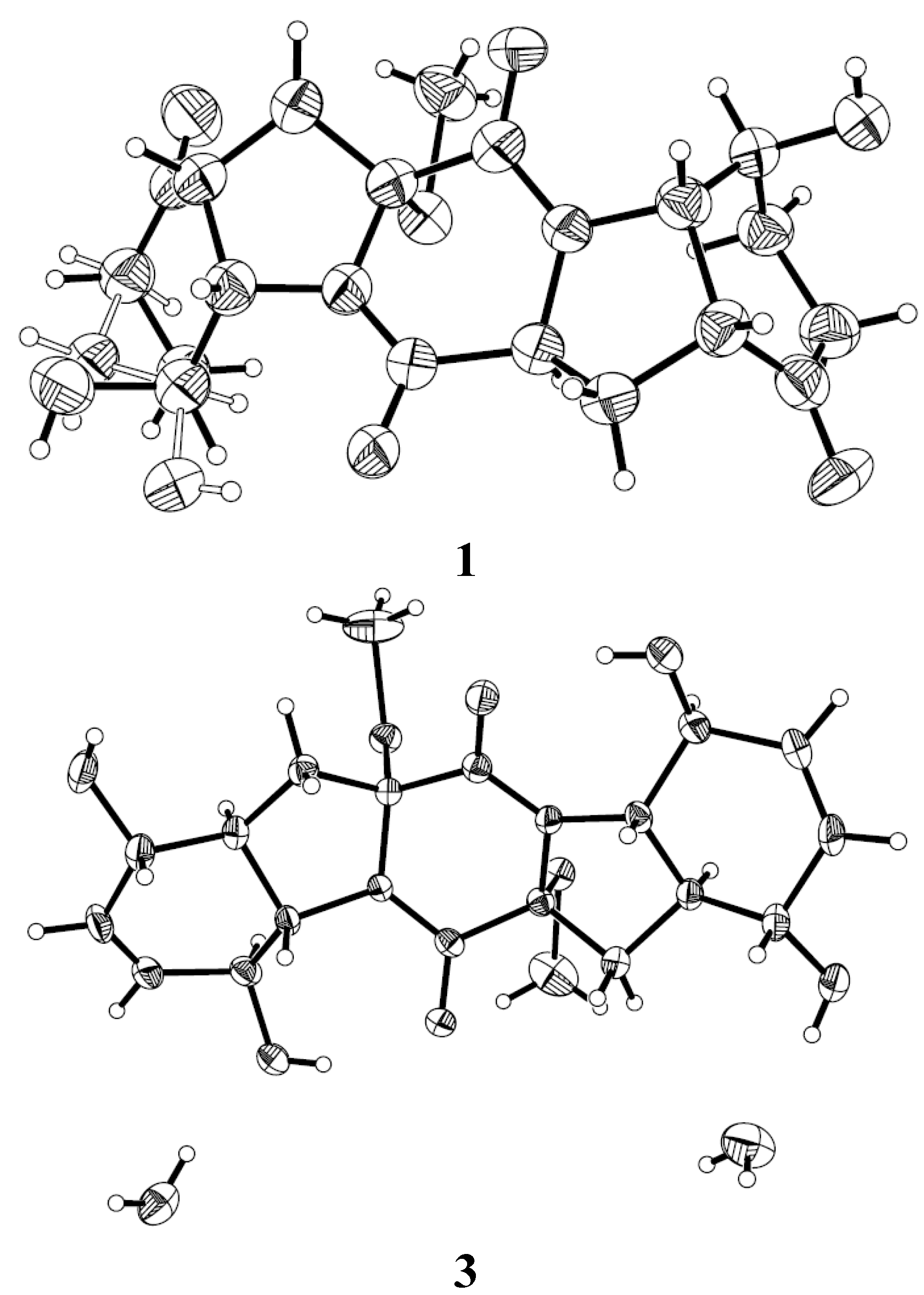
3.5. X-Ray Crystallographic Analysis of Compounds 1 and 3 [15]

All crystallographic data were collected on a Bruker Smart-1000 CCD diffractometer equipped with a graphite-monochromatic Cu Kα radiation (λ = 1.54178 Å) for 1 and Mo Kα radiation (λ = 0.71073 Å) for 3 at 293(2) K. The data were corrected for absorption by using the program SADABS [16]. The structures were solved by direct methods with the SHELXTL software package [17]. All non-hydrogen atoms were refined anisotropically. The H atoms were located by geometrical calculations, and their positions and thermal parameters were fixed during the structure refinement. The structure was refined by full-matrix least-squares techniques [18].
Crystal data for compound 1: C19H23N2O6S, F.W. (Formula Weight) = 407.45, orthorhombic space group, P2(1)2(1)2(1), unit cell dimensions a = 6.4278(12) Å, b = 13.001(3) Å, c = 23.565(4) Å, V = 1969.3(6) Å3, α = β = γ = 90°, Z = 4, dcalcd = 1.374 mg/m3, crystal dimensions 0.11 × 0.08 × 0.05 mm, μ = 1.800 mm1, F(000) = 860. The 3943 measurements yielded 2894 independent reflections after equivalent data were averaged, and Lorentz and polarization corrections were applied. The final refinement gave R1 = 0.1273 and wR2 = 0.3072 [I > 2σ(I)]. The Flack parameter was 0.02(12) in the final refinement for all 3943 reflections with 2894 Friedel pairs.
Crystal data for compound 3: C20H30N2O8S2, F.W. = 490.58, orthorhombic space group, P2(1)2(1)2(1), unit cell dimensions a = 8.3513(7) Å, b = 11.8611(12) Å, c = 24.024(3) Å, V = 2379.7(4) Å3, α = β = γ = 90°, Z = 4, dcalcd = 1.369 mg/m3, crystal dimensions 0.38 × 0.32 × 0.10 mm, μ = 0.271 mm1, F(000) = 1040. The 11941 measurements yielded 4180 independent reflections after equivalent data were averaged, and Lorentz and polarization corrections were applied. The final refinement gave R1 = 0.0445 and wR2 = 0.0893 [I > 2σ(I)].

3.6. Cytotoxicity Assay

The cytotoxic activity of the isolated compounds against nine tumor cell lines including Du145 (human carcinoma of prostate cell line), HeLa (human cervix carcinoma cell line), HepG2 (human liver hepatocellular cells), MCF-7 (human breast carcinoma cell line), NCI-H460 (human large cell lung carcinoma cell line), SGC-7901 (human gastric carcinoma cell line), SW1990 (human pancreatic cancer cell line), SW480 (human colon carcinoma cancer), and U251 (human glioma cells) were determined according to previously reported methods [19].

3.7. Antimicrobial Assay

Antimicrobial assay against human- and aqua-pathogenic microbes Aeromonas hydrophilia, Escherichia coli, Micrococcus luteus, Staphylococcus aureus, Vibrio arveyi, V. parahaemolyticus and plant pathogenic fungi Alternaria brassicae, Colletotrichum gloeosporioides, Fusarium graminearum, and Gaeumannomyces graminis was carried out using the well diffusion method [20]. Chloromycetin was used as a positive control for the bacteria, while amphotericin B was used as a positive control for the fungi.

4. Conclusions

Five new thiodiketopiperazine derivatives (15), along with one known analog (6), were isolated from the marine mangrove-derived endophytic fungus P. brocae MA-231. The structures and relative configurations were determined on the interpretation of the NMR data and the absolute configurations of all compounds were determined by CD comparison, while the structures of compounds 1 and 3 were confirmed by single-crystal X-ray diffraction analysis. Compounds 26 exhibited activity against some of the tested microbial strains.

Supplementary Files

Supplementary File 1

Acknowledgments

This work was financially supported by the National Natural Science Foundation of China (31270403) and by the Ministry of Science and Technology of China (2013AA092901 and 2010CB833802).

Author Contributions

Ling-Hong Meng and Peng Zhang performed the experiments for the isolation, structure elucidation, and antimicrobial evaluation; Ling-Hong Meng prepared the manuscript; Xiao-Ming Li performed the 1D and 2D NMR experiments; Bin-Gui Wang supervised the research work and revised the manuscript.

Conflicts of Interest

The authors declare no conflict of interest.

References

  1. Deffieux, G.; Baute, M.A.; Baute, R.; Filleau, M.J. New antibiotics from the fungus Epicoccum nigrum II. Epicorazine A: Structure elucidation and absolute configuration. J. Antibiot. 1978, 31, 1102–1105. [Google Scholar] [CrossRef] [PubMed]
  2. Guo, H.J.; Sun, B.D.; Gao, H.; Chen, X.L.; Liu, S.C.; Yao, X.S.; Liu, X.Z.; Che, Y.S. Diketopiperazines from the cordyceps-colonizing fungus Epicoccum nigrum. J. Nat. Prod. 2009, 72, 2115–2119. [Google Scholar] [CrossRef] [PubMed]
  3. Tan, R.X.; Jensen, P.R.; Williams, P.G.; Fenical, W. Isolation and structure assignments of rostratins A-D, cytotoxic disulfides produced by the marine-derived fungus Exserohilum rostratum. J. Nat. Prod. 2004, 67, 1374–1382. [Google Scholar] [CrossRef] [PubMed]
  4. Meng, L.H.; Li, X.M.; Lv, C.T.; Li, C.S.; Xu, G.M.; Huang, C.G.; Wang, B.G. Sulfur-containing cytotoxic curvularin macrolides from Penicillium sumatrense MA-92, a fungus obtained from the rhizosphere of the mangrove Lumnitzera racemosa. J. Nat. Prod. 201 2013, 76, 2145–2149. [Google Scholar] [CrossRef]
  5. An, C.Y.; Li, X.M.; Luo, H.; Li, C.S.; Wang, M.H.; Xu, G.M.; Wang, B.G. 4-Phenyl-3,4-dihydroquinolone derivatives from Aspergillus nidulans MA-143, an endophytic fungus isolated from the mangrove plant Rhizophora stylosa. J. Nat. Prod. 201 2013, 76, 1896–1901. [Google Scholar] [CrossRef]
  6. Wang, M.H.; Li, X.M.; Li, C.S.; Ji, N.Y.; Wang, B.G. Secondary metabolites from Penicillium pinophilum SD-272, a marine sediment-derived fungus. Mar. Drugs 2013, 11, 2230–2238. [Google Scholar] [CrossRef] [PubMed]
  7. Li, C.S.; Li, X.M.; Gao, S.S.; Lu, Y.H.; Wang, B.G. Cytotoxic anthranilic acid derivatives from deep sea sediment-derived fungus Penicillium paneum SD-44. Mar. Drugs 2013, 11, 3068–3076. [Google Scholar] [CrossRef] [PubMed]
  8. Liu, D.; Li, X.M.; Li, C.S.; Wang, B.G. Nigerasterols A and B, antiproliferative sterols from the mangrove-derived endophytic fungus Aspergillus niger MA-132. Helv. Chim. Acta 2013, 96, 1055–1061. [Google Scholar] [CrossRef]
  9. Du, F.Y.; Li, X.M.; Zhang, P.; Li, C.S.; Wang, B.G. Cyclodepsipeptides and O-containing heterocyclic metabolites from Beauveria felina EN-135, a marine-derived entomopathogenic fungus. Mar. Drugs 2014, 1, 2816–2826. [Google Scholar] [CrossRef]
  10. Meng, L.H.; Li, X.M.; Lv, C.T.; Huang, C.G.; Wang, B.G. Brocazines A–F, cytotoxic bisthiodiketopiperazine derivatives from Penicillium brocae MA-231, an endophytic fungus derived from the marine mangrove plant Avicennia marina. J. Nat. Prod. 201 2014, 77, 1921–1927. [Google Scholar] [CrossRef]
  11. Wang, J.M.; Ding, G.Z.; Fang, L.; Dai, J.G.; Yu, S.S.; Wang, Y.H.; Chen, X.G.; Ma, S.G.; Qu, J.; Xu, S.; et al. Thiodiketopiperazines produced by the endophytic fungus Epicoccum nigrum. J. Nat. Prod. 2010, 73, 1240–1249. [Google Scholar] [CrossRef] [PubMed]
  12. Wang, J.M.; Jiang, N.; Ma, J.; Yu, S.S.; Tan, R.X.; Dai, J.G.; Si, Y.K.; Ding, G.Z.; Ma, S.G.; Qu, J.; et al. Study on absolute configurations of α/α' chiral carbons of thiodiketopiperazines by experimental and calculated circular dichroism spectra. Tetrahedron 2013, 69, 1195–1201. [Google Scholar] [CrossRef]
  13. Kong, F.D.; Wang, Y.; Liu, P.P.; Dong, T.H.; Zhu, W.M. Thiodiketopiperazines from the marine-derived fungus Phoma sp. OUCMDZ-1847. J. Nat. Prod. 2014, 77, 132–137. [Google Scholar] [CrossRef] [PubMed]
  14. Wang, S.; Li, X.M.; Teuscher, F.; Li, D.L.; Diesel, A.; Ebel, R.; Proksch, P.; Wang, B.G. Chaetopyranin, a benzaldehyde derivative, and other related metabolites from Chaetomium globosum, an endophytic fungus derived from the marine red alga Polysiphonia urceolata. J. Nat. Prod. 2006, 69, 1622–1625. [Google Scholar] [CrossRef] [PubMed]
  15. Crystallographic Data of Compounds 1 and 3 have been Deposited in the Cambridge Crystallographic Data Centre as CCDC 1031878 for Compound 1 and CCDC 1031879 For Compound 3. Available online: http://www.ccdc.cam.ac.uk/data_request/cif (accessed on 31 October 2014).
  16. Sheldrick, G.M. SADABS, Software for Empirical Absorption Correction; University of Göttingen: Göttingen, Germany, 1996. [Google Scholar]
  17. Sheldrick, G.M. SHELXTL, Structure Determination Software Programs; Bruker Analytical X-ray System Inc.: Madison, WI, USA, 1997. [Google Scholar]
  18. Sheldrick, G.M. SHELXL-97 and SHELXS-97, Program for X-ray Crystal Structure Solution and Refinement; University of Göttingen: Göttingen, Germany, 1997. [Google Scholar]
  19. Bergeron, R.J.; Cavanaugh, P.F., Jr.; Kline, S.J.; Hughes, R.G., Jr.; Elliott, G.T.; Porter, C.W. Antineoplastic and antiherpetic activity of spermidine catecholamide iron chelators. Biochem. Biophys. Res. Commun. 1984, 121, 848–854. [Google Scholar] [CrossRef] [PubMed]
  20. Al-Burtamani, S.K.S.; Fatope, M.O.; Marwah, R.G.; Onifade, A.K.; Al-Saidi, S.H. Chemical composition, antibacterial and antifungal activities of the essential oil of Haplophyllum tuberculatum from Oman. J. Ethnopharmacol. 2005, 96, 107–112. [Google Scholar] [CrossRef] [PubMed]

