Article

Electrospinning Preparation, Structure, and Properties of Fe3O4/Tb(acac)3phen/Polystyrene Bifunctional Microfibers

1
Department of Mathematics and Physics, Luoyang Institute of Science and Technology, Luoyang 471023, China
2
School of Environmental Engineering and Chemistry, Luoyang Institute of Science and Technology, Luoyang 471023, China
*
Author to whom correspondence should be addressed.
Materials 2023, 16(12), 4409; https://doi.org/10.3390/ma16124409
Submission received: 27 March 2023 / Revised: 30 May 2023 / Accepted: 8 June 2023 / Published: 15 June 2023

Abstract

:
Compared to single functional materials, multifunctional materials with magnetism and luminescence are more attractive and promising; Thus, it has become an important subject. In our work, bifunctional Fe3O4/Tb(acac)3phen/polystyrene) microfibers with magnetic and luminescent properties (acac: acetylacetone, phen: 1,10-phenanthroline) were synthesized by simple electrospinning process. The doping of Fe3O4 and Tb(acac)3phen made the fiber diameter larger. The surface of pure polystyrene microfibers and microfibers doped only with Fe3O4 nanoparticles were chapped similar to bark, whereas the surface of the microfibers was smoother after doping with Tb(acac)3phen complexes. The luminescent properties of the composite microfibers were systematically studied in contrast to pure Tb(acac)3phen complexes, including excitation and emission spectra, fluorescence dynamics, and the temperature dependence of intensity. Compared with the pure complexes, the thermal activation energy and thermal stability of composite microfiber was significantly improved, and the luminescence of the unit mass of Tb(acac)3phen complexes in composite microfibers was stronger than that in pure Tb(acac)3phen complexes. The magnetic properties of the composite microfibers were also investigated using hysteresis loops, and an interesting experimental phenomenon was found that the saturation magnetization of the composite microfibers gradually increased with the increase in the doping proportion of terbium complexes.

1. Introduction

With the increasing demand for integrated devices, it is difficult for single-function materials to meet the needs of modern science and technology. Multifunctional materials have two or more functions. Therefore, Scientists are increasingly interested in multifunctional composite materials, owing to their wider application fields, such as nanosensors [1], drug delivery systems [2], biomedical engineering [3], and catalytic synthesis [4]. Luminescent–magnetic multifunctional materials have attracted wide attention, owing to their important applications in the medicine and biological fields [5,6,7]. Ghazimoradi et al. synthesized multifunctional core/double shells CoFe2O4/mSiO2-NH2/Poly(MAA-co-IA)/MF3 for the co-delivery of anticancer drugs (MTX and DOX) for breast cancer treatment [8]. Swain et al. synthesized multifunctional Fe3O4@BaMoO4:Eu3+ and used it for drug carrier [9]. Müssig et al. synthesized colorful luminescent magnetic supraparticles [10].
Rare earth luminescent materials exhibit unique chemical, electronic, and optical properties due to the f-f electron transition. Therefore, rare-earth luminescent materials have extensive applications on luminescence devices, displays, biolabeling, optical imaging photo-therapy, optical windows, miRNA detection [11,12,13], and so on. Magnetic nanomaterials have attracted increasing interest from scientists due to their potential applications, such as biomacromolecule separation, drug delivery and release, and MRI [14,15,16]. Among the magnetic materials, Fe3O4 has the advantages of high saturation magnetization, non-toxicity, good biocompatibility, and has a wide range of applications in the biomedical field [17,18,19]. Therefore, rare-earth luminescent materials and Fe3O4 nanoparticles have been employed as luminescent and magnetic materials in luminescent–magnetic composites [20,21].
Electrospinning is a relatively simple, straightforward, and versatile fiber-forming technology [22,23], which provides a unique way to fabricate continuous fibers or belts with 1D nanostructure [24,25]. This method shows an extensive application in filtration materials, optical fiber humidity sensors, electrode materials of Li-ion batteries, regenerative biology, cosmetics, skin regeneration, battery, filters, light-emitting diodes [20,26,27,28], and so on. In previous work, some groups have successfully fabricated luminescent–magnetic nanofibers [26,29,30] using the electrospinning method. Most of them are Janus structure fibers [20,24,26]. As far as we know, there is no report on the composite fibers of terbium complexes and Fe3O4 nanoparticles. Based on the above analysis, in this work, Fe3O4/Tb(acac)3phen/PS (polystyrene) multifunctional microfibers were synthesized via the single needle electrospinning process. The synthesis procedure of the multifunctional microfibers is shown in Figure 1. The structure and photoluminescent and magnetic properties of the multifunctional microfibers were investigated. This new kind of microfiber combines the advantages of rare-earth complexes and Fe3O4 NPs. The high-performance luminescent–magnetic bifunctional microfibers have potential applications in biomedical and biochemical fields.

2. Experimental Section

2.1. Materials

N,N-Dimethylformamide (DMF), anhydrous ethanol, ferric chloride hexahydrate (FeCl3·6H2O), ethylene glycol, anhydrous sodium acetate (NaAc), and ethylenediamine were purchased from Sinopharm Chemical Reagent Co., Ltd. (Shanghai, China). PS (molecular weight about 260,000), 1,10-phenanthroline (phen), Tetrabutylammonium chloride, acetyl acetone (acac), and terbium chloride hexahydrate (TbCl3·6H2O) were purchased from J&K Scientific (Beijing, China). The reagents used were all analytically pure and were not further purified before use. All chemicals were of analytical grade and were used as received without further purification.

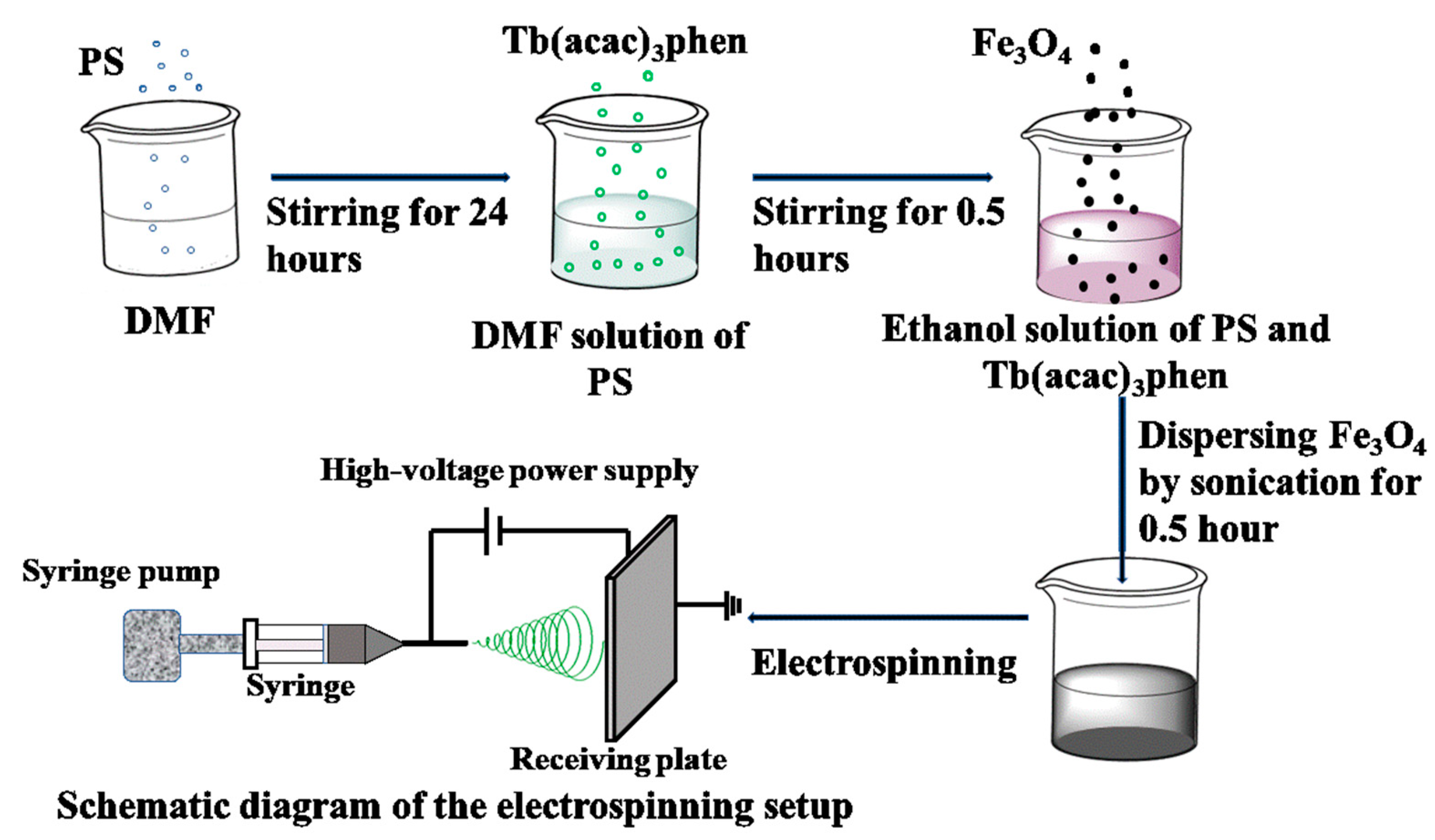
2.2. Electrospinning of Fe3O4/Tb(acac)3phen/PS Microfibers

Tb(acac)3phen complexes and Fe3O4 nanoparticles were synthesized according to the traditional method [31,32]. In the preparation of precursor solutions, N,N-dimethylformamide (DMF) was used as the solvent. In a typical synthesis, the electrospinning solution was prepared by dissolving 0.3 mg/mL PS in DMF. To prepare a uniform fiber by electrospinning, 0.015 g tetrabutylammonium chloride was added under vigorous stirring, and then the precursor was stirred for 24 h. A certain amount of Tb(acac)3phen complexes was dissolved in that solution under magnetic stirring for 10 min. The mass ratios of Tb(acac)3phen complexes to PS for various samples were 20%, 30%, 40%, 50%, respectively. A series of Fe3O4/Tb(acac)3phen/PS precursor solutions with different Tb(acac)3phen complex contents were prepared by dispersing 0.1 g Fe3O4 nanoparticles into the solution using ultrasonic method.
Functional microfibers were prepared by the electrospinning method. The electrospinning process was carried out at room temperature and relative humidity, similar to previous literatures [29,33]. The syringe was placed on a syringe pump and used at a flow rate of 0.6 mL/h−1. The voltage was 15 kV, and the distance between the collecting plate and the needle was 25 cm. The bifunctional microfibers with Tb(acac)3phen accounting for 20%, 30%, 40%, and 50% of PS mass were labeled as samples 20Fe20Tb, 20Fe30Tb, 20Fe40Tb, and 20Fe50Tb, respectively. For comparison, we prepared pure PS microfibers, microfibers containing only 20% Fe3O4 nanoparticles, and microfibers containing only 40% Tb(acac)3phen complexes. The three samples were labeled as PS, 20Fe0Tb, and 0Fe40Tb, respectively. The mass ratios of Tb(acac)3phen complexes and Fe3O4 nanoparticles to PS in different samples are shown in Table 1.

2.3. Characterization

The phase structure of Fe3O4 nanoparticles and the bifunctional microfibers was performed by X-ray diffraction (XRD) (D8 ADVANCE, Cu Kα, λ = 1.5418 Å, Bruker, Karlsruhe, Germany). The SEM (scanning electron microscope) images and EDAX (energy spectrum) was measured on a Hitachi SU8020 microscope (Hitachi, Ltd., Tokyo, Japan, 3 KV). TEM (transmission electron microscope) was performed using a FEI Tecnai G2 F30 S-TWIN electron microscope (FEI, Inc., Valley City, ND, USA, 300 KV). The IR absorption spectra were measured in the range 400–4000 cm−1 using an FT-IR (Fourier transform infrared) spectrophotometer (Nicolet Niccolet IS10, Thermo Scientific™, Waltham, MA, USA). The fluorescence dynamics, dependence of emission intensity on temperature (100 K, 150 K, 200 K, 250 K, 300 K, and 350 K), and excitation and emission spectra were recorded using an Edinburgh FLS1000 spectrophotometer (Edinburgh Instruments Ltd., Livingstone, UK). The step was 1 nm, and the power of xenon lamp was 300 W. The conductivity data were measured on ST2643 ultra high resistance tester (Suzhou Jingge Electronic Co., Ltd., Suzhou, China) and the test voltage was 100 V. The hysteresis loops of samples (room temperature) were measured on a vibrating sample magnetometer (MPMS-3) manufactured by Quantum Design, Inc. (San Diego, CA, USA), and the range of magnetic field was −30,000 Oe to 30,000 Oe.

3. Results and Discussion

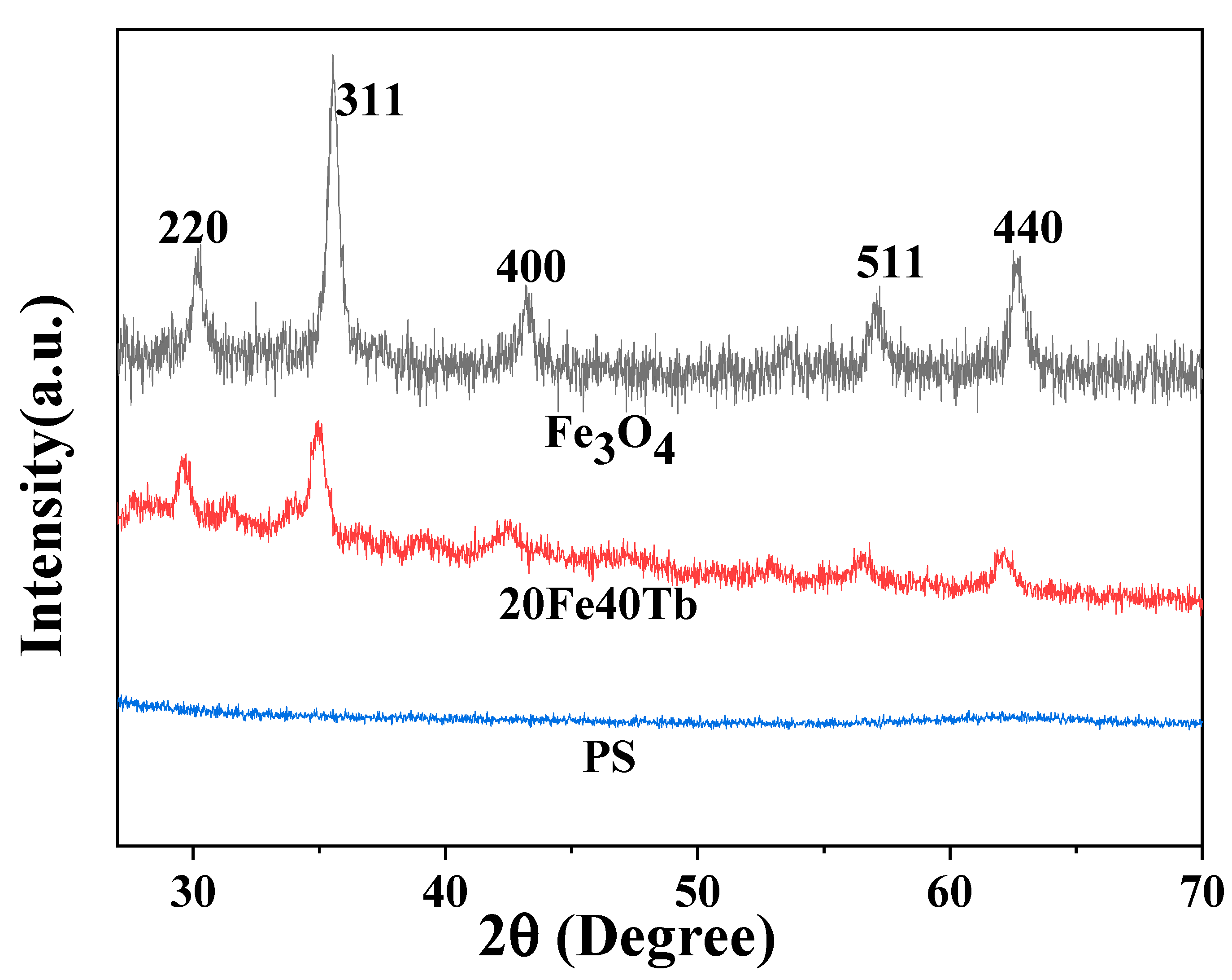
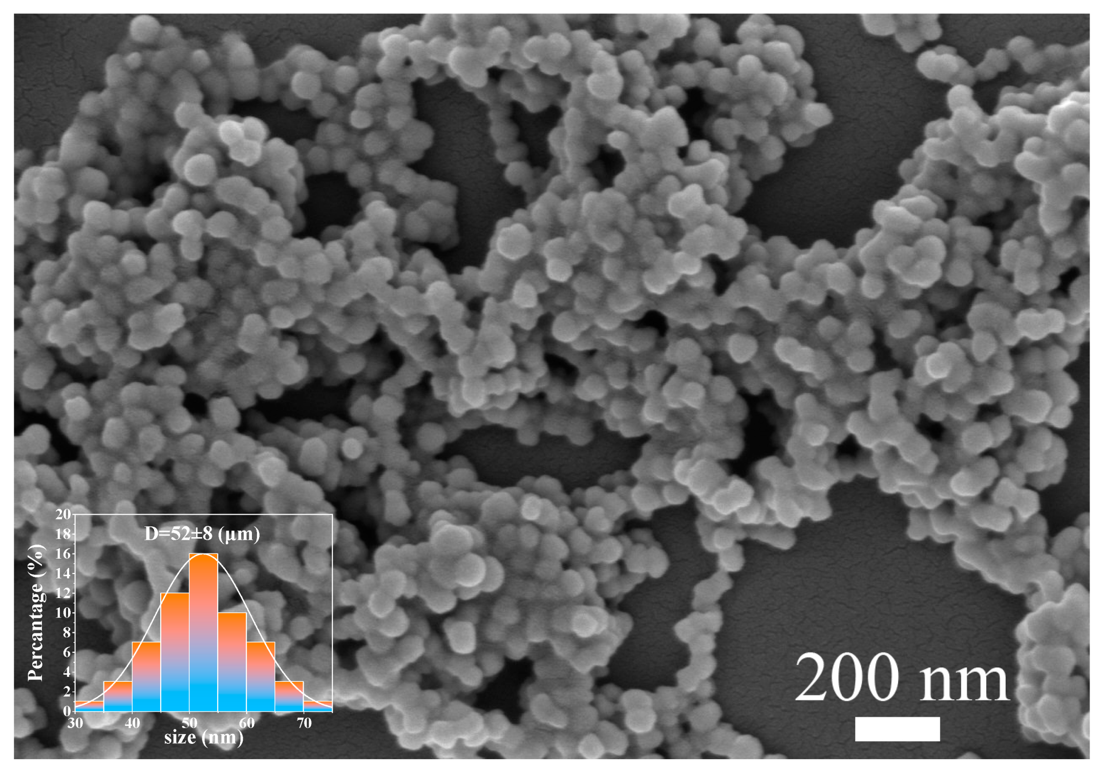
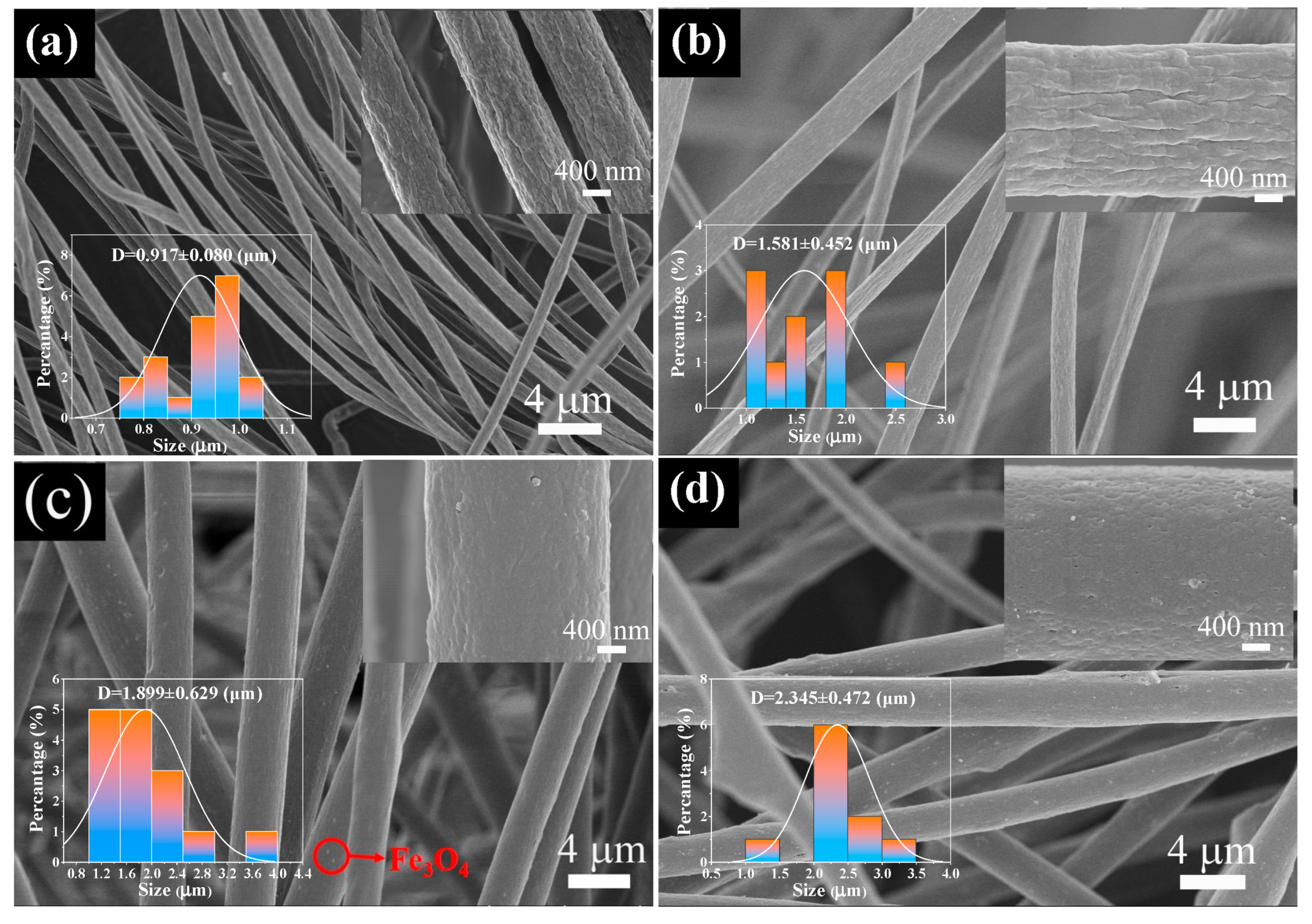
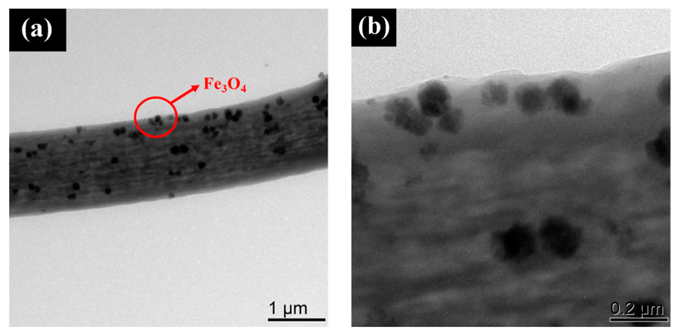
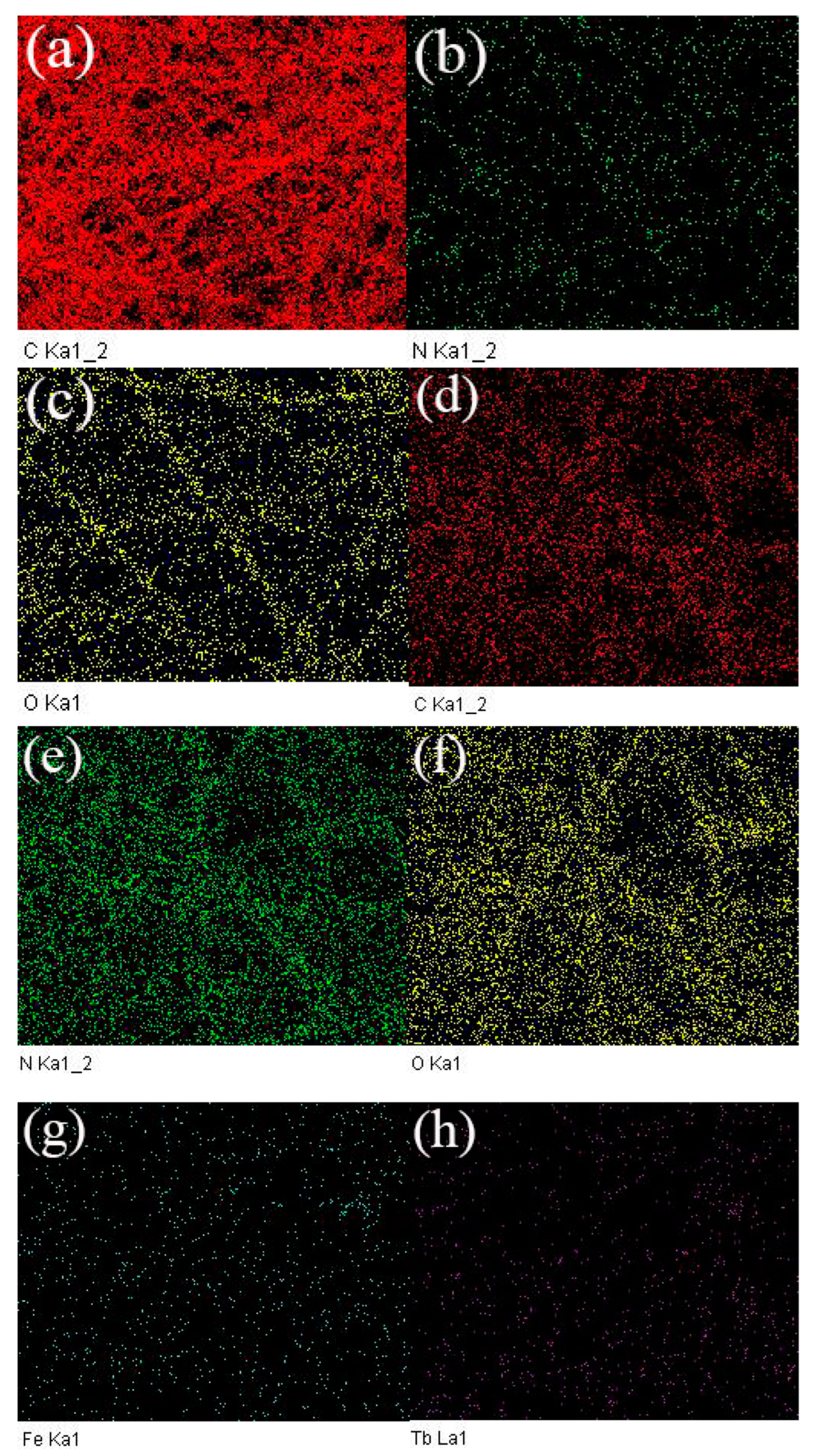
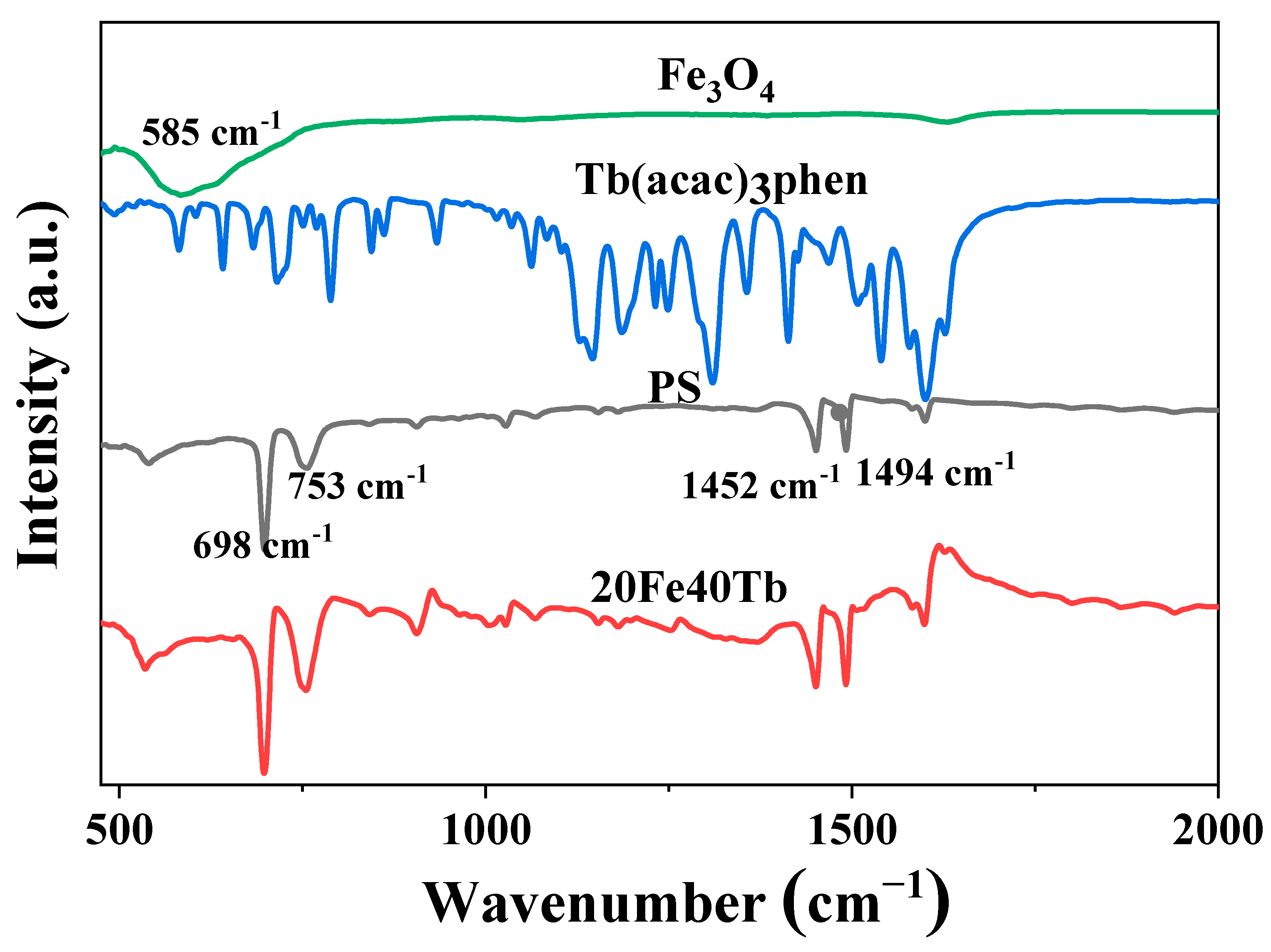
3.1. Structure and Morphology of Bifunctional Fe3O4/Tb(acac)3phen/PS Microfibers

The XRD patterns of sample 20Fe40Tb, Fe3O4 nanoparticles, and PS microfibers are shown in Figure 2. All diffraction peaks for Fe3O4 nanoparticles could be readily indexed to the face-centered cubic phase of Fe3O4, according to the Joint Committee on Powder Diffraction Standards (JCPDS) file on JCPDS 89-0688 [34]. The diffraction peaks of Fe3O4 were observed in sample 20Fe40Tb, which indicated that Fe3O4 nanoparticles were doped into the composite microfibers. The XRD pattern of PS microfibers showed no diffraction peaks, indicating that the PS microfibers were amorphous [29].
The SEM image of Fe3O4 nanoparticles are shown in Figure 3. The average diameter of Fe3O4 nanoparticles was estimated to be 52 nm. Figure 4 shows the SEM images of pure PS microfibers and samples 20Fe0Tb, 20Fe40Tb, and 20Fe50Tb. As can be seen from the figure, the diameters of pure PS microfibers were uniformly distributed, with an average diameter of about 0.9 µm. The average diameters of samples 20Fe0Tb, 20Fe40Tb, and 20Fe50Tb were about 1.6, 1.9, and 2.3 µm, respectively. The results indicated that the doping of Fe3O4 and Tb(acac)3phen made fiber diameters larger. It can be seen from the high-resolution SEM images of microfibers that the surfaces of pure PS fibers and fibers doped only with Fe3O4 nanoparticles were chapped similar to bark. The fiber surface became smooth, and the cracking phenomenon was weakened after doping with Tb(acac)3phen complexes. The electrospinning precursor solution conductivity values of the pure PS fibers and sample 0Fe40Tb were 4.00 × 10⁻⁶ and 1.23 × 10⁻⁷ S/cm, indicating that the doping of Tb(acac)3phen complexes reduced the conductivity of the electrospinning precursor solution, which influenced the fibers’ diameters and the fibers’ surface smoothness properties. As can be seen from Figure 4c,d, there are some white spots on the surface of fibers, which should be Fe3O4 nanoparticles doped into the fibers.
In order to further characterize the successful doping of Fe3O4 nanoparticles into the composite microfibers, TEM images of sample 20Fe40Tb are given in Figure 5. We could clearly see the Fe3O4 nanoparticles in the composite microfibers. The SEM-EDAX mapping of pure PS microfibers and sample 20Fe40Tb are shown in Figure 6, in which the distributions of C, N, O, Fe, and Tb elements were shown. The outline of the microfibers could be roughly seen from the element distribution of C, N, and O. Fe and Tb elements were almost evenly distributed in sample 20Fe40Tb, indicating the uniform distributions of Fe3O4 nanoparticles and Tb(acac)3phen complexes in the bifunctional fibers.
The FTIR spectra of pure Tb(acac)3phen complexes, PS fibers, Fe3O4 nanoparticles, and sample 20Fe40Tb are shown in Figure 7. The characteristic absorption band of the Fe-O bond was at 585 nm in Fe3O4 nanoparticles. The IR spectra of the bifunctional microfibers were similar to that of pure PS fibers, and no characteristic absorption peaks of the terbium complexes and Fe3O4 nanoparticles can be seen, indicating that most of the Tb(acac)3phen complexes and Fe3O4 nanoparticles in the microfibers were capped in the PS matrix [35,36]. For pure PS and bifunctional microfibers, the absorption peaks at 698, 753, 1452, and 1494 cm−1 were assigned to vibration absorption of benzene ring. These peaks maintained the same positions for the PS and bifunctional microfibers, indicating that Tb(acac)3phen complexes and Fe3O4 nanoparticles had little chemical interaction with PS and only physically mixed with it [35].

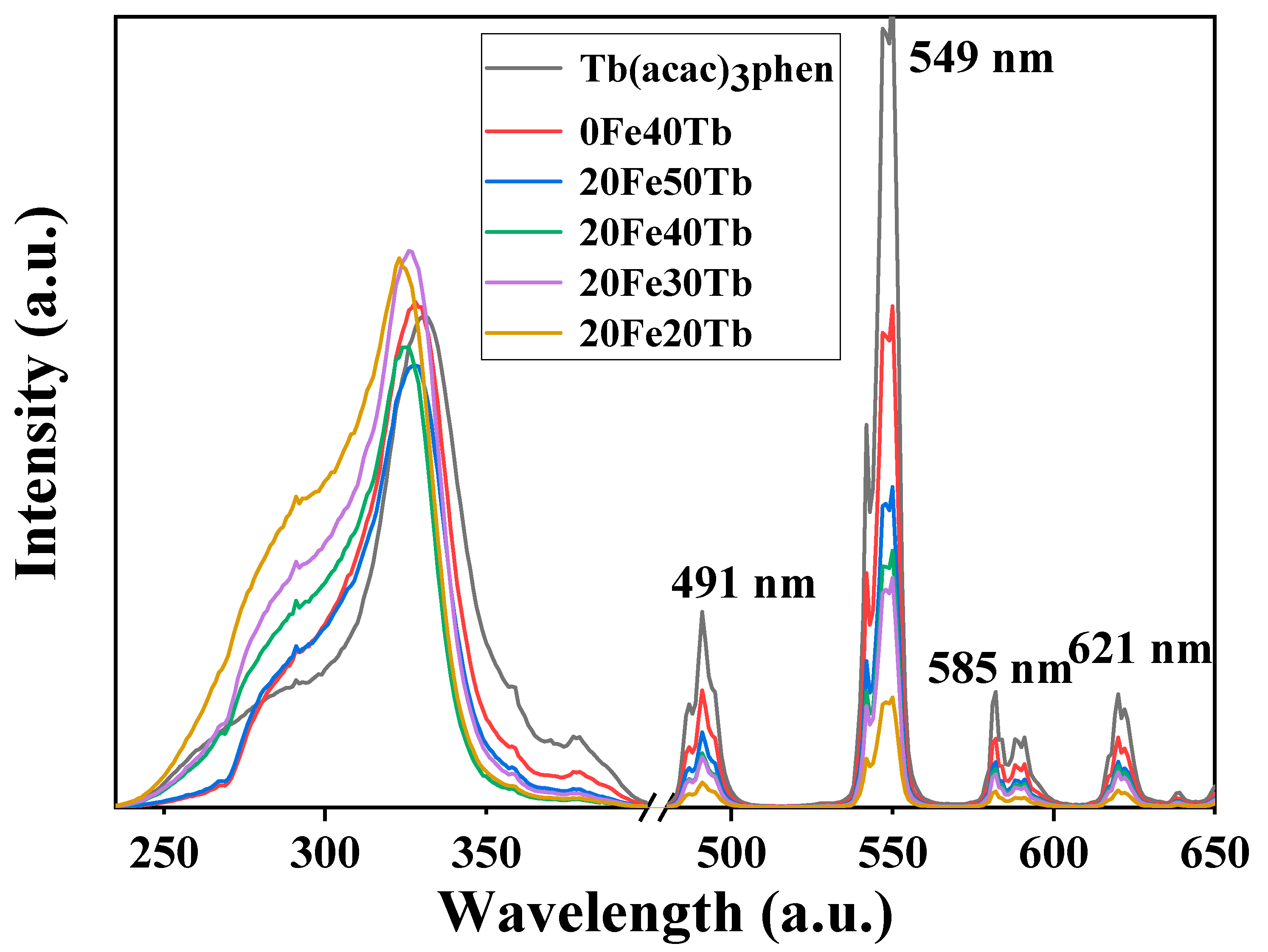
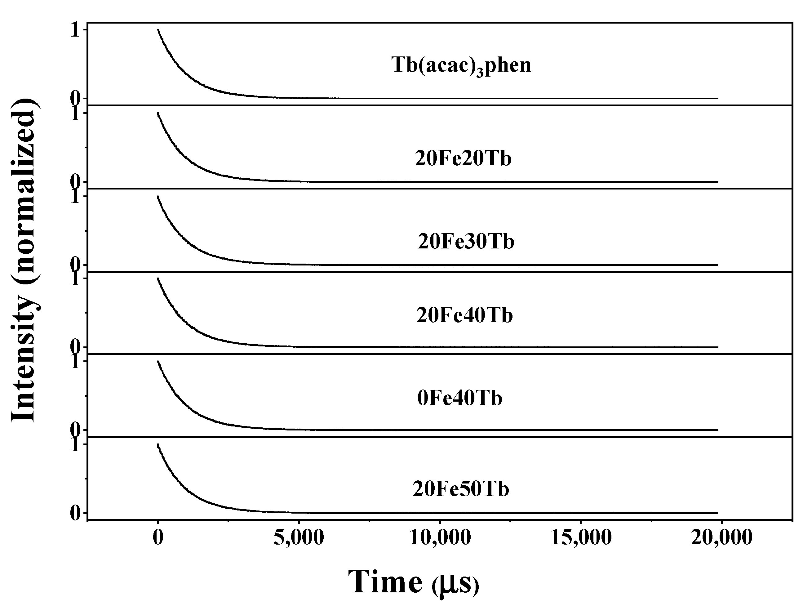
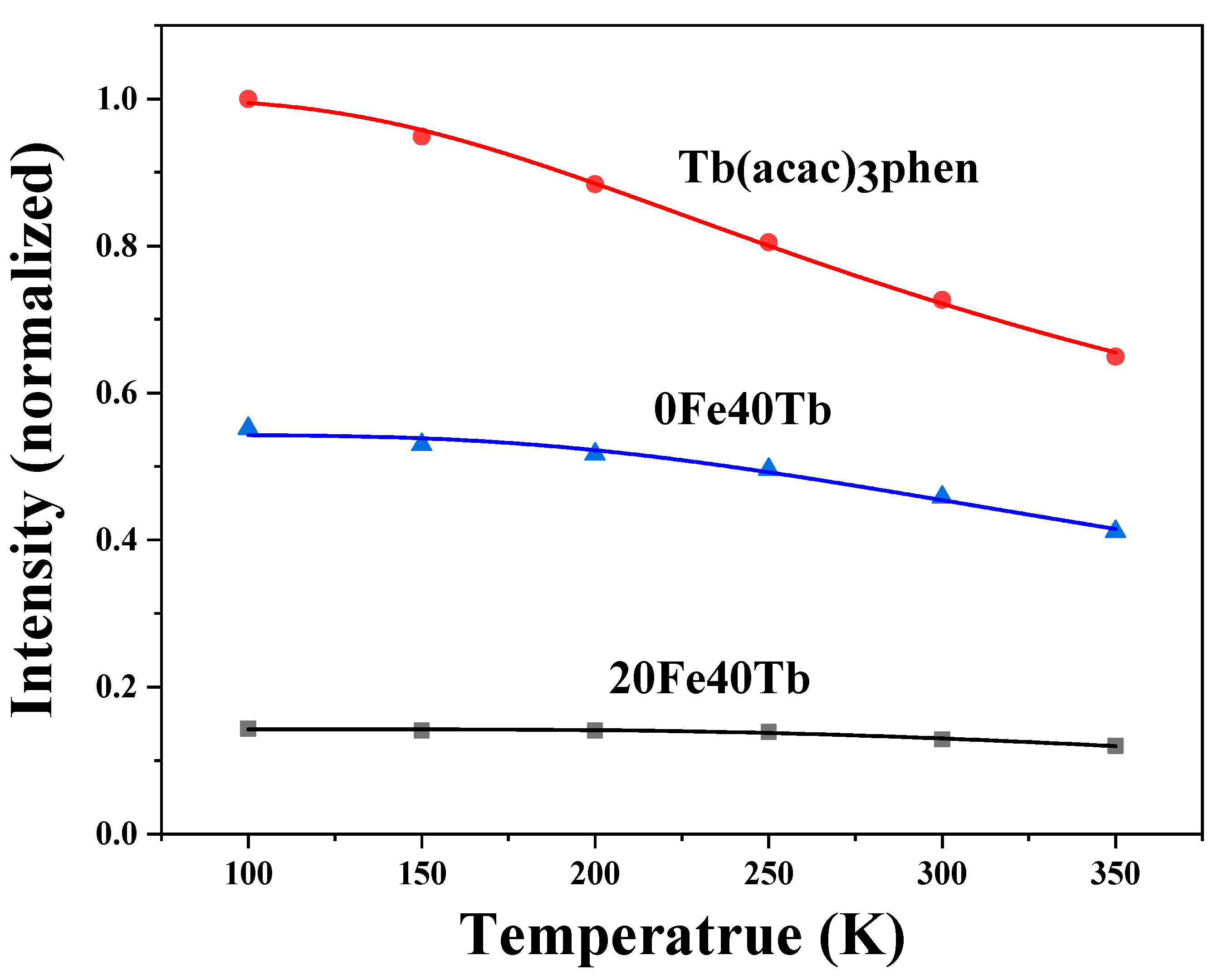
3.2. Photoluminescence Properties of Bifunctional Microfibers

The photoluminescence properties of samples with different doping concentrations of Tb(acac)3phen complexes were studied and compared them with pure terbium complexes and the bifunctional microfibers without Fe3O4 nanoparticles. Figure 8 shows the excitation and emission spectra of different samples. It could be seen from the excitation spectra that a broad excitation band ranging from 200 to 400 nm existed in all the excitation spectra, which corresponded to the π-π* electron transition of ligands [32]. The excitation peaks of the bifunctional fibers and the pure Tb(acac)3phen complexes did not completely overlap, and the excitation peaks of the bifunctional fibers blue shifted a small amount, which may have been caused by the change in the environment around the ligand [32]. The emission spectra indicated that all samples exhibited characteristic emission of terbium ion, and the main peak position was 549 nm, corresponding to the 5D4-7F5 transition of Tb3+. The shape and position of the emission spectra of all samples were similar, but the intensities were significantly different. The pure terbium complexes had the strongest emission spectrum, followed by the bifunctional fibers without the addition of Fe3O4 nanoparticles. Both ultraviolet and visible light can be absorbed by black Fe3O4 nanoparticles [20], indicating weaker emission spectra of the bifunctional fibers with the addition of Fe3O4 nanoparticles. When the doping ratios of the Fe3O4 nanoparticles were the same, the intensities of emission spectra gradually increased when adding more Tb(acac)3phen complexes.
In order to further investigate the photoluminescence properties of different samples, the relative luminescence intensities of samples 0Fe40Tb, 20Fe40Tb, and the pure Tb(acac)3phen complexes were calculated and are listed in Table 2. By comparing the intensities and concentration of different samples, we can see that the luminescence values of the unit mass of the Tb(acac)3phen complexes in samples 0Fe40Tb and 20Fe40Tb were stronger than that in the pure Tb(acac)3phen complexes, which meant that the outer luminescence efficiency values in samples 0Fe40Tb and 20Fe40Tb were improved [32]. Owing to the strong absorption of light by Fe3O4 nanoparticles, the luminescence of the unit mass of Tb(acac)3phen complexes in sample 20Fe40Tb was weaker than that in sample 0Fe40Tb but was still stronger than that in the pure Tb(acac)3phen complexes. In the pure complexes, a large part of excited-state energy was transferred into vibration transition energy because of strong electron–phonon coupling. When the Tb complexes were incorporated into the PS matrix, vibration transition was suppressed by PS. Therefore, more energy was transferred to Tb3+, leading to the improvement of photoluminescence.
Figure 9 shows the fluorescence decay curves of 5D4-7F5 emissions for Tb3+ under 305 nm excitation (room temperature). The 5D4-7F5 emissions decay exponentially in all samples. The exponential lifetimes for the 5D4 states are obtained to be 975 µs in the pure Tb(acac)3phen complexes, 965 µs in sample 20Fe20Tb, 989 µs in sample 20Fe30Tb, 977 µs in sample 20Fe40Tb, 984 µs in sample 0Fe40Tb, and 974 µs in sample 20Fe50Tb. The fluorescence lifetimes for the 5D4 state were almost the same in all samples. This phenomenon was contrary to previous references [32,35,37], which may have been caused by the large doping proportions of the terbium complexes in the bifunctional fibers.
In order to compare the thermal stability of the photoluminescence of the pure Tb complexes and the Tb complexes in PS fibers, the temperature dependence of the photoluminescence intensities of the pure Tb complexes and the samples 0Fe40Tb and 20Fe40Tb were measured under 326 nm excitation and in a temperature range of 100–350 K. The temperature-dependent emission intensities of different samples are shown in Figure 10. For all samples, the 5D4-7F5 emission intensity of Tb3+ decreased monotonically with increasing temperature in the 100–350 K range. Compared with the pure Tb complexes, the emission intensities of 5D4-7F5 of Tb3+ in samples 0Fe40Tb and 20Fe40Tb decreased more slowly. As a function of temperature, the emission intensity was well fitted by the thermal activation equation, which could be written as [38]
I T = I 0 1 + α e E A / K B T
where E A is the activation energy of the thermal quenching process, κ Β is Boltzmann’s constant, α is the proportional coefficient, I 0 is the emission intensity at 0 K, and T is the absolute temperature. By fitting the experimental data using the above Equation, the thermal activation energy E A values of the pure terbium complexes and the samples 0Fe40Tb and 20Fe40Tb were 56.5, 82.9, and 125 meV, respectively. Compared with pure terbium complexes, the thermal activation energy of the bifunctional fibers was significantly improved, which indicated that the photoluminescence temperature stability was higher than that of pure Tb complexes [36]. In the bifunctional fibers, the PS matrix limited the ligand vibrations and reduced the non-radiative transition caused by ligand vibrations, resulting the improved luminescence thermal stability of the terbium complexes in the bifunctional fibers.

3.3. Magnetic Properties of Bifunctional Microfibers

In order to characterize the magnetic properties of bifunctional fibers, Hysteresis loops of different samples are shown in Figure 11. The Y-axis value of pure Fe3O4 nanoparticles was contracted to a fifth of its original value. As can be seen from Figure 11, all samples were superparamagnetic. When the external magnetic field was added, the samples were magnetic, and the magnetism of the samples disappeared when the external magnetic field was removed, which made it possible for them to be applied in the biomedical field [39,40]. The saturation magnetization values of the Fe3O4 nanoparticles, samples 20Fe0Tb, 20Fe30Tb, 20Fe40Tb, and 20Fe50Tb, were 80.0, 1.1, 4.1, 6.1, and 7.4 emu/g, respectively. The reduced proportions of the Fe3O4 in bifunctional microfibers lead to smaller saturation magnetization properties. Interestingly, the saturation magnetization of the bifunctional microfibers with terbium complex doping was larger than that of bifunctional microfibers without terbium complex doping. Furthermore, the saturation magnetization of the bifunctional fibers gradually increased with the increase in the doping ratios of terbium complexes.

4. Conclusions

Fe3O4/Tb(acac)3phen/PS bifunctional microfibers with both magnetic and optical properties were prepared by electrospinning, and the structure, photoluminescence, and magnetic properties of the bifunctional microfibers were systematically studied. The experimental results indicated that Fe3O4 nanoparticles were successfully doped into PS microfibers. The characteristic emission peaks of Tb(acac)3phen complexes were observed in all bifunctional microfibers, which were located at 491, 549, 585, and 621 nm. The pure terbium complexes had the strongest emission spectrum, followed by the bifunctional fibers without the addition of Fe3O4 nanoparticles. The unit mass of the Tb(acac)3phen complexes in bifunctional microfibers was 1.2 times the luminescence intensity of that in pure Tb(acac)3phen complexes. The thermal activation energy E A of the pure terbium complexes and the bifunctional microfibers was 56.5 and 125 meV, indicating that the photoluminescence temperature stability was higher than that of pure Tb complexes. The saturation magnetization of the bifunctional fibers gradually increased with the increase in the doping ratios of terbium complexes. In our work, the optical and magnetic properties of Tb complexes and Fe3O4 nanoparticles were improved through combining, which made it possible for the bifunctional microfibers to be applied in biomedical and biochemical fields such as targeted drug delivery, magnetic resonance imaging, early diagnosis, and therapy of tumor cells.

Author Contributions

Formal analysis, R.Q. and K.W.; Investigation, J.W. and Y.W.; Data curation, X.W., H.W. and Y.C.; Writing—original draft, X.F.; Writing—review & editing, L.L. All authors have read and agreed to the published version of the manuscript.

Funding

This research was funded by the National Natural Science Foundation of China (Grant No. 11905096), the Natural Science Foundation of Henan Province (Grant Nos. 232300420124, 22A430030, 22A140024, 222300420241, 222300420240, and 23A140022).

Data Availability Statement

Not applicable.

Acknowledgments

The authors acknowledge the financial support from the National Natural Science Foundations of China (No. 11905096) and the Natural Science Foundations of Henan province (No. 232300420124, 22A430030, 22A140024, 222300420241, 222300420240, and 23A140022).

Conflicts of Interest

The authors declare no conflict of interest.

References

  1. Sun, F.; Qi, H.N.; Xie, Y.R.; Da, X.; Shao, H.; Yu, W.S.; Li, F.; Dong, X.T. Conjugated Electrospinning-made Heterostructured TiO2//Bi2WO6 Janus Nanofibers for Ethanol Gas Sensing. Mater. Lett. 2023, 336, 133904. [Google Scholar] [CrossRef]
  2. Tiwari, A.; Singh, A.; Debnath, A.; Kaul, A.; Garg, N.; Mathur, R.; Singh, A.; Randhawa, K. Multifunctional Magneto-fluorescent Nanocarriers for Dual Mode Imaging and Targeted Drug Delivery. ACS Appl. Nano Mater. 2019, 2, 3060–3072. [Google Scholar] [CrossRef]
  3. Santos, D.M.D.; Dias, L.M.; Surur, A.K.; Moraes, D.A.D.; Pavarina, A.C.; Fontana, C.R.; Correa, D.S. Electrospun Composite Bead-on-string Nanofibers Containing CaO2 Nanoparticles and MnO2 Nanosheets as Oxygen-release Systems for Biomedical Applications. ACS Appl. Nano Mater. 2022, 5, 14425–14436. [Google Scholar] [CrossRef]
  4. Lidston, C.A.L.; Severson, S.M.; Abel, B.A.; Coates, G.W. Multifunctional Catalysts for Ring-opening Copolymerizations. ACS Catal. 2022, 12, 11037–11070. [Google Scholar] [CrossRef]
  5. Zink, J.L.; Lin, F.C. Probing the Local Nanoscale Heating Mechanism of a Magnetic Core in Mesoporous Silica Drug-delivery Nanoparticles using Fluorescence Depolarization. J. Am. Chem. Soc. 2020, 142, 5212–5220. [Google Scholar]
  6. Xia, L.Y.; Li, X.L.; Zhu, F.J.; Hu, S.H.; Huang, L. Luminescent and Magnetic α-Fe2O3@Y2O3: Eu3+ Bifunctional Hollow Microspheres for Drug Delivery. J. Phys. Chem. C 2017, 121, 20279–20286. [Google Scholar] [CrossRef]
  7. Jiang, Z.Y.; Peng, H.X.; Chen, W.H.; Yu, F.B. A Novel Multifunctional Carrier with Magnetic-NIR Luminescent-microwave Heating Characteristics for Drug Delivery. J. Drug Deliv. Sci. Technol. 2023, 79, 104106. [Google Scholar] [CrossRef]
  8. Ghazimoradi, M.; Tarlani, A.; Alemi, A.; Hamishehkar, H.; Ghorbani, M. pH-Responsive, Magnetic-luminescent Core/Shell carriers for Co-delivery of Anticancer Drugs (MTX & DOX) for Breast Cancer Treatment. J. Alloys Compd. 2023, 936, 168257. [Google Scholar]
  9. Swain, S.K.; Phaomer, G.; Triphathy, S.K.; Yaiphaba, N.B.; Devi, R.B.; Nayak, S.; Parida, B.B. Effect of β-cyclodextrin Decoration on Structural, Optical and Magnetic Properties of Luminescent Magnetic Nanoparticles and its Application as a Drug Carrier. J. Mol. Struct. 2022, 1247, 131330. [Google Scholar] [CrossRef]
  10. Müssig, S.; Reichstein, J.; Miller, F. Colorful Luminescent Magnetic Supraparticles: Expanding the Applicability, Information Capacity, and Security of Micrometer-Scaled Identification Taggants by Dual-Spectral Encoding. Small 2022, 18, 2107511. [Google Scholar] [CrossRef]
  11. Wang, L.D.; Fang, P.Y.; Zhao, Z.F.; Huang, Y.Y.; Liu, Z.W.; Bian, Z.Q. Rare Earth Complexes with 5d–4f Transition: New Emitters in Organic Light-Emitting Diodes. J. Phys. Chem. Lett. 2022, 13, 2686–2694. [Google Scholar] [CrossRef]
  12. Liu, X.; Zhang, S.Q.; Cheng, Z.H.; Wei, X.; Yang, T.; Yu, Y.L.; Chen, M.L.; Wang, J.H. Highly Sensitive Detection of MicroRNA-21 with ICPMS via Hybridization Accumulation of Upconversion Nanoparticles. Anal. Chem. 2018, 90, 12116–12122. [Google Scholar] [CrossRef]
  13. Feng, P.F.; Yang, X.X.; Feng, X.X.; Zhao, G.D.; Li, X.C.; Gao, J.; Tang, Y.; Yan, C.H. Highly Stable Perovskite Quantum Dots Modified by Europium Complex for Dual-Responsive Optical Encoding. ACS Nano 2021, 15, 6266–6275. [Google Scholar] [CrossRef]
  14. Mohsin, Z.; Zaman, W.Q.; Aslam, S.M.; Shan, A.; Dai, Y.C.; Khan, I.M.; Niai, S.; Zhuang, Y.P.; Guo, M.J. Recent Advances of Magnetic Nanomaterials for Bioimaging, Drug Delivery, and Cell Therapy. ACS Appl. Nano Mater. 2022, 5, 10118–10136. [Google Scholar] [CrossRef]
  15. Chen, Z.W.; Zhu, C.D.; Yang, J.W.; Zhang, M.Y.; Yuan, J.M.; Shen, Y.; Zhou, J.W.; Huang, H.; Xu, D.S.; Crommen, J.; et al. Inside-Out Oriented Choline Phosphate-Based Biomimetic Magnetic Nanomaterials for Precise Recognition and Analysis of C-Reactive Protein. Anal. Chem. 2023, 95, 3532–3543. [Google Scholar] [CrossRef] [PubMed]
  16. Park, J.W.; Bae, K.H.; Kim, C.; Park, T.G. Clustered Magnetite Nanocrystals Cross-Linked with PEI for Efficient siRNA Delivery. Biomacromolecules 2011, 12, 457–465. [Google Scholar] [CrossRef] [PubMed]
  17. Bao, J.; Chen, W.; Liu, T.T.; Zhu, Y.L.; Jin, P.Y.; Wang, L.Y.; Liu, J.F.; Wei, Y.G.; Li, Y.D. Bifunctional Au-Fe3O4 Nanoparticles for Protein Separation. ACS Nano 2007, 1, 293–298. [Google Scholar] [CrossRef] [PubMed]
  18. Kim, J.R.; Tran, V.T.; Oh, S.; Kim, C.S.; Hong, J.C.; Kim, S.; Joo, Y.S.; Mun, S.; Kim, M.H.; Jung, J.W.; et al. Scalable Solvothermal Synthesis of Superparamagnetic Fe3O4 Nanoclusters for Bioseparation and Theragnostic Probes. ACS Appl. Mater. Interfaces 2018, 10, 41935–41946. [Google Scholar] [CrossRef]
  19. Lin, L.S.; Cong, Z.X.; Cao, J.B.; Ke, K.M.; Peng, Q.L.; Gao, J.H.; Yang, H.H.; Liu, G.; Chen, X.Y. Multifunctional Fe3O4@Polydopamine Core–Shell Nanocomposites for Intracellular mRNA Detection and Imaging-Guided Photothermal Therapy. ACS Nano 2014, 8, 3876–3883. [Google Scholar] [CrossRef] [Green Version]
  20. Tian, J.; Ma, Q.L.; Yu, W.S.; Li, D.; Dong, X.T.; Liu, G.X.; Wang, J.X. Preparation of Janus Microfibers with Magnetic and Fluorescence Functionality via Conjugate Electro-Spinning. Mater. Design 2019, 170, 107701. [Google Scholar] [CrossRef]
  21. Ma, Q.L.; Dong, X.T.; Li, D.; Yu, W.S.; Wang, J.X.; Liu, G.X. Peculiarly Structured Janus Nanofibers Display Synchronous and Tuned Trifunctionality of Enhanced Luminescence, Electrical Conduction, and Superparamagnetism. ChemPlusChem 2018, 83, 108–116. [Google Scholar]
  22. Qi, H.N.; Wang, G.Y.; Hu, Y.L.; Shao, H.; Ma, Q.L.; Li, D.; Yu, W.S.; Chang, L.M.; Zhang, X.J.; Dong, X.T. Conjugate ETectro-spinning Towards Janus Nanofibers Array Synchronously Endowed with Conductive Anisotropy, Magnetism and Luminescence. Mater. Today Commun. 2022, 33, 104765. [Google Scholar] [CrossRef]
  23. Bi, F.; Dong, X.T.; Wang, J.X.; Liu, G.X. Electrospinning preparation and photoluminescence properties of Y3Al5O12: Tb3+ nanostructures. Luminescence 2015, 30, 751–759. [Google Scholar] [CrossRef] [PubMed]
  24. Yang, X.L.; Tian, J.; Qi, H.N.; Sheng, Y.Q.; Xie, Y.R.; Yu, W.S.; Dong, X.T. Electrospun Aeolotropic Electrically Conductive Neoteric Janus Nanostrips Array Functionalized by Enhancive Up-conversion Luminescence and Magnetism. Mater. Today Commun. 2020, 24, 101035. [Google Scholar] [CrossRef]
  25. Wu, P.; Ma, Q.L.; Yu, W.S.; Wang, J.X.; Liu, G.X.; Dong, X.T. Enhanced Fluorescence Achieved by Introducing Benzoic Acid as Coligand onto Tb3+ Grafted PAN Nanofibers. Opt. Mater. 2021, 111, 110619. [Google Scholar] [CrossRef]
  26. Yang, L.; Hong, F.; Shao, H.; Qi, H.; Xie, Y.R.; Yu, W.S.; Dong, X.T.; Li, D.; Ma, Q.L.; Liu, G.X. Distinctive Sandwich-Type Composite Film and Deuterogenic Three-Dimensional Triwall Tubes Affording Concurrent Aeolotropic Conduction, Magnetism, and Up-/Down-Conversion Luminescence. ACS Omega 2022, 7, 14332–14344. [Google Scholar] [CrossRef]
  27. Shao, H.; Yin, D.D.; Li, D.; Ma, Q.L.; Yu, W.S.; Dong, X.T. Simultaneous Visual Detection and Removal of Cu2+ with Electrospun Self-Supporting Flexible Amidated Polyacrylonitrile/Branched Polyethyleneimine Nanofiber Membranes. ACS Appl. Mater. Interfaces 2021, 13, 49288–49300. [Google Scholar] [CrossRef]
  28. Ogueri, K.S.; Laurencin, C.T. Nanofiber Technology for Regenerative Engineering. ACS Nano 2020, 14, 9347–9363. [Google Scholar] [CrossRef]
  29. Qin, R.F.; Liu, L.N. Electrospinning Synthesis of Fe3O4/Eu(DBM)3phen/PVP Bifunctional Microfibers and Their Structure, Luminescent and Magnetic Properties. J. Mater. Sci. Mater. Electron. 2021, 32, 18741–18750. [Google Scholar]
  30. Hu, Y.L.; Qi, H.N.; Shao, H.; Yang, L.; Ma, Q.L.; Sheng, Y.Q.; Xie, Y.R.; Yu, W.S.; Li, D.; Dong, X.T. Topologically Designed Electrospun Peculiar Three-sided Walnut Shaped Microfiber Array Membrane Exhibits Superior Poly-function. Cmopos. Sci. Technol. 2023, 233, 109923. [Google Scholar] [CrossRef]
  31. Zhang, Y.H.; Su, Z.S.; Li, B.; Zhang, L.M.; Fan, D.; Ma, H.P. Recyclable Magnetic Mesoporous Nanocomposite with Improved Sensing Performance towards Nitrite. ACS Appl. Mater. Interfaces 2016, 8, 12344–12351. [Google Scholar] [CrossRef] [PubMed]
  32. Li, S.W.; Song, H.W.; Li, W.L.; Ren, X.G.; Lu, S.Z.; Pan, G.H.; Fan, L.B.; Yu, H.Q.; Zhang, H.; Qin, R.F.; et al. Improved Photoluminescence Properties of Ternary Terbium Complexes in Mesoporous Molecule Sieves. J. Phys. Chem. B 2006, 110, 23164–23169. [Google Scholar] [CrossRef] [PubMed]
  33. Diana, G.; Oldal, F.T.; Tibor, H.; Gyorgy, S. Green Electrospinning of Biodegradable Cellulose Acetate Nanofibrous Membranes with Tunable Porosity. ACS Sustain. Chem. Eng. 2023, 11, 994–1005. [Google Scholar]
  34. Tong, L.Z.; Shi, J.H.; Liu, D.M.; Li, Q.H.; Ren, X.Z.; Yang, H. Luminescent and Magnetic Properties of Fe3O4@SiO2@Y2O3:Eu3+ Composites with Core−Shell Structure. J. Phys. Chem. C 2012, 116, 7153–7157. [Google Scholar] [CrossRef]
  35. Zhang, H.; Song, H.W.; Dong, B.; Han, L.L.; Pan, G.H.; Bai, X.; Fan, L.B.; Lu, S.Z.; Zhao, H.F.; Wang, F. Electrospinning Preparation and Luminescence Properties of Europium Complex/Polymer Bifunctional Fibers. J. Phys. Chem. C 2008, 112, 9155–9162. [Google Scholar] [CrossRef]
  36. Zhang, H.; Song, H.W.; Yu, H.Q.; Bai, X.; Li, S.W.; Pan, G.H.; Dai, Q.L.; Wang, T.; Li, W.L.; Lu, S.Z.; et al. Electrospinning Preparation and Photoluminescence Properties of Rare-Earth Complex/Polymer Composite Fibers. Phys. Chem. C 2007, 111, 6524–6527. [Google Scholar] [CrossRef]
  37. Zhang, H.; Song, H.W.; Yu, H.Q.; Li, S.W.; Bai, X.; Pan, G.H.; Dai, Q.L.; Wang, T.; Li, W.L.; Lu, S.Z.; et al. Modified Photoluminescence Properties of Rare-Earth Complex/Polymer Composite Fibers Prepared by Electrospinning. Appl. Phys. Lett. 2007, 90, 103103. [Google Scholar] [CrossRef] [Green Version]
  38. Wang, C.Y.; Yang, F.C.; Gao, Y.H. The Highly-Efficient Light-Emitting Diodes Based on Transition Metal Dichalcogenides: From Architecture to Performance. Nanoscale Adv. 2020, 2, 4323–4340. [Google Scholar] [CrossRef]
  39. Farani, M.R.; Azarian, M.; Hossein, H.H.S.; Abdolvahabi, Z.; Abgarmi, Z.M.; Moradi, A.; Mousavi, S.M.; Ashrafizadeh, M.; Makvandi, P.; Saeb, S.M.; et al. Folic Acid-Adorned Curcumin-Loaded Iron Oxide Nanoparticles for Cervical Cancer. ACS Appl. Bio Mater. 2022, 5, 1305–1318. [Google Scholar] [CrossRef]
  40. Losito, D.W.; Araujo, D.R.; Bezzon, V.D.N.; Filho, P.L.O.; Fonseca, F.L.A.; Chagas, C.S.; Barbosa, E.; Oliveira, L.P.; Fantini, M.C.A.; Ferreira, F.F.; et al. Mesoporous Silica-Fe3O4 Nanoparticle Composites as Potential Drug Carriers. ACS Appl. Bio Mater. 2022, 5, 1305–1318. [Google Scholar] [CrossRef]
Figure 1. Schematic diagram of the synthetic procedure. Lina Liu et al.
Materials 16 04409 g001
Figure 2. XRD patterns of Fe3O4 nanoparticles, sample 20Fe40Tb, and PS microfibers. Lina Liu et al.
Materials 16 04409 g002
Figure 3. SEM image (inset: diameter distribution) of Fe3O4 nanoparticles. Lina Liu et al.
Materials 16 04409 g003
Figure 4. SEM images (inset: magnified view and diameter distribution) of (a) pure PS fibers; (b) sample 20Fe0Tb; (c) sample20Fe40Tb; (d) sample 20Fe50Tb. Lina Liu et al.
Materials 16 04409 g004
Figure 5. TEM images of sample 20Fe40Tb: (a) low resolution image; (b) high resolution image. Lina Liu et al.
Materials 16 04409 g005
Figure 6. SEM-EDAX mapping of (ac) pure PS microfibers and (dh) sample 20Fe40Tb. Lina Liu et al.
Materials 16 04409 g006
Figure 7. FTIR spectra of Tb(acac)3phen complexes, PS microfibers, Fe3O4 nanoparticles, and sample 20Fe40Tb. Lina Liu et al.
Materials 16 04409 g007
Figure 8. Excitation (λem = 550 nm) and emission spectra (λex = 326 nm) of pure Tb(acac)3phen complexes and samples 0Fe40Tb, 20Fe50Tb, 20Fe40Tb, 20Fe30Tb, and 20Fe20Tb. Lina Liu et al.
Materials 16 04409 g008
Figure 9. Fluorescence decay dynamics of the 5D4-7F5 transitions (λex = 305 nm) in samples 20Fe50Tb, 0Fe40Tb, 20Fe40Tb, 20Fe30Tb, 20Fe20Tb, and pure Tb(acac)3phen complexes (room temperature). Lina Liu et al.
Materials 16 04409 g009
Figure 10. Dependence of emission intensity of the 5D4-7F5 transitions on temperature in samples 0Fe40Tb, 20Fe40Tb, and pure Tb(acac)3phen complexes. Lina Liu et al.
Materials 16 04409 g010
Figure 11. The magnetic hysteresis loops of samples 20Fe50Tb, 20Fe40Tb, 20Fe30Tb, 20Fe0Tb, and pure Fe3O4 nanoparticles. Lina Liu et al.
Materials 16 04409 g011
Table 1. The mass ratios of Tb(acac)3phen complexes and Fe3O4 nanoparticles to PS in different samples.
SampleMass Ratio of Tb(acac)3phen Complexes to PS (%)Mass Ratio of Fe3O4 Nanoparticles to PS (%)
20Fe20Tb2020
20Fe30Tb3020
20Fe40Tb4020
20Fe50Tb5020
20Fe0Tb020
0Fe40Tb400
PS00
Table 2. The content of Tb(acac)3phen complexes and relative emission intensities of samples 0Fe40Tb, 20Fe40Tb, and pure Tb(acac)3phen complexes.
SampleTb(acac)3phen0Fe40Tb20Fe40Tb
Content of Tb(acac)3phen (mass %)10028.324.8
relative intensities of 5D4-7F512.151.27
Disclaimer/Publisher’s Note: The statements, opinions and data contained in all publications are solely those of the individual author(s) and contributor(s) and not of MDPI and/or the editor(s). MDPI and/or the editor(s) disclaim responsibility for any injury to people or property resulting from any ideas, methods, instructions or products referred to in the content.

