
Degradation Mechanism of a Sauce-Glazed Ware of the Song Dynasty Salvaged out of the Water at Dalian Island Wharf: Part I—The Effect of the Surface-Attached Composite Coagula

Abstract
:1. Introduction
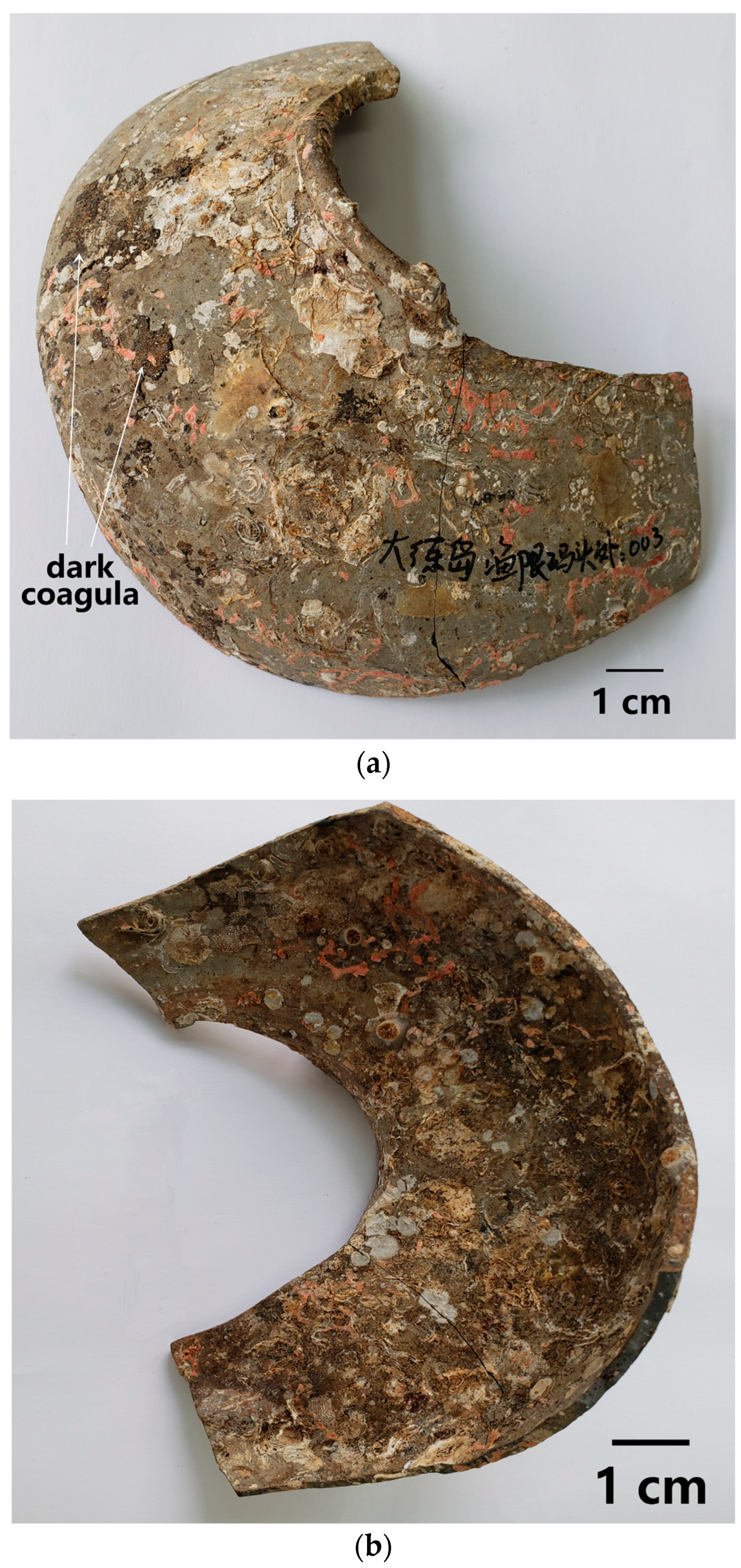
2. Sample
3. Research Method
4. Experimental Results
4.1. Chemical Composition of the Body and Glaze
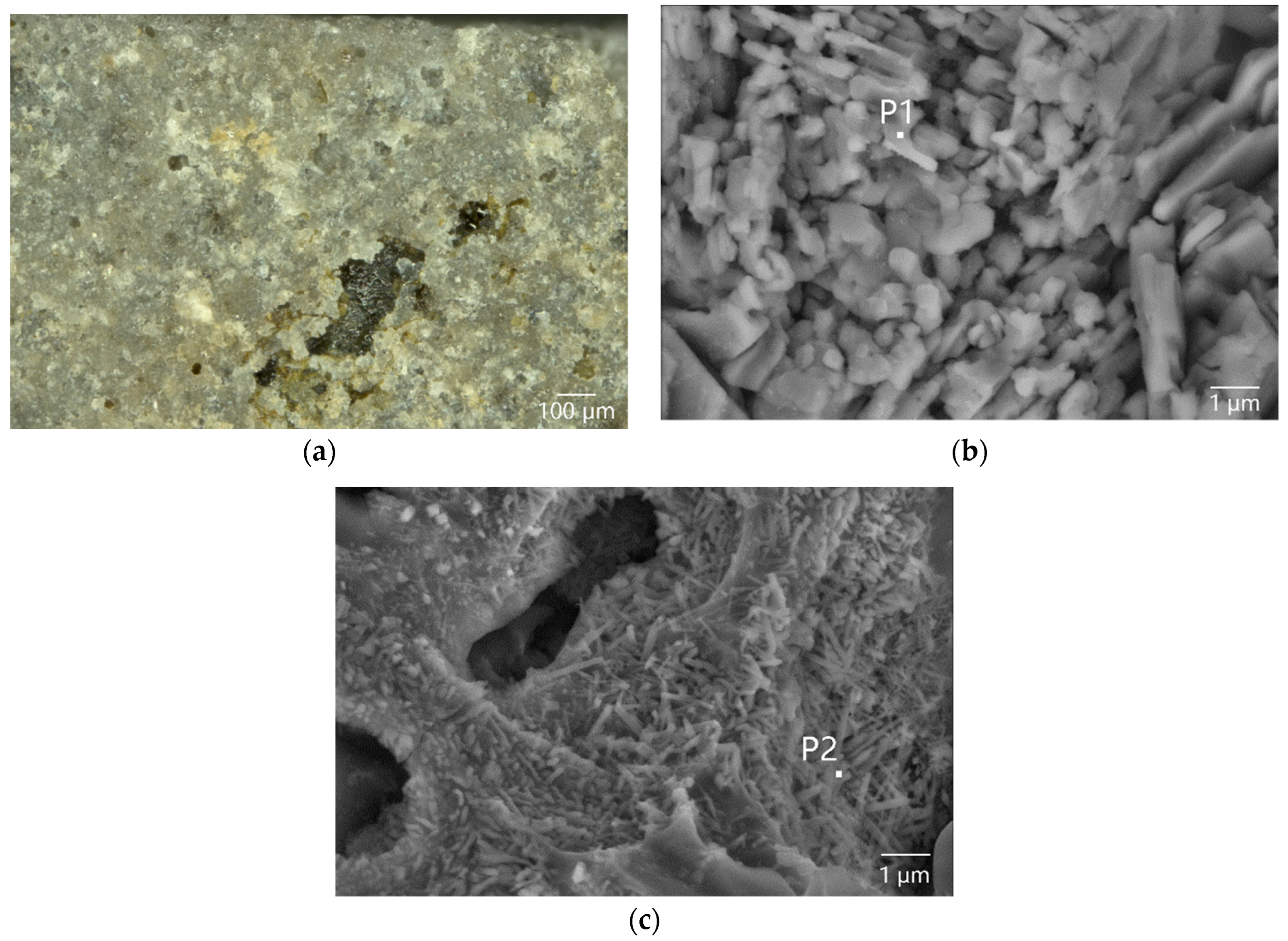
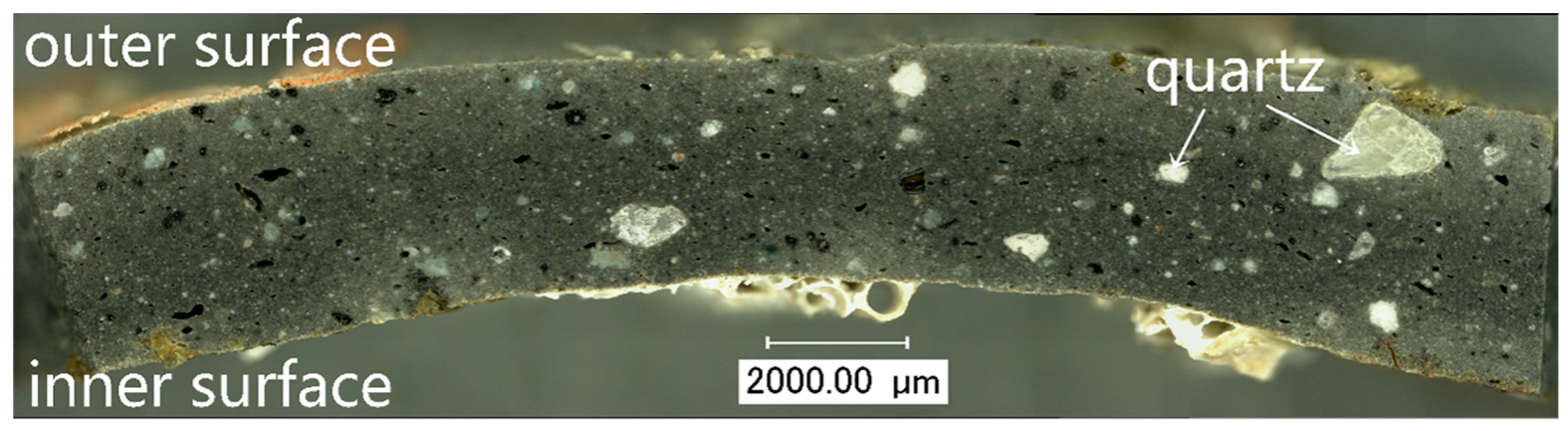
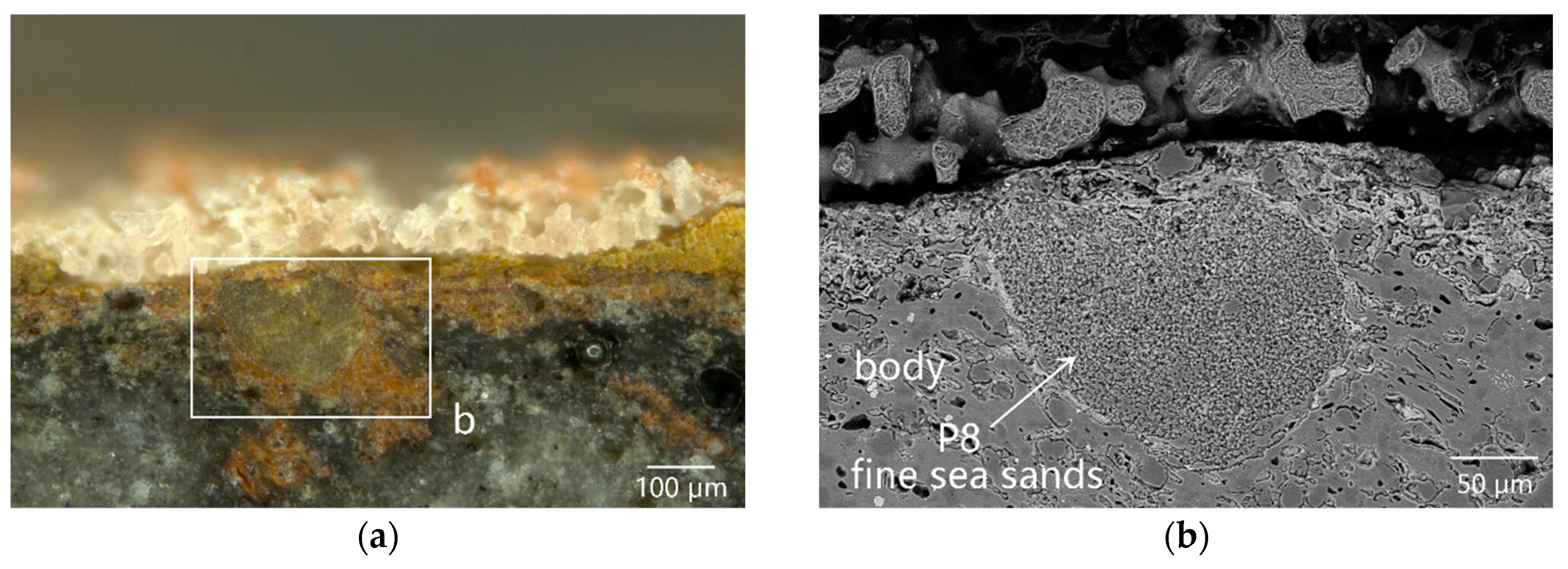
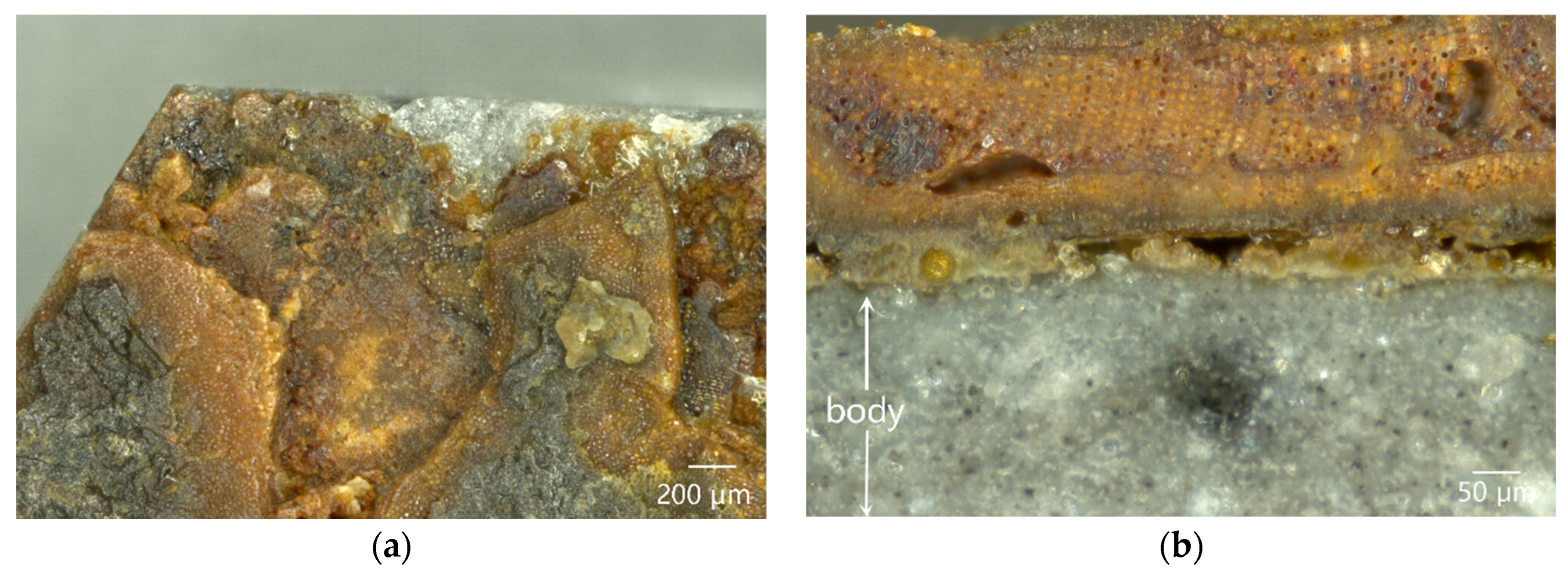
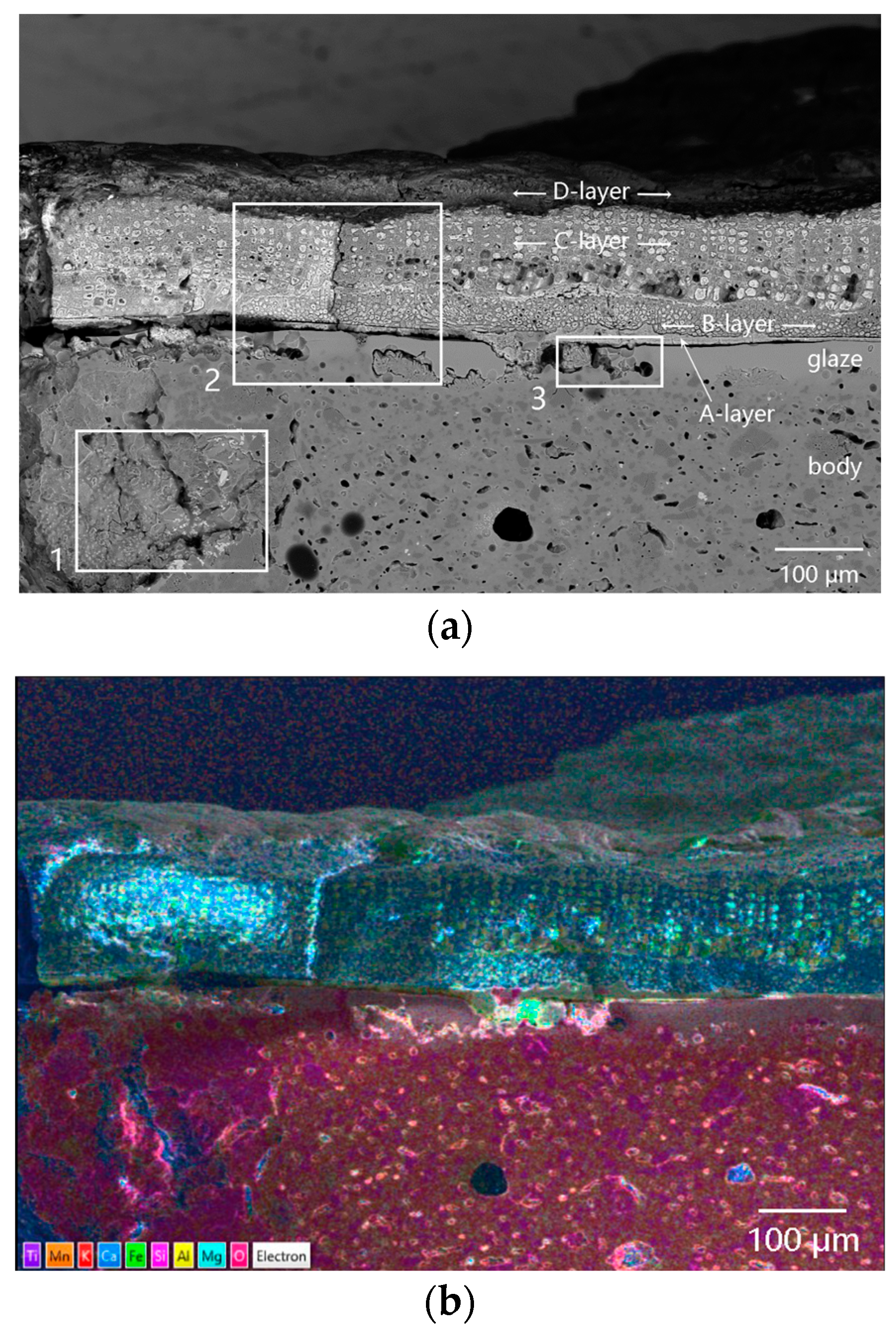
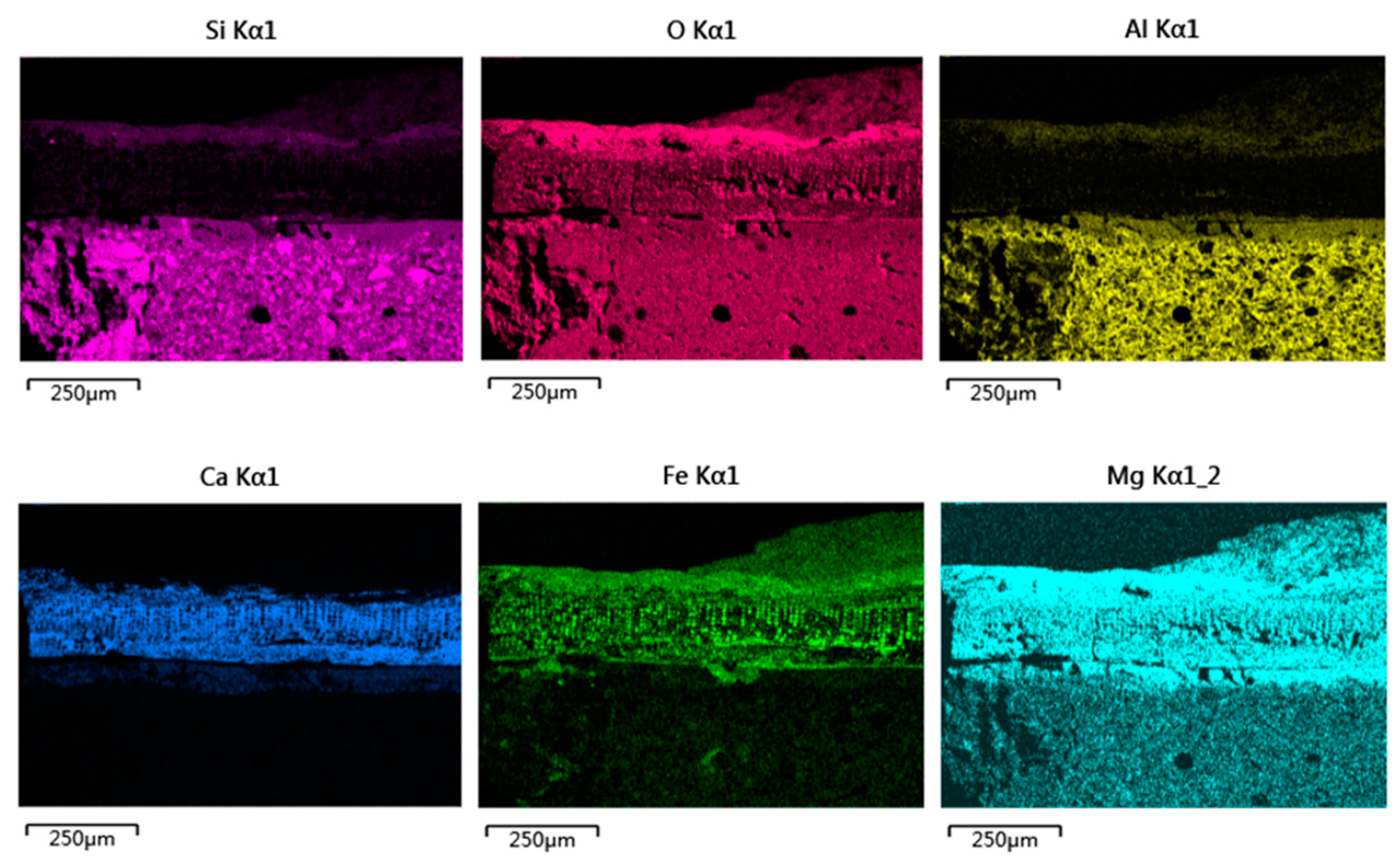
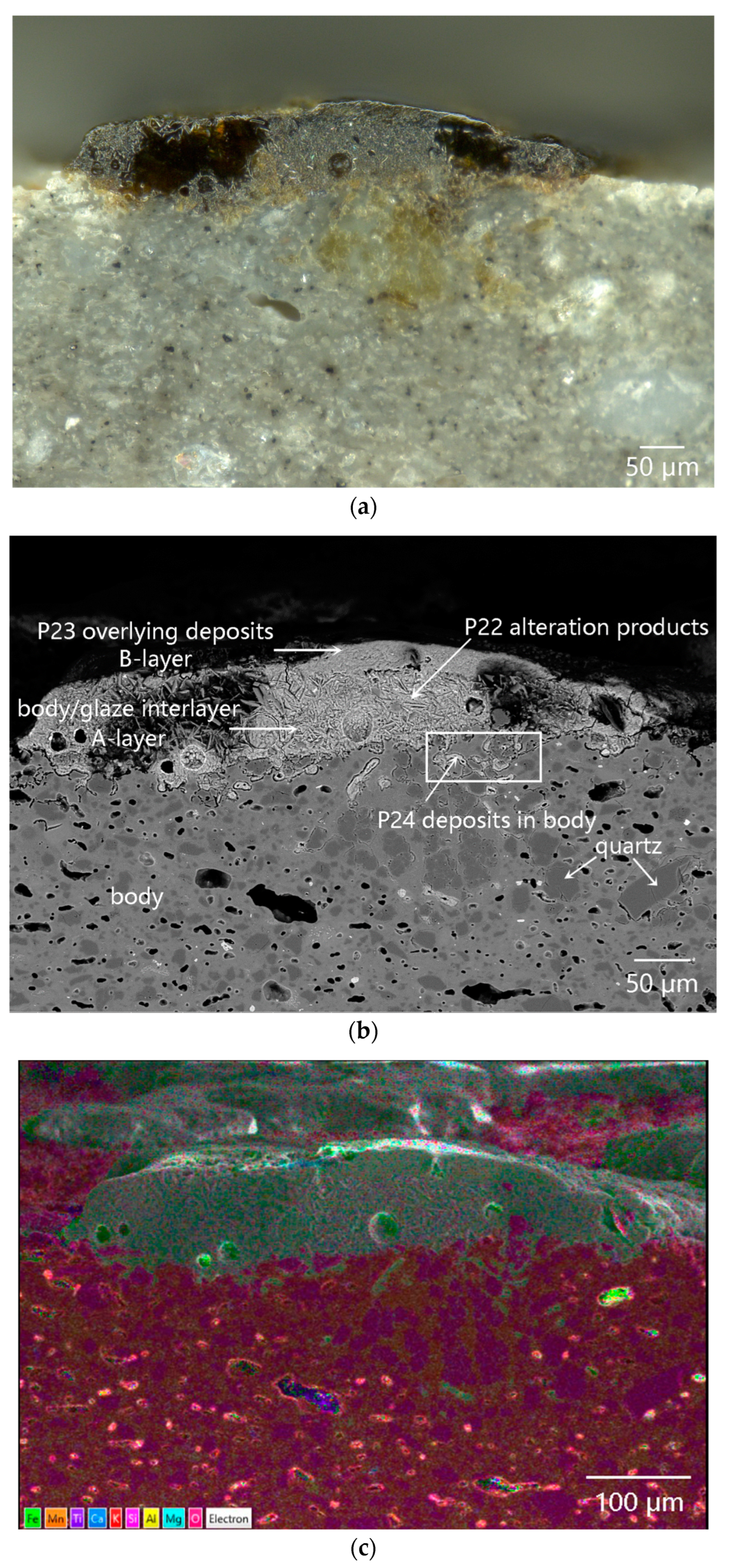
4.2. Morphology and Microstructure of the Samples
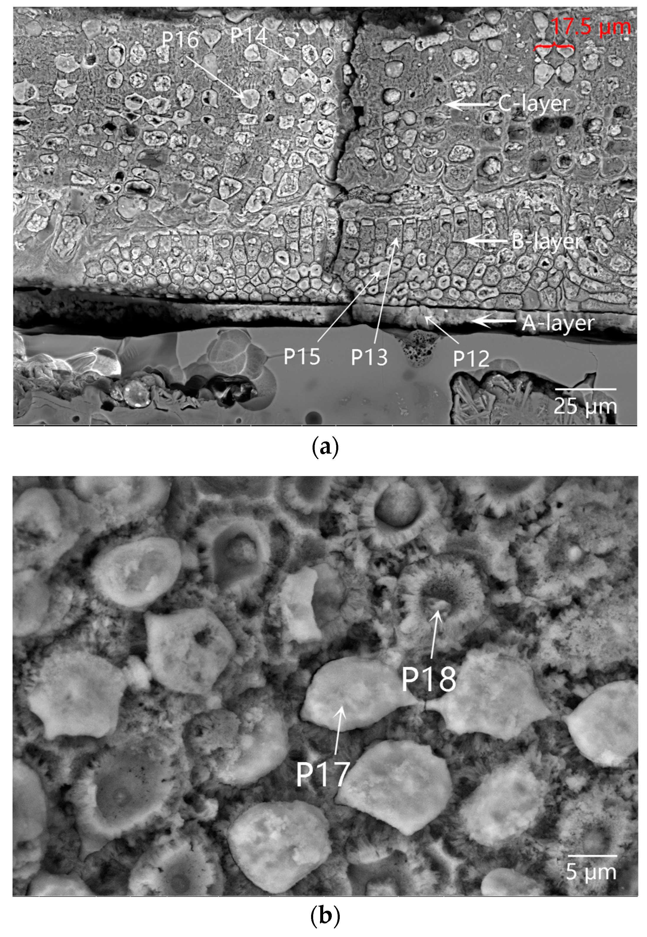
4.3. Analysis Results of the Cellular Coagula Attached to the Outer Surface of the Samples
4.3.1. Morphology of the Cellular Coagula
4.3.2. Microstructure and Microarea Composition of the Coagula

| Figure | Position | Na | Mg | Al | Si | K | Ca | Fe |
|---|---|---|---|---|---|---|---|---|
| Figure 2 | Rod-like crystals at the breakage position of the glaze (Anorthite, P1) | 0.63 | 0.46 | 27.64 | 43.11 | 1.20 | 23.03 | 3.94 |
| Sill-like crystal clusters composed of needle-like crystals in the naked body (Mullite, P2) | 0.44 | 1.61 | 32.71 | 49.98 | 5.08 | 1.47 | 8.72 | |
| Figure 4 | Residual glaze (P3) | 13.54 | 15.13 | 51.98 | 0.49 | 15.53 | 3.33 | |
| Deposits in the pores (P4) | 8.35 | 6.45 | 17.32 | 0.50 | 2.49 | 64.88 | ||
| Deposits in the pores (P5) | 10.51 | 7.04 | 27.65 | 0.52 | 1.76 | 52.52 | ||
| Deposits in the pores (P6) | 14.46 | 9.49 | 36.31 | 0.71 | 1.66 | 37.37 | ||
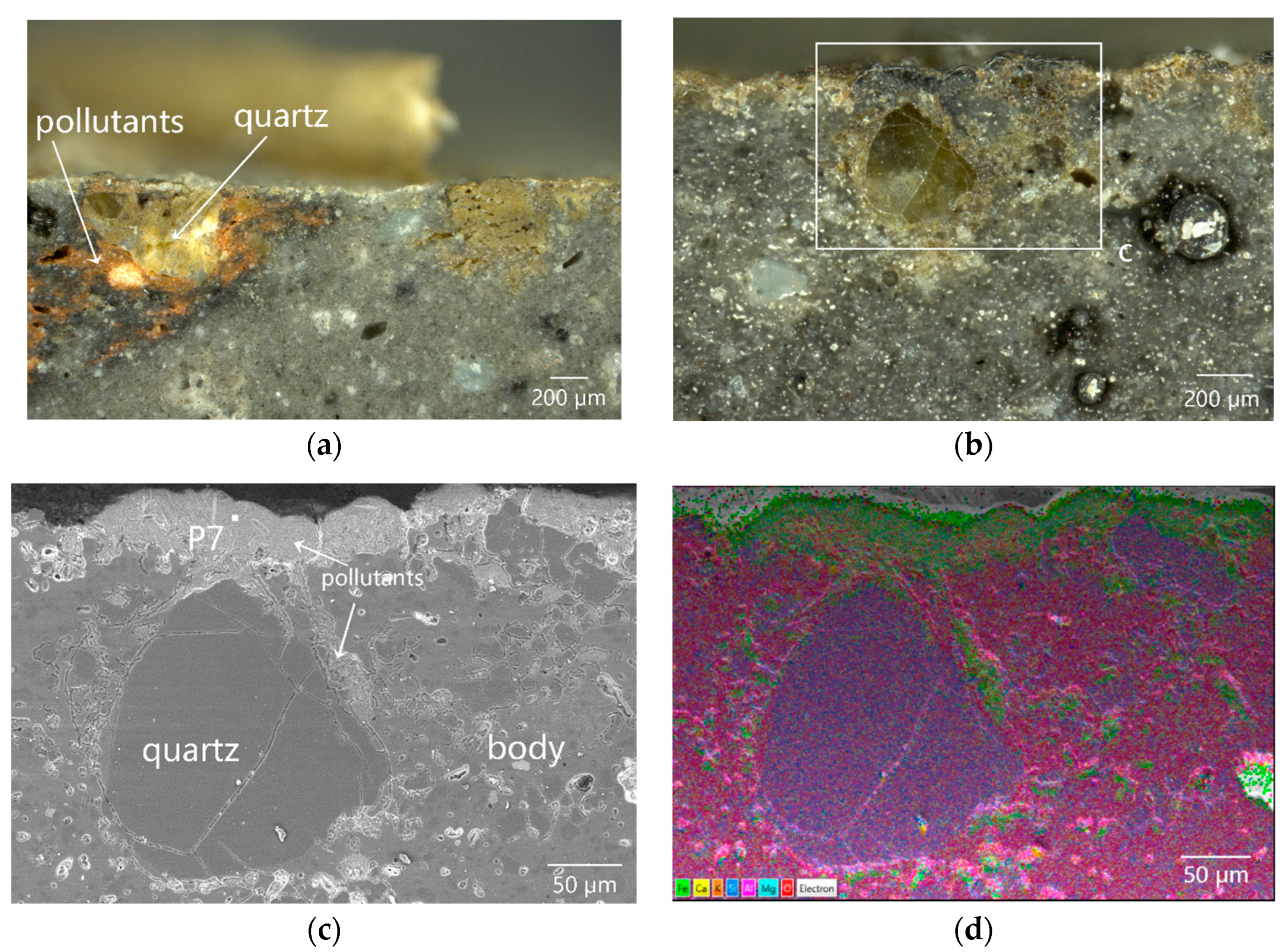
| Figure 5 | Pollutants deposited on the surface of the inner surface (P7) | 11.08 | 9.65 | 34.64 | 0.76 | 4.68 | 38.86 | |
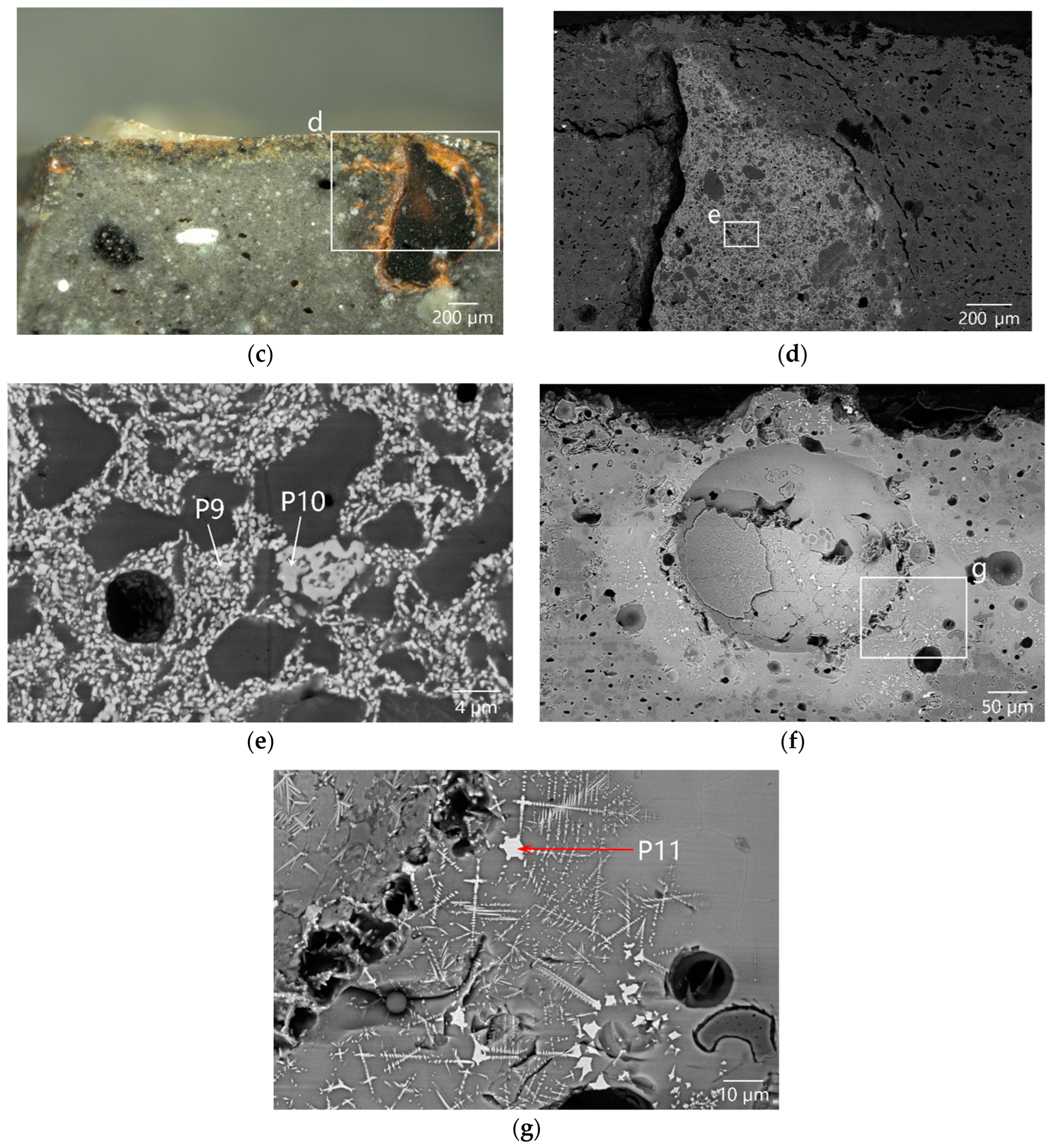
| Figure 6 | Deposits in the pores (P8) | 2.00 | 2.44 | 89.32 | 6.23 | |||
| Hexagonal crystals (P9) | 4.05 | 26.26 | 26.16 | 1.21 | 42.32 | |||
| Short-column crystals (P10) | 3.39 | 23.37 | 20.43 | 1.60 | 0.53 | 50.68 | ||
| Dendritic crystals (P11) | 2.42 | 38.27 | 1.37 | 57.93 | ||||
| Figure 10 | A-layer coagula (P12) | 2.56 | 6.11 | 21.92 | 1.10 | 5.88 | 62.44 | |
| B-layer coralline algae skeleton (P13) | 12.71 | 1.61 | 79.53 | 6.16 | ||||
| C-layer coralline algae skeleton (P14) | 0.61 | 8.45 | 0.50 | 86.11 | 4.33 | |||
| B-layer filler (P15) | 7.61 | 4.42 | 24.99 | 0.64 | 2.43 | 59.91 | ||
| C-layer filler (P16) | 6.45 | 2.44 | 23.79 | 0.67 | 2.41 | 64.24 | ||
| Filler (P17) | 10.23 | 5.22 | 31.66 | 0.93 | 2.43 | 49.53 | ||
| Coralline algae skeleton + Filler (P18) | 8.02 | 1.49 | 11.88 | 0.27 | 33.15 | 45.18 | ||
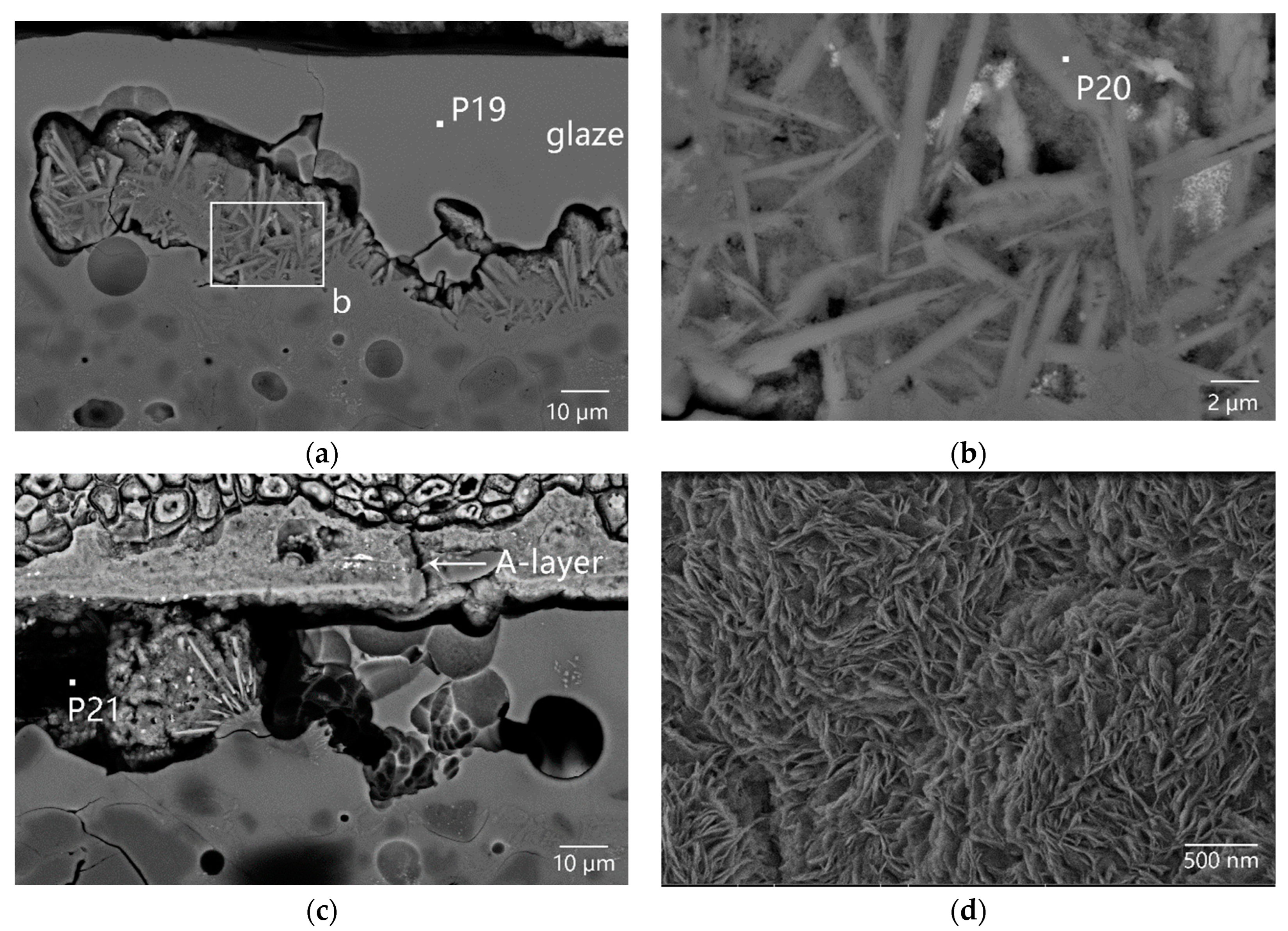
| Figure 11 | Glaze (P19) | 0.54 | 7.09 | 15.16 | 51.39 | 4.56 | 14.98 | 6.29 |
| Crystals in glaze-body interlayer (P20) | 2.05 | 2.23 | 22.66 | 50.80 | 2.46 | 14.92 | 4.88 | |
| Foreign pollutants in the pore (P21) | 0.88 | 0.95 | 2.40 | 0.22 | 1.02 | 94.52 | 0.88 |
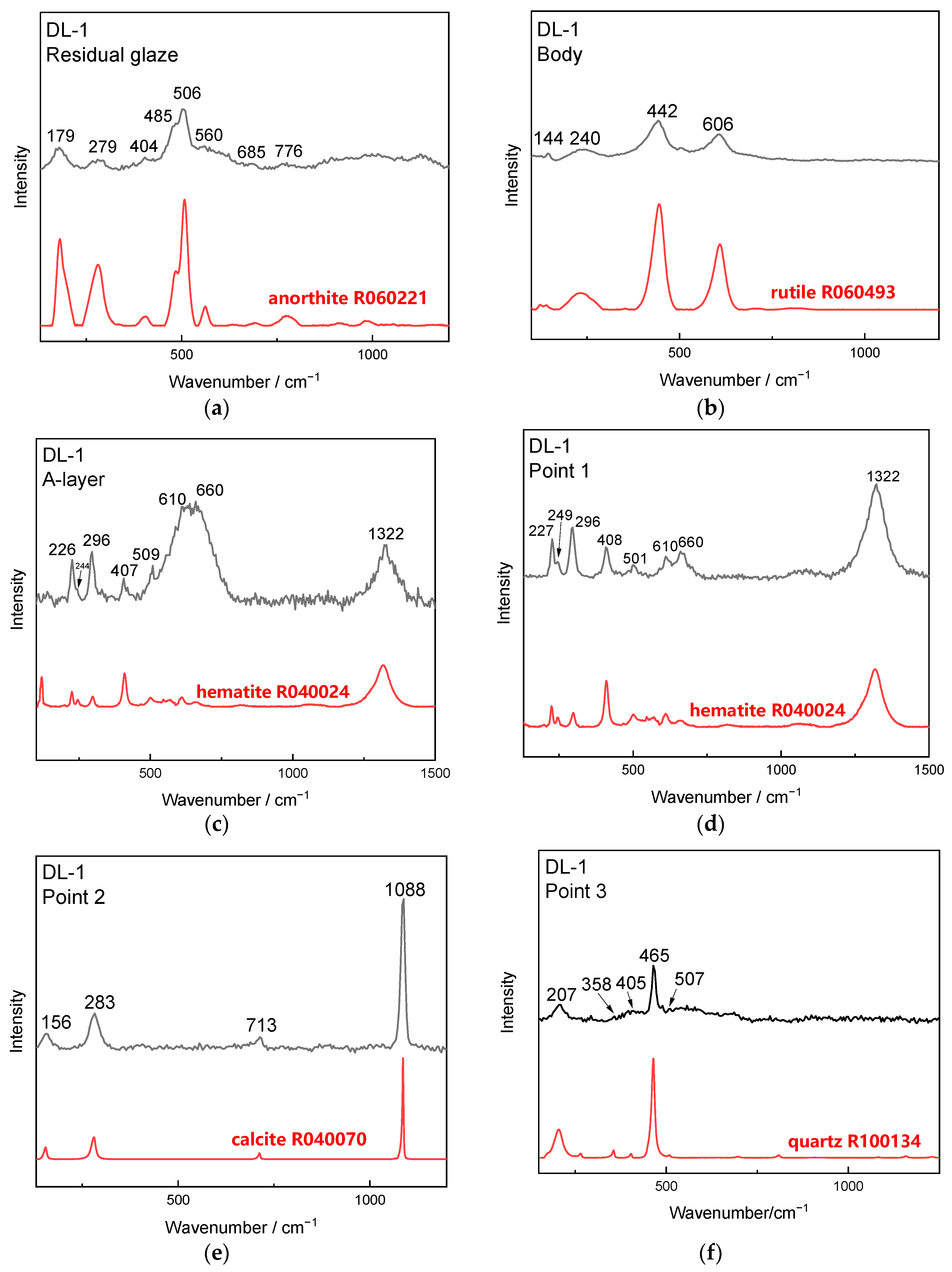
4.3.3. μ-Raman Analysis Results of the Coagula
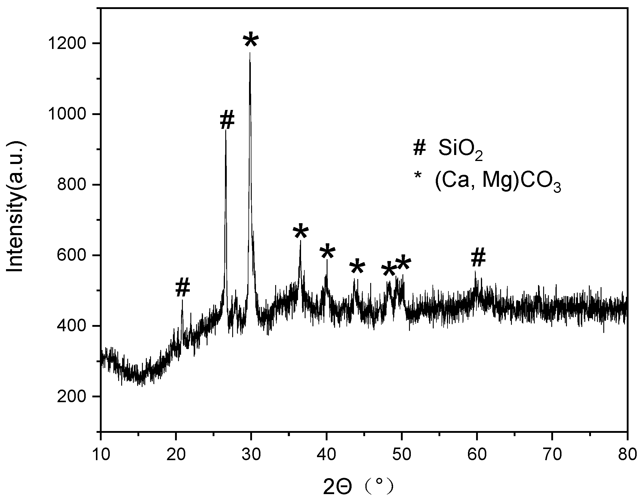
4.3.4. EDXRF and XRD Analysis Results of the Coagula
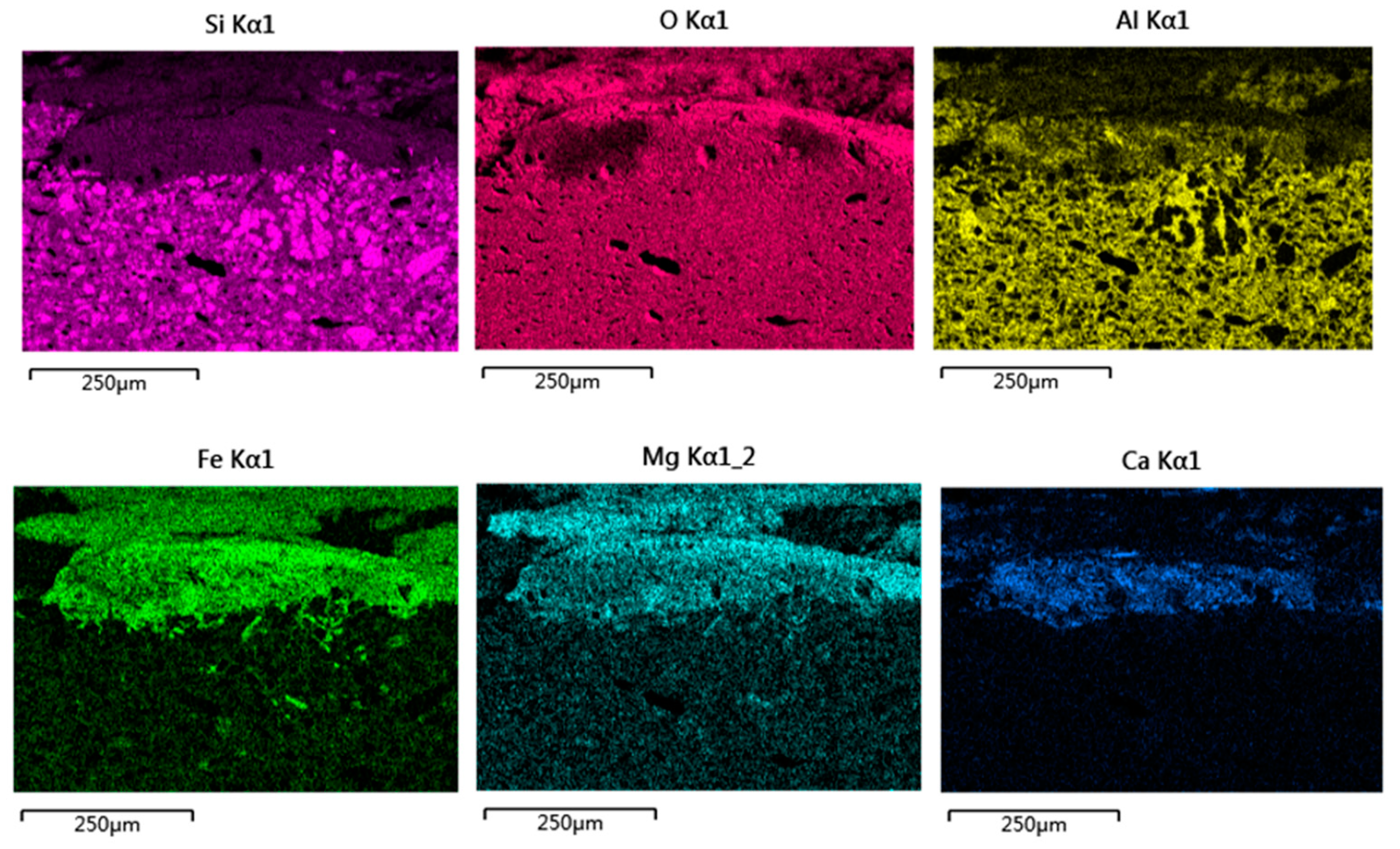
4.4. Microstructure and Micro-Area Composition of the Altered Glaze
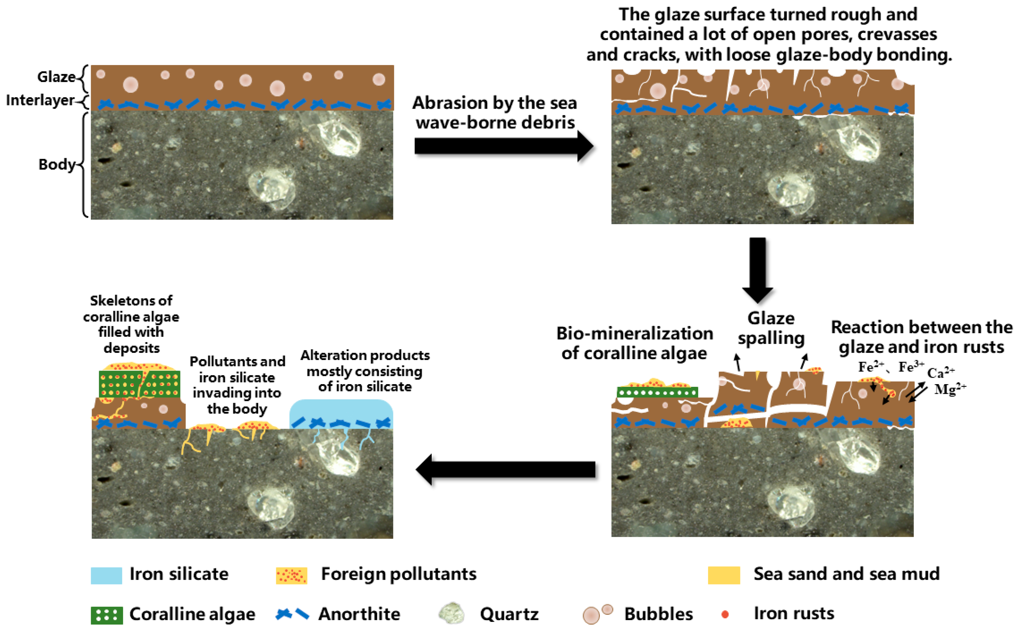
5. Discussion
6. Conclusions
Author Contributions
Funding
Institutional Review Board Statement
Informed Consent Statement
Data Availability Statement
Conflicts of Interest
References
- Li, N.S. Research on the Protection of Porcelain Excavated from the Ocean; Science Press: Beijing, China, 2016. (In Chinese) [Google Scholar]
- Song, J.M. Ocean Chemistry in China; Ocean Press: Beijing, China, 2000; pp. 53–68. (In Chinese) [Google Scholar]
- Bekic, L.; Curkovic, M.; Jelic, A.; Antonija, J.; Ivo, M.; Mladen, M.; Tanja, P.; Mladen, P. Conservation of Underwater Archaeological Finds Manual; Bekic, L., Ed.; International Centre for Underwater Archaeology in Zadar: Zadar, Croatia, 2014; pp. 18–38. [Google Scholar]
- Buys, S.; Oakley, V. The Conservation and Restoration of Ceramics; Routledge: New York, NY, USA, 2011; pp. 23–24. [Google Scholar]
- Xi, A.L. Comparative study on desalination technology of ceramic wares excavated from “Nan’ao I.”. Sci. Conserv. Archaeol. 2020, 32, 90–97. (In Chinese) [Google Scholar] [CrossRef]
- Ma, Y.R. A preliminary study on the desalination and conservation of ceramics from underwater archaeological excavations in China. Mus. Res. 2007, 1, 85–89. (In Chinese) [Google Scholar]
- Pearson, C. Conservation of Marine Archaeological Objects; Butterworth & Co., (Publishers) Ltd.: London, UK, 1987; pp. 16–22, 107–109, 261–264. [Google Scholar]
- Liu, W.; Zhang, Z.G.; Li, X.H.; Ma, Q.L. Scientific Analysis of Concretions on Ancient Marine Iron Recovered from Three Shipwrecks in the South China Sea. J. Natl. Mus. China 2011, 2, 145–156. (In Chinese) [Google Scholar]
- Yang, C.S.; Wang, J.L.; Zhang, Z.G. Research on Corrosion Products and Removal of Salts in Iron Ware from Huaguang Reef Ⅰ. CIESC J. 2011, 62, 2582–2587. (In Chinese) [Google Scholar]
- Hao, X.L.; Zhu, T.Q.; Xu, J.J.; Wang, Y.R.; Zhang, X.W. Microscopic Study on the Concretion of Ceramics in the “Nanhai I” Shipwreck of China, Southern Song Dynasty (1,127–1,279 A.D.). Microsc. Res. Tech. 2018, 81, 486–493. [Google Scholar] [CrossRef] [PubMed]
- Singley, K.; Hamilton, D.L. Basic Methods of Conserving Underwater Archaeological Material Culture. J. Am. Inst. Conserv. 1998, 37, 379–380. [Google Scholar] [CrossRef] [Green Version]
- Rodgers, B.A. The Archaeologist’s Manual for Conservation; Kluwer Academic/Plenum Publishers: New York, NY, USA, 2004; pp. 142–145. [Google Scholar]
- Wang, Y.R.; Zhu, T.Q.; Yang, G.C.; Tan, X.; Ye, D.Y.; Chen, H.T. The method to soften the concretions of ceramics in the “Nanhai I” Shipwreck of China Southern Song Dynasty (1127–1279AD). Herit. Sci. 2018, 6, 4. [Google Scholar] [CrossRef]
- Li, W.J.; Chen, Y.; Li, N.S.; Li, B.; Luo, W.G. A Study on Quantitative Evaluation of Damage in Conservation of Ceramics from Huaguangjiao Ⅰ Shipwreck with ICP-AES. Spectrosc Spectr Anal. 2015, 35, 772–776. (In Chinese) [Google Scholar] [CrossRef]
- Hu, D.B.; Zhang, H.Y. A Study on the Impacts of Commonly-used Porcelain Cleaning Agents. Sci. Conserv. Archaeol. 2010, 22, 49–59. (In Chinese) [Google Scholar] [CrossRef]
- Huang, H.; Yang, Q.F.; Wu, L.M. Safety Evaluation of Micro-nano Bubble Cleaning for Underwater Porcelain from Nanhai I Shipwreck. Sci. Conserv. Archaeol. 2017, 29, 30–37. (In Chinese) [Google Scholar]
- He, Y.; Li, W.D.; Li, J.A.; Xu, C.S.; Lu, X.K. Research on the Degradation of Ancient Longquan Celadons in the Dalian Island Shipwreck. NPJ Mater. Degrad. 2021, 6, 4. [Google Scholar] [CrossRef]
- He, Y.; Li, W.D.; Li, J.A.; Xu, C.S.; Lu, X.K. Corrosion of Longquan celadons in the marine environment: Study on the celadons from the Dalian Island shipwreck of the Yuan Dynasty. Herit. Sci. 2021, 9, 104. [Google Scholar] [CrossRef]
- He, Y.; Li, W.D.; Xu, C.S.; Lu, X.K.; Sun, X.M. Degradation Mechanism of the Ru Wares Unearthed from the Qingliangsi Site in Henan, China. Ceram. Int. 2022, 48, 17131–17142. [Google Scholar] [CrossRef]
- Li, J.Z. History of Science and Technology in China: Ceramic Volume; Science Press: Beijing, China, 1998. (In Chinese) [Google Scholar]
- Wray, J.L. Calcareous Algae; Elsevier Scientific Pub. Co: Amsterdam, The Netherlands, 1998; pp. 171–187. [Google Scholar]
- Yebra, D.M.; Kiil, S.; Dam-Johansen, K. Antifouling Technology-Past, Present and Future Steps towards Efficient and Environmentally Friendly Antifouling Coatings. Prog. Org. Coat. 2004, 50, 75–104. [Google Scholar] [CrossRef]
- Guenther, R.; Miklasz, K.; Carrington, E.; Martone, P.T. Macroalgal Spore Dysfunction: Ocean Acidification Delays and Weakens Adhesion. J. Phycol. 2018, 54, 153–158. [Google Scholar] [CrossRef] [PubMed]
- Saleh, S.A. Corrosion Mechanism of Iron Objects in Marine Environment an Analytical Investigation Study by Raman Spectrometry. Eur. J. Sci. Theol. 2017, 13, 185–206. [Google Scholar]
- Zhang, J.B.; Yin, Q.F.; Rong, Y.M.; Chen, C.L.; Sun, S.L. Corrosivity monitoring and evaluation of seawater and sedimentary environments around a shipwreck area in Pingtan sea, Fujian Province. J. Appl. Oceanogr. 2021, 40, 312–323. (In Chinese) [Google Scholar] [CrossRef]
- Johnson, M.D.; Carpenter, R.C. Ocean Acidification and Warming Decrease Calcification in the Crustose Coralline Alga Hydrolithon Onkodes and Increase Susceptibility to Grazing. J. Exp. Mar. Biol. Ecol. 2012, 434, 94–101. [Google Scholar] [CrossRef]
- Michelin, A.; Burger, E.; Leroy, E.; Foy, E.; Neff, D.; Benzerara, K.; Dillmann, P.; Gin, S. Effect of Iron Metal and Siderite on the Durability of Simulated Archeological Glassy Material. Corrosion Sci. 2013, 76, 403–414. [Google Scholar] [CrossRef]
- Michelin, A.; Burger, E.; Rebiscoul, D.; Neff, D.; Bruguier, F.; Drouet, E.; Dillmann, P.; Gin, S. Silicate Glass Alteration Enhanced by Iron: Origin and Long-Term Implications. Environ. Sci. Technol. 2013, 47, 750–756. [Google Scholar] [CrossRef]
- Palomar, T. Characterization of the Alteration Processes of Historical Glasses on the Seabed. Mater. Chem. Phys. 2018, 214, 391–401. [Google Scholar] [CrossRef]


| DL-1 | Mg | Al | Si | K | Ca | Ti | Fe | Mn | P | S |
|---|---|---|---|---|---|---|---|---|---|---|
| Body | 0.26 | 17.47 | 64.59 | 6.52 | 0.59 | 1.68 | 8.85 | 0.04 | ||
| Glaze (polluted and altered) | 7.07 | 3.73 | 15.65 | 0.40 | 27.23 | 0.12 | 38.44 | 6.75 | 0.29 | 0.32 |
| Na | Mg | Al | Si | S | Cl | K | Ca | Mn | Fe | |
|---|---|---|---|---|---|---|---|---|---|---|
| Dark coagula | 1.03 | 5.09 | 7.96 | 21.46 | 1.78 | 2.19 | 1.49 | 2.76 | 3.13 | 52.14 |
| Figure | Position | Mg | Al | Si | K | Ca | Fe |
|---|---|---|---|---|---|---|---|
| Figure 14 | Alteration products in the interlayer (P22) | 8.98 | 8.15 | 31.05 | 0.55 | 4.11 | 47.16 |
| Overlying deposits (B-layer, P23) | 11.29 | 4.58 | 32.00 | 1.70 | 50.42 | ||
| Deposits in the body (P24) | 8.25 | 7.29 | 29.79 | 0.77 | 1.16 | 52.72 |
Disclaimer/Publisher’s Note: The statements, opinions and data contained in all publications are solely those of the individual author(s) and contributor(s) and not of MDPI and/or the editor(s). MDPI and/or the editor(s) disclaim responsibility for any injury to people or property resulting from any ideas, methods, instructions or products referred to in the content. |
© 2023 by the authors. Licensee MDPI, Basel, Switzerland. This article is an open access article distributed under the terms and conditions of the Creative Commons Attribution (CC BY) license (https://creativecommons.org/licenses/by/4.0/).
Share and Cite
Ding, R.; Li, W.; Yang, Z.; Xu, C.; Lu, X. Degradation Mechanism of a Sauce-Glazed Ware of the Song Dynasty Salvaged out of the Water at Dalian Island Wharf: Part I—The Effect of the Surface-Attached Composite Coagula. Materials 2023, 16, 1176. https://doi.org/10.3390/ma16031176
Ding R, Li W, Yang Z, Xu C, Lu X. Degradation Mechanism of a Sauce-Glazed Ware of the Song Dynasty Salvaged out of the Water at Dalian Island Wharf: Part I—The Effect of the Surface-Attached Composite Coagula. Materials. 2023; 16(3):1176. https://doi.org/10.3390/ma16031176
Chicago/Turabian StyleDing, Rao, Weidong Li, Zelin Yang, Changsong Xu, and Xiaoke Lu. 2023. "Degradation Mechanism of a Sauce-Glazed Ware of the Song Dynasty Salvaged out of the Water at Dalian Island Wharf: Part I—The Effect of the Surface-Attached Composite Coagula" Materials 16, no. 3: 1176. https://doi.org/10.3390/ma16031176