
Biomedical Alloys and Physical Surface Modifications: A Mini-Review


1
Department of Orthopedics, Renmin Hospital, Wuhan University, Wuhan 430060, China
2
Key Laboratory of Artificial Micro- and Nano-Structures of Ministry of Education, School of Physics and Technology, Wuhan University, Wuhan 430072, China
*
Authors to whom correspondence should be addressed.
Materials 2022, 15(1), 66; https://doi.org/10.3390/ma15010066
Submission received: 30 October 2021
/
Revised: 3 December 2021
/
Accepted: 10 December 2021
/
Published: 22 December 2021
(This article belongs to the Special Issue Design and Manufacturing of Bioinspired Material and Structures)
Abstract
:Biomedical alloys are essential parts of modern biomedical applications. However, they cannot satisfy the increasing requirements for large-scale production owing to the degradation of metals. Physical surface modification could be an effective way to enhance their biofunctionality. The main goal of this review is to emphasize the importance of the physical surface modification of biomedical alloys. In this review, we compare the properties of several common biomedical alloys, including stainless steel, Co–Cr, and Ti alloys. Then, we introduce the principle and applications of some popular physical surface modifications, such as thermal spraying, glow discharge plasma, ion implantation, ultrasonic nanocrystal surface modification, and physical vapor deposition. The importance of physical surface modifications in improving the biofunctionality of biomedical alloys is revealed. Future studies could focus on the development of novel coating materials and the integration of various approaches.
1. Introduction
Biomaterials are currently widely used in biological systems for medical purposes [1] such as dental applications, surgery, and pharmaceutical. Some of these materials are already commercialized in applications related to tissue growth and drug delivery. We exhibit some applications of biomaterials in Figure 1. The specifics effects and applications are determined by biomaterial properties [2,3]. These days, the research direction is the targeted design and control of biomaterial properties to achieve specific biological responses. The most critical parameter of a biomaterial is its absolute non-toxicity. Biomaterial type should be chosen with care for a specific medical application. For example, for drug release, the biomaterials are typically based on novel polymers [4]; for dental implants and bone plates, biomedical alloys are the best choices [5].
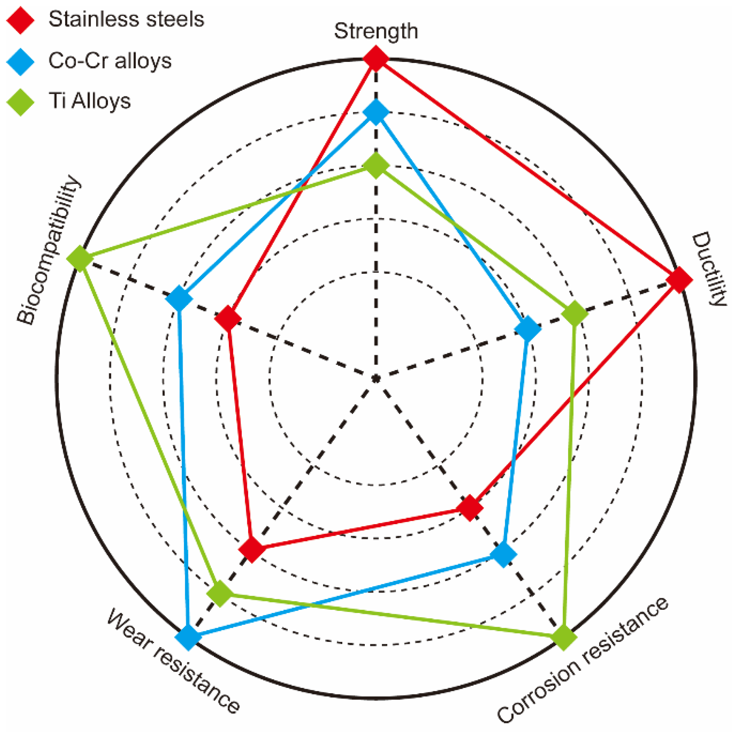
About 80% of all materials used for bio-implants are biomedical alloys. As the world population age at a fast rate, the demand for biomedical alloys is increasing rapidly. The most popular biomedical alloys include stainless steel [6], Co–Cr [7], and Ti alloys [8]; Figure 2 and Table 1 compare the properties of these three groups. Other biomedical alloys, based on Mg, Fe, Ta, and Nb alloys, are not as widely used [9]. Generally, the biofunctionality and the biological and mechanical biocompatibility of the currently used biomedical alloys should be improved to satisfy the growing variety of medical applications. Numerous efforts to improve the mechanical biocompatibility of all these alloys, including the strength, ductility, wear resistance, toughness, and corrosion, have been published in the scientific literature [10,11,12,13].
Figure 1.
(a) Poly(lactic-co-glycolic acid)-based bone-substitute materials for bone repairing and healing. (b) Biomedical application of Mg-based biomaterials and their corresponding physiological processes. (c) The fabrication and role of biomaterials in the delivery of cells, bioactive molecules, growth factors, and drugs for tissue engineering applications. (Reproduced with permission from [3,14,15]. Copyright (2021), Elsevier.)

Figure 2.
Comparison of strength, ductility, corrosion, and wear resistance as well as biocompatibility of stainless steel (red), Co–Cr (blue), and Ti (green) alloys.
Figure 2.
Comparison of strength, ductility, corrosion, and wear resistance as well as biocompatibility of stainless steel (red), Co–Cr (blue), and Ti (green) alloys.

The biofunctionality of biomedical alloys strongly depends on the properties of their superficial layers. Therefore, modification of these layers is a promising approach of tuning and improving various properties. Such surface modification methods, developed in recent decades [16,17], include mechanical, physical, chemical, and biochemical approaches [18]. The physical surface modification method involves a direct treatment of the superficial layers by thermal, kinetic, and electrical energy with almost no chemical modification to the original alloy matrix [19]. To fully understand the biofunctionality of biomedical alloys, a thorough analysis of their structures and physical surface modification mechanisms is essential.
Thus, this review first introduces common biomedical alloys (Co–Cr, Ti alloys, and stainless steels) and discusses their biomedical applications and potential improvements. Then, we present some physical surface modifications, including thermal spraying, glow discharge plasma, ion implantation, ultrasonic nanocrystal surface modifications, and physical vapor deposition. The same section discusses biomedical alloy applications modified by these methods. Finally, we compare various surface modifications methods and provide an outlook of the future progress in this field.

Table 1.
Summary of the properties, advantages, and disadvantages of the most common biomedical alloys.
Table 1.
Summary of the properties, advantages, and disadvantages of the most common biomedical alloys.
| Materials | Density | Tensile Strength | Elastic Modulus | Advantages | Disadvantages | Refs. |
|---|---|---|---|---|---|---|
| Stainless steels | ~7500 kg/m3 | ~620 MPa | 193~200 GPa | High strength, ductility | Harmful metal release, stress shielding | [20,21] |
| Co–Cr alloys | ~10,000 kg/m3 | ~150 MPa | 220~230 GPa | High strength and wear resistance | Harmful metal release | [22,23] |
| Ti Alloys | ~4500 kg/m3 | ~240 MPa | 55~114 GPa | High strength, corrosion resistance, biocompatibility, low elastic modulus | Stress shielding | [24,25] |
2. Methods
All the authors of this study performed an electronic search set from 1991 to 2021 using Web of Science. The following keywords were selected individually or combined: biomedical application, biomedical alloy, stainless steel, Co–Cr alloy, Ti alloy, physical surface modifications, thermal spraying, glow discharge plasma, ion implantation, ultrasonic nanocrystal surface modification, and physical vapor deposition. Review articles and related research articles were also sources of references to locate other articles. About 15~20 articles were selected in each section. The inclusion criterion was that an article should contain biomedical alloys and physical surface modifications. Articles not providing enough information about biomedical application were excluded.
3. Biomedical Alloys
3.1. Stainless Steel
The application of stainless steel in biomedicine has the longest history among metallic biomaterials. Mature modern metallurgy successfully fabricates stainless steel with excellent properties. Stainless steel is a desirable material because its manufacturing is a mature, easy-to-perform, and inexpensive technology. Additionally, stainless steel possesses high corrosion resistance and mechanical strength. Stainless steels exhibit higher ductility and cyclic twist strength than Co–Cr and Ti alloys. Moreover, 316L stainless steel (Cr–Ni–Mo, “L” represents low carbon) is the most common one and is widely used for temporary and permanent implants [26] because it limits the formation of Cr–C and enhances the corrosion resistance [20].
As an orthopedic implant material, stainless steel should be non-ferromagnetic. Austenitic 316L stainless steels meet this requirement, and thus are used widely. Austenitic stainless steels can be specified as ASTM F138, ASTM F1586, or ASTM F2581 [27]. The ASTM F1586 and ASTM F2581 steels exhibit better corrosive resistance. Terada et al. [28] found that electrochemical treatment could effectively increase the corrosion resistance of austenitic stainless steels. However, these steels contained Ni, which is harmful and bio-incompatible [29]. Ni-free stainless steels are essential for health reasons. Yang and Ren [30] fabricated high-nitrogen nickel-free stainless steel with even better mechanical properties and superior biocompatibility. Talha et al. [31] further studied Ni-free N-rich austenitic steel (which they fabricated in an induction furnace), especially how the cold-working affected its mechanical behavior, and reported its excellent ductility and mechanical strength. The strain-induced martensitic transformation (SIMT) (austenite to martensite) in austenitic stainless steel could be caused by mechanical impacts, which proved to be favorable to cellular activity and hydrophilicity [32].
The usual way to treat stainless steels is through chemical methods, e.g., acid immersion and electrochemical anodizing. Stainless steel after acid immersion and anodizing treatment can exhibit the lowest thickness of the fibrous capsule membrane [33]. Yang et al. [34] investigated the effect of nitric acid passivation on high-nitrogen nickel-free stainless steels. The corrosion rate can be decreased by this passivation. Aguilar et al. [35] used the poly(caffeic acid) to coat the surface of stainless steel. Hsu et al. [36] applied an electrochemical anodizing method to modify the surface of 316L stainless steel. The electrochemical anodizing method can form a nanoporous oxide layer of Cr2O3 which can induce cell adhesion and promote bone formation. In addition to the above chemical methods, there are many other ways to improve the biomedical performance of stainless steel. Recent studies [37,38] proved that additive manufacturing can improve the charge transfer resistance and breakdown potential of 316 L stainless steel for clinical use. The cold deformation method can enhance the surface diffusion and corrosion resistance, which contributes to the passive films on the surface [39]. Moreover, Yang et al. [40] developed a simple and environmentally friendly water treatment to treat high nitrogen nickel-free stainless steels. This method increased passive films and allowed the corrosion rate of stainless steels to dramatically reduce to 1/20 of the untreated ones. Trzaskowska et al. [41] prepared a stainless steel coated with non-toxic organic materials by electropolymerization. These special organic coatings can fill surface scratches and reduce fibrinogen adsorption.
3.2. Co–Cr Alloys
Co–Cr alloys exhibit significantly higher wear resistance, heat resistance, and strength than Ti alloys and stainless steels. Co–Cr alloys also possess better corrosion resistance than 316L stainless steel. Therefore, Co–Cr alloys are commonly used to prepare bone substitutes, usually surrounded by Cl-rich body fluids, which could cause stress- and corrosion-related cracking if 316L stainless steel is used instead of Co–Cr alloy. When Co–Cr alloys are enriched with Cr, stable Cr2O3 film forms and protects the alloy from Cl-ion attacks [23,42]. Yamanaka et al. [24] even used a Co–Cr-based cast alloy for dental applications because of its high strength (comparable to wrought Co–Cr alloys) without ductility loss.
Similar to stainless steel, phase transition also exists in the Co–Cr alloy. Zhu et al. [43] researched the SIMT process in Co–Cr–W–Ni alloys and the process conforms to Schmid’s law. Ueki et al. [44] studied the precipitates that were induced during γ-ε phase transformation in Co–Cr–Mo alloys. The successful fabrication of Co–Cr–Mo alloy single crystals was first reported by Kaita et al. [45]. Mori et al. [46] proposed a novel approach to control the γ-ε SIMT in a hot-rolled biomedical Co–Cr–Mo alloy by manipulating the initial dislocation structures. The SIMT process was suppressed by the carbides.
The selective laser melting (SLM) technique is widely used for Co–Cr alloys. The SLM can relieve the stress concentrations and decrease the fatigue crack growth rate. A Co–Cr–Mo–W alloy treated by SLM exhibited longer fatigue life, higher tensile strength, and higher ductility than the untreated one [47]. Zhou et al. [48] produced Co–Cr devices by SLM which met the requirements for application as dental prostheses. Moreover, Co–Cr–Mo–W alloy treated by SLM showed better corrosion resistance, which was caused by effective micro-cathodes of precipitates [49].
Further improvements of the corrosion and wear resistances, as well as biocompatibility, could be achieved by introducing certain modifications to Co–Cr-based alloys. For example, Yamanaka et al. [50] developed a novel Co–Cr–W biomedical alloy for dental restorations with excellent fabrication and mechanical properties. Kheimehsari et al. [51] added hydroxyapatite (HA) coating to improve the corrosion resistance of Co–Cr-based implants. In this case, the thickness and sintering conditions of this HA-layer significantly affected the corrosion resistance of the Co–Cr-based implants. Shirdar et al. [52] applied HA/TiO2 coatings to Co–Cr–Mo alloys to enhance their mechanical and electrochemical properties. Sawangrat et al. [53] applied the harmonic structure design to synthesize new biomedical Co–Cr–Mo alloys with much better mechanical properties than the conventional biomedical alloys. Trimble et al. [54] developed a finite element model to predict the orthogonal forces of biomedical grade Co–Cr–Mo alloy and to reduce the number of machining tests. Migita et al. [55] used solid-binding peptides to improve the biocompatibility of the Co–Cr–Mo alloys. Yamanaka et al. [56] prepared Co–Cr–Mo alloy rods with a small diameter by hot-caliber rolling which exhibited high strength and durability. Mahajan and Sidhu [57] investigated the performance and biological responses of a Co–Cr alloy by an electrical discharge machining method, which assisted in improving the design precision for enhanced clinical performance.
3.3. Ti Alloys
Ti is a unique member of the biomedical alloy group as it possesses superior biocompatibility and complete inertness to the physiological environment, high corrosion resistance and strength, as well as a low elastic modulus. The density of Ti-based alloys is below that of stainless steels and Co–Cr alloys [25]. Some harmful elements could be released from stainless steel and Co–Cr-based alloys if they corrode or become damaged by wear [58]. However, such damage in Ti alloys can be entirely mitigated by forming very inert passivating TiO2 film [26]. Assis and Costa [59] confirmed electrochemically, by studying various Ti alloys, that high corrosion resistance correlates with the barrier layer presence and properties. The low elastic modulus of Ti alloys can also avoid the problem of stress shielding. Therefore, Ti alloys are often the best solution to solve a variety of biomedical problems. Commercially pure Ti is classified into four grades, G1 to G4. Acid etched Ti G4 exhibited better surface structures and mechanical properties, making it an ideal implant for dentistry [60,61].
Except for these commercially pure Ti, there are many Ti-based implants in the form of binary and multiple alloys. A typical representative of binary Ti alloys is Ti–Nb alloy. Porous Ti–Nb alloys synthesized by electro-deoxidation are proposed to be the best-suited candidate of the materials for biomedical applications [62]. Ibrahim et al. [63] also prepared porous Ti–Nb shape memory alloys by microwave sintering with the most uniform pore shape. Kuroda et al. [64] prepared Ti–Nb alloy via arc-melting which exhibited good mechanical properties and no cytotoxic effects. Apart from Ti–Nb alloys, there are also other binary Ti alloys, e.g., Ti–Ni alloys [65] and Ti–Fe alloys [66]. The additional metal content plays an important role in the biomedical application of these Ti alloys. The content of martensite phase in β-type Ti–Nb–Sn alloys decreases with the increase of Nb content, which has influence on the low elastic modulus. Maya et al. [67] found that Ti alloys with more Nb content exhibited excellent osteoinductive properties. [68] Qi et al. [66] studied the effect of the Fe content on the Ti–Fe biomedical alloy. The Ti-Fe alloy with the addition of about 5 wt% Fe content displayed excellent corrosion resistance.
Recently, a new variety of multiple Ti alloy was developed. Chui et al. [69] fabricated a series of novel β-type Ti–Zr–Nb–Mo where the corrosion resistance was mainly determined by the Mo content. Quadros et al. [70] prepared a Ti–Ta–Zr alloy with a low elastic modulus, excellent corrosion resistance, and no cytotoxic effects. Yılmaz et al. [71] produced porous Ti–Nb–Zr–Ta alloys by the space-holding method. These porous Ti–Nb–Zr–Ta alloys possessed suitable mechanical properties for hard tissue implants. A new β-type Ti–Nb–Mo biomedical alloy could exhibit a low elastic modulus, good wear resistance, an anti-wear capability, and a long service life [72]. Zhu et al. [73] reported a Ti-based bulk glassy alloy with great potential for biomedical application. Zhao et al. [74] fabricated another novel Ti alloy with outstanding corrosion resistance and superior mechanical biocompatibility.
Different preparation methods were developed to improve the biomedical performance of Ti-based alloys. Gao et al. [75] modified Ti-based alloy surfaces and enhanced their biocompatibility and stability even further. Xu et al. [76] used arc melting and graphite mold casting to prepare a series of Ti–Mo–Nb alloys, which simultaneously exhibited high strength and a low elastic modulus. Yang et al. [77] introduced gel-casting to obtain near-shape porous Ti alloys and that could even directly fabricate customized implants. The releasing of Cu ions is beneficial to lower infection incidences in Cu-bearing Ti alloys [78].
4. Physical Surface Modification of Biomedical Alloys
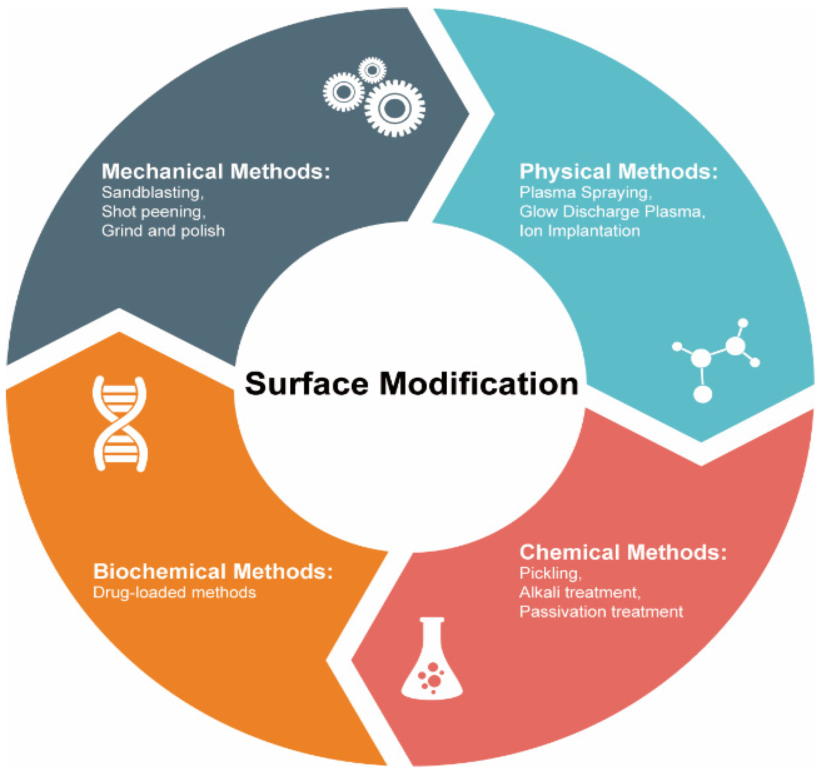
The degradation of the biomedical alloys based on stainless steel, Ti, and Co–Cr always starts on their surfaces. Thus, to improve or modify any properties of these materials (including corrosion and resistances as well as biocompatibility), a suitable surface modification approach must be used. These methods could be classified into treatment based on mechanical, physical, chemical, and biomedical techniques (see Figure 3). Below we discuss some of the physical surface modification methods.
4.1. Thermal Spraying
Thermal spray is an effective technology to improve wear resistance and biocompatibility through applying coatings. The thickness ranges from several microns to millimeters. The main methods of thermal spray are high velocity oxygen fuel spraying, flame spraying, plasma spraying, and so on. These methods can provide resistance to wear and corrosion which are favorable to biomedical applications [79,80,81,82,83]. Since these methods have similar principles, we mainly discuss plasma spraying in this review. Plasma spraying produces high temperatures (2700–11,700 °C). A typical energy source of this method is plasma arc heat. Metallic and ceramic powders can melt at such temperatures, which could be used to synthesize and apply a variety of coatings. The raw materials are heated or melted during the plasma spraying to coat the alloy surfaces at high speeds [84]. Liu and Ding [85] plasma-sprayed wollastonite coating and increased the bioactivity of Ti alloys. Zhou et al. [86] also used plasma-spraying and synthesized Ti alloys covered with thermal barrier coatings capable of withstanding very high temperatures. Sathish [87] developed a predictor of the tribological properties of 316L stainless steel treated by plasma nitriding, which is a significant achievement demonstrating the influence of physical surface modifications on the stainless steel properties.
Choosing the proper coatings is the key for the thermal spraying physical surface modification of biomedical alloys to target specific applications. For example, Ti alloys coated with specific composites showed better biocompatibility, wear resistance, and thermal stability than the uncoated ones [88]. Numerous coatings such as HA [89], CaO–SiO2 [90], CaSiO3 [91], TiO2 [92], and CaO–MgO–2SiO2 [93], were applied to biomedical alloys to realize or to improve their bioactivity. Among these coatings, HA coating is the most popular. Aruna et al. [94] prepared plasma sprayable grade HA powder which was non-toxic and beneficial for cells adherence. Pillai et al. [95] fabricated β-tricalcium phosphate (β-TCP) and HA/β-TCP composite coatings by a plasma spray process which could exhibit tunable solubility to satisfy specific biomedical applications. An advanced HA coating is fluoridated hydroxyapatite (FHA) coating. FHA coatings can be prepared by a suspension plasma spraying technique on a Ti substrate and greatly enhance the corrosion resistance [96].
However, the bond strength of biomedical composite alloys prepared by thermal spraying is weak due to the mismatched thermal expansion coefficients of the substrates and the coatings. The tensile forces often lead to cracking and/or delamination of such alloys [97]. To solve this problem, bond coatings, with the thermal expansion coefficient values between the substrates and original coatings, are used [97,98]. Moreover, there are some other improved plasma spray methods. Singh et al. [99] utilized atmospheric plasma spray (APS) to get the functionally gradient coating in a Ti–Al–V alloy which was assumed to promote early implant bonding with the host bone. Liu et al. [100] optimized the APS parameter to control the crystalline structure of HA coatings. Hameed et al. [101] developed a novel thermal spray process called axial suspension plasma spraying and the new method could lead to higher corrosion resistance.
4.2. Glow Discharge Plasma
Glow discharge plasma (categories of which include plasma surface modification [102], deposition [103], and polymerization [104]) also modifies the surfaces of biomaterials and the corresponding implants. It can clean and remove native oxides on the surfaces. Thus, glow discharge plasma can work at several nanometers. This approach is realized in an ultra-high vacuum and a high potential difference (~1 kV under the direct or alternating currents) between the corresponding electrodes [105] to produce an ionized gas, which determines the nature of the material being acquired.
Low-pressure plasma is a common treatment in glow discharge plasma. Truica-Marasescu and Wertheimer [106] used the low-pressure plasma polymerization of binary NH3/C2H4 mixtures to prepare N-rich organic coatings for biomedical applications. Yuvaraj et al. [107] modified the surface of carbon shell encapsulated HA by low-pressure plasma. The bioactivity of nanocarbon incorporated HA was enhanced. However, filamentary dielectric barrier discharge (FDBD) and atmospheric pressure glow discharge (APGD) have appeared as interesting alternatives. Sarra-Bournet et al. [108] discussed the potential of surface modifications realized with FDBD and APGD in different atmospheres (N2 + H2 and N2 + NH3 mixtures) which lead to very different surface chemistries.
Just as choosing the proper coatings in plasma spraying plays an important role, so too does the type of gas used in glow discharge plasma. The most common gas (air) has been used in glow discharge plasma for biomedical application [109]. Inert gas sources, e.g., Ar [110,111] and He [112] plasma, show a lot of advantages for biomedical applications: they are stable, low-cost, simple, and have enhanced plasma chemical activity. Apart from inert gas, Pandiyaraj et al. [113] investigated the surface properties of TiO2/PET films modified by O2 plasma which showed enhanced hydrophilicity, no cytotoxicity, and antibacterial activity. Jin et al. [114] co-implanted Zn and Ag into Ti alloys using plasma-immersion ion-implantation and obtained a material with excellent osteogenic and antibacterial ability, both of which are promising for applications related to orthopedic and dental implants.
Numerous papers have been published on the glow discharge plasma application to modify Ti alloys’ surfaces. Aronsson et al. [105] used glow discharge plasma with optimized parameters to remove surface contaminations and native oxides. Borgioli et al. [115] reported that glow discharge plasma was more efficient in hardening the Ti surface than simple annealing. Rossi et al. [116] prepared a nitrided Ti–Al–V alloy by glow discharge and reported a significantly better corrosion resistance than that of its unmodified counterparts. Plasma polymerization improves the immobilization of bioactive molecules and was implemented by Puleo et al. [117] to immobilize the bioactive molecules on a bioinert metal.
4.3. Ion Implantation
Ion implantation is considered an advanced physical surface modification method. The main application of ion implantation is improving resistance to wear, resistance, and fatigue. The depth range of ion implantation is about 1 μm. It is a low-temperature technique, during which ions of one element are accelerated into a solid target. Often, ion implantation is classified as a hybrid method consisting of a combination of beam-line and plasma immersion ion implantation approaches [118]. This method requires a vacuum to avoid contamination. Typical ion sources include ions of O, N, C, and metals. Tan and Crone [119] applied O-ion implantation to modify the surface of shape memory alloys to analyze their corrosion, wear, and biocompatibility. Li et al. [120] used ion-implantation to prepare Ti–Al–V alloys with Al in its oxidized state while V content could not be detected due to the stable outmost modified surface. The resulting alloy possessed significantly better wear and corrosion resistance as well as biocompatibility than its unmodified counterpart. However, ion implantation cannot be used for large-scale applications because of its high cost.
Research about ion implantation modifying surfaces has mainly concentrated on choosing the proper ion type. Ti-ion implantation could be utilized to modify pure Mg and improve the corrosion resistance and cytocompatibility [121]. Viviente et al. [122] applied C-ion implantation and increased the hardness of Ti alloys, while Rautray et al. [118] used N-ion implantation and increased the wear resistance of Ti–Al–V alloys. Dong et al. [123] applied Mn element implantation to the biomedical Mg surface and Mn ion and successfully modified the corrosion behavior. Jia et al. [124] conducted Zr-ion implantation to control the degradation of a biomedical Mg alloy. Different from these single elements, organic matter can also be chosen as an ion source. Wei et al. [125] introduced a method of carboxyl-ion (COOH+) implantation to reduce the degradation of Mg alloy and enhance the biofunctionality. Additionally, people have studied the collective effect of multiple ions. The incorporation of Ag and Cu ions increases the antibacterial activity of biomedical alloys [126,127]. Jörg et al. [128] modified Ti–Al–V surfaces by Ca- and P-ion implantation.
4.4. Ultrasonic Nanocrystal Surface Modification Techniques
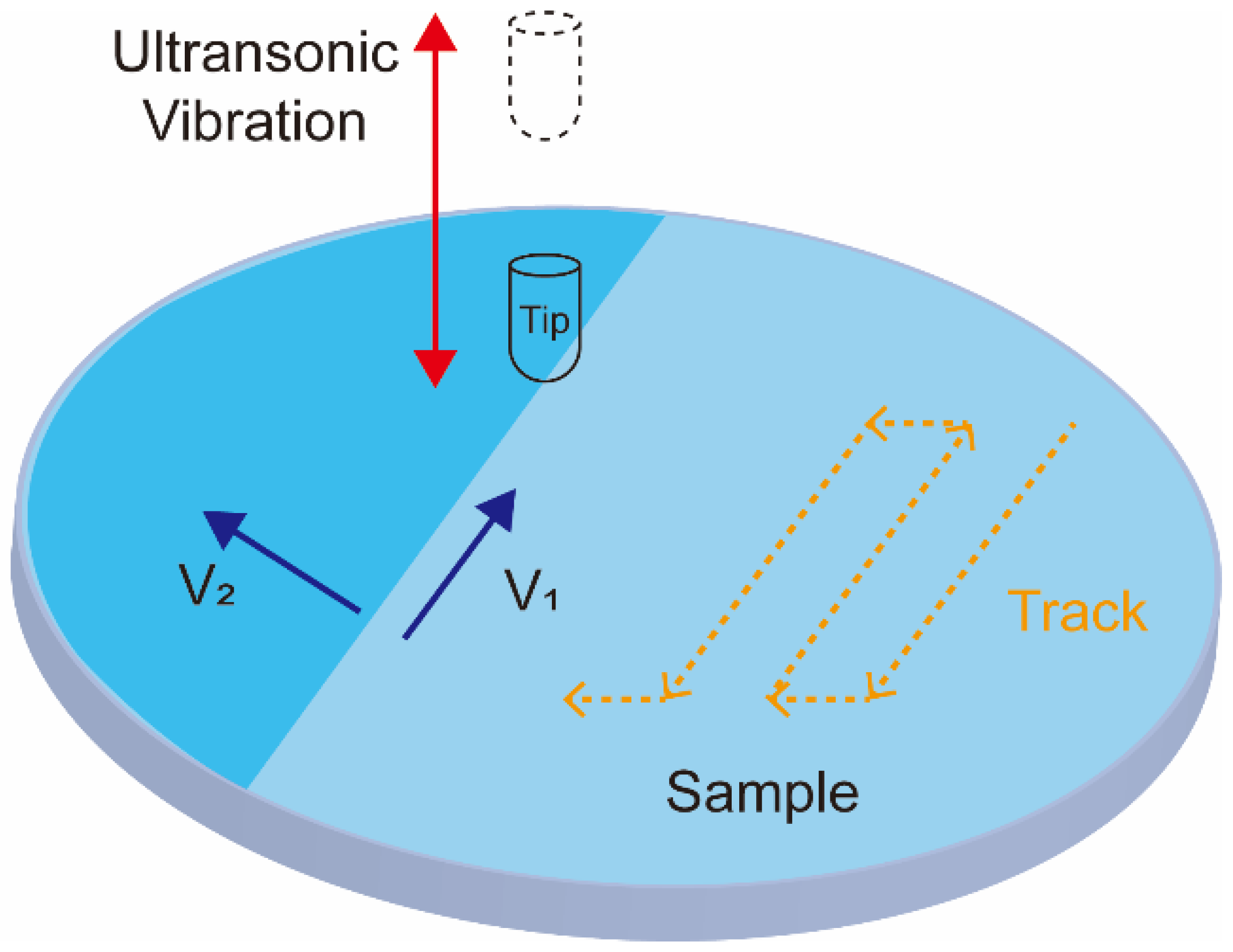
A newly developed ultrasonic nanocrystal surface modification (UNSM) method is currently attracting increasing attention in the field of biomedical applications. It can improve the fatigue strength, wear, and corrosion resistance of alloys. The UNSM process can affect the mechanical properties in a range of up to approximately 750 μm in depth. UNSM is a method that applies ultrasonic impacts to generate nanostructured surfaces. During UNSM, the ultrasonic waves are passed through a tungsten carbide tip at high frequencies (~20 kHz) and transposed onto a sample surface (see Figure 4) [129]. The UNSM is unique in that it can produce precise hierarchical surface patterns by accurately adjusting the operation parameters. The temperature of UNSM is also an important parameter. UNSM at different temperatures was used to treat a Co–Cr–Mo alloy manufactured by SLM [130]. The effect of UNSM at high temperature is stronger than that at ambient temperature. UNSM improves the sample surface finish and decreases its porosity, which translates into improved biocompatibility, corrosion, and mechanical properties.
UNSM is also widely used for biomedical device treatments. Hou et al. [131] fabricated a hierarchical surface structure on Ti–Ni alloys via UNSM to improve corrosion resistance and hardness. Ye et al. [132] applied UNSM to prepare Ni–Ti alloys with an amorphous surface layer. The resulting material demonstrated high wear resistance and excellent biocompatibility. Kheradmandfard et al. [133,134] UNSM-treated Ti–Nb–Ta–Zr-based implants and created micropatterns on their surfaces, which benefited the implant’s bioactivity and bone regeneration performance. UNSM treatment also improved the wear resistance, biocompatibility [135], surface finish, hardness, and corrosion resistance [136] of Ti alloys while it decreased surface porosity. Hou et al. [137] also reported that UNSM significantly improved the hardness, yield stress, and wear resistance of Mg alloys as well as the mechanical and tribological properties of 316L stainless steel tubing [138] used in various biomedical applications. Ma et al. [139] used UNSM to treat the poor surface finish of 3D-printed metals and obtained significantly improved corrosion, wear, and fatigue resistance of the resulting 3D parts.
Advanced and innovative UNSM treatments were also reported in the literature. For example, Zhang et al. [140] developed electrically-assisted ultrasonication nanocrystal surface modification (EA-UNSM), capable of generating smoother surfaces and lower porosities than those of conventional UNSM. Amanov and Pyun [141] combined UNSM with the local heat treatment (LHT) and achieved a very hard Ti–6Al–4V alloy.
4.5. Physical Vapor Deposition
Physical vapor deposition (PVD) is also an effective way to enhance biofunctionality through modifying the surface of alloy implants. Similar to the methods discussed above, PVD protect implants from corrosive environments by applying coatings [142]. Generally, PVD coatings are dense and uniform but time-consuming. PVD can easily control the Ca/Pa ratio and structure, which makes Ca–Pa-based coasting widely used in PVD, such as HA [143], Si–HA [144], C–HA [145], and Mg–HA [146]. However, the deposition rate of PVD is not satisfactory for biomedical applications. The most common PVD processes are magnetron sputtering [147,148,149] and vacuum evaporation [150,151,152]. The main biomedical application of PVD is improving hardness, biocompatibility, wear, and corrosion resistance. The working depth range is up to approximately 4 μm. There are some examples for applying PVD on biomedical alloys. Ben and Khlifi [153] developed TiN biocompatible coatings on Ti alloys by cathodic arc evaporation vacuum deposition. To enhance corrosion resistance and biocompatibility, Li et al. [154] prepared MgF2 coatings on a MgCa alloy via the vacuum evaporation deposition method. Gonzalez et al. [148] deposited Ti–Nb coatings on 316L stainless steel substrates by magnetron sputtering. Kim et al. [155] modified the morphologies of HA coatings on Ti–Ta–Zr alloys with superior wettability by radio-frequency magnetron sputtering and a cyclic voltammetry.
5. Conclusions
Biomedical alloys are used to solve a variety of medical problems, especially those related to bones. Typical biomedical alloys include stainless steel, Co–Cr alloys, and Ti alloys. These biomedical alloys possess excellent corrosion resistance and mechanical properties, which makes them excellent materials for future biomedical uses. However, there are still problems on their surfaces during service. Developing surface treatment methods has attracted increasing attention. Many efforts have proved that physical surface modifications are effective and stable ways to enhance surface biofunctionality. Common physical surface modifications are thermal spraying, glow discharge plasma, ion implantation, ultrasonic nanocrystal surface modification, and physical vapor deposition. Although these methods have achieved certain results, further improvement is still necessary to satisfy the increasing surgical requirements. The main process of physical surface modifications is applying coatings on the substrates. Thus, investigating novel coating materials will be helpful in the future. Moreover, combining these physical surface modifications in a reasonable way can have them support each other and overcome their individual disadvantages. In summary, more efforts are needed to develop physical surface modifications on biomedical alloys for medical application.
Author Contributions
Writing—initial draft preparation, review and editing, X.Y. and W.C.; data collection, X.Y. and H.L.; funding and proposal writing, X.Y. All authors have read and agreed to the published version of the manuscript.
Funding
Funding was provided by the National Natural Science Foundation of China through grant number 81802159.
Institutional Review Board Statement
Not applicable.
Informed Consent Statement
Not applicable.
Data Availability Statement
The data provided in this study could be released upon reasonable request.
Conflicts of Interest
The authors declare no competing interest.
References
- Peppas, N.A.; Langer, R. New challenges in biomaterials. Science 1994, 263, 1715. [Google Scholar] [CrossRef] [PubMed]
- Mooney, D.J.; Mikos, A.G. Growing new organs. Sci. Am. 1999, 280, 60–65. [Google Scholar] [CrossRef]
- Zhao, D.; Zhu, T.; Li, J.; Cui, L.; Zhang, Z.; Zhuang, X.; Ding, J. Poly(lactic-co-glycolic acid)-based composite bone-substitute materials. Bioact. Mater. 2021, 6, 346–360. [Google Scholar] [CrossRef]
- Langer, R.; Tirrell, D.A. Designing materials for biology and medicine. Nature 2004, 428, 487–492. [Google Scholar] [CrossRef] [PubMed]
- Niinomi, M.; Nakai, M.; Hieda, J. Development of new metallic alloys for biomedical applications. Acta Biomater. 2012, 8, 3888–3903. [Google Scholar] [CrossRef] [PubMed]
- Uggowitzer, P.J.; Magdowski, R.; Speidel, M.O. Nickel free high nitrogen austenitic steels. ISIJ Int. 1996, 36, 901–908. [Google Scholar] [CrossRef] [Green Version]
- Chiba, A.; Kumagai, K.; Takeda, H.; Nomura, N. Mechanical properties of forged low ni and c-containing co-cr-mo biomedical implant alloy. Mater. Sci. Forum 2005, 475–479, 2317–2322. [Google Scholar] [CrossRef]
- Wang, K. The use of titanium for medical applications in the USA. Mater. Sci. Eng. A 1996, 213, 134–137. [Google Scholar] [CrossRef]
- Niinomi, M. Recent metallic materials for biomedical applications. Metall. Mater. Trans. A 2002, 33, 477. [Google Scholar] [CrossRef]
- Jin, W.; Wang, G.; Lin, Z.; Feng, H.; Li, W.; Peng, X.; Qasim, A.M.; Chu, P.K. Corrosion resistance and cytocompatibility of tantalum-surface-functionalized biomedical zk60 mg alloy. Corros. Sci. 2017, 114, 45–56. [Google Scholar] [CrossRef]
- Al-Mangour, B.; Mongrain, R.; Irissou, E.; Yue, S. Improving the strength and corrosion resistance of 316l stainless steel for biomedical applications using cold spray. Surf. Coat. Technol. 2013, 216, 297–307. [Google Scholar] [CrossRef]
- Niinomi, M. Mechanical biocompatibilities of titanium alloys for biomedical applications. J. Mech. Behav. Biomed. Mater. 2008, 1, 30–42. [Google Scholar] [CrossRef]
- Saldívar-García, A.J.; López, H.F. Microstructural effects on the wear resistance of wrought and as-cast co-cr-mo-c implant alloys. J. Biomed. Mater. Res. Part A 2005, 74A, 269–274. [Google Scholar] [CrossRef] [PubMed]
- Zhou, H.; Liang, B.; Jiang, H.; Deng, Z.; Yu, K. Magnesium-based biomaterials as emerging agents for bone repair and regeneration: From mechanism to application. J. Magnes. Alloy 2021, 9, 779–804. [Google Scholar] [CrossRef]
- Ullah, S.; Chen, X. Fabrication, applications and challenges of natural biomaterials in tissue engineering. Appl. Mater. Today 2020, 20, 100656. [Google Scholar] [CrossRef]
- Deng, Z.N.; Liu, J.S.; He, Y.; Wang, S.Q.; Ma, J.F. Synthesis and properties of hydroxyapatite-containing porous titania coating on titanium by ultrasonic shot peening and micro-arc oxidation. Adv. Mater. Res. 2013, 690–693, 2081–2084. [Google Scholar] [CrossRef]
- Wang, Y.; Yu, H.; Chen, C.; Zhao, Z. Review of the biocompatibility of micro-arc oxidation coated titanium alloys. Mater. Des. 2015, 85, 640–652. [Google Scholar] [CrossRef]
- Zhang, L.-C.; Chen, L.-Y.; Wang, L. Surface modification of titanium and titanium alloys: Technologies, developments, and future interests. Adv. Eng. Mater. 2020, 22, 1901258. [Google Scholar] [CrossRef]
- Mauer, G.; Vaßen, R. Conditions for nucleation and growth in the substrate boundary layer at plasma spray-physical vapor deposition (ps-pvd). Surf. Coat. Technol. 2019, 371, 417–427. [Google Scholar] [CrossRef]
- Saravanan, M.; Devaraju, A.; Venkateshwaran, N.; Krishnakumari, A.; Saarvesh, J. A review on recent progress in coatings on aisi austenitic stainless steel. Mater. Today Proc. 2018, 5, 14392–14396. [Google Scholar] [CrossRef]
- Talha, M.; Behera, C.K.; Sinha, O.P. A review on nickel-free nitrogen containing austenitic stainless steels for biomedical applications. Mater. Sci. Eng. C 2013, 33, 3563–3575. [Google Scholar] [CrossRef]
- Al Jabbari, Y.S. Physico-mechanical properties and prosthodontic applications of co-cr dental alloys: A review of the literature. J. Adv. Prosthodont. 2014, 6, 138–145. [Google Scholar] [CrossRef] [Green Version]
- Yamanaka, K.; Mori, M.; Chiba, A. Developing high strength and ductility in biomedical co–cr cast alloys by simultaneous doping with nitrogen and carbon. Acta Biomater. 2016, 31, 435–447. [Google Scholar] [CrossRef] [Green Version]
- Mitragotri, S.; Lahann, J. Physical approaches to biomaterial design. Nat. Mater. 2009, 8, 15–23. [Google Scholar] [CrossRef] [Green Version]
- Antunes, R.A.; de Oliveira, M.C.L. Corrosion fatigue of biomedical metallic alloys: Mechanisms and mitigation. Acta Biomater. 2012, 8, 937–962. [Google Scholar] [CrossRef]
- Karamian, E.; Kalantar Motamedi, M.R.; Khandan, A.; Soltani, P.; Maghsoudi, S. An in vitro evaluation of novel nha/zircon plasma coating on 316l stainless steel dental implant. Prog. Nat. Sci. Mater. Int. 2014, 24, 150–156. [Google Scholar] [CrossRef] [Green Version]
- Sabará, E.W.F.; Pereira, V.; Molisani, A.L.; Caldeira, L.; Souza, R.C.; Simões, T.A.; Bueno, A.H.S. Electrochemical behaviour and microstructural characterization of different austenitic stainless steel for biomedical applications. Mater. Res. Express 2020, 7, 105402. [Google Scholar] [CrossRef]
- Terada, M.; Antunes, R.A.; Padilha, A.F.; Costa, I. Corrosion resistance of three austenitic stainless steels for biomedical applications. Mater. Corros. 2007, 58, 762–766. [Google Scholar] [CrossRef]
- Uggowitzer, P.J. Metal injection molding of nickel-free stainless steels. Adv. Powder Met. Part. Mater. 1997, 3, 113–121. [Google Scholar]
- Yang, K.; Ren, Y. Nickel-free austenitic stainless steels for medical applications. Sci. Technol. Adv. Mater. 2010, 11, 014105. [Google Scholar] [CrossRef]
- Talha, M.; Behera, C.K.; Sinha, O.P. Effect of nitrogen and cold working on structural and mechanical behavior of ni-free nitrogen containing austenitic stainless steels for biomedical applications. Mater. Sci. Eng. C 2015, 47, 196–203. [Google Scholar] [CrossRef]
- Challa, V.S.A.; Nune, K.C.; Gong, N.; Misra, R.D.K. The significant impact of mechanically-induced phase transformation on cellular functionality of biomedical austenitic stainless steel. J. Mech. Behav. Biomed. Mater. 2020, 108, 103815. [Google Scholar] [CrossRef]
- Peng, C.; Izawa, T.; Zhu, L.; Kuroda, K.; Okido, M. Tailoring surface hydrophilicity property for biomedical 316l and 304 stainless steels: A special perspective on studying osteoconductivity and biocompatibility. ACS Appl. Mater. Interfaces 2019, 11, 45489–45497. [Google Scholar] [CrossRef]
- Yang, Y.; Wang, Q.; Li, J.; Tan, L.; Yang, K. Enhancing general corrosion resistance of biomedical high nitrogen nickel-free stainless steel by nitric acid passivation. Acta Metall. Sin. 2020, 33, 307–312. [Google Scholar] [CrossRef] [Green Version]
- Aguilar, L.E.; Lee, J.Y.; Park, C.H.; Kim, C.S. Biomedical grade stainless steel coating of polycaffeic acid via combined oxidative and ultraviolet light-assisted polymerization process for bioactive implant application. Polymers 2019, 11, 584. [Google Scholar] [CrossRef] [Green Version]
- Hsu, H.-J.; Wu, C.-Y.; Huang, B.-H.; Tsai, C.-H.; Saito, T.; Ou, K.-L.; Chuo, Y.-C.; Lin, K.-L.; Peng, P.-W. Surface characteristics and cell adhesion behaviors of the anodized biomedical stainless steel. Appl. Sci. 2020, 10, 6275. [Google Scholar] [CrossRef]
- Teo, A.Q.; Yan, L.; Chaudhari, A.; O’Neill, G.K. Post-processing and surface characterization of additively manufactured stainless steel 316l lattice: Implications for biomedical use. Materials 2021, 14, 1376. [Google Scholar] [CrossRef]
- Lodhi, M.J.K.; Deen, K.M.; Greenlee-Wacker, M.C.; Haider, W. Additively manufactured 316l stainless steel with improved corrosion resistance and biological response for biomedical applications. Addit. Manuf. 2019, 27, 8–19. [Google Scholar] [CrossRef]
- Talha, M.; Ma, Y.; Lin, Y.; Pan, Y.; Kong, X.; Sinha, O.P.; Behera, C.K. Corrosion performance of cold deformed austenitic stainless steels for biomedical applications. Corros. Rev. 2019, 37, 283–306. [Google Scholar] [CrossRef]
- Yang, Y.; Wang, Q.; Li, J.; Tan, L.; Yang, K. Enhancing general corrosion resistance of biomedical high nitrogen nickel-free stainless steel by water treatment. Mater. Lett. 2019, 251, 196–200. [Google Scholar] [CrossRef]
- Trzaskowska, P.A.; Kuźmińska, A.; Butruk-Raszeja, B.; Rybak, E.; Ciach, T. Electropolymerized hydrophilic coating on stainless steel for biomedical applications. Colloids Surf. B Biointerfaces 2018, 167, 499–508. [Google Scholar] [CrossRef] [PubMed]
- Kocijan, A.; Milošev, I.; Pihlar, B. Cobalt-based alloys for orthopaedic applications studied by electrochemical and xps analysis. J. Mater. Sci. Mater. Med. 2004, 15, 643–650. [Google Scholar] [CrossRef] [PubMed]
- Zhu, Z.-Y.; Meng, L.; Chen, L. Strain-induced martensitic transformation in biomedical co–cr–w–ni alloys. Rare Met. 2020, 39, 241–249. [Google Scholar] [CrossRef]
- Ueki, K.; Kasamatsu, M.; Ueda, K.; Koizumi, Y.; Wei, D.; Chiba, A.; Narushima, T. Precipitation during γ-ε phase transformation in biomedical co-cr-mo alloys fabricated by electron beam melting. Metals 2020, 10, 71. [Google Scholar] [CrossRef] [Green Version]
- Kaita, W.; Hagihara, K.; Rocha, L.A.; Nakano, T. Plastic deformation mechanisms of biomedical co–cr–mo alloy single crystals with hexagonal close-packed structure. Scr. Mater. 2018, 142, 111–115. [Google Scholar] [CrossRef]
- Mori, M.; Yamanaka, K.; Sato, S.; Tsubaki, S.; Satoh, K.; Kumagai, M.; Imafuku, M.; Shobu, T.; Chiba, A. Tuning strain-induced γ-to-ε martensitic transformation of biomedical co–cr–mo alloys by introducing parent phase lattice defects. J. Mech. Behav. Biomed. Mater. 2019, 90, 523–529. [Google Scholar] [CrossRef] [PubMed]
- Dong, X.; Zhou, Y.; Sun, Q.; Qu, Y.; Shi, H.; Liu, W.; Peng, H.; Zhang, B.; Xu, S.; Yan, J.; et al. Fatigue behavior of biomedical co–cr–mo–w alloy fabricated by selective laser melting. Mater. Sci. Eng. A 2020, 795, 140000. [Google Scholar] [CrossRef]
- Zhou, Y.; Wei, W.; Yan, J.; Liu, W.; Li, N.; Li, H.; Xu, S. Microstructures and metal-ceramic bond properties of co-cr biomedical alloys fabricated by selective laser melting and casting. Mater. Sci. Eng. A 2019, 759, 594–602. [Google Scholar] [CrossRef]
- Dong, X.; Sun, Q.; Zhou, Y.; Qu, Y.; Shi, H.; Zhang, B.; Xu, S.; Liu, W.; Li, N.; Yan, J. Influence of microstructure on corrosion behavior of biomedical co-cr-mo-w alloy fabricated by selective laser melting. Corros. Sci. 2020, 170, 108688. [Google Scholar] [CrossRef]
- Yamanaka, K.; Mori, M.; Kuramoto, K.; Chiba, A. Development of new co–cr–w-based biomedical alloys: Effects of microalloying and thermomechanical processing on microstructures and mechanical properties. Mater. Des. 2014, 55, 987–998. [Google Scholar] [CrossRef]
- Kheimehsari, H.; Izman, S.; Shirdar, M.R. Effects of ha-coating on the surface morphology and corrosion behavior of a co-cr-based implant in different conditions. J. Mater. Eng. Perform. 2015, 24, 2294–2302. [Google Scholar] [CrossRef]
- Rezazadeh Shirdar, M.; Sudin, I.; Taheri, M.M.; Keyvanfar, A.; Yusop, M.Z.M.; Kadir, M.R.A. A novel hydroxyapatite composite reinforced with titanium nanotubes coated on co–cr-based alloy. Vacuum 2015, 122, 82–89. [Google Scholar] [CrossRef]
- Sawangrat, C.; Yamaguchi, O.; Vajpai, S.K.; Ameyama, K. Application of harmonic structure design to biomedical co–cr–mo alloy for improved mechanical properties. Mater. Trans. 2014, 55, 99–105. [Google Scholar] [CrossRef] [Green Version]
- Trimble, D.; Agarwal, A.; McDonnell, D.; Barron, S.; Ahearne, E.; O’Donnell, G.E. Finite element simulation of orthogonal machining of biomedical grade co–cr–mo alloy. CIRP J. Manuf. Sci. Technol. 2020, 28, 8–14. [Google Scholar] [CrossRef]
- Migita, S.; Sakashita, K.; Saito, Y.; Suyalatu; Yamazaki, T. Co–cr–mo alloy binding peptide as molecular glue for constructing biomedical surfaces. J. Appl. Biomater. Funct. Mater. 2020, 18, 2280800020924739. [Google Scholar] [CrossRef]
- Yamanaka, K.; Mori, M.; Yoshida, K.; Balvay, S.; Hartmann, D.; Fabrègue, D.; Chiba, A. Preparation of high-strength Co−Cr−Mo alloy rods via hot-caliber rolling. Materialia 2020, 12, 100729. [Google Scholar] [CrossRef]
- Mahajan, A.; Sidhu, S.S. Enhancing biocompatibility of co-cr alloy implants via electrical discharge process. Mater. Technol. 2018, 33, 524–531. [Google Scholar] [CrossRef]
- Okazaki, Y.; Gotoh, E. Comparison of metal release from various metallic biomaterials in vitro. Biomaterials 2005, 26, 11–21. [Google Scholar] [CrossRef]
- Assis, S.L.; Costa, I. Electrochemical evaluation of ti-13nb-13zr, ti-6al-4v and ti-6al-7nb alloys for biomedical application by long-term immersion tests. Mater. Corros. 2007, 58, 329–333. [Google Scholar] [CrossRef]
- Elias, C.N.; Fernandes, D.J.; Souza, F.M.d.; Monteiro, E.d.S.; Biasi, R.S.d. Mechanical and clinical properties of titanium and titanium-based alloys (ti g2, ti g4 cold worked nanostructured and ti g5) for biomedical applications. J. Mater. Res. Technol. 2019, 8, 1060–1069. [Google Scholar] [CrossRef]
- Elias, C.N.; Meirelles, L. Improving osseointegration of dental implants. Expert Rev. Med. Devices 2010, 7, 241–256. [Google Scholar] [CrossRef] [PubMed]
- Sri Maha Vishnu, D.; Sure, J.; Liu, Y.; Vasant Kumar, R.; Schwandt, C. Electrochemical synthesis of porous ti-nb alloys for biomedical applications. Mater. Sci. Eng. C 2019, 96, 466–478. [Google Scholar] [CrossRef]
- Ibrahim, M.K.; Hamzah, E.; Saud, S.N.; Nazim, E.M.; Bahador, A. Parameter optimization of microwave sintering porous ti-23%nb shape memory alloys for biomedical applications. Trans. Nonferrous Met. Soc. China 2018, 28, 700–710. [Google Scholar] [CrossRef]
- Kuroda, P.A.B.; da Silva, L.M.; Sousa, K.d.S.J.; Donato, T.A.G.; Grandini, C.R. Preparation, structural, microstructural, mechanical, and cytotoxic characterization of ti-15nb alloy for biomedical applications. Artif. Organs 2020, 44, 811–817. [Google Scholar] [CrossRef]
- Wang, Z.; Xu, X.-W.; Zhang, B. Hot compression deformation behavior of biomedical ni–ti alloy. Rare Met. 2019, 38, 609–619. [Google Scholar] [CrossRef]
- Qi, P.; Li, B.; Wang, T.; Zhou, L.; Nie, Z. Microstructure and properties of a novel ternary ti–6zr–xfe alloy for biomedical applications. J. Alloys Compd. 2021, 854, 157119. [Google Scholar] [CrossRef]
- Maya, A.E.A.; Grana, D.R.; Hazarabedian, A.; Kokubu, G.A.; Luppo, M.I.; Vigna, G. Zr–ti–nb porous alloys for biomedical application. Mater. Sci. Eng. C 2012, 32, 321–329. [Google Scholar] [CrossRef]
- Li, P.; Ma, X.; Wang, D.; Zhang, H. Microstructural and mechanical properties of β-type ti–nb–sn biomedical alloys with low elastic modulus. Metals 2019, 9, 712. [Google Scholar] [CrossRef] [Green Version]
- Chui, P.; Jing, R.; Zhang, F.; Li, J.; Feng, T. Mechanical properties and corrosion behavior of β-type ti-zr-nb-mo alloys for biomedical application. J. Alloys Compd. 2020, 842, 155693. [Google Scholar] [CrossRef]
- Quadros, F.d.F.; Kuroda, P.A.B.; Sousa, K.d.S.J.; Donato, T.A.G.; Grandini, C.R. Preparation, structural and microstructural characterization of ti-25ta-10zr alloy for biomedical applications. J. Mater. Res. Technol. 2019, 8, 4108–4114. [Google Scholar] [CrossRef]
- Yılmaz, E.; Gökçe, A.; Findik, F.; Gülsoy, H.Ö. Biomedical porous ti-16nb-10zr-(0–15)ta alloys: Paper presented at the “vii international congress of biomaterials, biomat’2018”, 14–16 march 2018, havana, cuba. Int. J. Mater. Res. 2018, 110, 375–378. [Google Scholar] [CrossRef]
- Li, P.; Ma, X.; Tong, T.; Wang, Y. Microstructural and mechanical properties of β-type ti–mo–nb biomedical alloys with low elastic modulus. J. Alloys Compd. 2020, 815, 152412. [Google Scholar] [CrossRef]
- Zhu, S.L.; Wang, X.M.; Qin, F.X.; Inoue, A. A new ti-based bulk glassy alloy with potential for biomedical application. Mater. Sci. Eng. A 2007, 459, 233–237. [Google Scholar] [CrossRef]
- Zhao, C.; Zhang, X.; Cao, P. Mechanical and electrochemical characterization of ti–12mo–5zr alloy for biomedical application. J. Alloys Compd. 2011, 509, 8235–8238. [Google Scholar] [CrossRef]
- Gao, A.; Hang, R.; Bai, L.; Tang, B.; Chu, P.K. Electrochemical surface engineering of titanium-based alloys for biomedical application. Electrochim. Acta 2018, 271, 699–718. [Google Scholar] [CrossRef]
- Xu, L.J.; Chen, Y.Y.; Liu, Z.G.; Kong, F.T. The microstructure and properties of ti–mo–nb alloys for biomedical application. J. Alloys Compd. 2008, 453, 320–324. [Google Scholar] [CrossRef]
- Yang, D.; Guo, Z.; Shao, H.; Liu, X.; Ji, Y. Mechanical properties of porous ti-mo and ti-nb alloys for biomedical application by gelcasting. Procedia Eng. 2012, 36, 160–167. [Google Scholar] [CrossRef] [Green Version]
- Bolzoni, L.; Yang, F. Development of cu-bearing powder metallurgy ti alloys for biomedical applications. J. Mech. Behav. Biomed. Mater. 2019, 97, 41–48. [Google Scholar] [CrossRef] [PubMed]
- Lima, R.S.; Khor, K.A.; Li, H.; Cheang, P.; Marple, B.R. Hvof spraying of nanostructured hydroxyapatite for biomedical applications. Mater. Sci. Eng. A 2005, 396, 181–187. [Google Scholar] [CrossRef] [Green Version]
- Wielage, B.; Wank, A.; Pokhmurska, H.; Grund, T.; Rupprecht, C.; Reisel, G.; Friesen, E. Development and trends in hvof spraying technology. Surf. Coat. Technol. 2006, 201, 2032–2037. [Google Scholar] [CrossRef]
- Gadow, R.; Killinger, A.; Stiegler, N. Hydroxyapatite coatings for biomedical applications deposited by different thermal spray techniques. Surf. Coat. Technol. 2010, 205, 1157–1164. [Google Scholar] [CrossRef]
- Stiegler, N.; Bellucci, D.; Bolelli, G.; Cannillo, V.; Gadow, R.; Killinger, A.; Lusvarghi, L.; Sola, A. High-velocity suspension flame sprayed (hvsfs) hydroxyapatite coatings for biomedical applications. J. Therm. Spray Technol. 2012, 21, 275–287. [Google Scholar] [CrossRef]
- Bansal, P.; Singh, G.; Sidhu, H.S. Improvement of surface properties and corrosion resistance of ti13nb13zr titanium alloy by plasma-sprayed ha/zno coatings for biomedical applications. Mater. Chem. Phys. 2021, 257, 123738. [Google Scholar] [CrossRef]
- Singh, H.; Sidhu, B.S.; Puri, D.; Prakash, S. Use of plasma spray technology for deposition of high temperature oxidation/corrosion resistant coatings—A review. Mater. Corros. 2007, 58, 92–102. [Google Scholar] [CrossRef]
- Liu, X.; Ding, C. Morphology of apatite formed on surface of wollastonite coating soaked in simulate body fluid. Mater. Lett. 2002, 57, 652–655. [Google Scholar] [CrossRef]
- Zhou, H.; Li, F.; He, B.; Wang, J.; Sun, B.-d. Air plasma sprayed thermal barrier coatings on titanium alloy substrates. Surf. Coat. Technol. 2007, 201, 7360–7367. [Google Scholar] [CrossRef]
- Sathish, T. Bonn technique: Tribological properties predictor for plasma nitrided 316l stainless steel. Mater. Today Proc. 2018, 5, 14545–14552. [Google Scholar] [CrossRef]
- Bacci, T.; Bertamini, L.; Ferrari, F.; Galliano, F.P.; Galvanetto, E. Reactive plasma spraying of titanium in nitrogen containing plasma gas. Mater. Sci. Eng. A 2000, 283, 189–195. [Google Scholar] [CrossRef]
- Liu, X.; Chu, P.K.; Ding, C. Surface modification of titanium, titanium alloys, and related materials for biomedical applications. Mater. Sci. Eng. R Rep. 2004, 47, 49–121. [Google Scholar] [CrossRef] [Green Version]
- Kokubo, T.; Ito, S.; Shigematsu, M.; Sanka, S.; Yamamuro, T. Fatigue and life-time of bioactive glass-ceramic a-w containing apatite and wollastonite. J. Mater. Sci. 1987, 22, 4067–4070. [Google Scholar] [CrossRef]
- De Aza, P.N.; Luklinska, Z.B.; Anseau, M.R.; Hector, M.; Guitián, F.; De Aza, S. Reactivity of a wollastonite–tricalcium phosphate bioeutectic® ceramic in human parotid saliva. Biomaterials 2000, 21, 1735–1741. [Google Scholar] [CrossRef]
- Zhang, W.; Gu, J.; Zhang, C.; Xie, Y.; Zheng, X. Preparation of titania coating by induction suspension plasma spraying for biomedical application. Surf. Coat. Technol. 2019, 358, 511–520. [Google Scholar] [CrossRef]
- Nonami, T.; Tsutsumi, S. Study of diopside ceramics for biomaterials. J. Mater. Sci. Mater. Med. 1999, 10, 475–479. [Google Scholar] [CrossRef]
- Aruna, S.T.R.; Shilpa, M.; Lakshmi, R.V.; Balaji, N.; Kavitha, V.; Gnanamani, A. Plasma sprayed hydroxyapatite bioceramic coatings from coprecipitation synthesized powder: Preparation, characterization and in vitro studies. Trans. Indian Ceram. Soc. 2018, 77, 90–99. [Google Scholar] [CrossRef]
- Pillai, R.S.; Frasnelli, M.; Sglavo, V.M. Ha/β-tcp plasma sprayed coatings on ti substrate for biomedical applications. Ceram. Int. 2018, 44, 1328–1333. [Google Scholar] [CrossRef]
- Bai, Y.; Zhou, S.-J.; Shi, L.; Ma, W.; Liu, C.-W. Fabrication and characterization of suspension plasma-sprayed fluoridated hydroxyapatite coatings for biomedical applications. J. Therm. Spray Technol. 2018, 27, 1322–1332. [Google Scholar] [CrossRef]
- Kurzweg, H.; Heimann, R.B.; Troczynski, T.; Wayman, M.L. Development of plasma-sprayed bioceramic coatings with bond coats based on titania and zirconia. Biomaterials 1998, 19, 1507–1511. [Google Scholar] [CrossRef]
- Chou, B.-Y.; Chang, E. Interface investigation of plasma-sprayed hydroxyapatite coating on titanium alloy with zro2 intermediate layer as bond coat. Scr. Mater. 2001, 45, 487–493. [Google Scholar] [CrossRef]
- Singh, J.; Chatha, S.S.; Singh, H. Synthesis and characterization of plasma sprayed functional gradient bioceramic coating for medical implant applications. Ceram. Int. 2021, 47, 9143–9155. [Google Scholar] [CrossRef]
- Liu, X.-M.; He, D.-Y.; Zhou, Z.; Wang, G.-H.; Wang, Z.-J.; Wu, X.; Tan, Z. Atmospheric plasma-sprayed hydroxyapatite coatings with (002) texture. J. Therm. Spray Technol. 2018, 27, 1291–1301. [Google Scholar] [CrossRef]
- Hameed, P.; Gopal, V.; Bjorklund, S.; Ganvir, A.; Sen, D.; Markocsan, N.; Manivasagam, G. Axial suspension plasma spraying: An ultimate technique to tailor ti6al4v surface with hap for orthopaedic applications. Colloids Surf. B Biointerfaces 2019, 173, 806–815. [Google Scholar] [CrossRef] [PubMed]
- Chu, P.K.; Chen, J.Y.; Wang, L.P.; Huang, N. Plasma-surface modification of biomaterials. Mater. Sci. Eng. R Rep. 2002, 36, 143–206. [Google Scholar] [CrossRef] [Green Version]
- Favia, P.; d’Agostino, R. Plasma treatments and plasma deposition of polymers for biomedical applications. Surf. Coat. Technol. 1998, 98, 1102–1106. [Google Scholar] [CrossRef]
- Cools, P.; De Geyter, N.; Vanderleyden, E.; Barberis, F.; Dubruel, P.; Morent, R. Adhesion improvement at the pmma bone cement-titanium implant interface using methyl methacrylate atmospheric pressure plasma polymerization. Surf. Coat. Technol. 2016, 294, 201–209. [Google Scholar] [CrossRef]
- Aronsson, B.O.; Lausmaa, J.; Kasemo, B. Glow discharge plasma treatment for surface cleaning and modification of metallic biomaterials. J. Biomed. Mater. Res. 1997, 35, 49–73. [Google Scholar] [CrossRef]
- Truica-Marasescu, F.; Wertheimer, M.R. Nitrogen-rich plasma-polymer films for biomedical applications. Plasma Process. Polym. 2008, 5, 44–57. [Google Scholar] [CrossRef]
- Yuvaraj, S.; Muthukumarasamy, N.; Flores, M.; Rajesh, G.; Paraskevopoulos, K.M.; Pouroutzidou, G.K.; Theodorou, G.S.; Ioannidou, K.; Lusvarghi, L.; Velauthapillai, D.; et al. Incorporation of nanosized carbon over hydroxyapatite (hap) surface using dc glow discharge plasma for biomedical application. Vacuum 2021, 190, 110300. [Google Scholar] [CrossRef]
- Sarra-Bournet, C.; Turgeon, S.; Mantovani, D.; Laroche, G. A study of atmospheric pressure plasma discharges for surface functionalization of ptfe used in biomedical applications. J. Phys. D Appl. Phys. 2006, 39, 3461–3469. [Google Scholar] [CrossRef]
- Wu, S.; Wand, Z.; Huang, Q.; Lu, X.; Pan, Y. Study on a room-temperature air plasma for biomedical application. IEEE Trans. Plasma Sci. 2011, 39, 1489–1495. [Google Scholar] [CrossRef] [Green Version]
- Lee, H.W.; Kim, M.S.; Won, I.H.; Yun, G.S.; Lee, J.K. Self-prevention of instability in a low-power microwave ar plasma jet for biomedical applications. J. Phys. D Appl. Phys. 2015, 48, 155203. [Google Scholar] [CrossRef]
- Li, J.; Lei, B.; Wang, J.; Xu, B.; Ran, S.; Wang, Y.; Zhang, T.; Tang, J.; Zhao, W.; Duan, Y. Atmospheric diffuse plasma jet formation from positive-pseudo-streamer and negative pulseless glow discharges. Commun. Phys. 2021, 4, 64. [Google Scholar] [CrossRef]
- Demkin, V.P.; Melnichuk, S.V.; Postnikov, A.V. Generating low-temperature glow discharge plasma in the atmospheric pressure helium after spark breakdown: Modelling plasma with the prescribed properties for biomedical applications. Phys. Plasmas 2018, 25, 083502. [Google Scholar] [CrossRef]
- Pandiyaraj, K.N.; Deshmukh, R.R.; Mahendiran, R.; Su, P.-G.; Yassitepe, E.; Shah, I.; Perni, S.; Prokopovich, P.; Nadagouda, M.N. Influence of operating parameters on surface properties of rf glow discharge oxygen plasma treated tio2/pet film for biomedical application. Mater. Sci. Eng. C 2014, 36, 309–319. [Google Scholar] [CrossRef]
- Jin, G.; Qin, H.; Cao, H.; Qian, S.; Zhao, Y.; Peng, X.; Zhang, X.; Liu, X.; Chu, P.K. Synergistic effects of dual zn/ag ion implantation in osteogenic activity and antibacterial ability of titanium. Biomaterials 2014, 35, 7699–7713. [Google Scholar] [CrossRef] [PubMed]
- Borgioli, F.; Galvanetto, E.; Galliano, F.P.; Bacci, T. Air treatment of pure titanium by furnace and glow-discharge processes. Surf. Coat. Technol. 2001, 141, 103–107. [Google Scholar] [CrossRef]
- Rossi, S.; Fedrizzi, L.; Bacci, T.; Pradelli, G. Corrosion behaviour of glow discharge nitrided titanium alloys. Corros. Sci. 2003, 45, 511–529. [Google Scholar] [CrossRef]
- Puleo, D.A.; Kissling, R.A.; Sheu, M.S. A technique to immobilize bioactive proteins, including bone morphogenetic protein-4 (bmp-4), on titanium alloy. Biomaterials 2002, 23, 2079–2087. [Google Scholar] [CrossRef]
- Rautray, T.R.; Narayanan, R.; Kim, K.-H. Ion implantation of titanium based biomaterials. Prog. Mater. Sci. 2011, 56, 1137–1177. [Google Scholar] [CrossRef] [Green Version]
- Tan, L.; Crone, W.C. Surface characterization of niti modified by plasma source ion implantation. Acta Mater. 2002, 50, 4449–4460. [Google Scholar] [CrossRef]
- Li, J.; Sun, M.; Ma, X. Structural characterization of titanium oxide layers prepared by plasma based ion implantation with oxygen on ti6al4v alloy. Appl. Surf. Sci. 2006, 252, 7503–7508. [Google Scholar] [CrossRef]
- Wu, H.; Xi, K.; Xiao, S.; Qasim, A.M.; Fu, R.K.Y.; Shi, K.; Ding, K.; Chen, G.; Wu, G.; Chu, P.K. Formation of self-layered hydrothermal coating on magnesium aided by titanium ion implantation: Synergistic control of corrosion resistance and cytocompatibility. Surf. Coat. Technol. 2020, 401, 126251. [Google Scholar] [CrossRef]
- Viviente, J.L.; Garcia, A.; Loinaz, A.; Alonso, F.; Oñate, J.I. Carbon layers formed on steel and ti alloys after ion implantation of c+ at very high doses. Vacuum 1999, 52, 141–146. [Google Scholar] [CrossRef]
- Dong, Q.; Jia, Y.; Ba, Z.; Tao, X.; Wang, Z.; Xue, F.; Bai, J. Exploring the corrosion behavior of mn-implanted biomedical mg. J. Alloys Compd. 2021, 873, 159739. [Google Scholar] [CrossRef]
- Jia, Y.; Ba, Z.; Chen, X.; Zhou, B.; Zhou, W.; Liu, H.; Dong, Q. Controlled surface mechanical property and corrosion resistance of zk60 magnesium alloy treated by zirconium ion implantation. Surf. Topogr. Metrol. Prop. 2020, 8, 025015. [Google Scholar] [CrossRef]
- Wei, X.; Liu, P.; Ma, S.; Li, Z.; Peng, X.; Deng, R.; Zhao, Q. Improvement on corrosion resistance and biocompability of zk60 magnesium alloy by carboxyl ion implantation. Corros. Sci. 2020, 173, 108729. [Google Scholar] [CrossRef]
- Cao, H.; Liu, X.; Meng, F.; Chu, P.K. Biological actions of silver nanoparticles embedded in titanium controlled by micro-galvanic effects. Biomaterials 2011, 32, 693–705. [Google Scholar] [CrossRef] [PubMed]
- Wan, Y.Z.; Raman, S.; He, F.; Huang, Y. Surface modification of medical metals by ion implantation of silver and copper. Vacuum 2007, 81, 1114–1118. [Google Scholar] [CrossRef]
- Jörg, F.; Betül, K.A.; Heiner, M.; Gerhard, K.; Erwin, B.R. Effects of ti6al4v surfaces manufactured through precision centrifugal casting and modified by calcium and phosphorus ion implantation on human osteoblasts. Metals 2020, 10, 1681. [Google Scholar] [CrossRef]
- Biffi, C.A.; Bassani, P.; Nematollahi, M.; Shayesteh Moghaddam, N.; Amerinatanzi, A.; Mahtabi, M.J.; Elahinia, M.; Tuissi, A. Effect of ultrasonic nanocrystal surface modification on the microstructure and martensitic transformation of selective laser melted nitinol. Materials 2019, 12, 3068. [Google Scholar] [CrossRef] [Green Version]
- Amanov, A. Effect of post-additive manufacturing surface modification temperature on the tribological and tribocorrosion properties of co-cr-mo alloy for biomedical applications. Surf. Coat. Technol. 2021, 421, 127378. [Google Scholar] [CrossRef]
- Hou, X.; Mankoci, S.; Walters, N.; Gao, H.; Zhang, R.; Li, S.; Qin, H.; Ren, Z.; Doll, G.L.; Cong, H.; et al. Hierarchical structures on nickel-titanium fabricated by ultrasonic nanocrystal surface modification. Mater. Sci. Eng. C 2018, 93, 12–20. [Google Scholar] [CrossRef]
- Ye, C.; Zhou, X.; Telang, A.; Gao, H.; Ren, Z.; Qin, H.; Suslov, S.; Gill, A.S.; Mannava, S.R.; Qian, D.; et al. Surface amorphization of niti alloy induced by ultrasonic nanocrystal surface modification for improved mechanical properties. J. Mech. Behav. Biomed. Mater. 2016, 53, 455–462. [Google Scholar] [CrossRef]
- Kheradmandfard, M.; Kashani-Bozorg, S.F.; Kim, C.-L.; Hanzaki, A.Z.; Pyoun, Y.-S.; Kim, J.-H.; Amanov, A.; Kim, D.-E. Nanostructured β-type titanium alloy fabricated by ultrasonic nanocrystal surface modification. Ultrason. Sonochem. 2017, 39, 698–706. [Google Scholar] [CrossRef]
- Kheradmandfard, M.; Kashani-Bozorg, S.F.; Kang, K.-H.; Penkov, O.V.; Zarei Hanzaki, A.; Pyoun, Y.-S.; Amanov, A.; Kim, D.-E. Simultaneous grain refinement and nanoscale spinodal decomposition of β phase in ti-nb-ta-zr alloy induced by ultrasonic mechanical impacts. J. Alloys Compd. 2018, 738, 540–549. [Google Scholar] [CrossRef]
- Kheradmandfard, M.; Kashani-Bozorg, S.F.; Lee, J.S.; Kim, C.-L.; Hanzaki, A.Z.; Pyun, Y.-S.; Cho, S.-W.; Amanov, A.; Kim, D.-E. Significant improvement in cell adhesion and wear resistance of biomedical β-type titanium alloy through ultrasonic nanocrystal surface modification. J. Alloys Compd. 2018, 762, 941–949. [Google Scholar] [CrossRef]
- Ma, C.; Andani, M.T.; Qin, H.; Moghaddam, N.S.; Ibrahim, H.; Jahadakbar, A.; Amerinatanzi, A.; Ren, Z.; Zhang, H.; Doll, G.L.; et al. Improving surface finish and wear resistance of additive manufactured nickel-titanium by ultrasonic nano-crystal surface modification. J. Mater. Process. Technol. 2017, 249, 433–440. [Google Scholar] [CrossRef]
- Hou, X.; Qin, H.; Gao, H.; Mankoci, S.; Zhang, R.; Zhou, X.; Ren, Z.; Doll, G.L.; Martini, A.; Sahai, N.; et al. A systematic study of mechanical properties, corrosion behavior and biocompatibility of az31b mg alloy after ultrasonic nanocrystal surface modification. Mater. Sci. Eng. C 2017, 78, 1061–1071. [Google Scholar] [CrossRef]
- Amanov, A.; Lee, S.W.; Pyun, Y.S. Low friction and high strength of 316l stainless steel tubing for biomedical applications. Mater. Sci. Eng. C 2017, 71, 176–185. [Google Scholar] [CrossRef]
- Ma, C.; Dong, Y.; Ye, C. Improving surface finish of 3d-printed metals by ultrasonic nanocrystal surface modification. Procedia CIRP 2016, 45, 319–322. [Google Scholar] [CrossRef] [Green Version]
- Zhang, H.; Zhao, J.; Liu, J.; Qin, H.; Ren, Z.; Doll, G.L.; Dong, Y.; Ye, C. The effects of electrically-assisted ultrasonic nanocrystal surface modification on 3d-printed ti-6al-4v alloy. Addit. Manuf. 2018, 22, 60–68. [Google Scholar] [CrossRef]
- Amanov, A.; Pyun, Y.-S. Local heat treatment with and without ultrasonic nanocrystal surface modification of ti-6al-4v alloy: Mechanical and tribological properties. Surf. Coat. Technol. 2017, 326, 343–354. [Google Scholar] [CrossRef]
- Qadir, M.; Li, Y.; Wen, C. Ion-substituted calcium phosphate coatings by physical vapor deposition magnetron sputtering for biomedical applications: A review. Acta Biomater. 2019, 89, 14–32. [Google Scholar] [CrossRef]
- Surmenev, R.A.; Surmeneva, M.A.; Evdokimov, K.E.; Pichugin, V.F.; Peitsch, T.; Epple, M. The influence of the deposition parameters on the properties of an rf-magnetron-deposited nanostructured calcium phosphate coating and a possible growth mechanism. Surf. Coat. Technol. 2011, 205, 3600–3606. [Google Scholar] [CrossRef]
- Surmeneva, M.A.; Chaikina, M.V.; Zaikovskiy, V.I.; Pichugin, V.F.; Buck, V.; Prymak, O.; Epple, M.; Surmenev, R.A. The structure of an rf-magnetron sputter-deposited silicate-containing hydroxyapatite-based coating investigated by high-resolution techniques. Surf. Coat. Technol. 2013, 218, 39–46. [Google Scholar] [CrossRef]
- Sima, L.E.; Stan, G.E.; Morosanu, C.O.; Melinescu, A.; Ianculescu, A.; Melinte, R.; Neamtu, J.; Petrescu, S.M. Differentiation of mesenchymal stem cells onto highly adherent radio frequency-sputtered carbonated hydroxylapatite thin films. J. Biomed. Mater. Res. Part A 2010, 95A, 1203–1214. [Google Scholar] [CrossRef]
- Hong, Z.; Mello, A.; Yoshida, T.; Luan, L.; Stern, P.H.; Rossi, A.; Ellis, D.E.; Ketterson, J.B. Osteoblast proliferation on hydroxyapatite coated substrates prepared by right angle magnetron sputtering. J. Biomed. Mater. Res. Part A 2010, 93A, 878–885. [Google Scholar] [CrossRef] [PubMed]
- Yi, P.; Peng, L.; Huang, J. Multilayered tialn films on ti6al4v alloy for biomedical applications by closed field unbalanced magnetron sputter ion plating process. Mater. Sci. Eng. C 2016, 59, 669–676. [Google Scholar] [CrossRef]
- Gonzalez, E.D.; Afonso, C.R.M.; Nascente, P.A.P. Influence of nb content on the structure, morphology, nanostructure, and properties of titanium-niobium magnetron sputter deposited coatings for biomedical applications. Surf. Coat. Technol. 2017, 326, 424–428. [Google Scholar] [CrossRef]
- Nemati, A.; Saghafi, M.; Khamseh, S.; Alibakhshi, E.; Zarrintaj, P.; Saeb, M.R. Magnetron-sputtered tixny thin films applied on titanium-based alloys for biomedical applications: Composition-microstructure-property relationships. Surf. Coat. Technol. 2018, 349, 251–259. [Google Scholar] [CrossRef]
- Choukourov, A.; Gordeev, I.; Polonskyi, O.; Artemenko, A.; Hanyková, L.; Krakovský, I.; Kylián, O.; Slavínská, D.; Biederman, H. Poly(ethylene oxide)-like plasma polymers produced by plasma-assisted vacuum evaporation. Plasma Process. Polym. 2010, 7, 445–458. [Google Scholar] [CrossRef]
- Choukourov, A.; Gordeev, I.; Arzhakov, D.; Artemenko, A.; Kylián, O.; Kousal, J.; Polonskyi, O.; Pešička, J.; Slavínská, D.; Biederman, H. Nanocomposite gold/poly(ethylene oxide)-like plasma polymers prepared by plasma-assisted vacuum evaporation and magnetron sputtering. Surf. Coat. Technol. 2011, 205, 2830–2837. [Google Scholar] [CrossRef]
- Raguraman, V.; Stanley Abraham, L.; Mubarak Ali, D.; Narendrakumar, G.; Thirugnanasambandam, R.; Kirubagaran, R.; Thajuddin, N. Unraveling rapid extraction of fucoxanthin from padina tetrastromatica: Purification, characterization and biomedical application. Process Biochem. 2018, 73, 211–219. [Google Scholar] [CrossRef]
- Ben Aissa, C.; Khlifi, K. Cae-pvd synthesis and characterization of titanium-based biocompatible coatings deposited on titanium alloy for biomedical application. Mater. Today Proc. 2021, 42, A10–A17. [Google Scholar] [CrossRef]
- Li, N.; Li, Y.D.; Wang, Y.B.; Li, M.; Cheng, Y.; Wu, Y.H.; Zheng, Y.F. Corrosion resistance and cytotoxicity of a mgf2 coating on biomedical mg–1ca alloy via vacuum evaporation deposition method. Surf. Interface Anal. 2013, 45, 1217–1222. [Google Scholar] [CrossRef]
- Kim, H.-J.; Jeong, Y.-H.; Choe, H.-C.; Brantley, W.A. Hydroxyapatite formation on biomedical ti–ta–zr alloys by magnetron sputtering and electrochemical deposition. Thin Solid Films 2014, 572, 119–125. [Google Scholar] [CrossRef]
Figure 3.
Commonly used methods to modify surfaces of biomedical alloys.

Figure 4.
Schematics showing the ultrasonic nanocrystal surface modification process.

Publisher’s Note: MDPI stays neutral with regard to jurisdictional claims in published maps and institutional affiliations. |
© 2021 by the authors. Licensee MDPI, Basel, Switzerland. This article is an open access article distributed under the terms and conditions of the Creative Commons Attribution (CC BY) license (https://creativecommons.org/licenses/by/4.0/).
Share and Cite
MDPI and ACS Style
Yan, X.; Cao, W.; Li, H. Biomedical Alloys and Physical Surface Modifications: A Mini-Review. Materials 2022, 15, 66. https://doi.org/10.3390/ma15010066
AMA Style
Yan X, Cao W, Li H. Biomedical Alloys and Physical Surface Modifications: A Mini-Review. Materials. 2022; 15(1):66. https://doi.org/10.3390/ma15010066
Chicago/Turabian StyleYan, Xinxin, Wei Cao, and Haohuan Li. 2022. "Biomedical Alloys and Physical Surface Modifications: A Mini-Review" Materials 15, no. 1: 66. https://doi.org/10.3390/ma15010066
Note that from the first issue of 2016, this journal uses article numbers instead of page numbers. See further details here.