Synthesis of Silver Modified Bioactive Glassy Materials with Antibacterial Properties via Facile and Low-Temperature Route

 , , , , and
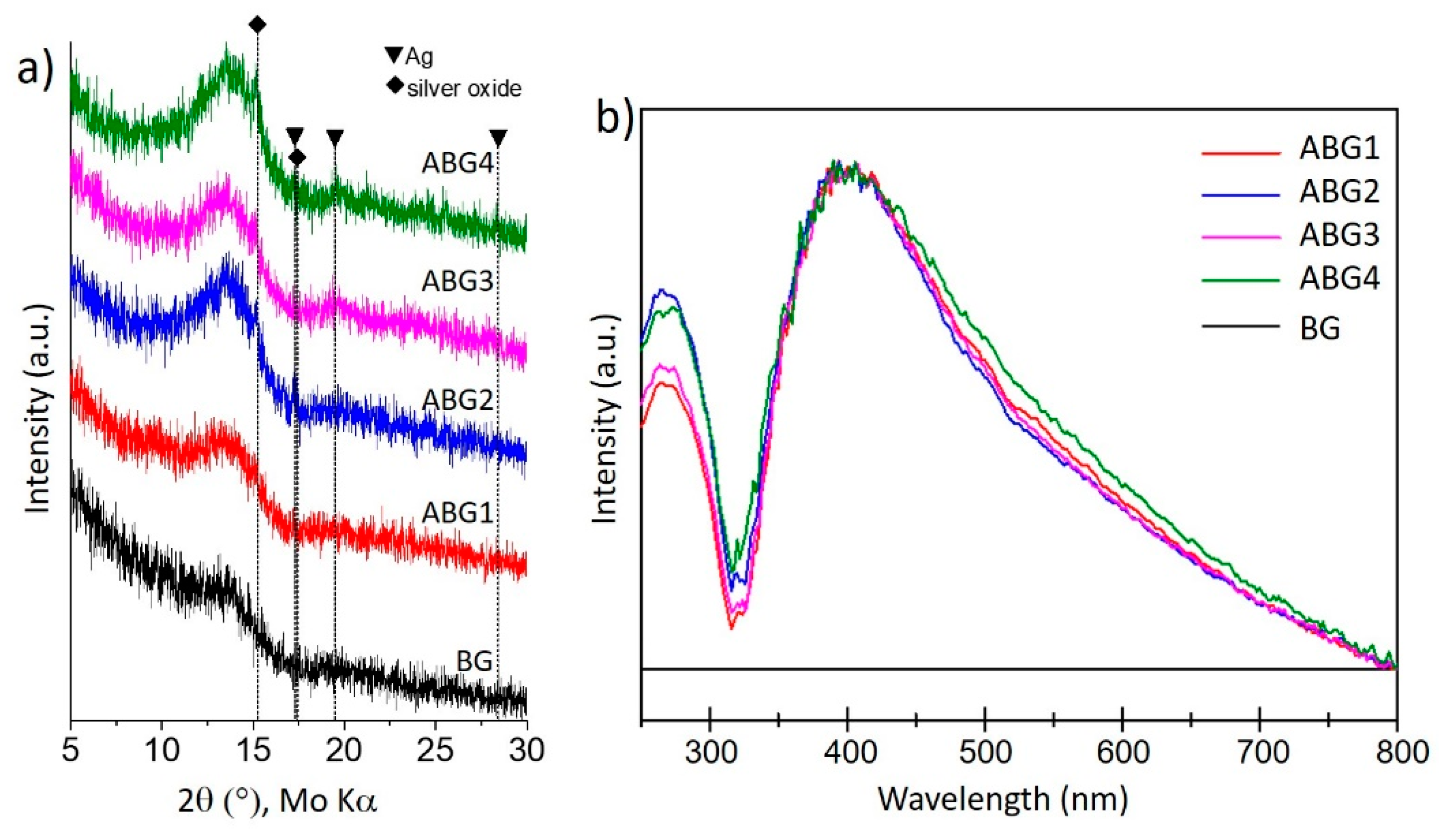
Abstract
:1. Introduction
2. Materials and Methods
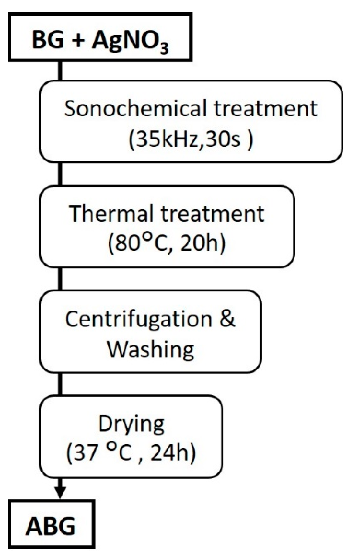
2.1. Preparation of Silver-Modified Glass Materials
2.2. Physicochemical Characterization
2.3. In Vitro Acellular Mineralization Tests
2.4. Antibacterial Activity
2.4.1. Analysis of Bacterial Growth Inhibition
2.4.2. Analysis of Bactericidal Action over Time
3. Results and Discussion
3.1. Chemical and Microstructural Characterization of Silver Modifed Glasses (ABG)
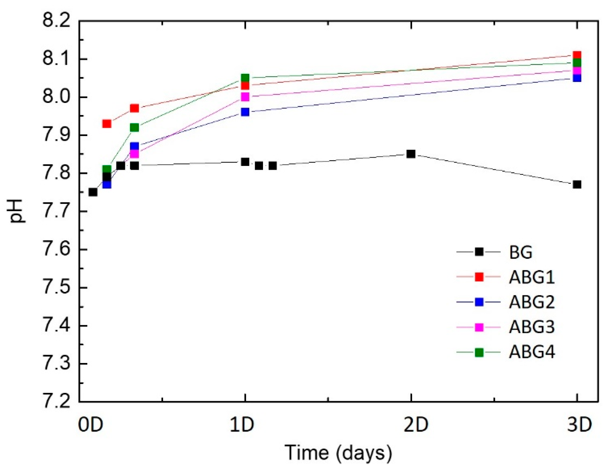
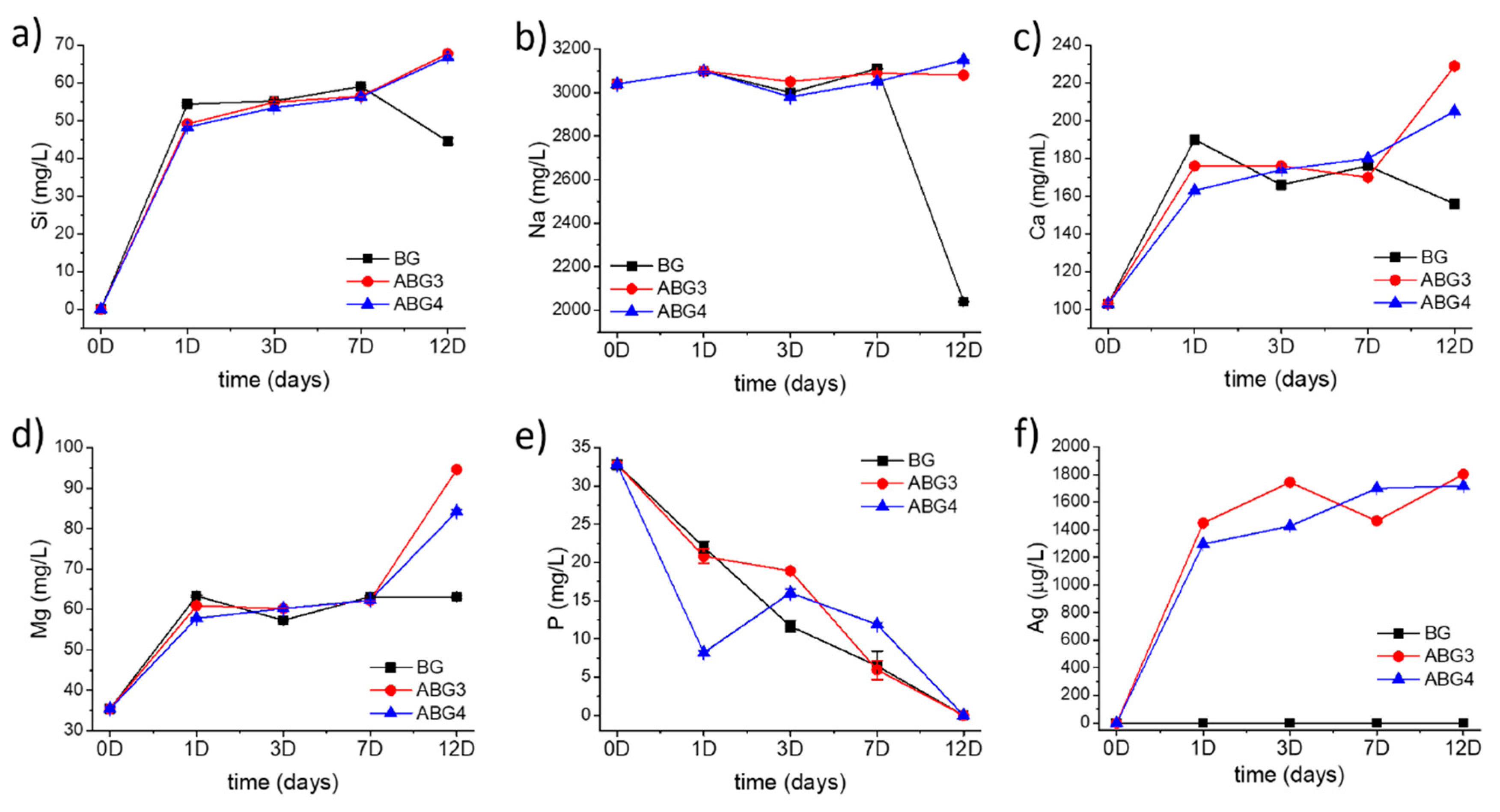
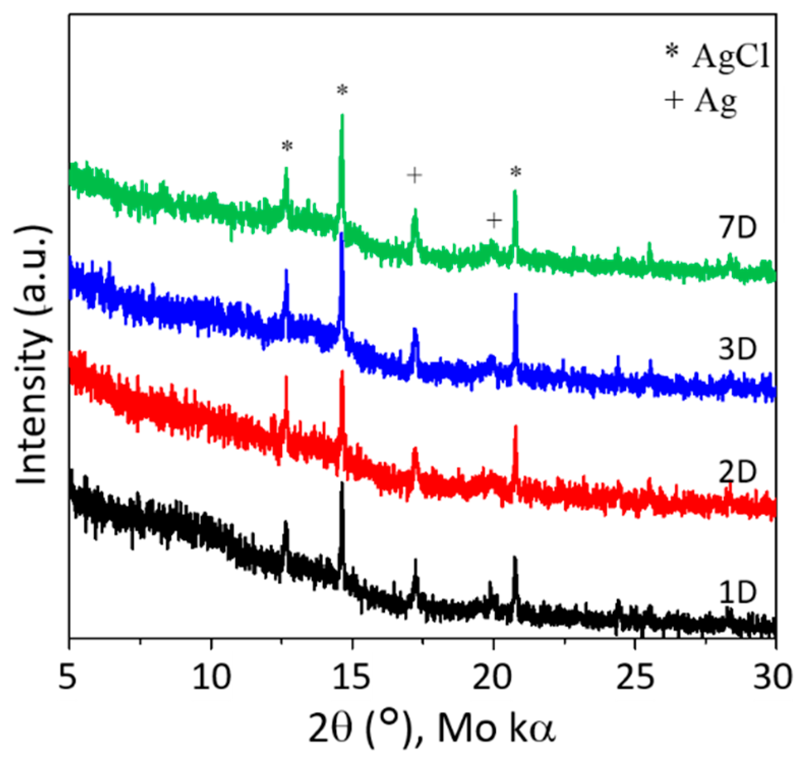
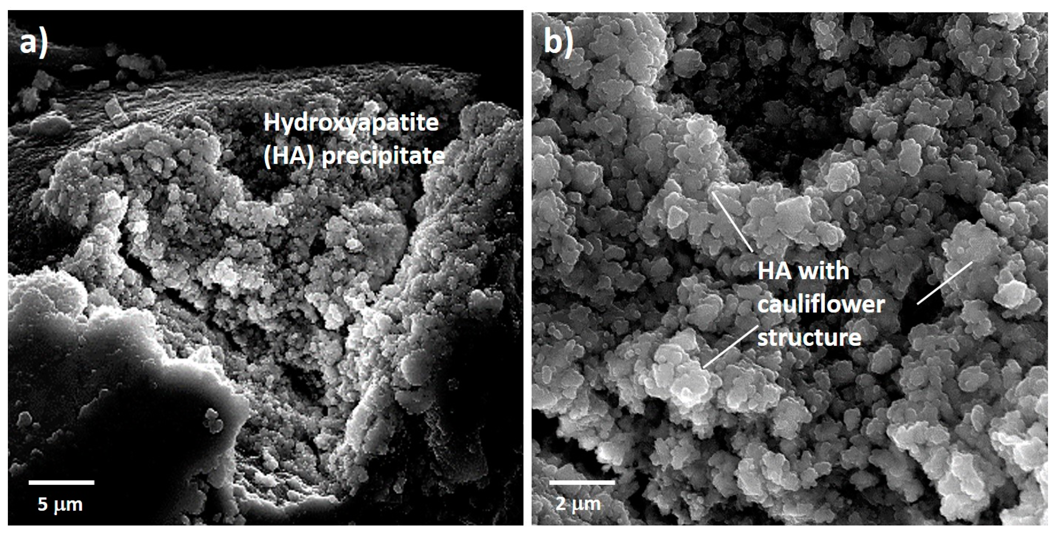
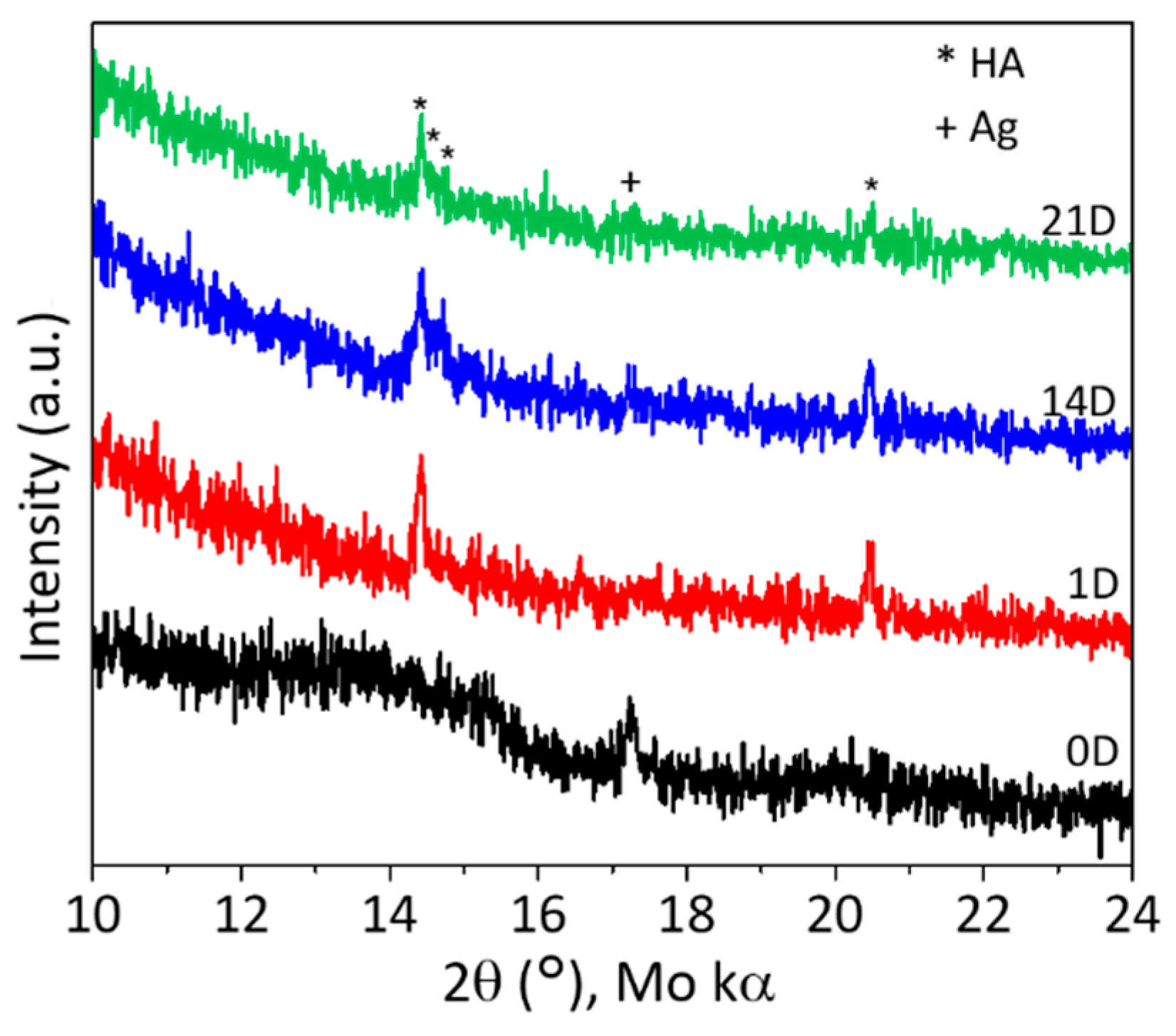
3.2. In Vitro Acellular Mineralization Assessments of the Silver Modified Glasses
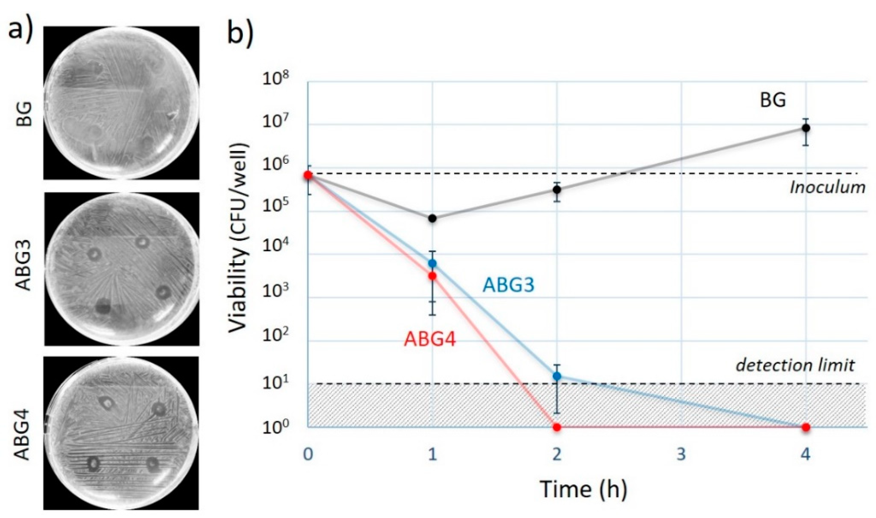
3.3. Bactericidal Activity of the As-Prepared of the Silver Modified Glasses
4. Conclusions
Author Contributions
Funding
Acknowledgments
Conflicts of Interest
References
- Best, S.M.; Porter, A.E.; Thian, E.S.; Huang, J. Bioceramics: Past, present and for the future. J. Eur. Ceram. Soc. 2008, 28, 1319–1327. [Google Scholar] [CrossRef]
- Day, R.M. Bioactive glass stimulates the secretion of angiogenic growth factors and angiogenesis in vitro. Tissue Eng. 2005, 11, 768–777. [Google Scholar] [CrossRef]
- Clare, A. Bio-Glasses. An Introduction; Jones, J.R., Clare, A.G., Eds.; Wiley: Chichester, UK, 2012; ISBN 978-0-470-71161-3. [Google Scholar]
- Lu, H.; Liu, Y.; Guo, J.; Wu, H.; Wang, J.; Wu, G. Biomaterials with Antibacterial and Osteoinductive Properties to Repair Infected Bone Defects. Int. J. Mol. Sci. 2016, 17, 334. [Google Scholar] [CrossRef]
- van de Belt, H.; Neut, D.; Schenk, W.; van Horn, J.R.; van der Mei, H.C.; Busscher, H.J. Infection of orthopedic implants and the use of antibiotic-loaded bone cements. A review. Acta Orthop. Scand. 2001, 72, 557–571. [Google Scholar] [CrossRef] [PubMed]
- Swathy, J.R.; Sankar, M.U.; Chaudhary, A.; Aigal, S.; Anshup; Pradeep, T. Antimicrobial silver: An unprecedented anion effect. Sci. Rep. UK 2014, 4, 7161. [Google Scholar] [CrossRef] [PubMed] [Green Version]
- You, C.; Han, C.; Wang, X.; Zheng, Y.; Li, Q.; Hu, X.; Sun, H. The progress of silver nanoparticles in the antibacterial mechanism, clinical application and cytotoxicity. Mol. Biol. Rep. 2012, 39, 9193–9201. [Google Scholar] [CrossRef] [PubMed]
- Chaloupka, K.; Malam, Y.; Seifalian, A.M. Nanosilver as a new generation of nanoproduct in biomedical applications. Trends Biotechnol. 2010, 28, 580–588. [Google Scholar] [CrossRef]
- Kim, J.S.; Kuk, E.; Yu, K.N.; Kim, J.-H.; Park, S.J.; Lee, H.J.; Kim, S.H.; Park, Y.K.; Park, Y.H.; Hwang, C.-Y.; et al. Antimicrobial effects of silver nanoparticles. Nanomed. Nanotechnol. Biol. Med. 2007, 3, 95–101. [Google Scholar] [CrossRef]
- Kaya, S.; Cresswell, M.; Boccaccini, A.R. Mesoporous silica-based bioactive glasses for antibiotic-free antibacterial applications. Mat. Sci. Eng. C-Mater. 2018, 83, 99–107. [Google Scholar] [CrossRef]
- Negas, T.; Hilfiker, D.; Bartkowski, S. Simple methods to incorporate silver and copper generate antimicrobial glasses and porous glass-bonded ceramics. Am. Ceram. Soc. Bull. 2017, 96, 26–31. [Google Scholar]
- Balagna, C.; Vitale-Brovarone, C.; Miola, M.; Verné, E.; Canuto, R.A.; Saracino, S.; Muzio, G.; Fucale, G.; Maina, G. Biocompatibility and Antibacterial Effect of Silver Doped 3D-Glass-Ceramic Scaffolds for Bone Grafting. J. Biomater. Appl. 2011, 25, 595–617. [Google Scholar] [CrossRef] [PubMed] [Green Version]
- Verné, E.; Ferraris, S.; Miola, M.; Fucale, G.; Maina, G.; Martinasso, G.; Canuto, R.A.; Di Nunzio, S.; Vitale-Brovarone, C. Synthesis and characterisation of bioactive and antibacterial glass–ceramic Part 1—Microstructure, properties and biological behaviour. Adv Appl. Ceram. 2008, 107, 234–244. [Google Scholar] [CrossRef]
- Bellantone, M.; Williams, H.D.; Hench, L.L. Broad-spectrum bactericidal activity of Ag2O-doped bioactive glass. Antimicrob. Agents. Chemother. 2002, 46, 1940–1945. [Google Scholar] [CrossRef] [PubMed] [Green Version]
- Vernè, E.; Di Nunzio, S.; Bosetti, M.; Appendino, P.; Vitale Brovarone, C.; Maina, G.; Cannas, M. Surface characterization of silver-doped bioactive glass. Biomaterials 2005, 26, 5111–5119. [Google Scholar] [CrossRef]
- Xu, H.; Zeiger, B.W.; Suslick, K.S. Sonochemical synthesis of nanomaterials. Chem. Soc. Rev. 2013, 42, 2555–2567. [Google Scholar] [CrossRef] [Green Version]
- Mănoiu, V.S.; Aloman, A. Obtaining silver nanoparticles by sonochemical methods. UPB. Sci. Bull. B 2010, 72, 179–186. [Google Scholar]
- Cheng, J.; Yao, S.; Zhang, W.; Zou, Y. Preparation and characterization of silver colloids with different morphologies under ultrasonic field. Front. Chem. 2006, 1, 418–422. [Google Scholar] [CrossRef]
- He, C.; Liu, L.; Fang, Z.; Li, J.; Guo, J.; Wei, J. Formation and characterization of silver nanoparticles in aqueous solution via ultrasonic irradiation. Ultrason. Sonochem. 2014, 21, 542–548. [Google Scholar] [CrossRef]
- Tulyaganov, D.U.; Agathopoulos, S.; Valerio, P.; Balamurugan, A.; Saranti, A.; Karakassides, M.A.; Ferreira, J.M.F. Synthesis, bioactivity and preliminary biocompatibility studies of glasses in the system CaO-MgO-SiO2-Na2O-P2O5-CaF2. J. Mater. Sci. Mater. Med. 2011, 22, 217–227. [Google Scholar] [CrossRef]
- Tulyaganov, D.U.; Makhkamov, M.E.; Urazbaev, A.; Goel, A.; Ferreira, J.M.F. Synthesis, processing and characterization of a bioactive glass composition for bone regeneration. Ceram. Int. 2013, 39, 2519–2526. [Google Scholar] [CrossRef]
- Agathopoulos, S.; Tulyaganov, D.U.; Ventura, J.M.G.; Kannan, S.; Saranti, A.; Karakassides, M.A.; Ferreira, J.M.F. Structural analysis and devitrification of glasses based on the CaO–MgO–SiO2 system with B2O3, Na2O, CaF2 and P2O5 additives. J. Non-Cryst. Solids 2006, 352, 322–328. [Google Scholar] [CrossRef]
- Hill, R.G.; Brauer, D.S. Predicting the bioactivity of glasses using the network connectivity or split network models. J. Non-Cryst. Solids 2011, 357, 3884–3887. [Google Scholar] [CrossRef]
- Macon, A.L.; Kim, T.B.; Valliant, E.M.; Goetschius, K.; Brow, R.K.; Day, D.E.; Hoppe, A.; Boccaccini, A.R.; Kim, I.Y.; Ohtsuki, C.; et al. A unified in vitro evaluation for apatite-forming ability of bioactive glasses and their variants. J. Mater. Sci. Mater. Med. 2015, 26, 115. [Google Scholar] [CrossRef] [PubMed] [Green Version]
- Beesk, W.; Jones, P.G.; Rumpel, H.; Schwarzmann, E.; Sheldrick, G.M. X-Ray crystal structure of Ag6O2. J. Chem. Soc. Chem. Commun. 1981, 664–665. [Google Scholar] [CrossRef]
- Standke, B.; Jansen, M. Ag3O4, the First Silver (II, III) Oxide. Angew. Chem. Int. Ed. Engl. 1986, 25, 77–78. [Google Scholar] [CrossRef]
- Norby, P.; Dinnebier, R.; Fitch, A.N. Decomposition of Silver Carbonate; the Crystal Structure of Two High-Temperature Modifications of Ag2CO3. Inorg. Chem. 2002, 41, 3628–3637. [Google Scholar] [CrossRef]
- Suh, I.K.; Ohta, H.; Waseda, Y. High-temperature thermal expansion of six metallic elements measured by dilatation method and X-ray diffraction. J. Mater. Sci. 1988, 23, 757–760. [Google Scholar] [CrossRef]
- Amendola, V.; Bakr, O.M.; Stellacci, F. A Study of the Surface Plasmon Resonance of Silver Nanoparticles by the Discrete Dipole Approximation Method: Effect of Shape, Size, Structure, and Assembly. Plasmonics 2010, 5, 85–97. [Google Scholar] [CrossRef]
- Hu, S.; Chang, J.; Liu, M.; Ning, C. Study on antibacterial effect of 45S5 Bioglass®. J. Mater. Sci.-Mater. Med. 2009, 20, 281–286. [Google Scholar] [CrossRef]
- Brauer, D.S. Bioactive Glasses—Structure and Properties. Angew. Chem. Int. Ed. 2015, 54, 4160–4181. [Google Scholar] [CrossRef]
- Wang, X.; Wu, H.-F.; Kuang, Q.; Huang, R.-B.; Xie, Z.-X.; Zheng, L.-S. Shape-Dependent Antibacterial Activities of Ag2O Polyhedral Particles. Langmuir 2010, 26, 2774–2778. [Google Scholar] [CrossRef] [PubMed]
- Evanoff, D.D.; Chumanov, G. Size-Controlled Synthesis of Nanoparticles. 1. “Silver-Only” Aqueous Suspensions via Hydrogen Reduction. J. Phys. Chem. B 2004, 108, 13948–13956. [Google Scholar] [CrossRef]
- Aronne, A.; Sigaev, V.N.; Champagnon, B.; Fanelli, E.; Califano, V.; Usmanova, L.Z.; Pernice, P. The origin of nanostructuring in potassium niobiosilicate glasses by Raman and FTIR spectroscopy. J. Non-Cryst. Solids 2005, 351, 3610–3618. [Google Scholar] [CrossRef]
- Omori, K. Analysis of the Infrared Absorption Spectrum of Diopside. Am. Mineral. 1971, 56, 1607–1616. [Google Scholar]
- Stoch, L.; Środa, M. Infrared spectroscopy in the investigation of oxide glasses structure. J. Mol. Struct. 1999, 511, 77–84. [Google Scholar] [CrossRef]
- Begum, S.; Johnson, W.E.; Worthington, T.; Martin, R.A. The influence of pH and fluid dynamics on the antibacterial efficacy of 45S5 Bioglass. Biomed. Mater. 2016, 11, 15006. [Google Scholar] [CrossRef] [Green Version]
- Vecstaudza, J.; Gasik, M.; Locs, J. Amorphous calcium phosphate materials: Formation, structure and thermal behaviour. J. Eur. Ceram. Soc. 2019, 39, 1642–1649. [Google Scholar] [CrossRef]
- Božanić, D.K.; Trandafilović, L.V.; Luyt, A.S.; Djoković, V. ‘Green’ synthesis and optical properties of silver–chitosan complexes and nanocomposites. React. Funct. Polym. 2010, 70, 869–873. [Google Scholar] [CrossRef]
- Villars, P.; Cenzual, K. Pearson’s Crystal Data-Crystal Structure Database for Inorganic Compounds; ASM International: Materials Park, OH, USA, 2010. [Google Scholar]
- Lellouche, J.; Kahana, E.; Elias, S.; Gedanken, A.; Banin, E. Antibiofilm activity of nanosized magnesium fluoride. Biomaterials 2009, 30, 5969–5978. [Google Scholar] [CrossRef]
- Marquis, R.E. Antimicrobial actions of fluoride for oral bacteria. Can. J. Microbiol. 1995, 41, 955–964. [Google Scholar] [CrossRef]


Publisher’s Note: MDPI stays neutral with regard to jurisdictional claims in published maps and institutional affiliations. |
© 2020 by the authors. Licensee MDPI, Basel, Switzerland. This article is an open access article distributed under the terms and conditions of the Creative Commons Attribution (CC BY) license (http://creativecommons.org/licenses/by/4.0/).
Share and Cite
Gonzalo-Juan, I.; Xie, F.; Becker, M.; Tulyaganov, D.U.; Ionescu, E.; Lauterbach, S.; De Angelis Rigotti, F.; Fischer, A.; Riedel, R. Synthesis of Silver Modified Bioactive Glassy Materials with Antibacterial Properties via Facile and Low-Temperature Route. Materials 2020, 13, 5115. https://doi.org/10.3390/ma13225115
Gonzalo-Juan I, Xie F, Becker M, Tulyaganov DU, Ionescu E, Lauterbach S, De Angelis Rigotti F, Fischer A, Riedel R. Synthesis of Silver Modified Bioactive Glassy Materials with Antibacterial Properties via Facile and Low-Temperature Route. Materials. 2020; 13(22):5115. https://doi.org/10.3390/ma13225115
Chicago/Turabian StyleGonzalo-Juan, Isabel, Fangtong Xie, Malin Becker, Dilshat U. Tulyaganov, Emanuel Ionescu, Stefan Lauterbach, Francesca De Angelis Rigotti, Andreas Fischer, and Ralf Riedel. 2020. "Synthesis of Silver Modified Bioactive Glassy Materials with Antibacterial Properties via Facile and Low-Temperature Route" Materials 13, no. 22: 5115. https://doi.org/10.3390/ma13225115