Zn/La Mixed Oxides Prepared by Coprecipitation: Synthesis, Characterization and Photocatalytic Studies


 , ,
, , 
Abstract
:1. Introduction
2. Materials and Methods
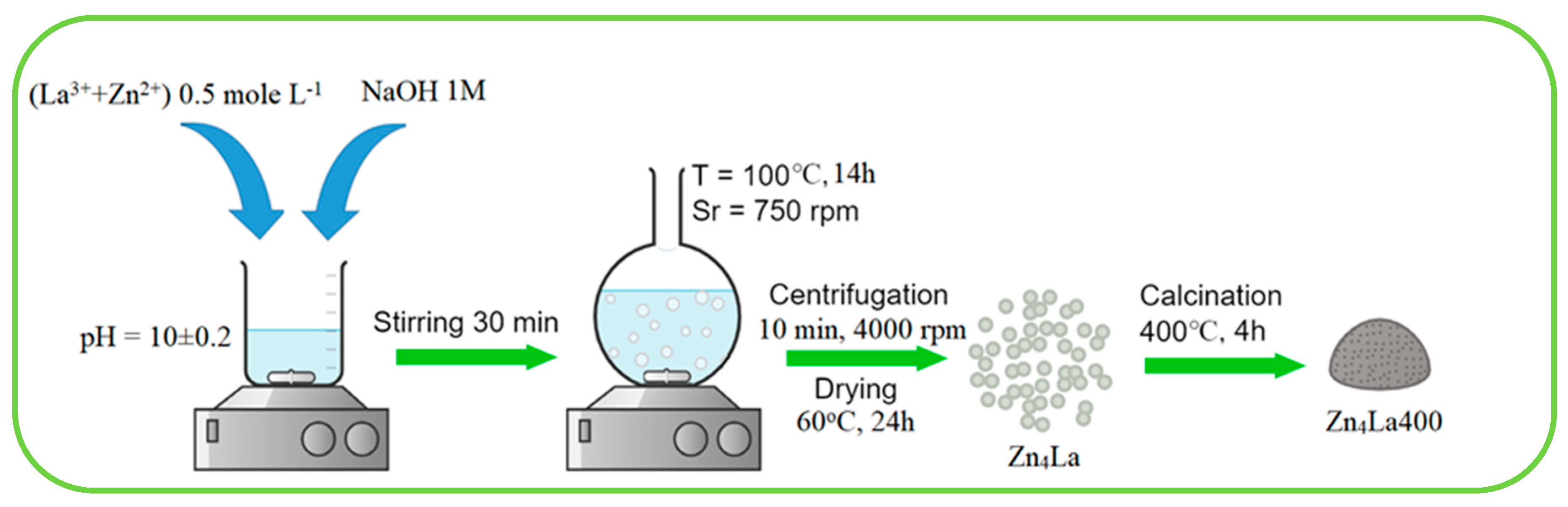
2.1. Materials Synthesis and Characterization
2.2. Photocatalytic Tests
3. Results and Discussion
3.1. Structural and Morphologic Characterization
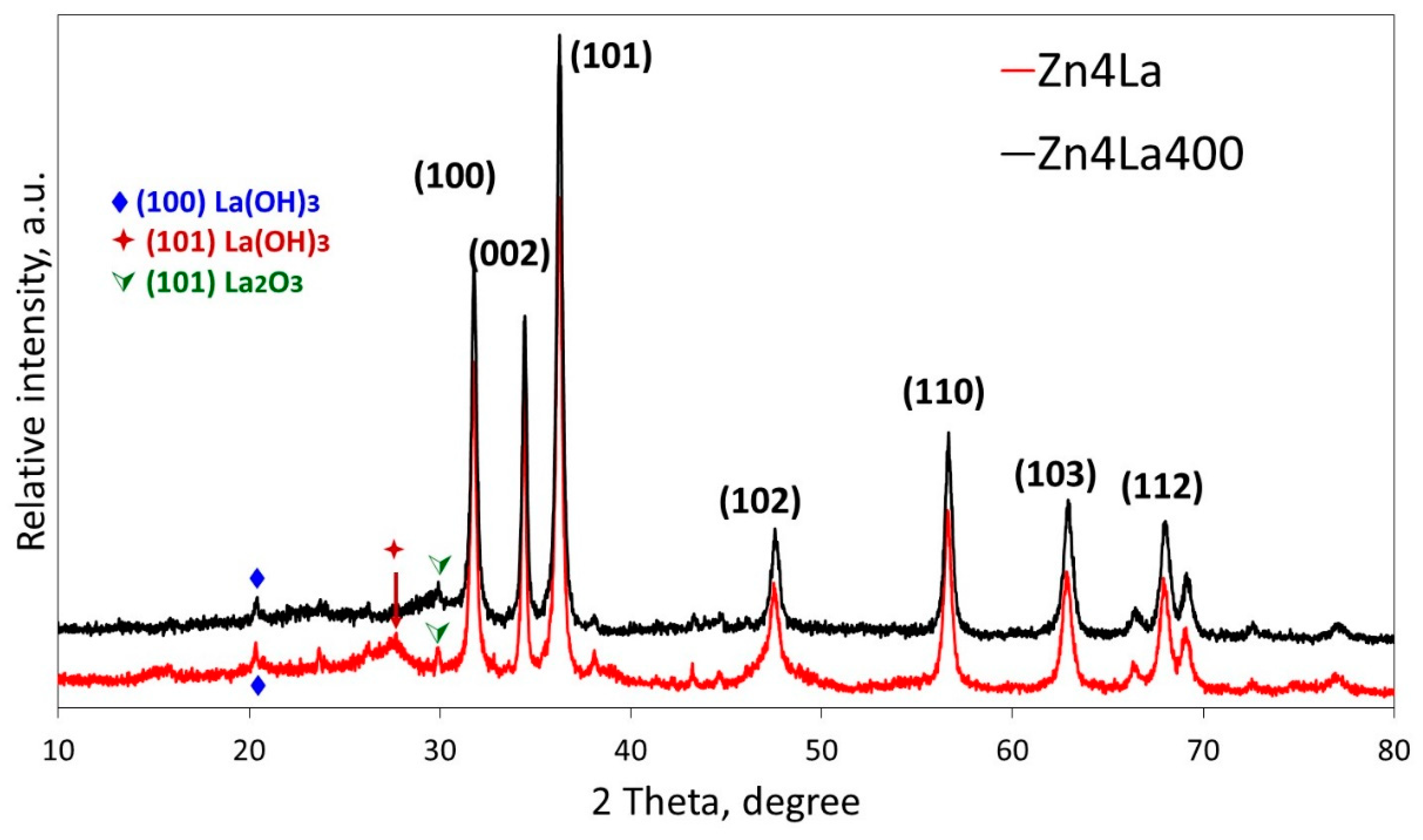
3.1.1. Powder XRD
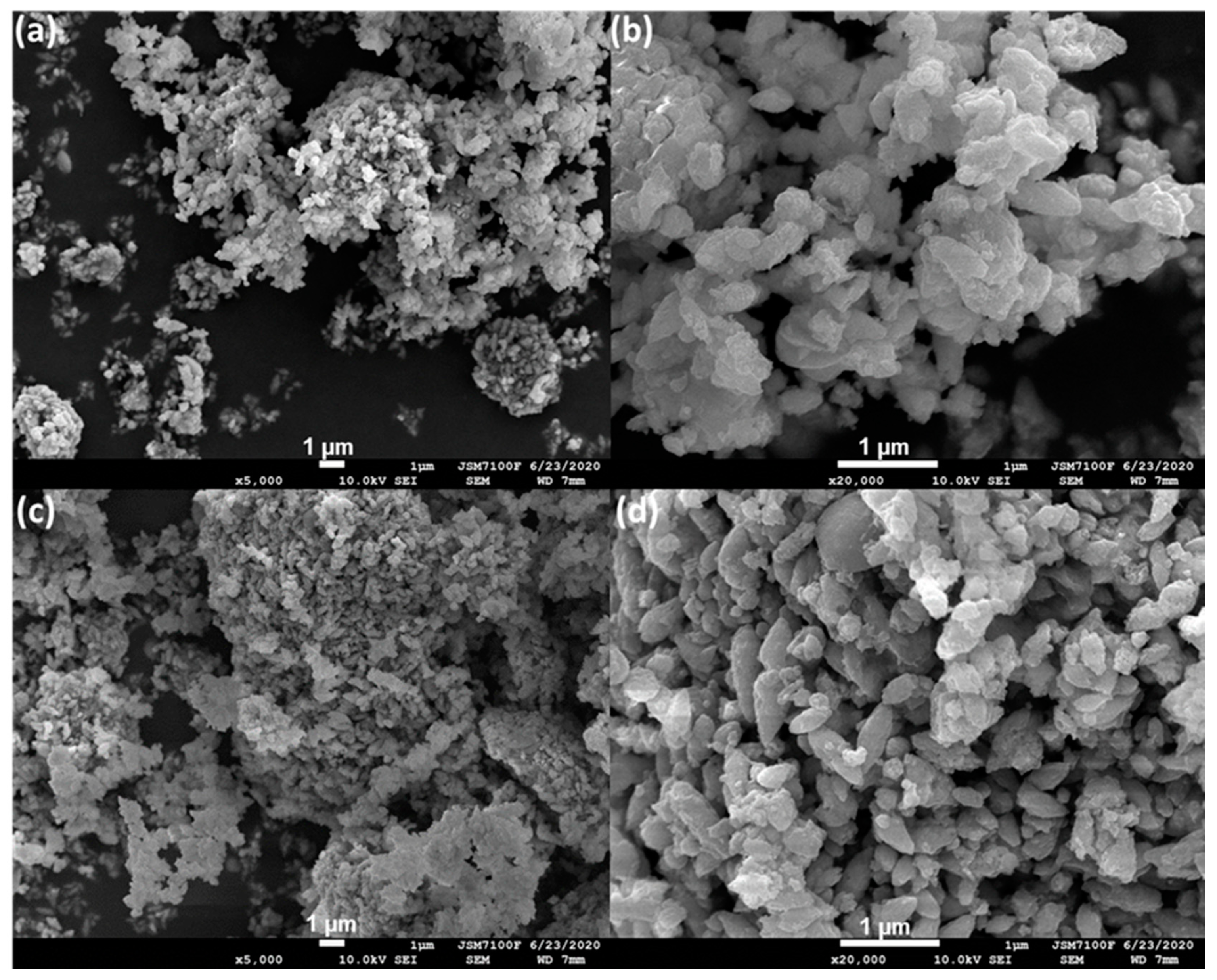
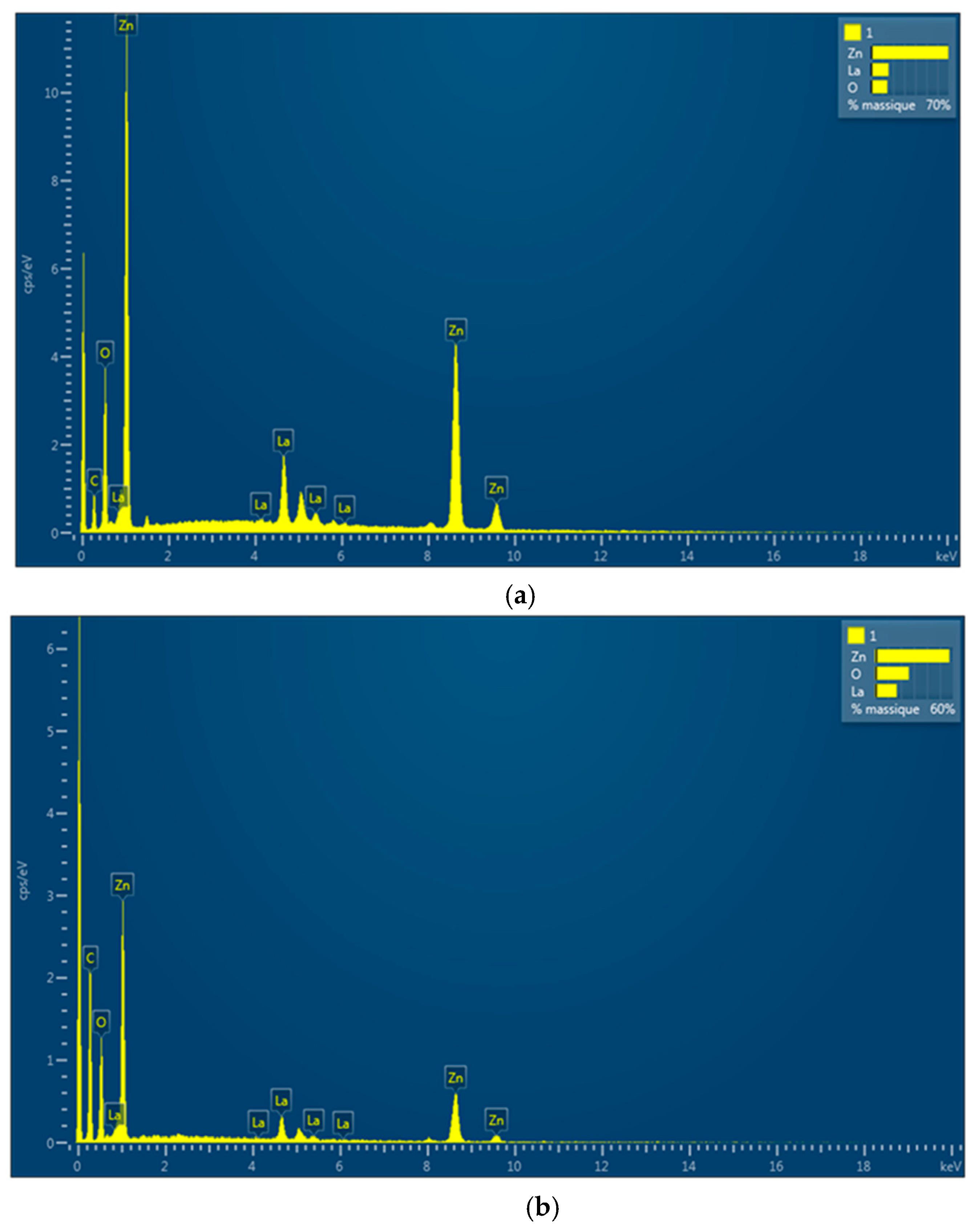
3.1.2. SEM/EDS Analysis
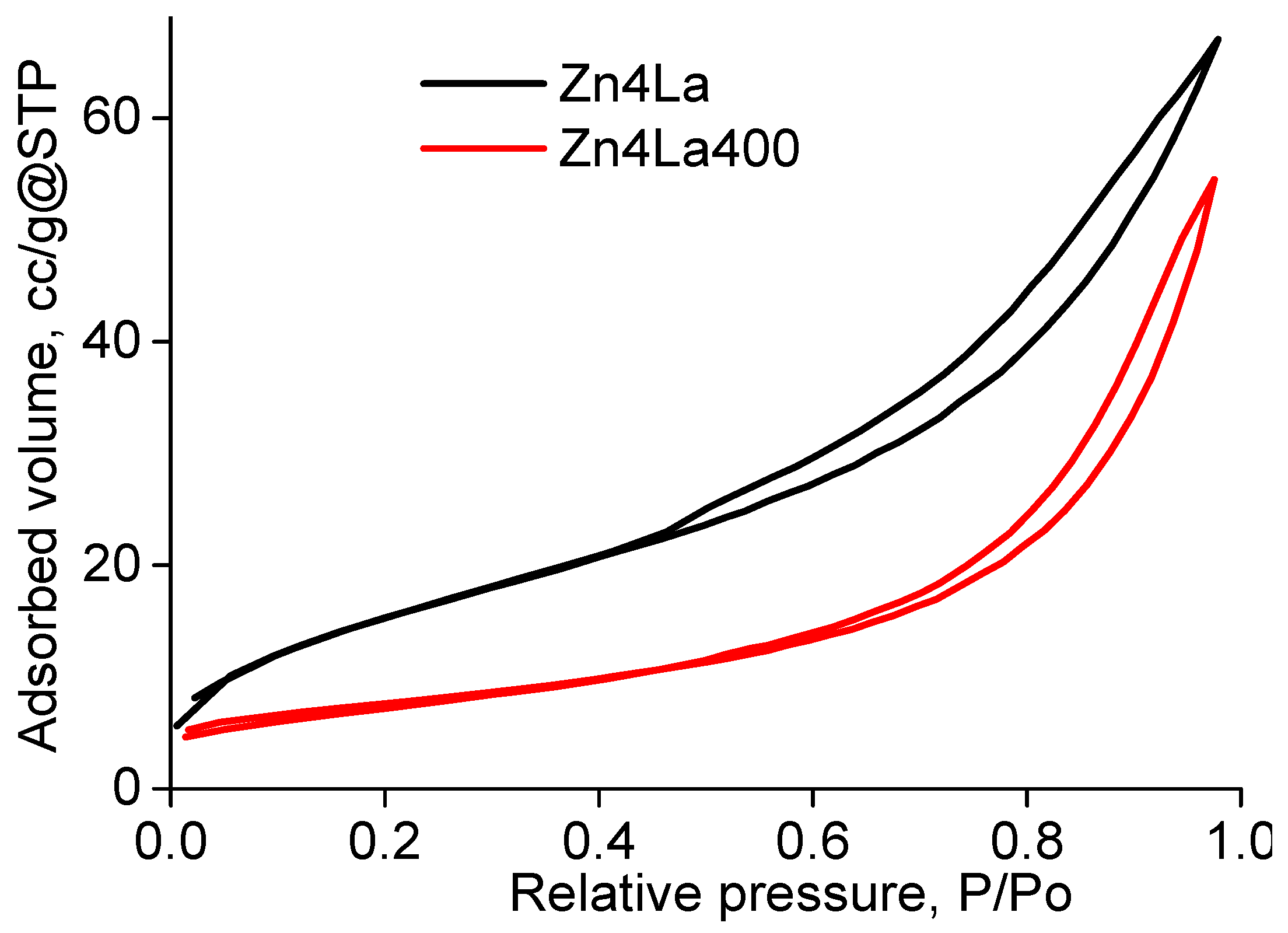
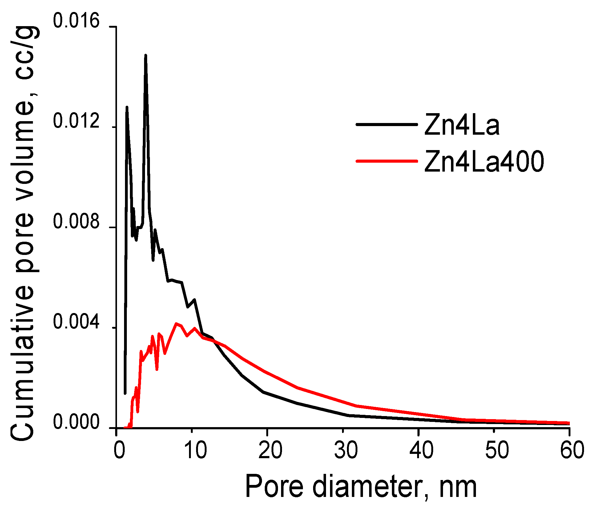
3.2. BET Analysis
3.3. FTIR Analysis
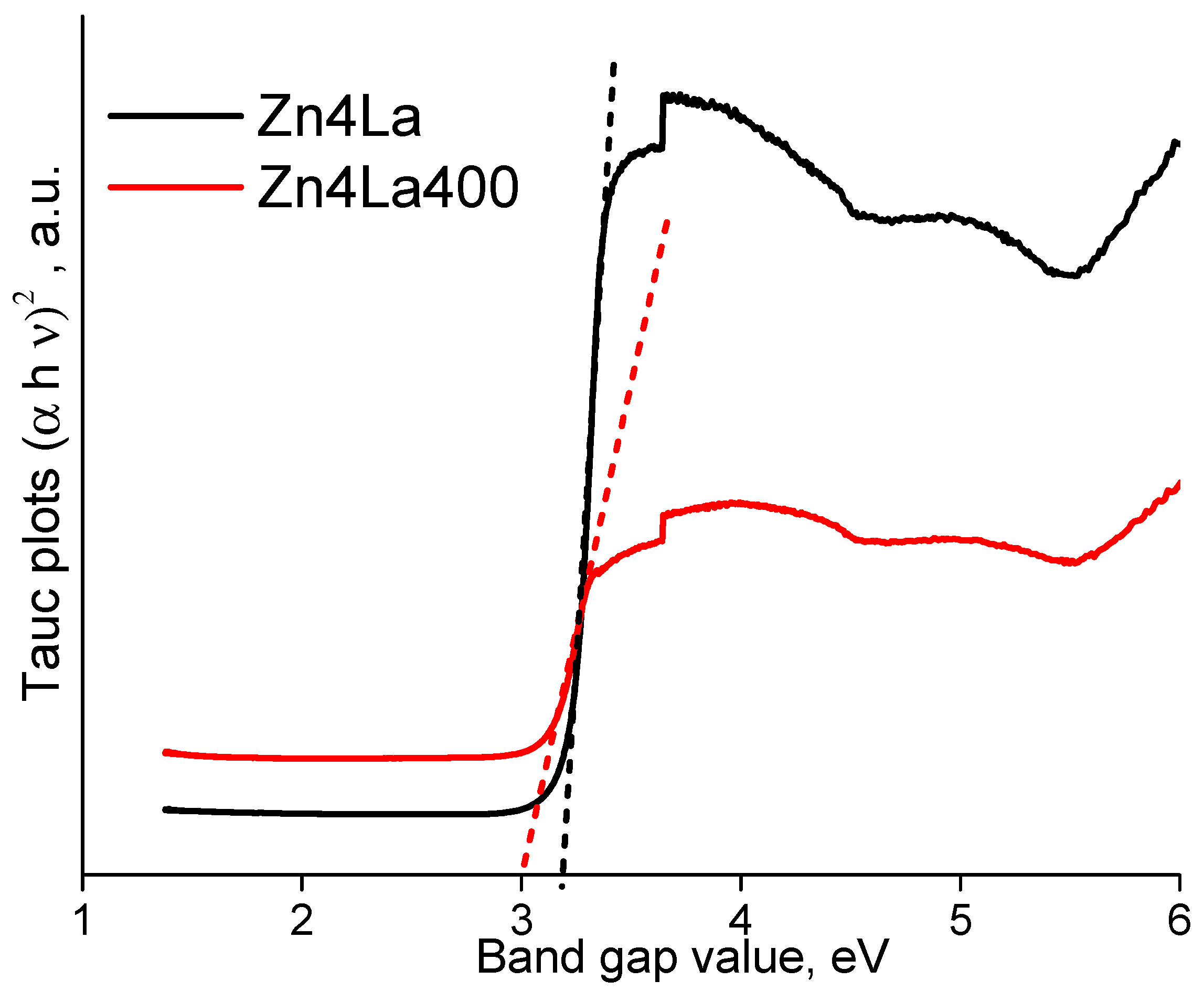
3.4. Band Gap
3.5. Photocatalytic Activity Investigations
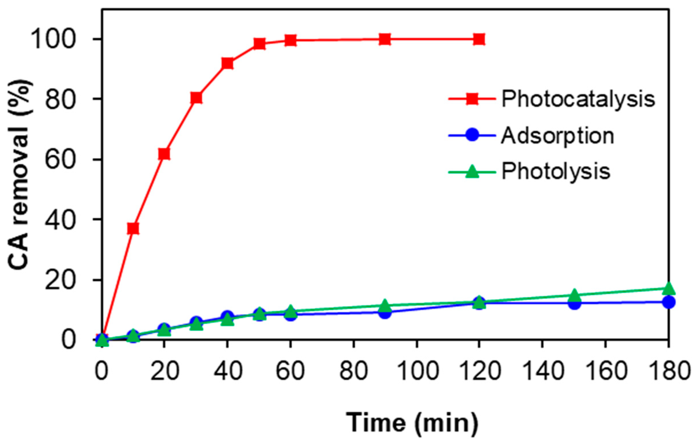
3.5.1. Preliminary Experiments: Role of Adsorption and Photolysis
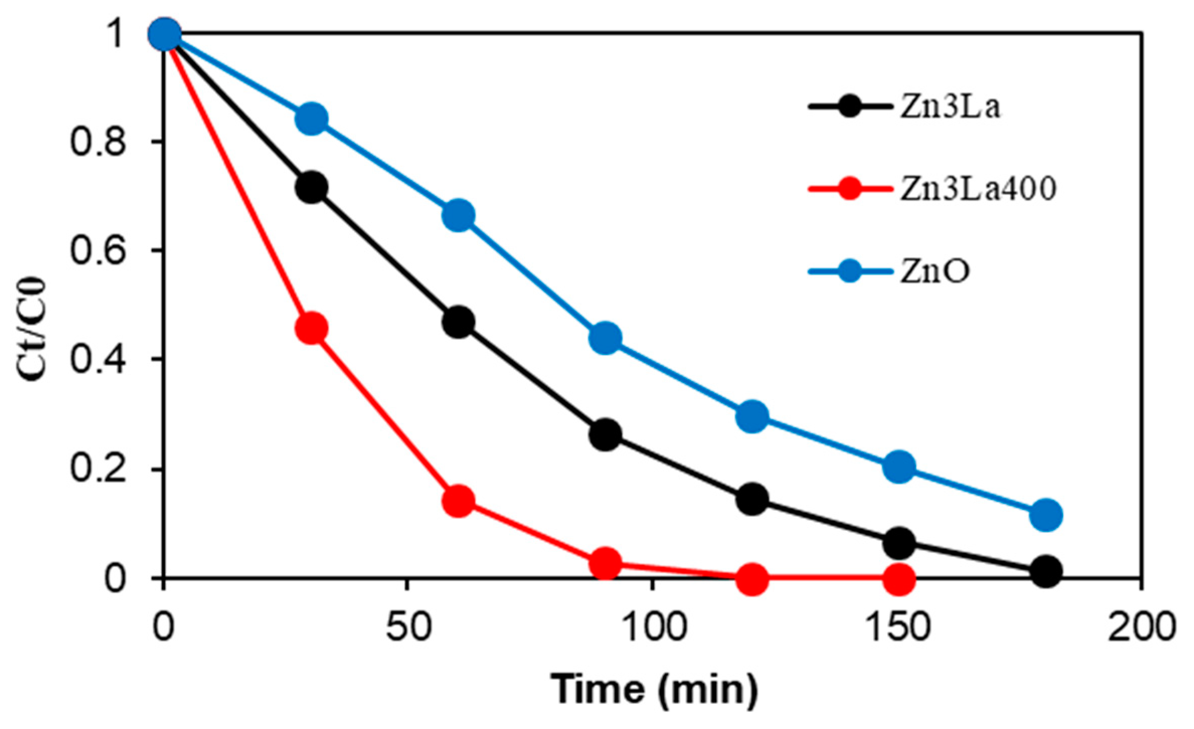
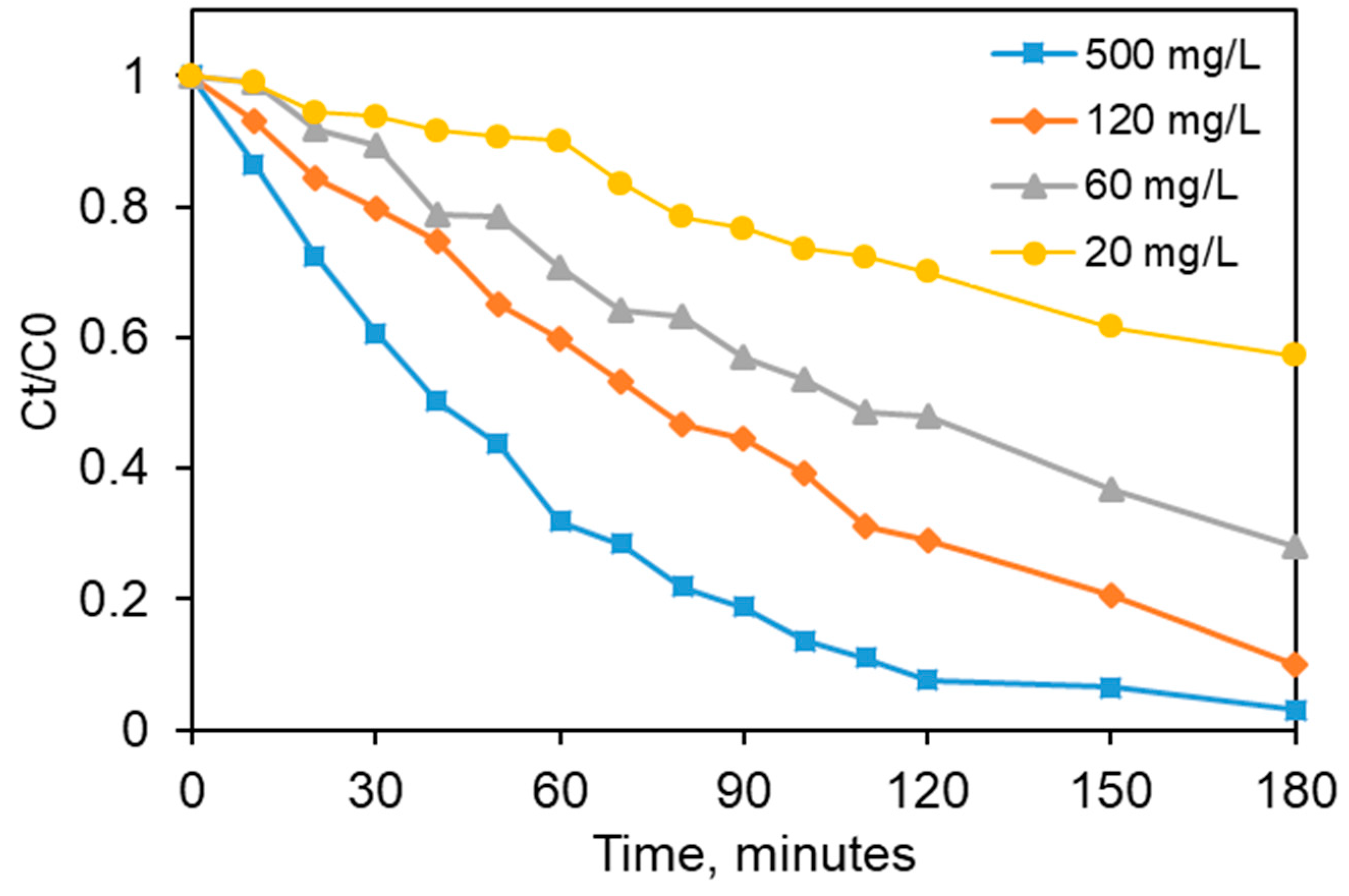
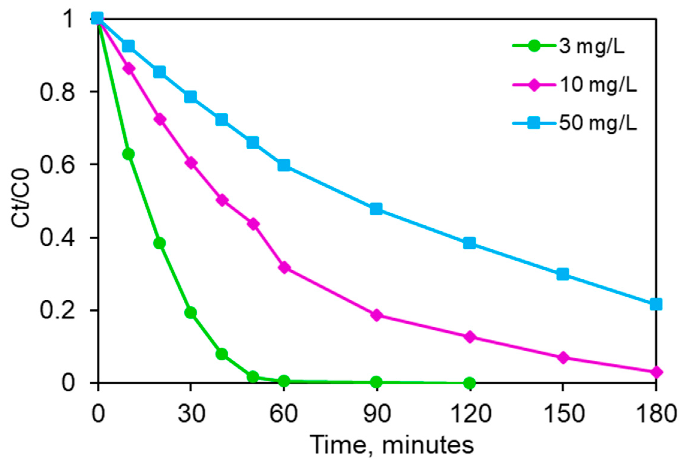
3.5.2. Effects of Some Operating Conditions on the Clofibric Acid Elimination with Zn4La400
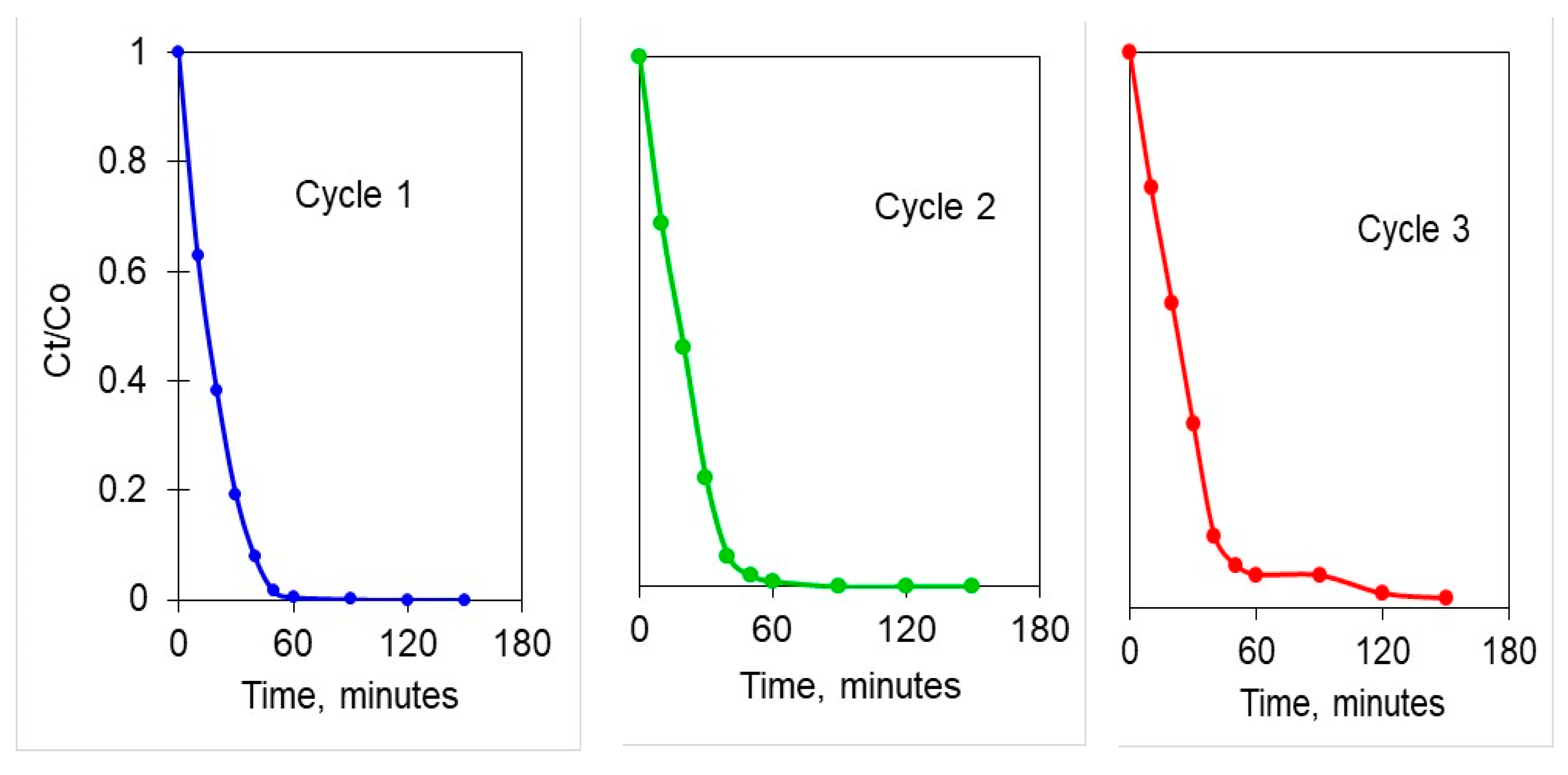
3.6. Investigation of the Catalyst Recyclability
4. Conclusions
Author Contributions
Funding
Conflicts of Interest
References
- Yu, J.; Jaroniec, M.; Jiang, C. (Eds.) Surface Science of Photocatalysis; Academic Press: Cambridge, MA, USA, 2020. [Google Scholar]
- Rueda-Marquez, J.J.; Levchuk, I.; Ibañez, P.F.; Sillanpää, M. A critical review on application of photocatalysis for toxicity reduction of real wastewaters. J. Clean. Prod. 2020, 120694. [Google Scholar] [CrossRef]
- Buema, G.; Lupu, N.; Chiriac, H.; Ciobanu, G.; Kotova, O.; Harja, M. Modeling of solid-fluid non-catalytic processes for nickel ion removal. Rev. Chim. 2020, 71, 4–15. [Google Scholar] [CrossRef]
- Harja, M.; Ciobanu, G. Ecofriendly Nano-adsorbents for Pollutant Removal from Wastewaters. In Handbook of Nanomaterials and Nanocomposites for Energy and Environmental Applications; Springer International Publishing: Cham, Switzerland, 2020. [Google Scholar]
- Stefan, M.I. (Ed.) Advanced Oxidation Processes for Water Treatment: Fundamentals and Applications; IWA Publishing: London, UK, 2017. [Google Scholar]
- Duduman, C.N.; Harja, M.; Barrena Pérez, M.I.; de Castro, C.G.; Lutic, D.; Kotova, O.; Cretescu, I. Preparation and characterization of nanocomposite material based on TiO2-Ag for environmental applications. Environ. Eng. Manag. J. 2018, 17, 925–936. [Google Scholar]
- De Castro, C.G.; Duduman, C.N.; Harja, M.; Lutic, D.; Juzsakova, T.; Cretescu, I. New TiO2-Ag nanoparticles used for organic compounds degradation. Environ. Eng. Manag. J. 2019, 18, 1755–1763. [Google Scholar]
- Favier, L.; Harja, M. TiO2/fly ash nanocomposite for photodegradation of persistent organic pollutant. In Handbook of Nanomaterials and Nanocomposites for Energy and Environmental Applications; Kharissova, O., Martínez, L., Kharisov, B., Eds.; Springer International Publishing: Cham, Switzerland, 2020. [Google Scholar]
- Sescu, A.M.; Favier, L.; Lutic, D.; Soto-Donoso, N.; Ciobanu, G.; Harja, M. TiO2 doped with noble metals as an efficient solution for the photodegradation of hazardous organic water pollutants at ambient conditions. Water 2020. under revision. [Google Scholar]
- Al-Sabahi, J.; Bora, T.; Al-Abri, M.; Dutta, J. Controlled Defects of Zinc Oxide Nanorods for Efficient Visible Light Photocatalytic Degradation of Phenol. Materials 2016, 9, 238. [Google Scholar] [CrossRef] [Green Version]
- Zhu, D.; Zhou, Q. Action and mechanism of semiconductor photocatalysis on degradation of organic pollutants in water treatment: A review. Environ. Nanotechnol. Monit. Manag. 2019, 12, 100255. [Google Scholar] [CrossRef]
- Ganguly, A.; Anjaneyulu, O.; Ojha, K.; Ganguli, A.K. Oxide-based nanostructures for photocatalytic and electrocatalytic applications. CrystEngComm 2015, 17, 8978–9001. [Google Scholar] [CrossRef]
- Nguyen, L.T.T.; Duong, A.T.T.; Nguyen, B.D.; Hai, N.Q.; Chu, V.H.; Nguyen, T.D.; Bach, L.G. Preparation, Characterization and Photocatalytic Activity of La-Doped Zinc Oxide Nanoparticles. Materials 2019, 12, 1195. [Google Scholar] [CrossRef] [Green Version]
- Govindaraj, R.; Govindan, R.; Geetha, M.; Anbarasan, P.M. Structural, morphological and luminescence studies on pristine and La doped zinc oxide (ZnO) nanoparticles. Optik 2015, 126, 1555–1558. [Google Scholar] [CrossRef]
- Liu, X.; Fu, F.; Zuo, H. Lanthanum ions-induced synthesis of ZnO nanostructures from zinc foil: Morphology change and photocatalytic activity. Surf. Interfaces 2016, 1, 29–34. [Google Scholar] [CrossRef]
- Shakir, M.; Faraz, M.; Sherwani, M.A.; Al-Resayes, S.I. Photocatalytic degradation of the Paracetamol drug using Lanthanum doped ZnO nanoparticles and their in-vitro cytotoxicity assay. J. Lumin. 2016, 176, 159–167. [Google Scholar] [CrossRef]
- Manikandan, A.; Meenatchi, B.; Vadivel, S.; Jaganathan, S.; Ladchumananandasivam, R.; Henini, M.; Maaza, M.; Aanand, J.S. Rare earth element (REE) lanthanum doped zinc oxide (La: ZnO) nanomaterials: Synthesis structural optical and antibacterial studies. J. Alloys Compd. 2017, 723, 1155–1161. [Google Scholar] [CrossRef] [Green Version]
- Goel, S.; Sinha, N.; Yadav, H.; Joseph, A.J.; Kumar, B. Experimental investigation on the structural, dielectric, ferroelectric and piezoelectric properties of La doped ZnO nanoparticles and their application in dye-sensitized solar cells. Phys. E Low Dimens. Syst. Nanostruct. 2017, 91, 72–81. [Google Scholar] [CrossRef]
- Dudek, S.; Kołodyńska, D. Enhanced Arsenic (V) Removal on an Iron-Based Sorbent Modified by Lanthanum (III). Materials 2020, 13, 2553. [Google Scholar] [CrossRef]
- Habashi, F. A new look at the periodic table. Interdiscip. Sci. Rev. 1997, 22, 53–60. [Google Scholar] [CrossRef]
- Muhammad, W.; Ullah, N.; Haroon, M.; Abbasi, B.H. Optical, morphological and biological analysis of zinc oxide nanoparticles (ZnO NPs) using Papaver somniferum L. RSC Adv. 2019, 9, 29541–29548. [Google Scholar] [CrossRef] [Green Version]
- Lutic, D.; Coromelci-Pastravanu, C.; Cretescu, I.; Poulios, I.; Stan, C.-D. Photocatalytic Treatment of Rhodamine 6G in Wastewater Using Photoactive ZnO. Int. J. Photoenergy 2012, 2012, 1–8. [Google Scholar] [CrossRef] [Green Version]
- Chen, J.; Wang, J.; Zhang, F.; Zhang, G.; Wu, Z.; Yan, P. The effect of La doping concentration on the properties of zinc oxide films prepared by the sol–gel method. J. Cryst. Growth 2008, 310, 2627–2632. [Google Scholar] [CrossRef]
- Yokoi, T.; Tsukada, K.; Terasaka, S.; Kamitakahara, M.; Matsubara, H. Morphological control of layered double hydroxide through a biomimetic approach using carboxylic and sulfonic acids. J. Asian Ceram. Soc. 2015, 3, 230–233. [Google Scholar] [CrossRef] [Green Version]
- Sumari, S.M. Adsorption of Anionic dyes from Aqueous Solutions by Calcined and Uncalcined Mg/Al Layered Double Hydroxide. Malays. J. Anal. Sci. 2016, 20, 777–792. [Google Scholar] [CrossRef]
- Yang, K.; Li, X.; Yu, C.; Zeng, D.; Chen, F.; Zhang, K.; Huang, W.; Ji, H. Review on heterophase/homophase junctions for efficient photocatalysis: The case of phase transition construction. Chin. J. Catal. 2019, 40, 796–818. [Google Scholar] [CrossRef]
- Pan, D.; Ge, S.; Zhao, J.; Tian, J.; Shao, Q.; Guo, L.; Mai, X.; Wu, T.; Murugadoss, V.; Liu, H.; et al. Synthesis and Characterization of ZnNiIn Layered Double Hydroxides Derived Mixed Metal Oxides with Highly Efficient Photoelectrocatalytic Activities. Ind. Eng. Chem. Res. 2018, 58, 836–848. [Google Scholar] [CrossRef]
- You, Y.; Zhao, H.; Vance, G.F. Surfactant-enhanced adsorption of organic compounds by layered double hydroxides. Colloids Surf. A Physicochem. Eng. Asp. 2002, 205, 161–172. [Google Scholar] [CrossRef]
- Carmichael, M.; Vidu, R.; Maksumov, A.; Palazoglu, A.; Stroeve, P. Using Wavelets to Analyze AFM Images of Thin Films: Surface Micelles and Supported Lipid Bilayers. Langmuir 2004, 20, 11557–11568. [Google Scholar] [CrossRef]
- Quinlan, F.T.; Vidu, R.; Predoana, L.; Zaharescu, M.; Gartrner, M.; Groza, J.; Stroeve, P. Lithium Cobalt Oxide (LiCoO2) Nanocoatings by Sol−Gel Methods. Ind. Eng. Chem. Res. 2004, 43, 2468–2477. [Google Scholar] [CrossRef]
- Sing, K.S.W. Reporting physisorption data for gas/solid systems with special reference to the determination of surface area and porosity (Recommendations 1984). Pure Appl. Chem. 1985, 57, 603–619. [Google Scholar] [CrossRef]
- Rouquerol, J.; Rouquerol, F.; Llewellyn, P.; Maurin, G.; Sing, K.S. Adsorption by Powders and Porous Solids: Principles, Methodology and Applications; Academic Press: Cambridge, MA, USA, 2013. [Google Scholar]
- Thommes, M.; Kaneko, K.; Neimark, A.V.; Olivier, J.P.; Rodriguez-Reinoso, F.; Rouquerol, J.; Sing, K.S. Physisorption of gases, with special reference to the evaluation of surface area and pore size distribution (IUPAC Technical Report). Pure Appl. Chem. 2015, 87, 1051–1069. [Google Scholar] [CrossRef] [Green Version]
- Villarroel-Rocha, J.; Barrera, D.; Sapag, K. Introducing a self-consistent test and the corresponding modification in the Barrett, Joyner and Halenda method for pore-size determination. Microporous Mesoporous Mater. 2014, 200, 68–78. [Google Scholar] [CrossRef]
- Pathan, A.A.; Desai, K.R.; Vajapara, S.; Bhasin, C.P. Conditional Optimization of Solution Combustion Synthesis for Pioneered La2O3 Nanostructures to Application as Future CMOS and NVMS Generations. Adv. Nanoparticles 2018, 7, 28–35. [Google Scholar] [CrossRef] [Green Version]
- Karthikeyan, S.; Raj, A.D.; Irudayaraj, A.A.; Raj, D.M.A. Effect of Temperature on the Properties of La2O3 Nanostructures. Mater. Today Proc. 2015, 1021–1025. [Google Scholar] [CrossRef]
- NIST Chemistry WebBook. Available online: https://webbook.nist.gov/cgi/cbook.cgi?Formula=ZnO&NoIon=on&Units=SI (accessed on 15 August 2020).
- Tauc, J.; Grigorovici, R.; Vancu, A. Optical Properties and Electronic Structure of Amorphous Germanium. Phys. Status Solidi 1966, 15, 627–637. [Google Scholar] [CrossRef]
- Suram, S.K.; Newhouse, P.F.; Gregoire, J.M. High Throughput Light Absorber Discovery, Part 1: An Algorithm for Automated Tauc Analysis. ACS Comb. Sci. 2016, 18, 673–681. [Google Scholar] [CrossRef]
- D’Amato, C.A.; Giovannetti, R.; Zannotti, M.; Rommozzi, E.; Minicucci, M.; Gunnella, R.; Di Cicco, A. Band Gap Implications on Nano-TiO2 Surface Modification with Ascorbic Acid for Visible Light-Active Polypropylene Coated Photocatalyst. Nanomaterials 2018, 8, 599. [Google Scholar] [CrossRef] [Green Version]
- Cai, J.; Xin, W.; Liu, G.; Lin, D.; Zhu, D. Effect of calcination temperature on structural properties and photocatalytic activity of Mn-C-codoped TiO. Mater. Res. 2016, 19, 401–407. [Google Scholar] [CrossRef] [Green Version]
- Chen, Y.; Dionysiou, D.D. Effect of calcination temperature on the photocatalytic activity and adhesion of TiO2 films prepared by the P-25 powder-modified sol–gel method. J. Mol. Catal. A Chem. 2006, 244, 73–82. [Google Scholar] [CrossRef]
- Fan, J.; Zhao, L.; Yu, J.; Liu, G. The effect of calcination temperature on the microstructure and photocatalytic activity of TiO2-based composite nanotubes prepared by an In Situ template dissolution method. Nanoscale 2012, 4, 6597–6603. [Google Scholar] [CrossRef]
- Nasirian, M.; Bustillo-Lecompte, C.F.; Mehrvar, M. Photocatalytic efficiency of Fe2O3/TiO2 for the degradation of typical dyes in textile industries: Effects of calcination temperature and UV-assisted thermal synthesis. J. Environ. Manag. 2017, 196, 487–498. [Google Scholar] [CrossRef]
- Vrinceanu, N.; Hlihor, R.; Simion, A.I.; Rusu, L.; Fekete-Kertész, I.; Barka, N.; Favier, L. New Evidence of the Enhanced Elimination of a Persistent Drug Used as a Lipid Absorption Inhibitor by Advanced Oxidation with UV-A and Nanosized Catalysts. Catalysts 2019, 9, 761. [Google Scholar] [CrossRef] [Green Version]
- Gavrila, L.; Simion, A.-I.; Grigoras, C.-G.; Favier, L. An Eco-Friendly Solution for the Efficient Elimination of Pentoxifylline from Water: An Operational Performance Investigation. Rev. Chim. 2020, 71, 59–69. [Google Scholar] [CrossRef]
- Harja, M.; Sescu, A.-M.; Favier, L.; Lutic, D. Doping Titanium Dioxide with Palladiun for Enhancing thePhotocatalytic Decontamination and Mineralization of a Refractory Water Pollutant. Rev. Chim. 2020, 71, 145–152. [Google Scholar] [CrossRef]
- Favier, L.; Rusu, L.; Simion, A.I.; Hlihor, R.; Pacala, M.L.; Augustyniak, A. Efficient degradation of clofibric acid through a heterogeneous photocatalytic oxidation process. Environ. Eng. Manag. J. 2019, 18, 1683–1692. [Google Scholar] [CrossRef]
- Li, S.; Clarizia, L.; Satyro, S.; Spasiano, D.; Marotta, R.; Andreozzi, R. A kinetic study of the simultaneous removal of EDDS and cupric ions from acidic aqueous solutions by TiO2-based photocatalysis under artificial solar light irradiation and deaerated batch conditions. Chem. Eng. J. 2015, 270, 519–527. [Google Scholar] [CrossRef]
- Makama, A.; Salmiaton, A.; Choong, T.; Hamid, M.; Abdullah, N.; Saion, E. Influence of parameters and radical scavengers on the visible-light-induced degradation of ciprofloxacin in ZnO/SnS2 nanocomposite suspension: Identification of transformation products. Chemosphere 2020, 253, 126689. [Google Scholar] [CrossRef]
- Nasirian, M.; Mehrvar, M. Modification of TiO2 to enhance photocatalytic degradation of organics in aqueous solutions. J. Environ. Chem. Eng. 2016, 4, 4072–4082. [Google Scholar] [CrossRef]
- Ounnar, A.; Favier, L.; Bouzaza, A.; Bentahar, F.; Trari, M. Kinetic study of spiramycin removal from aqueous solution using heterogeneous photocatalysis. Kinet. Catal. 2016, 57, 200–206. [Google Scholar] [CrossRef]
- Favier, L.; Harja, M.; Simion, A.I.; Rusu, L.; Kadmi, Y.; Pacala, M.L.; Bouzaza, A. Advanced oxidation process for the removal of chlorinated phenols in aqueous suspensions. J. Environ. Prot. Ecol. 2016, 17, 1132–1141. [Google Scholar]
- Petala, A.; Noe, A.; Frontistis, Z.; Drivas, C.; Kennou, S.; Mantzavinos, D.; Kondarides, D.I. Synthesis and characterization of CoOx/BiVO4 photocatalysts for the degradation of propyl paraben. J. Hazard. Mater. 2019, 372, 52–60. [Google Scholar] [CrossRef]
- Nguyen, T.B.; Huang, C.P.; Doong, R.A. Photocatalytic degradation of bisphenol A over a ZnFe2O4/TiO2 nanocomposite under visible light. Sci. Total Environ. 2019, 646, 745–756. [Google Scholar] [CrossRef]
- Brzezińska, M.; García-Muñoz, P.; Ruppert, A.M.; Keller, N. Photoactive ZnO Materials for Solar Light-Induced CuxO-ZnO Catalyst Preparation. Materials 2018, 11, 2260. [Google Scholar] [CrossRef] [PubMed] [Green Version]
- Burlacu, I.F.; Favier, L.; Matei, E.; Predescu, C.; Deak, G. Successful elimination of a refractory emergent organic compound from aqueous system using different catalytic materials. UPB Sci. Bull. 2019, 81, 217–225. [Google Scholar]
- Nguyen, C.H.; Fu, C.-C.; Juang, R.-S. Degradation of methylene blue and methyl orange by palladium-doped TiO2 photocatalysis for water reuse: Efficiency and degradation pathways. J. Clean. Prod. 2018, 202, 413–427. [Google Scholar] [CrossRef]
- Silva, C.G.; Faria, J.L. Photocatalytic Oxidation of Phenolic Compounds by Using a Carbon Nanotube-Titanium Dioxide Composite Catalyst. ChemSusChem 2010, 3, 609–618. [Google Scholar] [CrossRef] [PubMed]
- Haw, C.; Chiu, W.; Rahman, S.A.; Khiew, P.; Radiman, S.; Shukor, R.A.; Hamid, M.A.A.; Ghazali, N. The design of new magnetic-photocatalyst nanocomposites (CoFe2O4–TiO2) as smart nanomaterials for recyclable-photocatalysis applications. New J. Chem. 2016, 40, 1124–1136. [Google Scholar] [CrossRef]
- Elhalil, A.; Elmoubarki, R.; Sadiq, M.; Abdennouri, M.; Kadmi, Y.; Favier, L.; Qourzal, S.; Barka, N. Enhanced photocatalytic degradation of caffeine as a model pharmaceutical pollutant by Ag-ZnO-Al2O3 nanocomposite. Desalin. Water Treat. 2017, 94, 254–262. [Google Scholar] [CrossRef] [Green Version]
- Predescu, A.M.; Matei, E.; Berbecaru, A.C.; Pantilimon, C.; Drăgan, C.; Vidu, R.; Predescu, C.; Kuncser, V. Synthesis and characterization of dextran-coated iron oxide nanoparticles. R. Soc. Open Sci. 2018, 5, 171525. [Google Scholar] [CrossRef] [Green Version]


| Species | Atomic Radius, Å | Ionic Radius, Å |
|---|---|---|
| Zn | 1.53 | 0.74 |
| La | 2.74 | 1.16 |
| Pollutant Concentration, (mg/L) | TOC Removal, (%) |
|---|---|
| 50 | 29.5 |
| 10 | 44.2 |
| 3 | 72.1 |
Publisher’s Note: MDPI stays neutral with regard to jurisdictional claims in published maps and institutional affiliations. |
© 2020 by the authors. Licensee MDPI, Basel, Switzerland. This article is an open access article distributed under the terms and conditions of the Creative Commons Attribution (CC BY) license (http://creativecommons.org/licenses/by/4.0/).
Share and Cite
Sescu, A.M.; Harja, M.; Favier, L.; Berthou, L.O.; Gomez de Castro, C.; Pui, A.; Lutic, D. Zn/La Mixed Oxides Prepared by Coprecipitation: Synthesis, Characterization and Photocatalytic Studies. Materials 2020, 13, 4916. https://doi.org/10.3390/ma13214916
Sescu AM, Harja M, Favier L, Berthou LO, Gomez de Castro C, Pui A, Lutic D. Zn/La Mixed Oxides Prepared by Coprecipitation: Synthesis, Characterization and Photocatalytic Studies. Materials. 2020; 13(21):4916. https://doi.org/10.3390/ma13214916
Chicago/Turabian StyleSescu, Amalia Maria, Maria Harja, Lidia Favier, Laurence Oughebbi Berthou, Consuelo Gomez de Castro, Aurel Pui, and Doina Lutic. 2020. "Zn/La Mixed Oxides Prepared by Coprecipitation: Synthesis, Characterization and Photocatalytic Studies" Materials 13, no. 21: 4916. https://doi.org/10.3390/ma13214916