In Vitro Re-Hardening of Bleached Enamel Using Mineralizing Pastes: Toward Preventing Bacterial Colonization

 ,
,  , , and
Abstract
:1. Introduction
2. Materials and Methods
2.1. Specimen Preparation
2.2. Bleaching Procedure and Remineralization
- —
- subgroup 1a: intact enamel (no treatment was done),
- —
- subgroup 1b: enamel + Perfect Bleach Office+,
- —
- subgroup 2a: intact enamel (no treatment was done),
- —
- subgroup 2b: enamel + Perfect Bleach,
- —
- subgroup 3a: intact enamel (no treatment was done),
- —
- subgroup 3b: enamel + Perfect Bleach Office+ + Remin Pro,
- —
- subgroup 4a: intact enamel (no treatment was done),
- —
- subgroup 4b: enamel + Perfect Bleach + Remin Pro.
2.3. Surface Micro-Hardness (SMH) Measurements
3. Results
4. Discussion
5. Conclusions
Author Contributions
Funding
Acknowledgments
Conflicts of Interest
References
- Sasaki, R.T.; Arcanjo, A.J.; Flório, F.M.; Basting, R.T. Micromorphology and microhardness of enamel after treatment with home-use bleaching agents containing 10% carbamide peroxide and 7.5% hydrogen peroxide. J. Appl. Oral Sci. 2009, 17, 611–616. [Google Scholar] [CrossRef] [PubMed] [Green Version]
- Mondelli, R.F.; de Azevedo, J.F.; Francisconi, P.A.; Ishikiriama, S.K.; Mondelli, J. Wear and surface roughness of bovine enamel submitted to bleaching. Eur. J. Esthet. Dent. 2009, 4, 396–403. [Google Scholar]
- Nagi, S.M.; Hassan, S.N.; Abd El-Alim, S.H.; Elmissiry, M.M. Remineralization potential of grape seed extract hydrogels on bleached enamel compared to fluoride gel: An in vitro study. J. Clin. Exp. Dent. 2019, 11, 401–407. [Google Scholar] [CrossRef] [PubMed]
- Vidhya, S.; Srinivasulu, S.; Sujatha, M.; Mahalaxmi, S. Effect of grape seed extract on the bond strength of bleached enamel. Oper. Dent. 2011, 36, 433–438. [Google Scholar] [CrossRef] [PubMed]
- Faraoni-Romano, J.J.; Silveira, A.G.; Turssi, C.P.; Serra, M.C. Bleaching agents with varying concentrations of carbamide and⁄or hydrogen peroxides: Effect on dental microhardness and roughness. J. Esthet. Restor. Dent. 2008, 20, 395–404. [Google Scholar] [CrossRef] [PubMed]
- Carey, C.M. Tooth whitening: What we now know. J. Evid. Dent Pract. 2014, 14, 70–76. [Google Scholar] [CrossRef] [Green Version]
- Sulieman, M.A. An overview of tooth-bleaching techniques: Chemistry, safety and efficacy. Periodontol 2000 2008, 48, 148–169. [Google Scholar] [CrossRef]
- Goldberg, M.; Grootveld, M.; Lynch, E. Undesirable and adverse effects of tooth-whitening products: A review. Clin. Oral Investig. 2010, 14, 1–10. [Google Scholar] [CrossRef]
- Minoux, M.; Serfaty, R. Vital tooth bleaching: Biologic adverse effects—A review. Quintessence Int. 2008, 39, 645–659. [Google Scholar]
- Abouassi, T.; Wolkewitz, M.; Hahn, P. Effect of carbamide peroxide and hydrogen peroxide on enamel surface: An in vitro study. Clin. Oral Investig. 2011, 15, 673–680. [Google Scholar] [CrossRef]
- Cadenaro, M.; Navarra, C.O.; Mazzoni, A.; Nucci, C.; Matis, B.A.; Di Lenarda, R.; Breschi, L. An in vivo study of the effect of a 38 percent hydrogen peroxide in-office whitening agent on enamel. J. Am. Dent. Assoc. 2010, 141, 449–454. [Google Scholar] [CrossRef] [PubMed]
- Miranda, T.A.; Moura, S.K.; Amorim, V.H.; Terada, R.S.; Pascotto, R.C. Influence of exposure time to saliva and antioxidant treatment on bond strength to enamel after tooth bleaching: An insitu study. J. Appl. Oral Sci. 2013, 21, 567–574. [Google Scholar] [CrossRef] [PubMed] [Green Version]
- Whang, H.J.; Shin, D.H. Effects of applying antioxidants on bond strength of bleached bovine dentin. Resto. Dent. Endod. 2015, 40, 37–43. [Google Scholar] [CrossRef] [Green Version]
- Efeoglu, N.; Wood, D.; Efeoglu, C. Microcomputerised tomography evaluation of 10% carbamide peroxide applied to enamel. J. Dent. 2005, 33, 561–567. [Google Scholar] [CrossRef]
- Martin, J.M.; Almeida, J.B.; Rosa, E.A.R.; Soares, P.; Torno, V.; Rached, R.N.; Mazur, R.F. Effect of fluoride therapies on the surface roughness of human enamel exposed to bleaching agents. Quintessence Int. 2010, 41, 71–78. [Google Scholar]
- Soldani, P.; Amaral, C.M.; Rodrigues, J.A. Microhardness evaluation of in situ vital bleaching and thickening agents on human dental enamel. Int. J. Periodontics Rest. Dent. 2010, 30, 203–211. [Google Scholar]
- Ferreira, S.S.; Arau´jo, J.L.; Morhy, O.N.; Tapety, C.M.; Youssef, M.N.; Sobral, M.A. The effect of fluoride therapies on the morphology of bleached human dental enamel. Microsc. Res. Tech. 2011, 74, 512–516. [Google Scholar] [CrossRef]
- Haywood, V.B. Treating sensitivity during tooth whitening. Compend. Contin. Educ. Dent. 2005, 26, 11–20. [Google Scholar]
- Borges, B.C.; Borges, J.S.; de Melo, C.D.; Pinheiro, I.V.; Santos, A.J.; Braz, R.; Montes, M.A. Efficacy of a novel at-home bleaching technique with carbamide peroxides modified by CPP-ACP and its effect on the microhardness of bleached enamel. Oper. Dent. 2011, 36, 521–528. [Google Scholar] [CrossRef] [Green Version]
- Fukuyama, M.; Kawamoto, C.; Saikaew, P.; Matsuda, Y.; Carvalho, R.M.; Selimovic, D. Effect of topical fluoride application on enamel after in-office bleaching, as evaluated using a novel hardness tester and a transverse microradiography method. Eur. J. Oral Sci. 2017, 125, 471–478. [Google Scholar] [CrossRef]
- Burgmaier, G.M.; Schulze, I.M.; Attin, T. Fluoride uptake and development of artificial erosions in bleached and fluoridated enamel. J. Oral Rehabil. 2002, 29, 799–804. [Google Scholar] [CrossRef] [PubMed]
- Philip, N. State of the Art Enamel Remineralization Systems: The Next Frontier in Caries Management. Caries Res. 2019, 53, 284–295. [Google Scholar] [CrossRef] [PubMed]
- Ricucci, D.; Siqueira, J.F., Jr.; Loghin, S.; Berman, L.H. The cracked tooth: Histopathologic and histobacteriologic aspects. J. Endod. 2015, 41, 34352. [Google Scholar] [CrossRef] [PubMed]
- Arciola, C.R.; Campoccia, D.; Montanaro, L. Implant infections: Adhesion, biofilm formation and immune evasion. Nat. Rev. Microbiol. 2018, 16, 397–409. [Google Scholar] [CrossRef] [PubMed]
- Campoccia, D.; Mirzaei, R.; Montanaro, L.; Arciola, C.R. Hijacking of immune defences by biofilms: A multifront strategy. Biofouling 2019, 25, 1–20. [Google Scholar] [CrossRef]
- Sato, T.; Kikuchi, M.; Aizawa, M. Preparation of hydroxyapatite/collagen injectable bone paste with an anti-washout property utilizing sodium alginate. Part 1: Influences of excess supplementation of calcium compounds. J. Mater. Sci. Mater. Med. 2017, 28, 49. [Google Scholar] [CrossRef]
- Eliaz, N.; Metoki, N. Calcium Phosphate Bioceramics: A Review of Their History, Structure, Properties, Coating Technologies and Biomedical Applications. Materials 2017, 10, 334. [Google Scholar] [CrossRef] [Green Version]
- Arciola, C.R.; Montanaro, L.; Moroni, A.; Giordano, M.; Pizzoferrato, A.; Donati, M.E. Hydroxyapatite-coated orthopaedic screws as infection resistant materials: In vitro study. Biomaterials 1999, 20, 323–327. [Google Scholar] [CrossRef]
- Pajor, K.; Pajchel, L.; Kolmas, J. Hydroxyapatite and Fluorapatite in Conservative Dentistry and Oral Implantology-A Review. Materials 2019, 12, 2683. [Google Scholar] [CrossRef] [Green Version]
- Pearson, J.J.; Gerken, N.; Bae, C.; Lee, K.B.; Satsangi, A.; McBride, S.; Appleford, M.R.; Dean, D.D.; Hollinger, J.O.; Ong, J.L.; et al. In vivo hydroxyapatite scaffold performance in infected bone defects. J. Biomed. Mater. Res. Part B Appl. Biomater. 2019, 107, 1–10. [Google Scholar] [CrossRef]
- Imataki, R.; Shinonaga, Y.; Nishimura, T.; Abe, Y.; Arita, K. Mechanical and Functional Properties of a Novel Apatite-Ionomer Cement for Prevention and Remineralization of Dental Caries. Materials 2019, 12, 3998. [Google Scholar] [CrossRef] [PubMed] [Green Version]
- Farina, M.; Schemmel, A.; Weissmuller, G.; Cruz, R.; Kachar, B.; Bisch, P.M. Atomic force microscopy study of tooth sufaces. J. Struct. Biol. 1999, 125, 39–49. [Google Scholar] [CrossRef] [PubMed]
- Lombardini, M.; Ceci, M.; Colombo, M.; Bianchi, S.; Poggio, C. Preventive effect of different toothpastes on enamel erosion: AFM and SEM studies. Scanning 2014, 36, 401–410. [Google Scholar] [CrossRef] [PubMed]
- McNally, L.M.; Barbour, M.E.; O’Sullivan, D.J.; Jagger, D.C. An in vitro investigation of the effect of some analgesics on human enamel. J. Oral Rehabil. 2006, 33, 529–532. [Google Scholar] [CrossRef]
- Badr, S.B.; Ibrahim, M.A. Protective effect of three different fluoride pretreatments on artificially induced dental erosion in primary and permanent teeth. J. Am. Sci. 2010, 6, 442–451. [Google Scholar]
- Hemingway, C.A.; Parker, D.M.; Addy, M.; Barbour, M.E. Erosion of enamel by non-carbonated soft drinks with and without toothbrushing abrasion. Br. Dent. J. 2006, 201, 447–450. [Google Scholar] [CrossRef] [Green Version]
- Poggio, C.; Grasso, N.; Ceci, M.; Beltrami, R.; Colombo, M.; Chiesa, M. Ultrastructural evaluation of enamel surface morphology after tooth bleaching followed by the application of protective pastes. Scanning 2016, 38, 221–226. [Google Scholar] [CrossRef] [Green Version]
- Tantbirojin, D.; Huang, A.; Ericson, M.D.; Poolthong, S. Change in surface hardness of enamel by a cola drink and a CPP-ACP paste. J. Dent. 2008, 36, 74–79. [Google Scholar] [CrossRef]
- Colombo, M.; Poggio, C.; Lasagna, A.; Chiesa, M.; Scribante, A. Vickers Micro-Hardness of New Restorative CAD/CAM Dental Materials: Evaluation and Comparison after Exposure to Acidic Drink. Materials 2019, 12, 1246. [Google Scholar] [CrossRef] [Green Version]
- Kwon, Y.H.; Huo, M.S.; Kim, K.H.; Kim, S.K.; Kim, Y.J. Effects of hydrogen peroxide on the light reflectance and morphology of bovine enamel. J. Oral Rehabil. 2002, 29, 473–477. [Google Scholar] [CrossRef]
- Özkan, P.; Kansu, G.; Özak, S.T.; Kurtulmuș-Yilmaz, S.; Kansu, P. Effect of bleaching agents and whitening dentifrices on the surface roughness of human teeth enamel. Acta Odontol. Scand. 2013, 71, 488–497. [Google Scholar] [CrossRef]
- Polydorou, O.; Scheitza, S.; Spraul, M.; Vach, K.; Hellwig, E. The effect of long-term use of tooth bleaching products on the human enamel surface. Odontology 2018, 106, 64–72. [Google Scholar] [CrossRef] [PubMed]
- Lussi, A.; Hellwig, E.; Zero, D.; Jaeggi, T. Erosive tooth wear: Diagnosis, risk factors and prevention. Am. J. Dent. 2006, 19, 319–325. [Google Scholar] [PubMed]
- Oshiro, M.; Yamaguchi, K.; Takamizawa, T.; Inage, H.; Watanabe, T.; Irokawa, A.; Ando, S.; Miyazaki, M. Effect of CPP-ACP paste on tooth mineralization: An FE-SEM study. J. Oral Sci. 2007, 49, 115–120. [Google Scholar] [CrossRef] [PubMed] [Green Version]
- Llena, C.; Esteve, I.; Rodríguez-Lozano, F.J.; Forner, L. The application of casein phosphopeptide and amorphous calcium phosphate with fluoride (CPP-ACPF) for restoring mineral loss after dental bleaching with hydrogen or carbamide peroxide: An in vitro study. Ann. Anat. Anz. 2019, 225, 48–53. [Google Scholar] [CrossRef] [PubMed]
- Da Costa Soares, M.U.; Araújo, N.C.; Borges, B.C.; Sales Wda, S.; Sobral, A.P. Impact of remineralizing agents on enamel microhardness recovery after in-office tooth bleaching therapies. Acta Odontol. Scand. 2013, 71, 343–348. [Google Scholar] [CrossRef]
- De Abreu, D.R.; Sasaki, R.T.; Amaral, F.L.; Florio, F.M.; Basting, R.T. Effect of home-use and in-office bleaching agents containing hydrogen peroxide associated with amorphous calcium phosphate on enamel microhardness and surface roughness. J. Esthet. Restor. Dent. 2011, 23, 158–168. [Google Scholar] [CrossRef]
- Kamath, U.; Sheth, H.; Mullur, D.; Soubhagya, M. The effect of Remin Pro on bleached enamel hardness: An in-vitro study. Indian J. Dent. Res. 2013, 24, 690–693. [Google Scholar]
- Coceska, E.; Gjorgievska, E.; Coleman, N.J.; Gabric, D.; Slipper, I.J.; Stevanovic, M.; Nicholson, J.W. Enamel alteration following tooth bleaching and remineralization. J. Microsc. 2016, 262, 232–244. [Google Scholar] [CrossRef]
- Kemaloglu, H.; Tezel, H.; Ergucu, Z. Does post-bleaching fluoridation affect the further demineralization of bleached enamel? An in vitro study. BMC Oral Health 2014, 14, 113. [Google Scholar] [CrossRef] [Green Version]
- Uthappa, R.; Suprith, M.L.; Bhandary, S.; Dash, S. A comparative study of different bleaching agents on the morphology of human enamel: An in vitro SEM study. J. Contemp. Dent. Pract. 2012, 13, 756–759. [Google Scholar] [CrossRef] [PubMed]
- Jiang, T.; Ma, X.; Wang, Y.; Tong, H.; Shen, X.; Hu, Y.; Hu, J. Investigation of the effects of 30% hydrogen peroxide on human tooth enamel by Raman scattering and laser-induced fluorescence. J. Biomed. Opt. 2008, 13, 014019. [Google Scholar] [CrossRef]
- Dominguez, J.A.; Bittencourt, B.; Michel, M.; Sabino, N.; Gomes, J.C.; Gomes, O.M. Ultrastructural Evaluation of enamel after dental bleaching associated with fluoride. Microsc. Res. Tech. 2012, 75, 1093–1098. [Google Scholar] [CrossRef] [PubMed]
- Attin, T.; Betke, H.; Schippan, F.; Wiegand, A. Potential of fluoridated carbamide peroxide gels to support post-bleaching enamel re-hardening. J. Dent. 2007, 35, 755–759. [Google Scholar] [CrossRef] [PubMed]


| Group | Material | Type | Composition | Manufactured | Batch Number |
|---|---|---|---|---|---|
| 1 | Perfect Bleach Office+ | Professional whitening agent | Hydrogen peroxide 35% (activated gel) | Voco GmbH, Cuxhaven, Germany | 1902200 |
| 2 | Perfect Bleach | Home whitening agent | Carbamide peroxide 16% (activated gel) | Voco GmbH, Cuxhaven, Germany | 1903699 |
| 3 | Remin Pro | Prophylaxis paste | Hydroxyapatite, sodium fluoride (1450 ppm fluoride), xylitol | Voco GmbH, Cuxhaven, Germany | 1809775 |
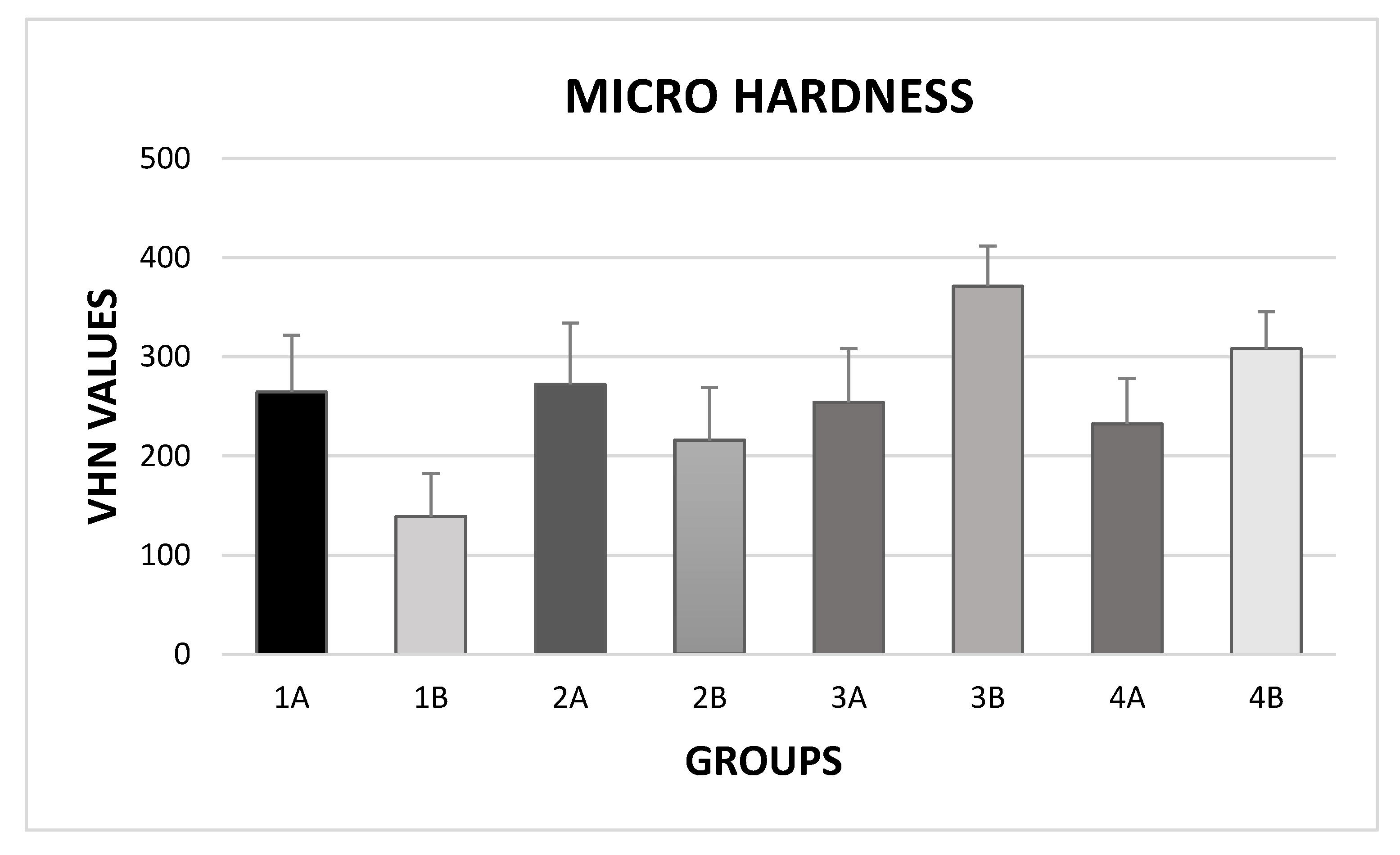
| Group | Enamel | Mean (SD) (kgp/mm2) | Minimum Value (kgp/mm2) | Median (kgp/mm2) | Maximum Value (kgp/mm2) |
|---|---|---|---|---|---|
| 1a | Intact | 264.74 (57.10) a | 151.70 | 263.18 | 365.00 |
| 1b | H2O2 35% | 138.96 (43.48) b | 64.20 | 136.80 | 231.20 |
| 2a | Intact | 272.21 (61.68) a,f | 112.50 | 292.05 | 335.90 |
| 2b | CH6N2O3 16% | 215.79 (53.13) c,e | 92.30 | 222.35 | 313.60 |
| 3a | Intact | 254.26 (53.95) a,e | 146.80 | 252.45 | 350.50 |
| 3b | H2O2 35% + Hydroxyapatite | 371.08 (40.70) d | 293.50 | 369.70 | 453.70 |
| 4a | Intact | 232.29 (46.04) a,e | 150.10 | 229.75 | 335.20 |
| 4b | CH6N2O3 16% + Hydroxyapatite | 308.24 (36.97) d,f | 240.30 | 303.70 | 382.10 |
© 2020 by the authors. Licensee MDPI, Basel, Switzerland. This article is an open access article distributed under the terms and conditions of the Creative Commons Attribution (CC BY) license (http://creativecommons.org/licenses/by/4.0/).
Share and Cite
Scribante, A.; Poggio, C.; Gallo, S.; Riva, P.; Cuocci, A.; Carbone, M.; Arciola, C.R.; Colombo, M. In Vitro Re-Hardening of Bleached Enamel Using Mineralizing Pastes: Toward Preventing Bacterial Colonization. Materials 2020, 13, 818. https://doi.org/10.3390/ma13040818
Scribante A, Poggio C, Gallo S, Riva P, Cuocci A, Carbone M, Arciola CR, Colombo M. In Vitro Re-Hardening of Bleached Enamel Using Mineralizing Pastes: Toward Preventing Bacterial Colonization. Materials. 2020; 13(4):818. https://doi.org/10.3390/ma13040818
Chicago/Turabian StyleScribante, Andrea, Claudio Poggio, Simone Gallo, Paolo Riva, Antonella Cuocci, Manuel Carbone, Carla Renata Arciola, and Marco Colombo. 2020. "In Vitro Re-Hardening of Bleached Enamel Using Mineralizing Pastes: Toward Preventing Bacterial Colonization" Materials 13, no. 4: 818. https://doi.org/10.3390/ma13040818