Article

Lack of Skeletal Effects in Mice with Targeted Disruptionof Prolyl Hydroxylase Domain 1 (Phd1) Gene Expressed in Chondrocytes

1
Musculoskeletal Disease Center, Loma Linda VA Healthcare System, Loma Linda, CA 92357, USA
2
Department of Medicine, Loma Linda University, Loma Linda, CA 92354, USA
*
Author to whom correspondence should be addressed.
Life 2023, 13(1), 106; https://doi.org/10.3390/life13010106
Submission received: 30 November 2022 / Revised: 16 December 2022 / Accepted: 28 December 2022 / Published: 30 December 2022
(This article belongs to the Special Issue Feature Paper in Physiology and Pathology)

Abstract

:
The critical importance of hypoxia-inducible factor (HIF)s in the regulation of endochondral bone formation is now well established. HIF protein levels are closely regulated by the prolyl hydroxylase domain-containing protein (PHD) mediated ubiquitin-proteasomal degradation pathway. Of the three PHD family members expressed in bone, we previously showed that mice with conditional disruption of the Phd2 gene in chondrocytes led to a massive increase in the trabecular bone mass of the long bones. By contrast, loss of Phd3 expression in chondrocytes had no skeletal effects. To investigate the role of Phd1 expressed in chondrocytes on skeletal development, we conditionally disrupted the Phd1 gene in chondrocytes by crossing Phd1 floxed mice with Collagen 2α1-Cre mice for evaluation of a skeletal phenotype. At 12 weeks of age, neither body weight nor body length was significantly different in the Cre+; Phd1flox/flox conditional knockout (cKO) mice compared to Cre; Phd1flox/flox wild-type (WT) control mice. Micro-CT measurements revealed significant gender differences in the trabecular bone volume adjusted for tissue volume at the secondary spongiosa of the femur and the tibia for both genotypes, but no genotype differences were found for any of the trabecular bone measurements of either femur or tibia. Similarly, cortical bone parameters were not affected in the Phd1 cKO mice compared to control mice. Histomorphometric analyses revealed no significant differences in bone area, bone formation rate or mineral apposition rate in the secondary spongiosa of femurs between cKO and WT control mice. Loss of Phd1 expression in chondrocytes did not affect the expression of markers of chondrocytes (collage 2, collagen 10) or osteoblasts (alkaline phosphatase, bone sialoprotein) in the bones of cKO mice. Based on these and our published data, we conclude that of the three PHD family members, only Phd2 expressed in chondrocytes regulates endochondral bone formation and development of peak bone mass in mice.

1. Introduction

Previous studies have uncovered that the prolyl hydroxylase domain-containing proteins (PHDs) are negative regulators of the hypoxia-inducible transcription factor (HIF)1α [1,2]. The hydroxylation of specific proline residues (Pro-402 and Pro-564) in the C-terminal oxygen-dependent degradation domains of the HIF1α by PHDs, primarily the PHD2 isoform, leads to the targeting of HIF1α for ubiquitination through an E3 ligase complex initiated by the binding of the Von Hippel Lindau protein (pVHL) and subsequent proteasomal degradation [1,2]. When the oxygen level is low in the cells, the Phd gene expression is suppressed, and the HIF1α degradation is reduced and the protein is accumulated in the cytoplasm from where it traffics to nucleus and binds to HIF regulatory elements in the promoter regions of the hypoxia-responsive genes including VEGF, Runx2 and osterix to regulate the target gene expression and subsequently bone formation [3,4]. In mammals, PHD enzymes include PHD1, PHD2, and PHD3 [5]. Both PHD1 and PHD2 contain more than 400 amino acid residues while PHD3 has less than 250. All three members contain the highly conserved hydroxylase domain in the catalytic carboxy-terminal region and are expressed in bones. However, PHD1 and PHD2 preferably hydroxylate the N-terminal oxygen-dependent degradation domains (NODD) but are less active for the C-terminal oxygen-dependent degradation domains (CODD) whereas PHD3 almost exclusively hydroxylates the CODD [6,7]. Mice with deletion of Phd1 and Phd3 genes grow normal, but Phd2 gene knockout (KO) in mice causes embryonic lethality because the placenta is underdeveloped [8]. The structural difference among the PHD proteins and the data from mouse genetic studies suggest they may have tissue specific functions.
We previously unveiled that PHD2 was highly expressed in bone cells and contributed to an indispensable role in regulating bone homeostasis by upregulating the transcription of genes critical for osteoblast differentiation and function [9]. Mice with targeted deletion of Phd2 in osteoblasts were smaller and died 12 to 14 weeks after birth. Bone mineral density (BMD) in femurs and the ratio of trabecular bone volume to the tissue volume (BV/TV) in the secondary spongiosa regions of the long bones of the osteoblast-specific conditional knockout (cKO) mice were dramatically low [9]. Mice lacking PHD2 protein in chondrocytes born normally, but the growth after birth were retarded because of elevated mineralization of the cartilage matrix. The chondrocyte-specific cKO mice manifested an increased endochondral bone formation in the femur, tibia and spine, resulting from increased HIF signaling in chondrocytes [10]. While the expression level of Phd3 in the bones in chondrocyte specific Phd2 KO mice was dramatically elevated, loss of Phd3 in chondrocytes did not affect endochondral bone formation and skeletal phenotypes [11]. To investigate the role of Phd1 expressed in chondrocytes on skeletal development, we conditionally disrupted the Phd1 gene in chondrocytes by crossing Phd1 floxed mice with Col2α1-Cre mice for evaluation of skeletal phenotypes.

2. Materials and Methods

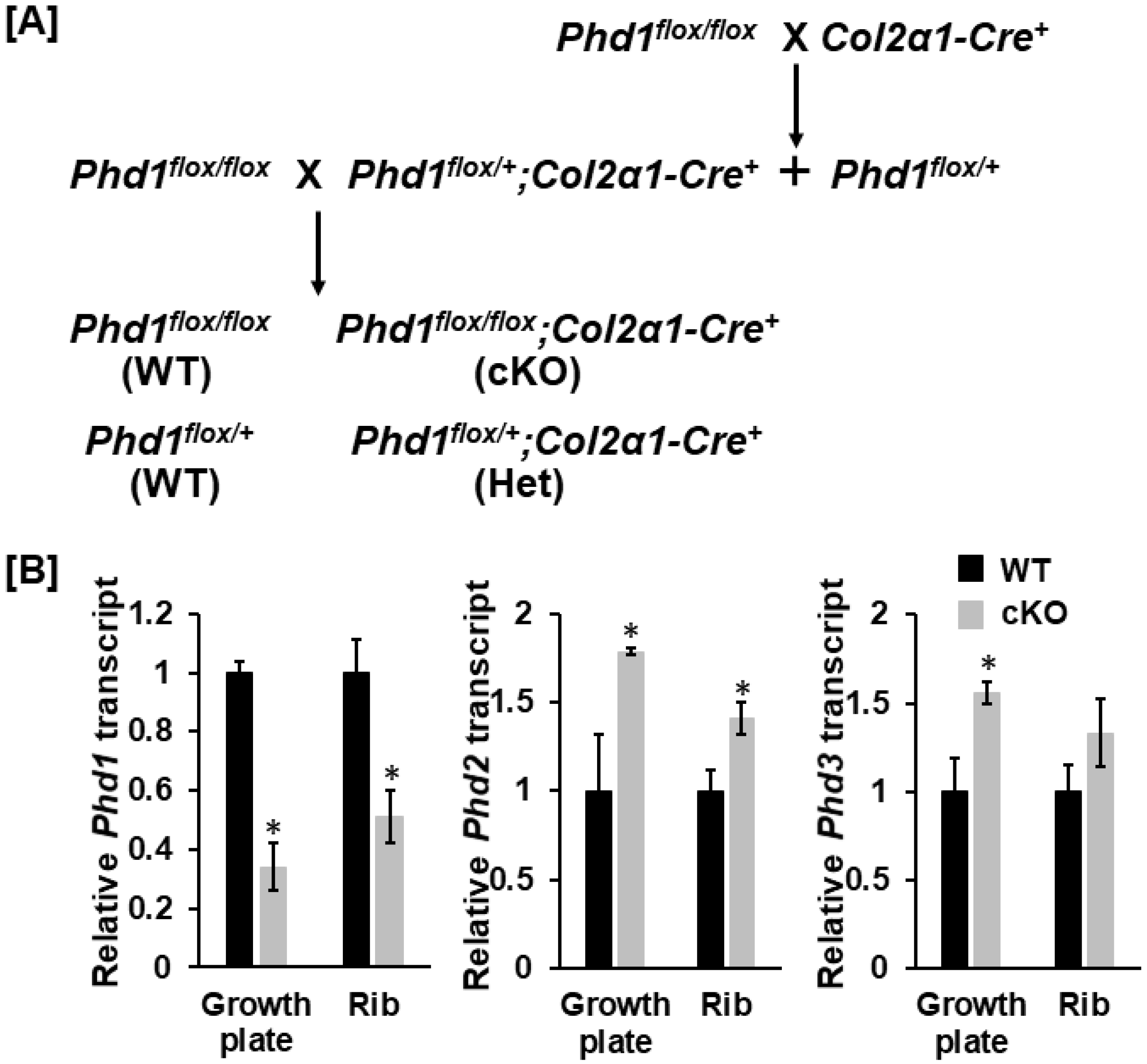
2.1. Breeding Strategy of cKO Mice

Phd1 floxed mice were bred with mice overexpressing Cre under the control of the Collagen 2α1 (Col2α1) promoter to produce Cre positive, Phd1 floxed heterozygous mice (Phd1flox/+; Col2α1-Cre+) according to the breeding strategy described previously [11,12,13]. The Phd1flox/+; Col2α1-Cre+ mice were then backcrossed with Phd1flox/flox mice to generate Cre-positive, loxP-homozygous (Phd1flox/flox; Col2α1-Cre+) cKO and Cre-negative, Phd1 loxP-homozygous or heterozygous (Phd1flox/flox; Phd1flox/+) wild-type (WT) littermates (Figure 1A). The genetic background of these mice is C57BL/6. Both sex mice were used in this study. Mice were housed at the Loma Linda VA Healthcare System (Loma Linda, CA, USA) at 22 °C and with 14 h light and 10 h dark, as well as free access to food and water. Experiments were carried out according to the protocol approved by the Institutional Animal Care and Use Committee (IACUC) of the Loma Linda VA Healthcare System (CA, USA). Mice were anesthetized using isoflurane before tail clipping. Mice were euthanized by exposing to CO2 gas proceeded by cervical dislocation.

2.2. Evaluation of Bone Phenotypes

Areal BMD of the total body, long and lumbar bones (L4–6) of 12-week-old mice were quantified by the FAXITRON UltraFocusDXA 1000 as reported [11,14,15]. Trabecular and cortical bones of the femur and the tibia were scanned and quantified by microcomputed tomography (µCT) in 12-week old mice described previously [16]. The formalin-fixed bones in PBS were scanned by µCT with 55 kVp volts and a voxel size of 10.5 µm. A 1.05 mm cortical bone in the mid-diaphysis of the femur and the tibia were analyzed for cortical bone parameters. A 2.1 mm of the secondary spongiosa of the distal femur and the proximal tibia beginning 0.3675 mm from the growth plates were assessed for TV(mm3), BV(mm3), and BV/TV, as described [17,18,19].

2.3. Double Labeling and Histomorphometric Analyses

Twelve-week-old mice were injected intraperitoneally with calcein (20 mg/kg) eight and two days before euthanization by CO2 to label mineralizing bone surfaces. Mouse right femurs were fixed in 10% formalin for 3 days, washed 3 times with PBS, dehydrated, and embedded in methyl methacrylate resin for sectioning. The sampling sites and histomorphometric analyses were performed as described [19]. The first and the second calcein labeling of the trabecular bone in the secondary spongiosa region of the distal femurs were blindly quantified with OsteoMeasure V3.1.0.2 computer software (OsteoMetrics, Decatur, GA, USA) [20,21]. The mineral apposition rate (MAR) and bone formation rate/bone surface (BFR/BS) were calculated as described previously [22].

2.4. Primary Chondrocyte Culture

Primary chondrocytes isolated from the rib cartilage and the growth plates of the femurs and the tibias of 10-day old WT and cKO mice (3 female and 3 male littermate mice) were cultured as previously described [23]. Cells were grown in DMEM/F12 medium containing 10% fetal bovine serum (FBS), penicillin (100 U/mL), and streptomycin (100 μg/mL) to approximately 90% confluence before harvesting for RNA extraction.

2.5. RNA Extraction and Real-Time PCR

Total RNA was extracted from the femurs and the tibias of the WT and cKO mice or primary chondrocyte cultures derived from WT and cKO mice with the Trizol as described [24,25]. An aliquot of RNA (300 ng) was reverse-transcribed into cDNA in 20 µL volume of reaction by oligo(dT)12–18 primer. A real-time PCR contained 0.5 µL template cDNA, 1x SYBR GREEN master mix (Qiagen), and 100 nM of specific forward and reverse primers in a 25 μL volume of reaction. Primers used for real-time PCR are listed in Table 1. Relative gene expression was calculated by using the ΔΔCT method [26].

2.6. Statistical Analysis

Student’s t-test was used for data analyses. Data are Mean ± SEM (n = 6–10).

3. Results

3.1. Expression of Phd1 Was Partially Disrupted in Chondrocytes in cKO Mice

To test if loss of Phd1 expression in chondrocytes impairs endochondral bone formation, we produced chondrocyte-specific Phd1 cKO mice by breeding the Phd1 floxed mice with the Col2α-Cre mice, in which the Cre recombinase is overexpressed in Col2α-expressing chondrocytes [13,27]. After 2 generations of breeding, the Phd1 floxed, Cre+ cKO mice (Phd1flox/flox; Col2α-Cre+) were produced and compared to Cre negative WT littermates (Phd1flox/flox or Phd1lox/+; Cre). The cKO mice born and developed normally. To investigate if PHD1 protein exists in bone cells of cKO mice, RNA was isolated from chondrocytes derived from the growth plates of the femurs and the tibias, and the ribs of 10-day old WT and cKO mice for real-time PCR with specific primers to Phd1, 2 and 3. As shown in Figure 1B, the expression levels of Phd1 transcript were reduced by 66% and 45% in the growth plate and rib chondrocytes, respectively, in the cKO mice compared to WT mice. By comparison, Phd2 was increased in chondrocytes of both growth plates and ribs of Phd1 cKO mice by 79% and 41%, respectively. While the expression levels of Phd3 were 56% and 33% higher, respectively, in the growth plate and rib chondrocytes of Phd1 cKO mice, only Phd3 expression level in growth plate chondrocytes was significantly higher compared to WT mice.

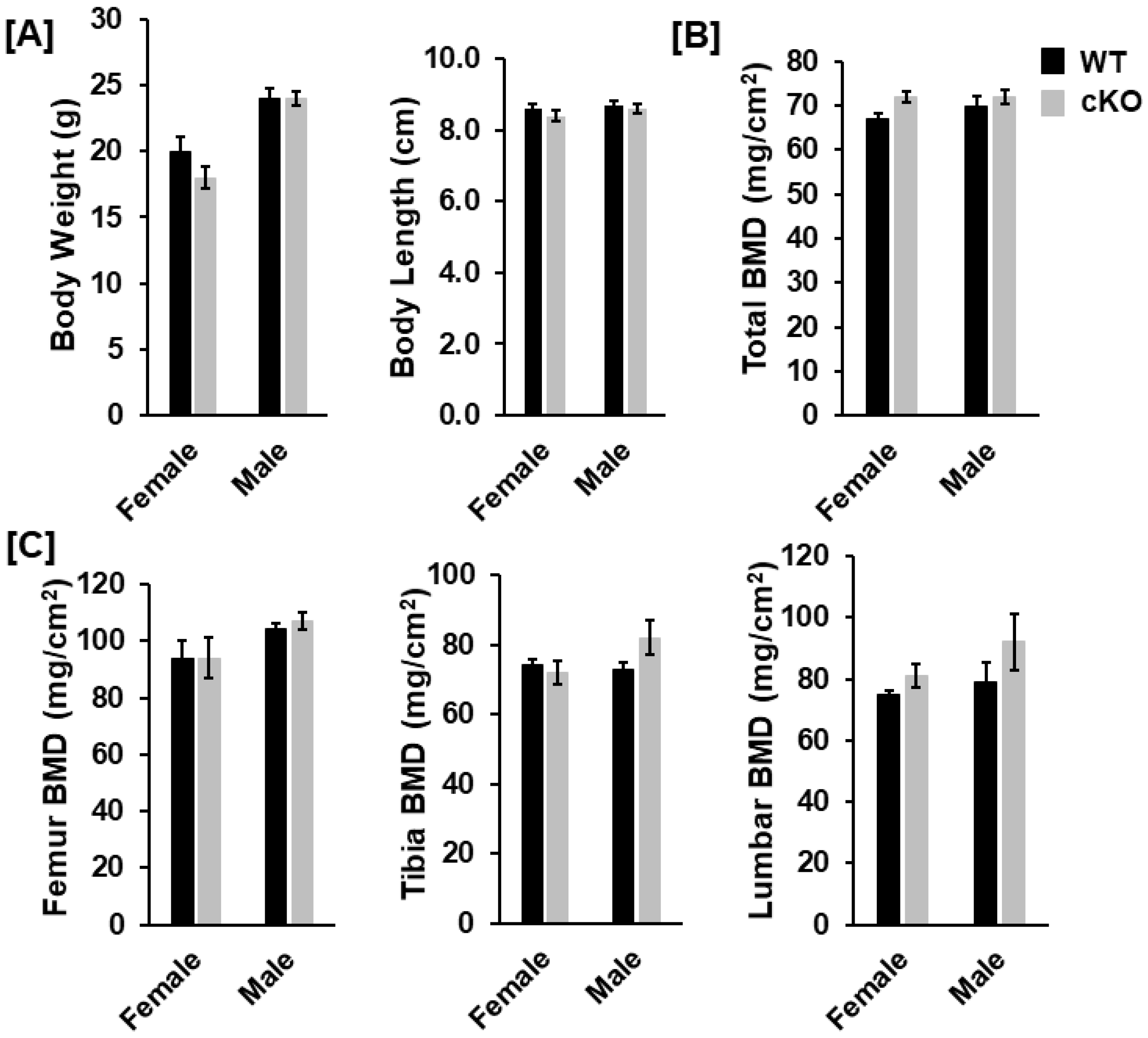
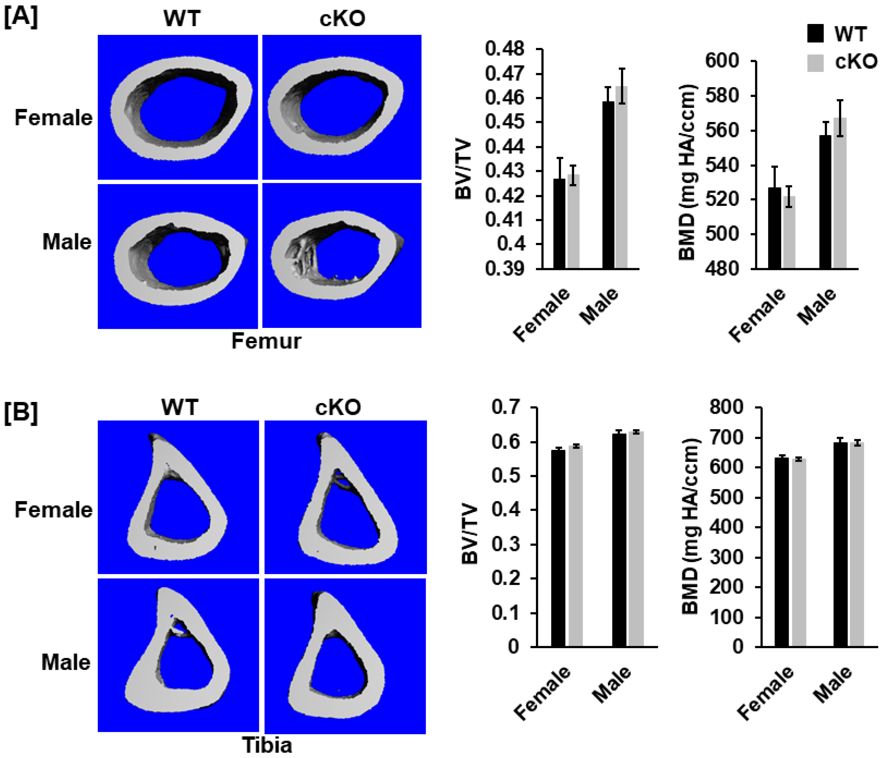
3.2. Deletion of Phd1 in Col2α-Expressing Chondrocytes Does Not Affect Skeletal Growth in Mice at 12 Weeks of Age

To analyze the bone phenotypes, we performed DXA screening and μCT scanning. At 12 weeks after birth, neither body weight nor body length was significantly different in the cKO mice compared to gender-matched control mice for either gender (Figure 2A). DXA measurements revealed no significant changes in total body, femur, tibia, and lumbar BMDs between the two genotypes for either gender (Figure 2B,C). Concurred with DXA data, µCT scanning of the femoral trabecular bone uncovered no significant changes in either BV/TV or any of the trabecular bone parameters including BMD, trabecular number (Tb. N), trabecular thickness (Tb. Th) and trabecular spacing (Tb. Sp) in Phd1 cKO from the gender-matched WT mice for either gender (Figure 3A–C). The tibial BMD, BV/TV, Tb. N, Tb. Th, and Tb. Sp in cKO mice were also comparable to WT mice in either gender mice (Figure 4A–C). Deletion of the Phd1 gene in chondrocytes had no impact on either cortical BV/TV ratio or BMD (Figure 5A). The deficiency of PHD1 expression had no effect on cortical BV/TV and BMD in the tibia either (Figure 5B).

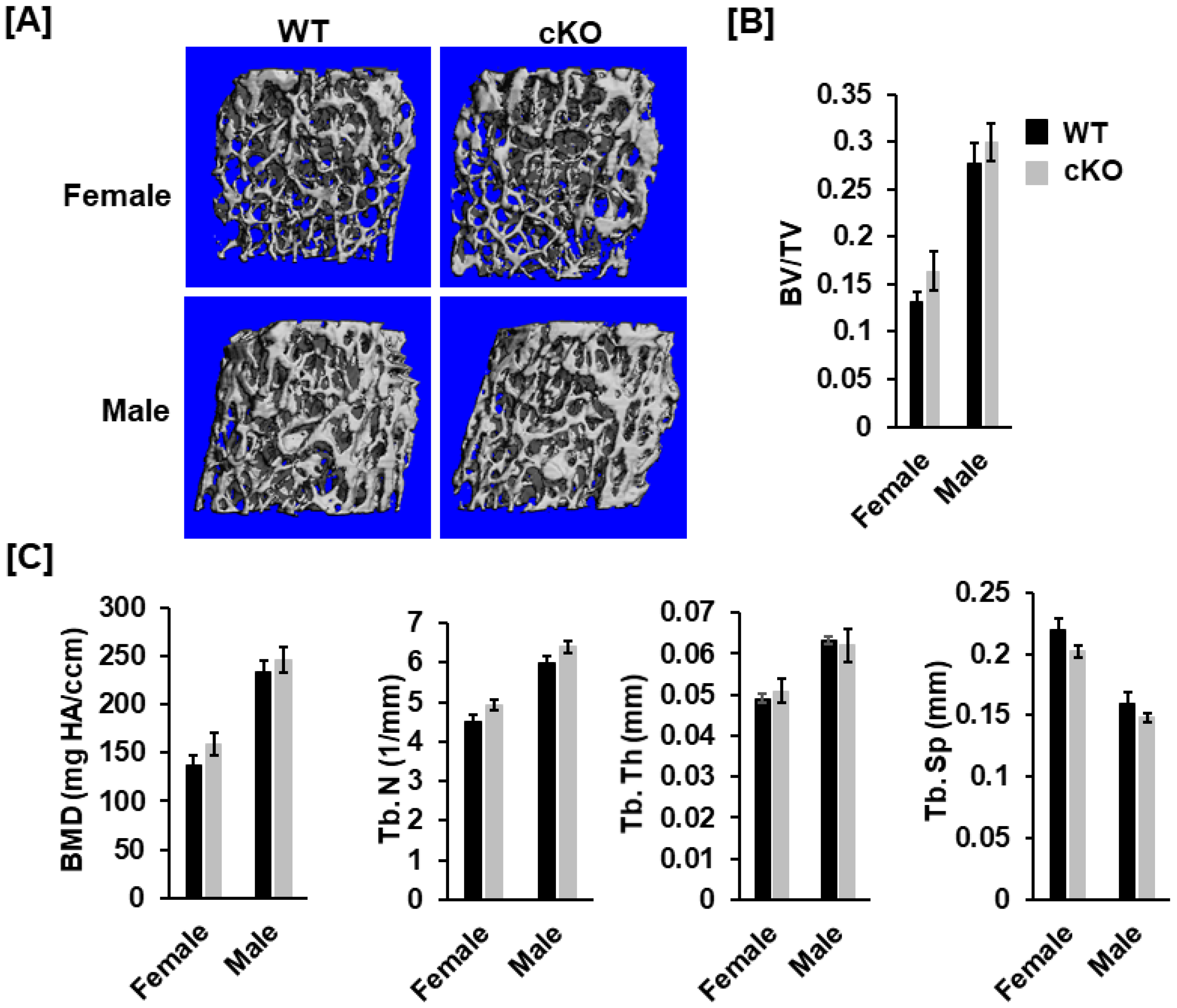
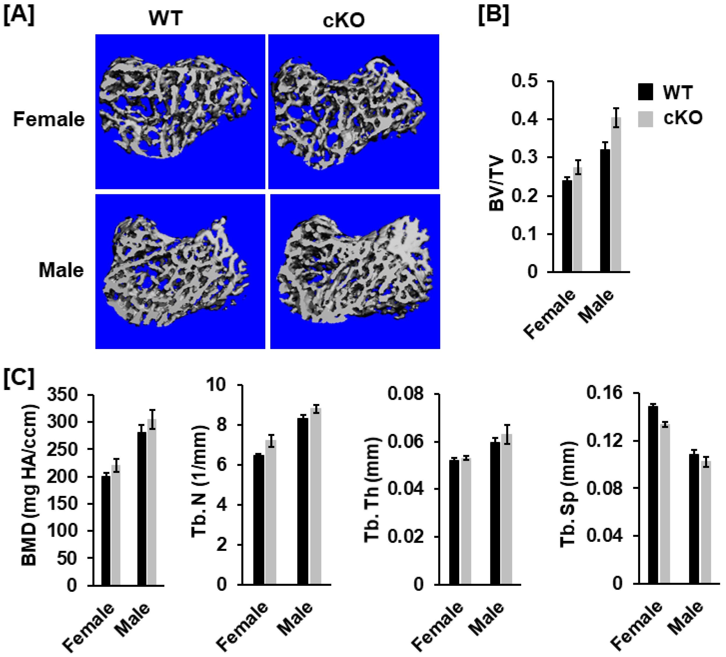
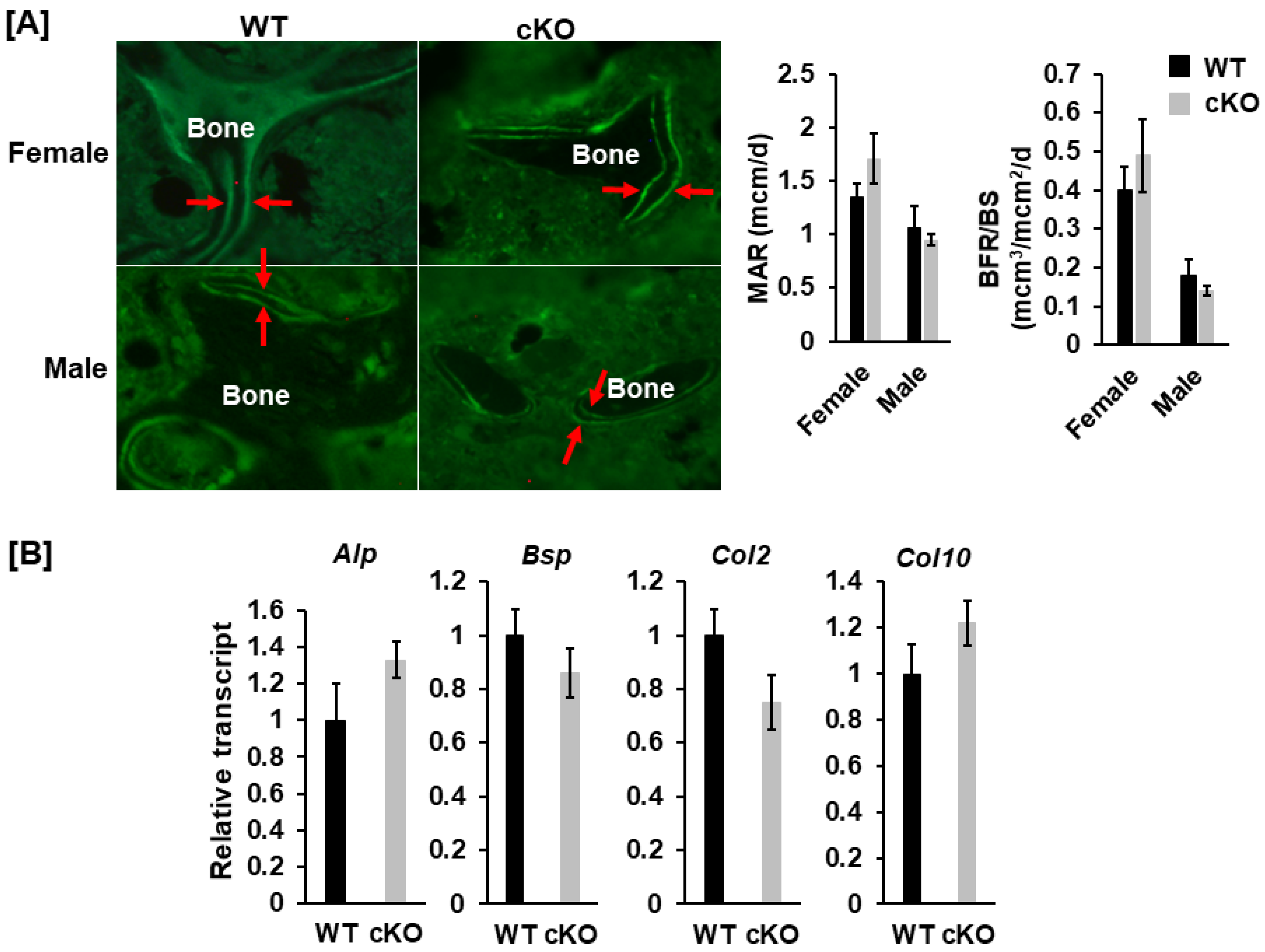
3.3. Knockout of Phd1 in Chondrocytes Neither Influences Bone Formation Nor Expression of Marker Genes of Osteoblast/Chondrocyte Differentiation

To determine if the deletion of Phd1 in Col2α1 expressing cells impacts osteoblast formation, and trabecular bone formation in the femur, we performed histomorphometry analyses and examined the bone marker genes expression in long bones of the cKO mice. We uncovered that knockdown of Phd1 expression in chondrocytes neither affected the MAR) nor the BFR/BS in cKO mice as compared to the WT control littermates (Figure 6A). Consistent with the histomorphometric data, lack of Phd1 in chondrocytes had no impact on the differentiation of both osteoblasts and chondrocytes as evidenced by comparable expression levels of marker genes, alkaline phosphatase (Alp), bone sialoprotein (Bsp), collagen 2 (Col2), and collagen 10 (Col10) in long bones in the cKO mice compared with the WT mice (Figure 6B).

4. Discussion

Of three family members, the PHD2 protein is the most abundant in bones [28]. PHD2 is believed to be the critical oxygen sensor during hypoxia, which is emphasized by the fact that mice with global deletion of the Phd2 gene are embryonically lethal [8]. By contrast, mice with global disruption of either the Phd1 or Phd3 gene develop normally. Consistent with an important role for PHD2 in bones, we and others have shown that disruption of the Phd2 gene in osteoblasts and chondrocytes influenced bone formation and development of peak bone mass [9,27,29]. Mice with deletion of Phd2 in osteoblasts were smaller and died twelve to fourteen weeks after birth. Femoral BMD and trabecular BV/TV of osteoblast-specific cKO mice were notably diminished. By contrast, mice lacking Phd2 were born normally, but the development was retarded after birth resulted from abnormal mineralization of the cartilage matrix. Endochondral bone formation was enhanced in the femur, tibia, and spine of the Phd2 chondrocyte-specific cKO mice [10]. While the expression level of Phd3 elevated 7-fold in chondrocytes of Phd2-cKO mice, targeted disruption of Phd3 gene in mice had no impact on bone cell differentiation, endochondral bone formation, and bone development [11]. Our previous studies indicate that Phd3, unlike Phd2, does not play an important role in regulating chondrocyte differentiation and bone growth.
PHD enzymes function through hydroxylation of the specific proline and asparagine residues of HIF-α and negatively regulate HIF-α protein stability [30]. Both HIF1α and HIF2α contain two prolyl hydroxylation sites in a central degradation domain of HIF1α. Hydroxylation of these sites promotes HIF1α interaction with the ubiquitin ligase for ubiquitination and subsequent degradation [5,31]. Hydroxylation of an asparagine residue in the C-terminals prevents HIFα transcription factor from cooperation with the co-activator, p300/CBP, leading to HIF1α inactivation [32]. Recent studies suggest that the PHD1/2 proteins specifically and preferentially hydroxylate their substrates. Although both PHD1 and PHD2 are active on CODD and NODD, PHD1 appears to act more effectively on substrate HIF2α, whereas PHD2 more actively hydroxylates on substrate HIF1α [7]. These studies indicate the PHD1 vs. PHD2 hydroxylate HIFα in a CODD sequence-dependent manner. Congruent with these studies, Phd2 deletion mice had an increased level of HIF1α in the liver and the kidneys but no increase in the HIF2α protein was noted. In contrast, PHD1/3 double deficient mice had elevated level of HIF2α protein only in the liver [28,33]. On the other hand, loss of Hif1α in osteoblasts impaired skeletal growth [34,35]. Mice without HIF1α protein in the condensing mesenchymal stem cells had shorter bones, impaired mineralized skulls and wider sutures because of severe chondrocyte apoptosis and impaired chondrocyte proliferation in the growth plate [34]. By contrast, loss of HIF2α protein in mice only resulted in a modest decrease in trabecular BV [36]. However, recent mouse genetics studies demonstrated that HIF2 is a negative regulator of osteoblastogenesis and bone mass accrual by upregulating the transcription factor SOX9 to impair osteoblast differentiation [37]. Loss of HIF2 in mesenchymal progenitors increases bone mass by promoting bone formation without affecting bone resorption [37,38]. Since SOX9 is also a master transcription factor in chondrocyte differentiation, we assumed that if the PHD1/HIF/SOX9 signaling axis in chondrocytes is important in endochondral bone formation, then loss of the Phd1 gene in chondrocytes should influence trabecular bone mass because PHD1 hydroxylates target HIF proteins and promotes ubiquitin-mediated protein degradation. To test the hypothesis, Phd1 was deleted in chondrocytes by breeding Phd1 floxed mice with Col2α1-Cre mice, and the effects of knocking out the Phd1 gene in chondrocytes on the development of peak bone mass was evaluated. Surprisingly, no significant changes in either body weight or body length was observed in the cKO mice compared to gender- and age-matched WT littermates. Micro-CT measurements unveiled significant gender differences in the trabecular BV/TV at the metaphysis of either the femur or the tibia of WT and cKO mice. We did not observe a genotype difference for any of the trabecular measurements of the long bones. Similarly, cortical bone parameters were not affected in the Phd1 cKO mice compared to control mice. Histomorphometric analyses observed no significant differences in bone formation rate or mineral apposition rate in the secondary spongiosa of femurs between cKO and WT control mice. These data suggest that Phd1 expressed in chondrocytes exert no major role in regulating the skeletal phenotype.
We found that Phd1 expression was reduced only by 66% and 45%, respectively, in cultured growth plate and rib chondrocytes derived from the long bones of 10-day old cKO mice. The magnitude of reduction in Phd1 expression in growth plate chondrocytes of Phd1 cKO mice was similar to the 60% reduction in Phd2 expression reported previously in the growth plate chondrocytes of Phd2 cKO mice [27]. One potential explanation for the partial reduction in Phd1 expression in the cKO mice is the possibility that the cultures used were not entirely homogeneous for chondrocytes and might contain other cell types (fibroblasts, osteoblasts) which remains to be examined. In any case, our data show that 66%-45% loss of Phd1 transcript in the growth plate and rib chondrocytes had no impact on the transcription of chondrocyte markers, Col2 and Col10, or osteoblast markers, Alp, Bsp2, in the bones of cKO mice. By contrast, we found that the expression levels of Phd2 and Phd3 were increased in the chondrocytes derived from Phd1 cKO mice which could represent a compensatory response to the loss of Phd1 expression. In previous studies, we reported that PHD2 was a negative regulator of chondrocyte differentiation since disruption of Phd2 gene in chondrocytes, promoted chondrocyte differentiation and increased trabecular bone formation [27,39]. We, therefore, anticipated an increased Phd2 expression to reduce chondrocyte differentiation, and trabecular bone volume in the Phd1 cKO mice. However, that was not the case. Further studies comparing the skeletal phenotypes Phd1, Phd2 and Phd1/2 cKO mice are needed to verify if the compensatory increase in Phd2 expression has any role in the Phd1 cKO mice. While expression of Phd3 was elevated by 56% in the growth plate chondrocytes, this compensatory increase in the expression of Phd3 is unlikely to play a significant role in regulating bone formation based on our previous findings on the lack of skeletal phenotype in chondrocyte specific Phd3 cKO mice. Consistent with our interpretation, Wu et al. found that the trabecular bone phenotype was unaffected in mice with disruption of both phd1 and Phd3 genes in osterix expressing cells [40]. Our data, together with our previous reports, imply that Phd2 transcribed in chondrocytes is a major contributor to endochondral bone formation [27]. PHD2 expressed in chondrocytes can functionally compensate for the loss of PHD1 in Phd1 cKO mice.

5. Conclusions

Phd1 expressed in chondrocytes does not regulate endochondral bone formation. Of the Phd1/2/3 genes, only Phd2 transcribed in chondrocytes contributes to the endochondral bone formation and the peak bone mass in mice.

Author Contributions

Conceptualization, S.M.; methodology, D.L., S.P., W.T. and G.A.G.; formal analysis, W.X., S.P., W.T. and E.K.L.; data curation, W.X., S.P., W.T. and E.K.L.; writing—original draft preparation, W.X.; writing—review and editing, W.X. and S.M.; supervision, S.M.; funding acquisition, S.M. All authors have read and agreed to the published version of the manuscript.

Funding

This work was supported by funding from Department of Veterans Affairs (VA) BLR&D merit review grants, 1-101-BX-001396, 1-101-BX-005263 and IK6BX005381 awarded to S.M.; S.M. is a recipient of Senior Research Career Scientist Award from the VA. All work was performed in facilities provided by the VA.

Institutional Review Board Statement

The study was conducted according to the guidelines of the National Institutes of Health (USA) and approved by the Institutional Animal Care and Use Committee of the Jerry L. Pettis Memorial Veterans Affairs Medical Center (0047/1165).

Informed Consent Statement

Not applicable.

Data Availability Statement

Raw data are available upon request.

Acknowledgments

The authors thank Donna Strong for her proofreading.

Conflicts of Interest

The authors declare no conflict of interest.

References

  1. Lando, D.; Peet, D.J.; Whelan, D.A.; Gorman, J.J.; Whitelaw, M.L. Asparagine hydroxylation of the HIF transactivation domain a hypoxic switch. Science 2002, 295, 858–861. [Google Scholar] [CrossRef] [PubMed]
  2. Berra, E.; Ginouves, A.; Pouyssegur, J. The hypoxia-inducible-factor hydroxylases bring fresh air into hypoxia signalling. EMBO Rep. 2006, 7, 41–45. [Google Scholar] [CrossRef] [PubMed] [Green Version]
  3. Huang, L.E.; Bunn, H.F. Hypoxia-inducible factor and its biomedical relevance. J. Biol. Chem. 2003, 278, 19575–19578. [Google Scholar] [CrossRef] [PubMed] [Green Version]
  4. Wan, C.; Shao, J.; Gilbert, S.R.; Riddle, R.C.; Long, F.; Johnson, R.S.; Schipani, E.; Clemens, T.L. Role of HIF-1alpha in skeletal development. Ann. N. Y. Acad. Sci. 2010, 1192, 322–326. [Google Scholar] [CrossRef] [Green Version]
  5. Epstein, A.C.; Gleadle, J.M.; McNeill, L.A.; Hewitson, K.S.; O’Rourke, J.; Mole, D.R.; Mukherji, M.; Metzen, E.; Wilson, M.I.; Dhanda, A.; et al. C. elegans EGL-9 and mammalian homologs define a family of dioxygenases that regulate HIF by prolyl hydroxylation. Cell 2001, 107, 43–54. [Google Scholar] [CrossRef] [Green Version]
  6. Villar, D.; Vara-Vega, A.; Landazuri, M.O.; Del Peso, L. Identification of a region on hypoxia-inducible-factor prolyl 4-hydroxylases that determines their specificity for the oxygen degradation domains. Biochem. J. 2007, 408, 231–240. [Google Scholar] [CrossRef] [PubMed] [Green Version]
  7. Appelhoff, R.J.; Tian, Y.M.; Raval, R.R.; Turley, H.; Harris, A.L.; Pugh, C.W.; Ratcliffe, P.J.; Gleadle, J.M. Differential function of the prolyl hydroxylases PHD1, PHD2, and PHD3 in the regulation of hypoxia-inducible factor. J. Biol. Chem. 2004, 279, 38458–38465. [Google Scholar] [CrossRef] [PubMed] [Green Version]
  8. Takeda, K.; Ho, V.C.; Takeda, H.; Duan, L.J.; Nagy, A.; Fong, G.H. Placental but not heart defects are associated with elevated hypoxia-inducible factor alpha levels in mice lacking prolyl hydroxylase domain protein 2. Mol. Cell. Biol. 2006, 26, 8336–8346. [Google Scholar] [CrossRef] [Green Version]
  9. Cheng, S.; Xing, W.; Pourteymoor, S.; Mohan, S. Conditional disruption of the prolyl hydroxylase domain-containing protein 2 (Phd2) gene defines its key role in skeletal development. J. Bone Miner. Res. 2014, 29, 2276–2286. [Google Scholar] [CrossRef] [PubMed]
  10. Laperre, K.F.P.; Van Looveren, R.; Bouillon, R.; Carmeliet, P.; Carmeliet, G. Deletion of the oxygen-sensor PHD2 in chondrocytes results in increased cartilage and bone mineralization. J. Bone Miner. Res. 2008, 23 (Suppl. S1), S44. [Google Scholar]
  11. Xing, W.; Pourteymoor, S.; Gomez, G.A.; Chen, Y.; Mohan, S. Prolyl Hydroxylase Domain-Containing Protein 3 Gene Expression in Chondrocytes Is Not Essential for Bone Development in Mice. Cells 2021, 10, 2200. [Google Scholar] [CrossRef] [PubMed]
  12. Duan, L.J.; Takeda, K.; Fong, G.H. Hematological, hepatic, and retinal phenotypes in mice deficient for prolyl hydroxylase domain proteins in the liver. Am. J. Pathol. 2014, 184, 1240–1250. [Google Scholar] [CrossRef] [PubMed] [Green Version]
  13. Ovchinnikov, D.A.; Deng, J.M.; Ogunrinu, G.; Behringer, R.R. Col2a1-directed expression of Cre recombinase in differentiating chondrocytes in transgenic mice. Genesis 2000, 26, 145–146. [Google Scholar] [CrossRef]
  14. Xing, W.; Pourteymoor, S.; Chen, Y.; Mohan, S. Targeted Deletion of the Claudin12 Gene in Mice Increases Articular Cartilage and Inhibits Chondrocyte Differentiation. Front. Endocrinol. 2022, 13, 931318. [Google Scholar] [CrossRef]
  15. Xing, W.; Pourteymoor, S.; Kesavan, C.; Gomez, G.A.; Mohan, S. Mice with Targeted Knockout of Tetraspanin 3 Exhibit Reduced Trabecular Bone Mass Caused by Decreased Osteoblast Functions. Cells 2022, 11, 977. [Google Scholar] [CrossRef]
  16. Xing, W.; Liu, J.; Cheng, S.; Vogel, P.; Mohan, S.; Brommage, R. Targeted disruption of leucine-rich repeat kinase 1 but not leucine-rich repeat kinase 2 in mice causes severe osteopetrosis. J. Bone Miner. Res. 2013, 28, 1962–1974. [Google Scholar] [CrossRef]
  17. Bouxsein, M.L.; Boyd, S.K.; Christiansen, B.A.; Guldberg, R.E.; Jepsen, K.J.; Muller, R. Guidelines for assessment of bone microstructure in rodents using micro-computed tomography. J. Bone Miner. Res. 2010, 25, 1468–1486. [Google Scholar] [CrossRef]
  18. Xing, W.; Pourteymoor, S.; Mohan, S. Ascorbic acid regulates osterix expression in osteoblasts by activation of prolyl hydroxylase and ubiquitination-mediated proteosomal degradation pathway. Physiol. Genom. 2011, 43, 749–757. [Google Scholar] [CrossRef] [Green Version]
  19. Xing, W.; Kim, J.; Wergedal, J.; Chen, S.T.; Mohan, S. Ephrin B1 regulates bone marrow stromal cell differentiation and bone formation by influencing TAZ transactivation via complex formation with NHERF1. Mol. Cell. Biol. 2010, 30, 711–721. [Google Scholar] [CrossRef] [Green Version]
  20. Beamer, W.G.; Donahue, L.R.; Rosen, C.J.; Baylink, D.J. Genetic variability in adult bone density among inbred strains of mice. Bone 1996, 18, 397–403. [Google Scholar] [CrossRef]
  21. Qin, X.; Wergedal, J.E.; Rehage, M.; Tran, K.; Newton, J.; Lam, P.; Baylink, D.J.; Mohan, S. Pregnancy-associated plasma protein-A increases osteoblast proliferation in vitro and bone formation in vivo. Endocrinology 2006, 147, 5653–5661. [Google Scholar] [CrossRef] [PubMed]
  22. Parfitt, A.M.; Drezner, M.K.; Glorieux, F.H.; Kanis, J.A.; Malluche, H.; Meunier, P.J.; Ott, S.M.; Recker, R.R. Bone histomorphometry: Standardization of nomenclature, symbols, and units: Report of the ASBMR Histomorphometry Nomenclature Committee. J. Bone Miner. Res. 1987, 2, 595–610. [Google Scholar] [CrossRef] [PubMed]
  23. Cheng, S.; Xing, W.; Zhou, X.; Mohan, S. Haploinsufficiency of osterix in chondrocytes impairs skeletal growth in mice. Physiol. Genom. 2013, 45, 917–923. [Google Scholar] [CrossRef] [PubMed] [Green Version]
  24. Xing, W.; Singgih, A.; Kapoor, A.; Alarcon, C.M.; Baylink, D.J.; Mohan, S. Nuclear factor-E2-related factor-1 mediates ascorbic acid induction of osterix expression via interaction with antioxidant-responsive element in bone cells. J. Biol. Chem. 2007, 282, 22052–22061. [Google Scholar] [CrossRef] [PubMed] [Green Version]
  25. Xing, W.; Baylink, D.; Kesavan, C.; Hu, Y.; Kapoor, S.; Chadwick, R.B.; Mohan, S. Global gene expression analysis in the bones reveals involvement of several novel genes and pathways in mediating an anabolic response of mechanical loading in mice. J. Cell. Biochem. 2005, 96, 1049–1060. [Google Scholar] [CrossRef] [PubMed]
  26. Livak, K.J.; Schmittgen, T.D. Analysis of relative gene expression data using real-time quantitative PCR and the 2(-Delta Delta C(T)) Method. Methods 2001, 25, 402–408. [Google Scholar] [CrossRef] [PubMed]
  27. Cheng, S.; Xing, W.; Pourteymoor, S.; Schulte, J.; Mohan, S. Conditional Deletion of Prolyl Hydroxylase Domain-containing Protein 2 (Phd2) Gene Reveals its Essential Role in Chondrocyte Function and Endochondral Bone Formation. Endocrinology 2015, 157, en20151473. [Google Scholar] [CrossRef] [Green Version]
  28. Takeda, K.; Aguila, H.L.; Parikh, N.S.; Li, X.; Lamothe, K.; Duan, L.J.; Takeda, H.; Lee, F.S.; Fong, G.H. Regulation of adult erythropoiesis by prolyl hydroxylase domain proteins. Blood 2008, 111, 3229–3235. [Google Scholar] [CrossRef] [Green Version]
  29. Zhu, K.; Song, P.; Lai, Y.; Liu, C.; Xiao, G. Prolyl hydroxylase domain proteins regulate bone mass through their expression in osteoblasts. Gene 2016, 594, 125–130. [Google Scholar] [CrossRef]
  30. Fong, G.H.; Takeda, K. Role and regulation of prolyl hydroxylase domain proteins. Cell Death Differ. 2008, 15, 635–641. [Google Scholar] [CrossRef] [Green Version]
  31. Bruick, R.K.; McKnight, S.L. A conserved family of prolyl-4-hydroxylases that modify HIF. Science 2001, 294, 1337–1340. [Google Scholar] [CrossRef] [PubMed]
  32. Metzen, E.; Berchner-Pfannschmidt, U.; Stengel, P.; Marxsen, J.H.; Stolze, I.; Klinger, M.; Huang, W.Q.; Wotzlaw, C.; Hellwig-Burgel, T.; Jelkmann, W.; et al. Intracellular localisation of human HIF-1 alpha hydroxylases: Implications for oxygen sensing. J. Cell Sci. 2003, 116 Pt 7, 1319–1326. [Google Scholar] [CrossRef] [Green Version]
  33. Takeda, K.; Cowan, A.; Fong, G.H. Essential role for prolyl hydroxylase domain protein 2 in oxygen homeostasis of the adult vascular system. Circulation 2007, 116, 774–781. [Google Scholar] [CrossRef]
  34. Provot, S.; Zinyk, D.; Gunes, Y.; Kathri, R.; Le, Q.; Kronenberg, H.M.; Johnson, R.S.; Longaker, M.T.; Giaccia, A.J.; Schipani, E. Hif-1alpha regulates differentiation of limb bud mesenchyme and joint development. J. Cell. Biol. 2007, 177, 451–464. [Google Scholar] [CrossRef] [PubMed]
  35. Wan, C.; Gilbert, S.R.; Wang, Y.; Cao, X.; Shen, X.; Ramaswamy, G.; Jacobsen, K.A.; Alaql, Z.S.; Eberhardt, A.W.; Gerstenfeld, L.C.; et al. Activation of the hypoxia-inducible factor-1alpha pathway accelerates bone regeneration. Proc. Natl. Acad. Sci. USA 2008, 105, 686–691. [Google Scholar] [CrossRef] [PubMed] [Green Version]
  36. Shomento, S.H.; Wan, C.; Cao, X.; Faugere, M.C.; Bouxsein, M.L.; Clemens, T.L.; Riddle, R.C. Hypoxia-inducible factors 1alpha and 2alpha exert both distinct and overlapping functions in long bone development. J. Cell. Biochem. 2010, 109, 196–204. [Google Scholar] [CrossRef] [PubMed]
  37. Merceron, C.; Ranganathan, K.; Wang, E.; Tata, Z.; Makkapati, S.; Khan, M.P.; Mangiavini, L.; Yao, A.Q.; Castellini, L.; Levi, B.; et al. Hypoxia-inducible factor 2alpha is a negative regulator of osteoblastogenesis and bone mass accrual. Bone Res. 2019, 7, 7. [Google Scholar] [CrossRef] [PubMed] [Green Version]
  38. Lee, S.Y.; Park, K.H.; Yu, H.G.; Kook, E.; Song, W.H.; Lee, G.; Koh, J.T.; Shin, H.I.; Choi, J.Y.; Huh, Y.H.; et al. Controlling hypoxia-inducible factor-2alpha is critical for maintaining bone homeostasis in mice. Bone Res. 2019, 7, 14. [Google Scholar] [CrossRef] [Green Version]
  39. Cheng, S.; Pourteymoor, S.; Alarcon, C.; Mohan, S. Conditional Deletion of the Phd2 Gene in Articular Chondrocytes Accelerates Differentiation and Reduces Articular Cartilage Thickness. Sci. Rep. 2017, 7, 45408. [Google Scholar] [CrossRef]
  40. Wu, C.; Rankin, E.B.; Castellini, L.; Alcudia, J.F.; LaGory, E.L.; Andersen, R.; Rhodes, S.D.; Wilson, T.L.; Mohammad, K.S.; Castillo, A.B.; et al. Oxygen-sensing PHDs regulate bone homeostasis through the modulation of osteoprotegerin. Genes Dev. 2015, 29, 817–831. [Google Scholar] [CrossRef]
Figure 1. Breeding strategy of Phd1 conditional knockout (cKO) mice. (A) A breeding strategy of Phd1 cKO mice, heterozygous (Het) and wild-type (WT) mice. (B) Phd1 expression was partially disrupted in chondrocyte cultures derived from the cKO mice. Total RNA was extracted from primary chondrocytes derived from the femur growth plates and the ribs of 10-day old mice for quantitative PCR (n = 3). Star (*): p < 0.01.
Life 13 00106 g001
Figure 2. Bone parameters were not significantly altered in the Phd1 cKO mice at 12 weeks after birth. (A) Body weight and length of the mice, as indicated in the figure. (B,C) Bone mineral density (BMD) for total body, the femur, the tibia, and the lumbar bone, respectively, quantified by DXA (n = 6–10).
Life 13 00106 g002
Figure 3. Trabecular parameters were not significantly altered in the femur of the Phd1 cKO mice at 12 weeks after birth. (A) Representative µCT images of the distal femurs. (B,C) Trabecular bone data of the femurs (BV/TV, BMD, Tb.N, Tb. Th, and Tb. Sp) measured by µCT (n = 6–10). BV, bone volume; TV, tissue volume; Tb. N, trabecular number; Tb. Th, trabecular thickness; Th. Sp, Trabecular spacing.
Life 13 00106 g003
Figure 4. No changes in trabecular parameters were found in the proximal metaphysis of the tibia in the Phd1 cKO mice at 12 weeks after birth. (A) Representative µCT images of the proximal tibias. (B,C) Trabecular bone data of the tibias (BV/TV, BMD, Tb.N, Tb. Th, and Tb. Sp) measured by µCT (n = 6–10).
Life 13 00106 g004
Figure 5. No changes were found in cortical bone parameters of the femur and the tibia in the Phd1 cKO mice at 12 weeks after birth. (A) Representative µCT images of the cortical bone of the femurs and the quantitative data of the cortical bones of the femurs (BV/TV, BMD). (B) Images of the cortical bone of the tibias and the cortical bone data of the tibias (BV, BMD) (n = 6–10).
Life 13 00106 g005
Figure 6. Knockout of Phd1 in chondrocytes neither influences bone formation nor expression of markers of osteoblast/chondrocyte differentiation. (A) Representative images of the calcein-labeled trabecular bone of the distal femurs and the quantitative data of mineral apposition rate (MAR) and bone formation rate/bone surface (BFR/BS), respectively. The bone indicated by two red arrows is the double-labeled, newly formed bone (n = 6). (B) Expression levels of the marker genes of osteoblast and chondrocyte differentiation in long bones measured by RT-real-time PCR. Alp, alkaline phosphatase; Bsp, bone sialoprotein; Col2, collagen 2; Col10: collagen 10.
Life 13 00106 g006
Table 1. Primer Sequence for Real-Time PCR.
GeneForward PrimerReverse Primer
Ppia5′-CCATGGCAAATGCTGGACCA5′-TCCTGGACCCAAAACGCTCC
Phd15′-GGAACCCACATGAGGTGAAG5′-AACACCTTTCTGTCCCGATG
Phd35′-GGGACGCCAAGTTACACGGA5′-GGGCTCCACGTCTGCTACAA
Phd25′-GAAGCTGGGCAACTACAGGA5′-CATGTCACGCATCTTCCATC
Alp5′-ATGGTAACGGGCCTGGCTACA5′-AGTTCTGCTCATGGACGCCGT
Bsp5′-AACGGGTTTCAGCAGACAACC5′-TAAGCTCGGTAAGTGTCGCCA
Col25′-TGGCTTCCACTTCAGCTATG5′-AGGTAGGCGATGCTGTTCTT
Col105′-ACGGCACGCCTACGATGT5′-CCATGATTGCACTCCCTGAA
Note: Ppia, peptidylprolyl isomerase A; Phd, prolyl hydroxylase domain-containing protein; Alp, alkaline phosphatase; Bsp, bone sialoprotein; Col2, collagen 2; Col10, collagen 10.
Disclaimer/Publisher’s Note: The statements, opinions and data contained in all publications are solely those of the individual author(s) and contributor(s) and not of MDPI and/or the editor(s). MDPI and/or the editor(s) disclaim responsibility for any injury to people or property resulting from any ideas, methods, instructions or products referred to in the content.

