
Rare Causes of Acute Coronary Syndrome: Carbon Monoxide Poisoning

 , , ,
, , ,  ,
,
Abstract
:
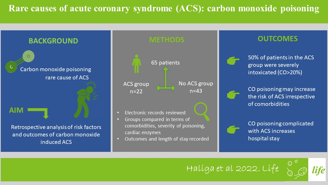
1. Introduction
2. Materials and Methods
2.1. Study Design and Selection of Patients
2.2. Outcomes
2.3. Blood Analysis
2.4. Treatment
2.5. Data Analysis
3. Results
3.1. Patients’ Characteristics
3.2. Severity of CO Poisoning
3.3. Cardiac Ischemic Events
3.4. Arrhythmias
4. Discussion
4.1. Cardiovascular Effects of CO Poisoning
4.2. Arrhythmias, Conduction Disorders
4.3. Study Limitations
5. Conclusions
Author Contributions
Funding
Institutional Review Board Statement
Informed Consent Statement
Data Availability Statement
Conflicts of Interest
References
- Collet, J.-P.; Thiele, H.; Barbato, E.; Barthélémy, O.; Bauersachs, J.; Bhatt, D.L.; Dendale, P.; Dorobantu, M.; Edvardsen, T.; Folliguet, T.; et al. 2020 ESC Guidelines for the management of acute coronary syndromes in patients presenting without persistent ST-segment elevation. Eur. Heart J. 2021, 42, 1289–1367. [Google Scholar] [CrossRef] [PubMed]
- Thygesen, K.; Alpert, J.S.; Jaffe, A.S.; Simoons, M.L.; Chaitman, B.R.; White, H.D.; Katus, H.A.; Lindahl, B.; Morrow, D.A.; Joint ESC/ACCF/AHA/WHF Task Force for the Universal Definition of Myocardial Infarction; et al. Third universal definition of myocardial infarction. Circulation 2012, 126, 2020–2035. [Google Scholar] [CrossRef] [PubMed] [Green Version]
- Smollin, C.; Olson, K. Carbon monoxide poisoning (acute). BMJ Clin. Evid. 2010, 2010, 2103. [Google Scholar] [PubMed]
- Mattiuzzi, C.; Lippi, G. Worldwide epidemiology of carbon monoxide poisoning. Hum. Exp. Toxicol. 2020, 39, 387–392. [Google Scholar] [CrossRef]
- Braubach, M.; Algoet, A.; Beaton, M.; Lauriou, S.; Héroux, M.E.; Krzyzanowski, M. Mortality associated with exposure to carbon monoxide in WHO European Member States. Indoor Air 2013, 23, 115–125. [Google Scholar] [CrossRef]
- Kaya, H.; Coşkun, A.; Beton, O.; Zorlu, A.; Kurt, R.; Yucel, H.; Gunes, H.; Yilmaz, M.B. COHgb levels predict the long-term development of acute myocardial infarction in CO poisoning. Am. J. Emerg. Med. 2016, 34, 840–844. [Google Scholar] [CrossRef] [PubMed]
- Rose, J.J.; Wang, L.; Xu, Q.; McTiernan, C.F.; Shiva, S.; Tejero, J.; Gladwin, M.T. Carbon Monoxide Poisoning: Pathogenesis, Management, and Future Directions of Therapy. Am. J. Respir. Crit. Care Med. 2017, 195, 596–606. [Google Scholar] [CrossRef] [Green Version]
- Lichtarska, D.; Feldman, R. Troponin positive acute coronary syndromes in the course of acute carbon monoxide poisoning as the factor exposing primary coronary heart disease previously undiagnosed. Prz. Lek. 2011, 68, 510–514. [Google Scholar]
- Zierle-Ghosh, A.; Jan, A. Physiology, Body Mass Index; StatPearls Publishing: Treasure Island, FL, USA, 2021. [Google Scholar]
- Palmeri, R.; Gupta, V. Carboxyhemoglobin Toxicity; StatPearls Publishing: Treasure Island, FL, USA, 2022. [Google Scholar]
- Gao, Y.; Yang, J.; Ma, L.; Zhang, Y.; Li, Z.; Wu, L.; Yang, L.; Wang, H. Non-ST elevation myocardial infarction induced by carbon monoxide poisoning: A case report. Medicine 2019, 98, e15151. [Google Scholar] [CrossRef]
- Koskela, R.S.; Mutanen, P.; Sorsa, J.A.; Klockars, M. Factors predictive of ischemic heart disease mortality in foundry workers exposed to carbon monoxide. Am. J. Epidemiol. 2000, 152, 628–632. [Google Scholar] [CrossRef] [PubMed] [Green Version]
- Henz, S.; Maeder, M. Prospective study of accidental carbon monoxide poisoning in 38 Swiss soldiers. Swiss Med. Wkly. 2005, 135, 398–408. [Google Scholar] [PubMed]
- Thiels, H.; Van Durme, J.P.; Vermeire, P.; Pannier, R. Modifications Electrocardiographiques Immédiates et Tardives au Cours de L’intoxication Oxycarbonée Aiguë. Lille Med. 1972, 17, 191–195. [Google Scholar] [PubMed]
- Balzan, M.V.; Cacciottolo, J.M.; Mifsud, S. Unstable angina and exposure to carbon monoxide. Postgrad. Med. J. 1994, 70, 699–702. [Google Scholar] [CrossRef] [PubMed] [Green Version]
- Satran, D.; Henry, C.R.; Adkinson, C.; Nicholson, C.I.; Bracha, Y.; Henry, T.D. Cardiovascular manifestations of moderate to severe carbon monoxide poisoning. J. Am. Coll. Cardiol. 2005, 45, 1513–1516. [Google Scholar] [CrossRef] [PubMed] [Green Version]
- Gallucci, G.; Tartarone, A.; Lerose, R.; Lalinga, A.V.; Capobianco, A.M. Cardiovascular risk of smoking and benefits of smoking cessation. J. Thorac. Dis. 2020, 12, 3866–3876. [Google Scholar] [CrossRef] [PubMed]
- Dorey, A.; Scheerlinck, P.; Nguyen, H.; Albertson, T. Acute and Chronic Carbon Monoxide Toxicity from Tobacco Smoking. Mil. Med. 2020, 185, e61–e67. [Google Scholar] [CrossRef] [PubMed]
- Anderson, R.F.; Allensworth, D.C.; DeGroot, W.J. Myocardial toxicity from carbon monoxide poisoning. Ann. Intern. Med. 1967, 67, 1172–1182. [Google Scholar] [CrossRef]
- Cha, Y.S.; Cha, K.C.; Kim, O.H.; Lee, K.H.; Hwang, S.O.; Kim, H. Features and predictors of myocardial injury in carbon monoxide poisoned patients. Emerg. Med. J. 2014, 31, 210–215. [Google Scholar] [CrossRef]
- Liu, Q.; Gao, X.; Xiao, Q.; Zhu, B.; Liu, Y.; Han, Y.; Wang, W. A combination of NLR and sST2 is associated with adverse cardiovascular events in patients with myocardial injury induced by moderate to severe acute carbon monoxide poisoning. Clin. Cardiol. 2021, 44, 401–406. [Google Scholar] [CrossRef]
- Kalay, N.; Ozdogru, I.; Cetinkaya, Y.; Eryol, N.K.; Dogan, A.; Gul, I.; Inanc, T.; Ikizceli, I.; Oguzhan, A.; Abaci, A. Cardiovascular effects of carbon monoxide poisoning. Am. J. Cardiol. 2007, 99, 322–324. [Google Scholar] [CrossRef]
- Dziewierz, A.; Ciszowski, K.; Gawlikowski, T.; Rakowski, T.; Kleczyński, P.; Surdacki, A.; Dudek, D. Primary angioplasty in patient with ST-segment elevation myocardial infarction in the setting of intentional carbon monoxide poisoning. J. Emerg. Med. 2013, 45, 831–834. [Google Scholar] [CrossRef]
- Kim, S.; Lim, J.H.; Kim, Y.; Oh, S.; Choi, W.G. A Case of Acute Carbon Monoxide Poisoning Resulting in an ST Elevation Myocardial Infarction. Korean Circ. J. 2012, 42, 133–135. [Google Scholar] [CrossRef]
- Dragelytė, G.; Plenta, J.; Chmieliauskas, S.; Jasulaitis, A.; Raudys, R.; Jovaiša, T.; Badaras, R. Myocardial Rupture following Carbon Monoxide Poisoning. Case Rep. Crit. Care 2014, 2014, 281701. [Google Scholar] [CrossRef] [PubMed]
- Cho, D.H.; Ko, S.M.; Son, J.W.; Park, E.J.; Cha, Y.S. Myocardial Injury and Fibrosis From Acute Carbon Monoxide Poisoning: A Prospective Observational Study. JACC Cardiovasc. Imaging 2021, 14, 1758–1770. [Google Scholar] [CrossRef] [PubMed]
- Pach, J.; Hubalewska-Hoła, A.; Pach, D.; Szpak, D. Usefulness of rest and forced perfusion scintigraphy (SPECT) to evaluate cardiotoxicity in acute carbon monoxide poisoning. Prz. Lek. 2001, 58, 297–300. [Google Scholar]
- Henry, T.D.; Satran, D. Acute Carbon Monoxide Poisoning and Cardiac Magnetic Resonance: The Future Is Now. JACC Cardiovasc. Imaging 2021, 14, 1771–1773. [Google Scholar] [CrossRef] [PubMed]
- Henry, C.R.; Satran, D.; Lindgren, B.; Adkinson, C.; Nicholson, C.I.; Henry, T.D. Myocardial injury and long-term mortality following moderate to severe carbon monoxide poisoning. JAMA 2006, 295, 398–402. [Google Scholar] [CrossRef] [Green Version]
- Aslan, S.; Erol, M.K.; Karcioglu, O.; Meral, M.; Cakir, Z.; Katirci, Y. The investigation of ischemic myocardial damage in patients with carbon monoxide poisoning. Anadolu Kardiyol. Derg. 2005, 5, 189–193. [Google Scholar] [PubMed]
- Aslan, S.; Uzkeser, M.; Seven, B.; Gundogdu, F.; Acemoglu, H.; Aksakal, E.; Varoglu, E. The evaluation of myocardial damage in 83 young adults with carbon monoxide poisoning in the East Anatolia region in Turkey. Hum. Exp. Toxicol. 2006, 25, 439–446. [Google Scholar] [CrossRef] [PubMed]
- Lee, F.Y.; Chen, W.K.; Lin, C.L.; Kao, C.H. Carbon monoxide poisoning and subsequent cardiovascular disease risk: A nationwide population-based cohort study. Medicine 2015, 94, e624. [Google Scholar] [CrossRef] [PubMed]
- Atif, B.; Osman, K.A.; Ilker, A.; Alpasla, U. Reversible left bundle-branch block due to carbon monoxide poisoning: A case report. Am. J. Emerg. Med. 2016, 34, 342.E1–342.E3. [Google Scholar]

| Demographic Data | Total (n = 65) | % |
|---|---|---|
| Age interval | ||
| 18–39 years | 26 | 40.0 |
| 40–59 years | 12 | 18.5 |
| 60–79 years | 19 | 29.2 |
| over 80 years | 8 | 12.3 |
| Gender | ||
| Male | 29 | 44.6 |
| Female | 36 | 55.4 |
| Residence | ||
| Urban | 40 | 61.5 |
| Rural | 25 | 38.5 |
| Smoking | ||
| Current smoker | 23 | 35.4 |
| Non-smoker | 42 | 64.6 |
| Body mass index | ||
| Normal | 21 | 32.3 |
| Overweight | 7 | 10.8 |
| Obesity | 4 | 6.2 |
| Not available | 33 | 50.8 |
| Patients’ comorbidities | ||
| Diabetes mellitus type 2 | 7 | 10.8 |
| Arterial hypertension | 12 | 18.5 |
| Ischemic heart disease | 9 | 13.8 |
| Previous MI | 1 | 1.5 |
| Heart failure | 9 | 13.8 |
| Severity of poisoning | ||
| Mild poisoning | 29 | 44.6 |
| Moderate poisoning | 13 | 20.0 |
| Severe poisoning | 23 | 35.4 |
| Parameter | Groups | χ2 Test | T Test | p Value | |
|---|---|---|---|---|---|
| Event (n = 22) | Non-Event (n = 43) | ||||
| Mean Age | 57.45 ± 24.27 | 48.65 ± 21.06 | 2.292 | 0.135 | |
| Smoking status | 0.440 | 0.507 | |||
| Current smoker | 9 (40.9%) | 14 (32.6%) | |||
| Non-smoker | 13 (59.1%) | 29 (67.4%) | |||
| Angina | 3 (13.6%) | 1 (2.3%) | 3.029 | 0.082 | |
| Comorbidities | |||||
| Diabetes mellitus type 2 | 2 (9.1%) | 5 (11.6%) | 0.100 | 0.752 | |
| Arterial hypertension | 6 (27.3%) | 6 (14.0%) | 1.646 | 0.200 | |
| Ischemic heart disease | 4 (18.2%) | 5 (11.6%) | 0.507 | 0.477 | |
| Previous myocardial infarction | 0 (0.0%) | 1 (2.3%) | 0.834 | 0.361 | |
| Heart failure | 4 (18.2%) | 5 (11.6%) | 0.507 | 0.477 | |
| Severity of poisoning | 3.153 | 0.207 | |||
| Mild poisoning | 8 (36.4%) | 21 (48.8%) | |||
| Moderate poisoning | 3 (13.6%) | 10 (23.3%) | |||
| Severe poisoning | 11 (50.0%) | 12 (27.9%) | |||
| Troponin | 10.396 | 0.006 | |||
| Increased | 2 (9.1%) | 1 (2.3%) | |||
| Normal | 16 (72.7%) | 17 (39.5%) | |||
| NA | 4 (18.2%) | 25 (58.1%) | |||
| CK-MB | 9.009 | 0.011 | |||
| Increased | 5 (22.7%) | 13 (30.2%) | |||
| Normal | 16 (72.7%) | 17 (39.5%) | |||
| NA | 1 (4.5%) | 13 (30.2%) | |||
| Orotracheal intubation | 3 (13.6%) | 5 (11.6%) | 0.054 | 0.817 | |
| Length of admission | 4.067 | 0.131 | |||
| 1–3 days | 7 (31.8%) | 24 (55.8%) | |||
| 4–7 days | 10 (45.5%) | 15 (34.9%) | |||
| Over 7 days | 5 (22.7%) | 4 (9.3%) | |||
| Outcome | 4.260 | 0.119 | |||
| Favourable | 17 (77.3%) | 24 (55.8%) | |||
| Death | 2 (9.1%) | 3 (7.0%) | |||
| Discharged against medical advice | 3 (13.6%) | 16 (37.2%) | |||
| Parameter | Other Features of Ischaemia (n = 9) | STEMI (n = 4) | NSTEMI (n = 9) | p Value for T Test | p Value for Cfi Square Test |
|---|---|---|---|---|---|
| Mean Age | 58.56 ± 24.42 | 27.75 ± 9.74 | 69.56 ± 17.53 | 0.009 | |
| Smoking status | 0.308 | ||||
| Current smoker | 3 (33.3%) | 2 (50.0%) | 3 (33.3%) | ||
| Non-smoker | 6 (66.7%) | 2 (50.0%) | 6 (66.7%) | ||
| Comorbidities | |||||
| Diabetes mellitus type 2 | 1 (11.1%) | 0 (0.0%) | 1 (11.1%) | 0.655 | |
| Arterial hypertension | 4 (44.4%) | 0 (0.0%) | 2 (22.2%) | 0.144 | |
| Ischemic heart disease | 3 (33.3%) | 0 (0.0%) | 1 (11.1%) | 0.210 | |
| Previous myocardial infarction | 0 (0.0%) | 0 (0.0%) | 0 (0.0%) | - | |
| Heart failure | 2 (22.2%) | 0 (0.0%) | 2 (22.2%) | 0.408 | |
| Severity of poisoning | 0.482 | ||||
| Mild poisoning | 4 (44.4%) | 2 (50.0%) | 2 (22.2%) | ||
| Moderate poisoning | 2 (22.2%) | 0 (0%) | 1 (11.1%) | ||
| Severe poisoning | 3 (33.3%) | 2 (50.0%) | 6 (66.7%) | ||
| Troponin | 0.855 | ||||
| Increased | 1 (11.1%) | 0 (0.0%) | 1 (11.1%) | ||
| Normal | 7 (77.8%) | 2 (50.0%) | 6 (66.7%) | ||
| NA | 1 (11.1%) | 2 (50.0%) | 2 (22.2%) | ||
| CK-MB | 0.095 | ||||
| Increased | 0 (0.0%) | 2 (50.0%) | 3 (33.3%) | ||
| Normal | 8 (88.9%) | 2 (50.0%) | 6 (66.7%) | ||
| NA | 1 (11.1%) | 0 (0.0%) | 0 (0.0%) | ||
| Orotracheal intubation | 1 (11.1%) | 2 (50.0%) | 1 (11.1%) | 0.791 | |
| Length of admission | 0.495 | ||||
| 1–3 days | 2 (22.2%) | 3 (75.0%) | 3 (33.3%) | ||
| 4–7 days | 5 (55.6%) | 1 (25.0%) | 3 (33.3%) | ||
| Over 7 days | 2 (22.2%) | 0 (0.0%) | 3 (33.3%) | ||
| Outcome | 0.604 | ||||
| Favourable | 6 (66.7%) | 3 (75.0%) | 7 (77.8%) | ||
| Death | 1 (11.1%) | 1 (25.0%) | 1 (11.1%) | ||
| Discharged against medical advice | 2 (22.2%) | 0 (0.0%) | 1 (11.1%) | ||
| Severity of Poisoning | p-Value | ||||
|---|---|---|---|---|---|
| Mild Poisoning (n = 29) | Moderate Poisoning (n = 13) | Severe Poisoning (n = 23) | |||
| ECG | Arrhythmias | 5 (17.2%) | 1 (7.7%) | 2 (8.7%) | 0.552 |
| Conduction disorders | 5 (17.2%) | 2 (15.4%) | 4 (17.4%) | 0.986 | |
Publisher’s Note: MDPI stays neutral with regard to jurisdictional claims in published maps and institutional affiliations. |
© 2022 by the authors. Licensee MDPI, Basel, Switzerland. This article is an open access article distributed under the terms and conditions of the Creative Commons Attribution (CC BY) license (https://creativecommons.org/licenses/by/4.0/).
Share and Cite
Haliga, R.E.; Morărașu, B.C.; Șorodoc, V.; Lionte, C.; Sîrbu, O.; Stoica, A.; Ceasovschih, A.; Constantin, M.; Șorodoc, L. Rare Causes of Acute Coronary Syndrome: Carbon Monoxide Poisoning. Life 2022, 12, 1158. https://doi.org/10.3390/life12081158
Haliga RE, Morărașu BC, Șorodoc V, Lionte C, Sîrbu O, Stoica A, Ceasovschih A, Constantin M, Șorodoc L. Rare Causes of Acute Coronary Syndrome: Carbon Monoxide Poisoning. Life. 2022; 12(8):1158. https://doi.org/10.3390/life12081158
Chicago/Turabian StyleHaliga, Raluca Ecaterina, Bianca Codrina Morărașu, Victorița Șorodoc, Cătălina Lionte, Oana Sîrbu, Alexandra Stoica, Alexandr Ceasovschih, Mihai Constantin, and Laurentiu Șorodoc. 2022. "Rare Causes of Acute Coronary Syndrome: Carbon Monoxide Poisoning" Life 12, no. 8: 1158. https://doi.org/10.3390/life12081158