
Diagnostic Performance of the Rapid Antigen Test as a Screening Tool for SARS-CoV-2 Infection in the Emergency Department

 , , , ,
Abstract
:1. Introduction
2. Materials and Methods
2.1. Study Design and Population
2.2. Setting and Sample
2.3. Statistical Analysis
3. Results
4. Discussion
5. Conclusions
Author Contributions
Funding
Institutional Review Board Statement
Informed Consent Statement
Data Availability Statement
Acknowledgments
Conflicts of Interest
References
- Zhu, N.; Zhang, D.; Wang, W.; Li, X.; Yang, B.; Song, J.; Zhao, X.; Huang, B.; Shi, W.; Lu, R.; et al. A Novel Coronavirus from Patients with Pneumonia in China, 2019. N. Engl. J. Med. 2020, 382, 727–733. [Google Scholar] [CrossRef] [PubMed]
- Li, Q.; Guan, X.; Wu, P.; Wang, X.; Zhou, L.; Tong, Y.; Ren, R.; Leung, K.S.M.; Lau, E.H.Y.; Wong, J.Y.; et al. Early Transmission Dynamics in Wuhan, China, of Novel Coronavirus–Infected Pneumonia. N. Engl. J. Med. 2020, 382, 1199–1207. [Google Scholar] [CrossRef] [PubMed]
- World Health Organization. WHO Coronavirus Disease (COVID-19) Dashboard with Vaccination Data|WHO Coronavirus (COVID-19) Dashboard With Vaccination Data. Available online: https://covid19.who.int/region/searo/country/bd (accessed on 10 June 2022).
- Karim, S.S.A.; Karim, Q.A. Omicron SARS-CoV-2 Variant: A New Chapter in the COVID-19 Pandemic. Lancet 2021, 398, 2126–2128. [Google Scholar] [CrossRef]
- Wee, L.E.; Fua, T.P.; Chua, Y.Y.; Ho, A.F.W.; Sim, X.Y.J.; Conceicao, E.P.; Venkatachalam, I.; Tan, K.B.K.; Tan, B.H. Containing COVID-19 in the Emergency Department: The Role of Improved Case Detection and Segregation of Suspect Cases. Acad. Emerg. Med. 2020, 27, 379–387. [Google Scholar] [CrossRef]
- Hooker, E.A.; Mallow, P.J.; Oglesby, M.M. Characteristics and Trends of Emergency Department Visits in the United States (2010–2014). J. Emerg. Med. 2019, 56, 344–351. [Google Scholar] [CrossRef]
- Chen, Y.; Klein, S.L.; Garibaldi, B.T.; Li, H.; Wu, C.; Osevala, N.M.; Li, T.; Margolick, J.B.; Pawelec, G.; Leng, S.X. Aging in COVID-19: Vulnerability, Immunity and Intervention. Ageing Res. Rev. 2021, 65, 101205. [Google Scholar] [CrossRef]
- Taylor, J.; Rangaiah, J.; Narasimhan, S.; Clark, J.; Alexander, Z.; Manuel, R.; Balasegaram, S. Nosocomial COVID-19: Experience from a Large Acute NHS Trust in South-West London. J. Hosp. Infect. 2020, 106, 621–625. [Google Scholar] [CrossRef] [PubMed]
- Wu, S.Y.; Yau, H.S.; Yu, M.Y.; Tsang, H.F.; Chan, L.W.C.; Cho, W.C.S.; Shing Yu, A.C.; Yuen Yim, A.K.; Li, M.J.W.; Wong, Y.K.E.; et al. The Diagnostic Methods in the COVID-19 Pandemic, Today and in the Future. Expert Rev. Mol. Diagn. 2020, 20, 985–993. [Google Scholar] [CrossRef]
- Centers for Disease Control and Prevention. Overview of Testing for SARS-CoV-2, the Virus That Causes COVID-19. Available online: https://www.cdc.gov/coronavirus/2019-ncov/hcp/testing-overview.html (accessed on 18 June 2022).
- Mustafa Hellou, M.; Górska, A.; Mazzaferri, F.; Cremonini, E.; Gentilotti, E.; De Nardo, P.; Poran, I.; Leeflang, M.M.; Tacconelli, E.; Paul, M. Nucleic Acid Amplification Tests on Respiratory Samples for the Diagnosis of Coronavirus Infections: A Systematic Review and Meta-Analysis. Clin. Microbiol. Infect. 2021, 27, 341–351. [Google Scholar] [CrossRef]
- Lucero, A.; Sokol, K.; Hyun, J.; Pan, L.; Labha, J.; Donn, E.; Kahwaji, C.; Miller, G. Worsening of Emergency Department Length of Stay during the COVID-19 Pandemic. J. Am. Coll. Emerg. Physicians Open 2021, 2, e12489. [Google Scholar] [CrossRef]
- Guo, F.; Qin, Y.; Fu, H.; Xu, F. The Impact of COVID-19 on Emergency Department Length of Stay for Urgent and Life-Threatening Patients. BMC Health Serv. Res. 2022, 22, 696. [Google Scholar] [CrossRef] [PubMed]
- Xiong, X.; Wai, A.K.C.; Wong, J.Y.H.; Tang, E.H.M.; Chu, O.C.K.; Wong, C.K.H.; Rainer, T.H. Impact of Varying Wave Periods of COVID-19 on in-Hospital Mortality and Length of Stay for Admission through Emergency Department: A Territory-Wide Observational Cohort Study. Influ. Other Respir. Viruses 2022, 16, 193–203. [Google Scholar] [CrossRef]
- Peeling, R.W.; Olliaro, P.L.; Boeras, D.I.; Fongwen, N. Scaling up COVID-19 Rapid Antigen Tests: Promises and Challenges. Lancet Infect. Dis. 2021, 21, e290–e295. [Google Scholar] [CrossRef]
- Larremore, D.B.; Wilder, B.; Lester, E.; Shehata, S.; Burke, J.M.; Hay, J.A.; Tambe, M.; Mina, M.J.; Parker, R. Test Sensitivity Is Secondary to Frequency and Turnaround Time for COVID-19 Screening. Sci. Adv. 2021, 7, eabd5393. [Google Scholar] [CrossRef]
- Scohy, A.; Anantharajah, A.; Bodéus, M.; Kabamba-Mukadi, B.; Verroken, A.; Rodriguez-Villalobos, H. Low Performance of Rapid Antigen Detection Test as Frontline Testing for COVID-19 Diagnosis. J. Clin. Virol. 2020, 129, 104455. [Google Scholar] [CrossRef]
- World Health Organization. Antigen-Detection in the Diagnosis of SARS-CoV-2 Infection: Interim Guidance, 6 October 2021, World Health Organization. World Health Organization. 2021. Available online: https://apps.who.int/iris/handle/10665/345948 (accessed on 10 June 2022).
- Möckel, M.; Corman, V.M.; Stegemann, M.S.; Hofmann, J.; Stein, A.; Jones, T.C.; Gastmeier, P.; Seybold, J.; Offermann, R.; Bachmann, U.; et al. SARS-CoV-2 Antigen Rapid Immunoassay for Diagnosis of COVID-19 in the Emergency Department. Biomarkers 2021, 26, 213–220. [Google Scholar] [CrossRef]
- Carbonell-Sahuquillo, S.; Lázaro-Carreño, M.I.; Camacho, J.; Barrés-Fernández, A.; Albert, E.; Torres, I.; Bretón-Martínez, J.R.; Martínez-Costa, C.; Navarro, D. Evaluation of a Rapid Antigen Detection Test (PanbioTM COVID-19 Ag Rapid Test Device) as a Point-of-Care Diagnostic Tool for COVID-19 in a Pediatric Emergency Department. J. Med. Virol. 2021, 93, 6803–6807. [Google Scholar] [CrossRef]
- Turcato, G.; Zaboli, A.; Pfeifer, N.; Sibilio, S.; Tezza, G.; Bonora, A.; Ciccariello, L.; Ausserhofer, D. Rapid Antigen Test to Identify COVID-19 Infected Patients with and without Symptoms Admitted to the Emergency Department. Am. J. Emerg. Med. 2022, 51, 92–97. [Google Scholar] [CrossRef] [PubMed]
- Thirion-Romero, I.; Guerrero-Zúñiga, D.S.; Arias-Mendoza, D.A.; Cornejo-Juárez, D.D.P.; Meza-Meneses, D.P.; Torres-Erazo, D.D.S.; Hernández-Gilsoul, D.T.; Galindo-Fraga, D.A.; Villegas-Mota, D.I.; Sepúlveda-Delgado, D.J.; et al. Evaluation of Panbio Rapid Antigen Test for SARS-CoV-2 in Symptomatic Patients and Their Contacts: A Multicenter Study. Int. J. Infect. Dis. 2021, 113, 218–224. [Google Scholar] [CrossRef] [PubMed]
- Dinnes, J.; Deeks, J.J.; Adriano, A.; Berhane, S.; Davenport, C.; Dittrich, S.; Emperador, D.; Takwoingi, Y.; Cunningham, J.; Beese, S.; et al. Rapid, Point-of-Care Antigen and Molecular-Based Tests for Diagnosis of SARS-CoV-2 Infection. Cochrane Database Syst. Rev. 2020, 3. [Google Scholar] [CrossRef]
- Jegerlehner, S.; Suter-Riniker, F.; Jent, P.; Bittel, P.; Nagler, M. Diagnostic Accuracy of a SARS-CoV-2 Rapid Antigen Test in Real-Life Clinical Settings: Antigen Tests in Real-Life Clinical Settings. Int. J. Infect. Dis. 2021, 109, 118–122. [Google Scholar] [CrossRef] [PubMed]
- Al Bayat, S.; Mundodan, J.; Hasnain, S.; Sallam, M.; Khogali, H.; Ali, D.; Alateeg, S.; Osama, M.; Elberdiny, A.; Al-Romaihi, H.; et al. Can the Cycle Threshold (Ct) Value of RT-PCR Test for SARS-CoV-2 Predict Infectivity among Close Contacts? J. Infect. Public Health 2021, 14, 1201. [Google Scholar] [CrossRef] [PubMed]
- Khalid, M.F.; Selvam, K.; Jeffry, A.J.N.; Salmi, M.F.; Najib, M.A.; Norhayati, M.N.; Aziah, I. Performance of Rapid Antigen Tests for COVID-19 Diagnosis: A Systematic Review and Meta-Analysis. Diagnostics 2022, 12, 110. [Google Scholar] [CrossRef] [PubMed]
- Bernstein, S.L.; Aronsky, D.; Duseja, R.; Epstein, S.; Handel, D.; Hwang, U.; McCarthy, M.; McConnell, K.J.; Pines, J.M.; Rathlev, N.; et al. The Effect of Emergency Department Crowding on Clinically Oriented Outcomes. Acad. Emerg. Med. 2009, 16, 1–10. [Google Scholar] [CrossRef]
- Singer, A.J.; Thode, H.C.; Viccellio, P.; Pines, J.M. The Association between Length of Emergency Department Boarding and Mortality. Acad. Emerg. Med. 2011, 18, 1324–1329. [Google Scholar] [CrossRef]


| COVID-19 RT–PCR | |||
|---|---|---|---|
| Positive (n = 348) | Negative (n = 1527) | p Value * | |
| Age, median [25–75th percentile] | 49.5 (26–74) | 55 (28–72) | 0.410 |
| Age distribution, n (%) | 0.053 | ||
| <20 | 56 (16.1) | 183 (12.0) | |
| ≥20 to <65 | 162 (46.6) | 798 (52.3) | |
| ≥65 to <75 | 46 (13.2) | 211 (13.8) | |
| ≥75 to <85 | 47 (13.5) | 222 (14.5) | |
| ≥85 | 37 (10.6) | 113 (7.4) | |
| Sex, n (%) | 0.342 | ||
| Male | 175 (50.3) | 724 (47.4) | |
| Female | 173 (49.7) | 803 (52.6) | |
| Temperature at admission (°C) | 37.7 [36.9–38.4] | 37.0 [36.5–37.7] | <0.001 |
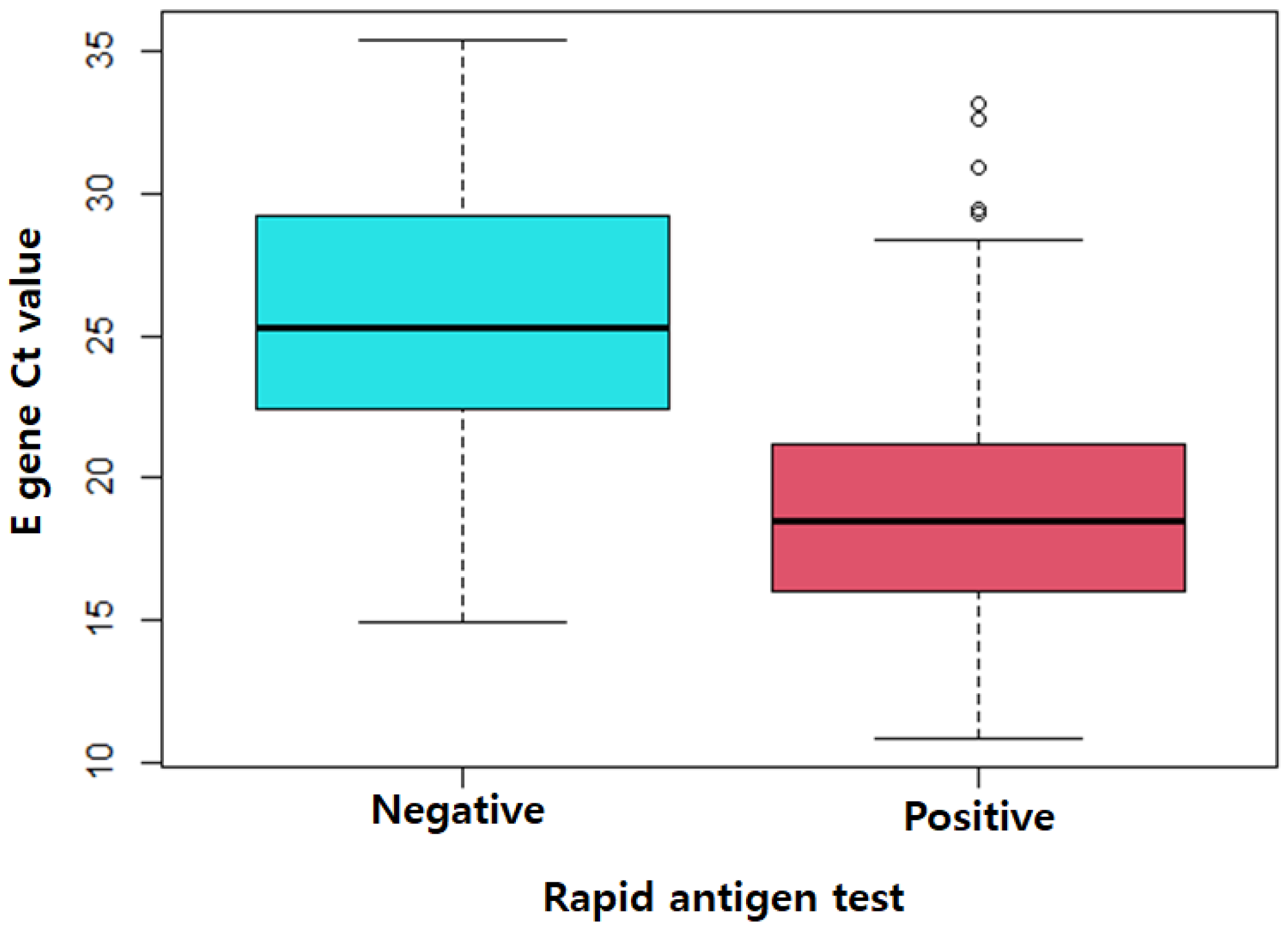
| Rapid antigen test, n (%) | <0.001 | ||
| Positive | 236 (67.8) | 1 (0.1) | |
| Negative | 112 (32.2) | 1526 (99.9) | |
| % | 95% CI | |
|---|---|---|
| Sensitivity | 67.8 | 62.6–72.7 |
| Specificity | 99.9 | 99.6–100.0 |
| PPV | 99.6 | 97.7–100.0 |
| NPV | 93.2 | 91.8–94.3 |
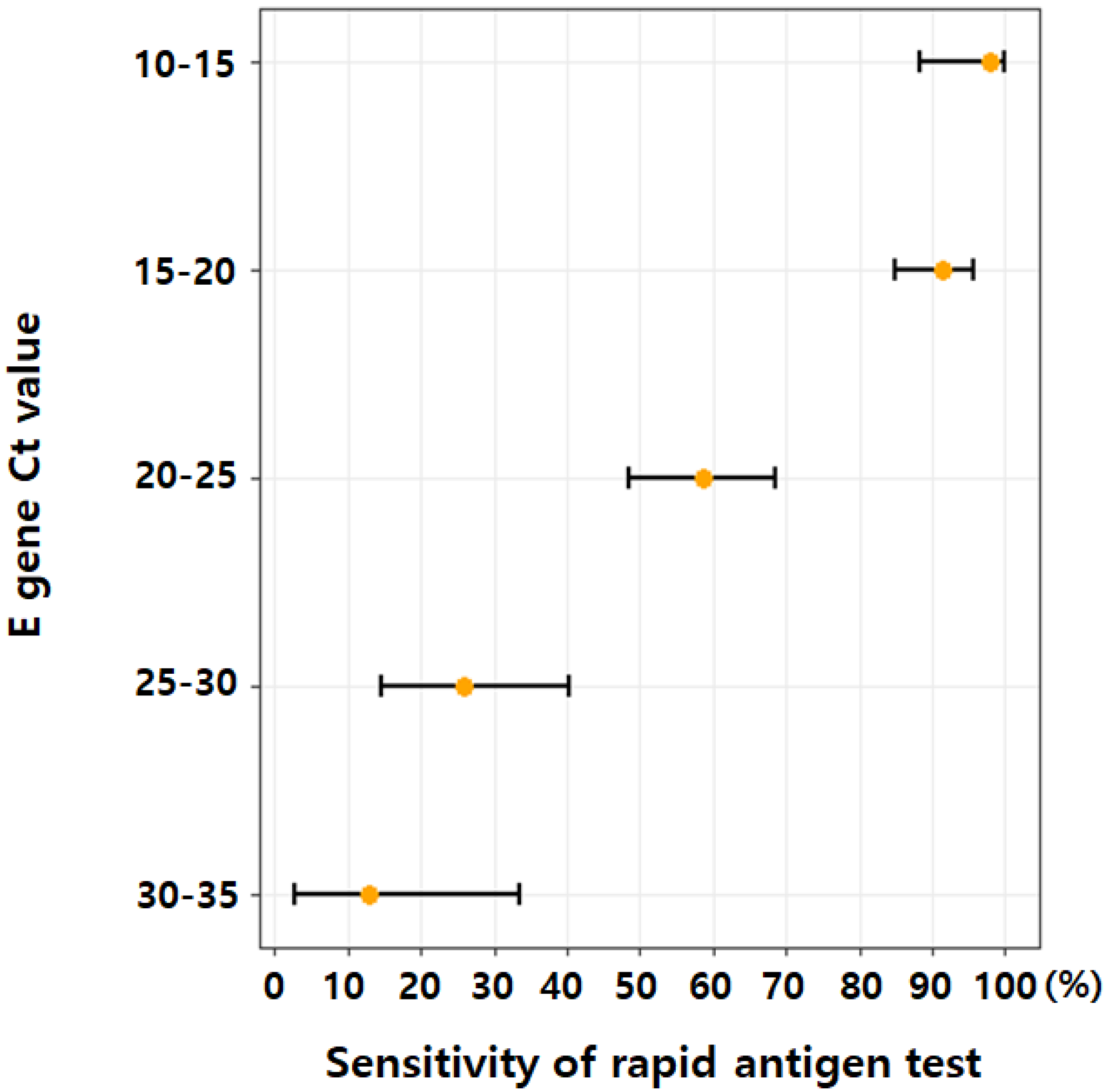
| E Gene Ct Value | Number of COVID-19 Positive Patients | Sensitivity (%) [95% CI] |
|---|---|---|
| Ct value category 1 | ||
| Ct ≤ 24.3 a | 259 | 82.2 [77.0–86.7] |
| Ct ≤ 20.1 b | 174 | 93.1 [88.3–96.4] |
| Ct ≤ 17.1 c | 86 | 96.5 [90.1–99.3] |
| Ct value category 2 d | ||
| Ct ≤ 30.0 | 324 | 71.9 [66.7–76.7] |
| Ct ≤ 25.0 | 274 | 80.3 [75.1–84.8] |
| Ct ≤ 20.0 | 172 | 93.0 [88.1–96.3] |
| Ct ≤ 15.0 | 45 | 97.8 [88.2–99.9] |
Publisher’s Note: MDPI stays neutral with regard to jurisdictional claims in published maps and institutional affiliations. |
© 2022 by the authors. Licensee MDPI, Basel, Switzerland. This article is an open access article distributed under the terms and conditions of the Creative Commons Attribution (CC BY) license (https://creativecommons.org/licenses/by/4.0/).
Share and Cite
Lee, H.; Kang, H.; Cho, Y.; Oh, J.; Lim, T.-H.; Ko, B.-S.; Lee, J. Diagnostic Performance of the Rapid Antigen Test as a Screening Tool for SARS-CoV-2 Infection in the Emergency Department. J. Pers. Med. 2022, 12, 1172. https://doi.org/10.3390/jpm12071172
Lee H, Kang H, Cho Y, Oh J, Lim T-H, Ko B-S, Lee J. Diagnostic Performance of the Rapid Antigen Test as a Screening Tool for SARS-CoV-2 Infection in the Emergency Department. Journal of Personalized Medicine. 2022; 12(7):1172. https://doi.org/10.3390/jpm12071172
Chicago/Turabian StyleLee, Heekyung, Hyunggoo Kang, Yongil Cho, Jaehoon Oh, Tae-Ho Lim, Byuk-Sung Ko, and Juncheol Lee. 2022. "Diagnostic Performance of the Rapid Antigen Test as a Screening Tool for SARS-CoV-2 Infection in the Emergency Department" Journal of Personalized Medicine 12, no. 7: 1172. https://doi.org/10.3390/jpm12071172