Article

Evaluation of Antifungal Selective Toxicity Using Candida glabrata ERG25 and Human SC4MOL Knock-In Strains

by
Keiko Nakano
1,
Michiyo Okamoto
1,
Azusa Takahashi-Nakaguchi
1,
Kaname Sasamoto
1,
Masashi Yamaguchi
1 and
Hiroji Chibana
1,2,3,*
1
Medical Mycology Research Center, Chiba University, Chiba 260-8673, Japan
2
School of Medicine, Niigata University, Niigata 951-8510, Japan
3
Faculty of Medicine, University of the Ryukyus, Okinawa 903-0125, Japan
*
Author to whom correspondence should be addressed.
J. Fungi 2023, 9(10), 1035; https://doi.org/10.3390/jof9101035
Submission received: 29 September 2023 / Revised: 17 October 2023 / Accepted: 18 October 2023 / Published: 20 October 2023
(This article belongs to the Special Issue Antifungal Drugs, 2nd Edition)

Abstract

:
With only four classes of antifungal drugs available for the treatment of invasive systemic fungal infections, the number of resistant fungi is increasing, highlighting the urgent need for novel antifungal drugs. Ergosterol, an essential component of cell membranes, and its synthetic pathway have been targeted for antifungal drug development. Sterol-C4-methyl monooxygenase (Erg25p), which is a greater essential target than that of existing drugs, represents a promising drug target. However, the development of antifungal drugs must consider potential side effects, emphasizing the importance of evaluating their selective toxicity against fungi. In this study, we knocked in ERG25 of Candida glabrata and its human ortholog, SC4MOL, in ERG25-deleted Saccharomyces cerevisiae. Utilizing these strains, we evaluated 1181-0519, an Erg25p inhibitor, that exhibited selective toxicity against the C. glabrata ERG25 knock-in strain. Furthermore, 1181-0519 demonstrated broad-spectrum antifungal activity against pathogenic Candida species, including Candida auris. The approach of utilizing a gene that is functionally conserved between yeast and humans and subsequently screening for molecular target drugs enables the identification of selective inhibitors for both species.

1. Introduction

The threat of antimicrobial resistance (AMR) has progressed in parallel with the COVID-19 pandemic caused by the SARS-CoV-2 virus [1]. These resistant pathogens include drug-resistant Candida [1,2,3]. In recent years, drug-resistant Candida species other than Candida albicans, called non-albicans Candida, have become more common, especially Candida glabrata. Therefore, the development of new antifungal agents against them is urgently needed [4,5,6].
Ergosterol and its synthetic pathway have served as sources for antifungal drug targets [5,7]. Polyenes act directly on ergosterol [8,9], while azoles inhibit lanosterol 14α-demethylase (Erg11p) [10,11,12,13]. Allylamines target squalene monooxygenase (Erg1p); morpholines affect C14 sterol reductase (Erg24p). Additionally, there may still be potential targets [14,15]. Interestingly, it has been demonstrated that the antifungal effects of targeting the ergosterol biosynthetic pathway are not solely attributable to ergosterol depletion. Instead, they result from the accumulation of abnormal sterols due to the interruption or bypass of the pathway. Therefore, even within the same pathway, the cellular effects vary depending on which enzyme is targeted [16].
As we can observe through the example of azoles, it is crucial to take into account the emergence of drug resistance when targeting the ergosterol synthesis pathway. In the case of C. glabrata, the primary mechanism driving azole resistance involves the upregulation of efflux pumps, particularly CDR1, which is mediated by a transcription factor, PDR1 [17,18,19,20,21,22,23,24]. Other studies have also highlighted that azole resistance coincides with stress induced by mitochondrial loss or dysfunction, leading to the activation of PDR1, and subsequently, the upregulation of CDR1 [25,26,27,28,29,30]. Furthermore, as a strategy for azole resistance in C. glabrata, our attention has been directed towards the role of host cholesterol uptake. This process enables the uptake of cholesterol from the host and its utilization as a substitute for ergosterol [16,31,32,33,34]. In a prior investigation, we demonstrated that out of the 12 genes involved in the ergosterol pathway, only the gene knockdown of ERG25 or ERG26 was not compensated by serum. Additionally, we conducted a comparative analysis of the ERG25 and ERG26 genes with their orthologs in other fungi and humans. The results indicated that the amino acid sequence of ERG25 is more conserved across fungi than ERG26 and exhibits less similarity to its human ortholog [16]. These insights have led us to place a stronger focus on ERG25.
A cell-free assay system is useful for evaluating target-molecule-specific activity [35]. However, Erg25p has been suggested to form a complex with the ER membrane proteins Erg28p, Erg27p, and Erg26p. This complex formation has not been fully clarified [36,37], and difficulties exist in accurately reconstructing the membrane assembly in vitro. Thus, we focused on the use of a knock-in strain of Saccharomyces cerevisiae. In total, 47% of genes in S. cerevisiae, including ERG25, have been reported to be complemented by human genes [38]. These genes can be used for the phenotypic examination of selective toxicity using knock-in strains.
Several drugs targeting Erg25p have been reported. For example, 6-amino-2-n-pentylthiobenzothiazole (APB) has been shown to inhibit mycelial formation in C. albicans [39], while PF1163A and PF1163B exhibit inhibitory effects against C. albicans and display a slight inhibitory activity against C. glabrata, C. krusei, C. parapsilosis, and Aspergillus fumigatus [40,41]. Additionally, diazabolins have been identified as inhibitors against S. cerevisiae [42]. It is worth noting that S. cerevisiae is vulnerable to 1181-0519 (N-[(2E)-2-[(4-nitrophenyl) hydrazinylidene]propyl] acetamide) [43,44]. As far as our knowledge extends, there have been no reports to date evaluating the activity of 1181-0519 against Candida species.
In this study, we demonstrated functional complementation between the genes C. glabrata ERG25 and human SC4MOL by replacing ERG25 in S. cerevisiae. Subsequently, we used these strains to assess the selective toxicity of an Erg25p inhibitor, 1181-0519. The use of this phenotypic in vitro evaluation system for target molecules presents significant advantages, as it has the potential to expedite and streamline drug development. Furthermore, this experimental system shows promise for evaluating the selective toxicity of Erg25p and can serve as a valuable tool for screening other inhibitors targeting either Erg25p or SC4MOL.

2. Materials and Methods

2.1. Strains, Plasmids, and Media

Escherichia coli ME9806 (iVEC3) (National Bio-Resource Project (NBRP), Mishima, Japan) was used as the cloning host. Candida auris CBS 10913, C. tropicalis CBS 94, C. parapsilosis CBS 604, and C. krusei CBS 573 were obtained through the ME9806 National Bio-Resource Project (NBRP), Chiba, Japan. The bacterial strains were grown in Luria Broth containing 50 µg/mL ampicillin (FUJIFILM Wako Pure Chemical Corporation, Osaka, Japan). The strains used in the present study are listed in Table 1. The YEp352-GAPII (containing TDH3 promoter and URA3) and the YEp351-GAPII plasmid (containing TDH3 promoter and LEU2) [45] were used to express recombinant proteins. All yeast strains were grown in YPD medium composed of 1% (w/v) Bacto Yeast Extract (Gibco, Miami, FL, USA), 2% (w/v) HIPOLYPEPTON (Nihon Pharmaceutical Co., Ltd., Osaka, Japan), and 2% (w/v) glucose or synthetic defined minimal medium (SD) (0.17% (w/v) Yeast Nitrogen Base without amino acids and ammonium sulfate, 5% (w/v) ammonium sulfate (Wako), 2% (w/v) glucose, and appropriate amino acids). The solid media were supplemented with 2% (w/v) agar (Wako). PF1163B (kindly provided by Meiji Seika Pharma Co., Ltd., Odawara, Kanagawa, Japan) and 1181-0519(N-[(2E)-2-[(4-nitrophenyl) hydrazinylidene] propyl] acetamide) (ChemDiv, San Diego, CA, USA) were used as growth inhibitors.

2.2. Construction of Sc(hERG25) and Sc(CgERG25)

The gene encoding SC4MOL (hERG25), which was optimized for expression in S. cerevisiae, was obtained from Eurofins Genomics KK (Tokyo, Japan) (Figure S1). A DNA fragment containing hERG25 was synthesized via polymerase chain reaction (PCR) using the primers hERG25-F1 and hERG25-R1 (all primers are listed in Table S1), and linear YEp352-GAPII containing TDH3 promoter and the URA3 gene was amplified using primers YEp352-GAPII-F and YEp352-GAPII-R. The DNA fragment was inserted into YEp352-GAPII by in vivo cloning using E. coli strain ME9806 (iVEC3) [46]. The resulting plasmid, YEp352-GAPII-hERG25, was transformed into Sc(erg25Δ/ERG25) cells as described previously [47]. Ura+ transformants were then inoculated on a solid sporulation medium containing 1% (w/v) potassium acetate and 2% (w/v) agar at 28 °C for 2 days. Spore formation was confirmed under a microscope, and spores were planted on the YPD agar medium. The resulting haploid strains were sub-cultured using the replica plating method on three types of media: YPD, SD-URA, and SD + G418 (200 µg/mL) (Figure S2). The strains that grew on all three media were designated Sc(hERG25).
A DNA fragment encoding CgERG25 was amplified using primers CgERG25-F1 and CgERG25-R1, with the genomic DNA of C. glabrata as a template (Supplementary Table S1). Linear YEp351-GAPII containing TDH3 promoter and LEU2 was amplified using the primers YEp352-GAPII-F3 and YEp352-GAPII-R3. The DNA fragment, including the region encoding CgERG25, was inserted into YEp351-GAPII by in vivo cloning using E. coli strain ME9806 (iVEC3). The resulting plasmid, YEp351-GAPII-CgERG25, was transformed into Sc(hERG25) cells and selected on SD medium without uracil and leucine. Transformants were selected on an SD plate containing 5-fluoroorotic acid (5-FOA) without leucine to obtain Sc(CgERG25).

2.3. Liquid Growth Assays and Determination of IC50

The strains were inoculated in YPD medium and grown to saturation overnight. Growth assays were performed in 96-well flat-bottom plates in 100 µL of SD medium with or without 20 mg/L uracil or 20 mg/L leucine. Growth rates were determined by measuring the optical density at 600 nm (OD600) after shaking at 1000 rpm for 10 s every 10 min for at least 288 cycles at 28 °C using an Infinite M200PRO (TECAN, Männedorf, Switzerland). Drugs were dissolved in dimethyl sulfoxide (DMSO) and dispensed into the plates. The final concentration of DMSO was adjusted to 0.1% (v/v). The growth rate of each strain was calculated as follows: (1) the first 10 OD readings were averaged and subtracted from all OD readings of the corresponding curve to set the baseline of the growth curve to zero, and (2) the area under the curve (AUC) was then calculated as the sum of all OD readings. ‘Relative growth’ was calculated as previously described [48] and as follows: (AUCcondition − AUCcontrol)/AUCcontrol, where AUCcontrol represents the growth rate of the reference condition that was assayed on the same microtiter plate. Half maximal (50%) inhibitory concentration (IC50) is the concentration of the drug that is 50% of the OD600 value of a well containing only medium and 0.1% (v/v) DMSO after 48 h of culture.

2.4. Spotting Assay

The strains were then inoculated on solid synthetic defined (SD) medium with or without 20 mg/L uracil or 20 mg/L leucine containing 2% (w/v) agar at 28 °C for 2 days. Two to three single colonies were suspended in saline solution (Otsuka Pharmaceutical Co., Ltd., Ltd., Tokyo, Japan), adjusted to 2 × 10⁶ cells/mL using a cell counter (WATSON Co., Ltd., Tokyo, Japan), serially diluted 1:10, and spotted (5 µL) on solid SD medium containing 2% (w/v) agar using an epMotion 96 (Eppendorf, Hamburg, Germany). After 48 h of incubation at 28 °C, pictures of the growth of cells were taken. Assays were repeated three times.

2.5. Determination of Minimum Inhibitory Concentration (MIC)

The determination of the minimum inhibitory concentration (MIC) of 1181-0519 was primarily conducted following the CLSI-M27A guidelines. Various strains were streaked onto YPD agar medium and incubated at 28 °C for 1 or 2 days until colonies became visible. Subsequently, five colonies with a diameter of approximately 1 mm were selected and suspended in tubes containing 1 mL of sterile saline solution (0.9% (w/v)). The resulting suspension was vigorously mixed by vortexing for 1 min. Cell counts were obtained using a blood cell counting board, and the final cell count was adjusted to 3 × 10³ cells/mL using Roswell Park Memorial Institute (RPMI) MOPS medium (pH 7) in all wells. The wells in the first column contained 32 µM of 1181-0519, 1 × 10³ cells/mL, and 0.5% (v/v) DMSO in 200 µL of RPMI MOPS. The other wells contained 1 × 10³ cells/mL and 0.5% (v/v) DMSO in 100 µL of RPMI MOPS. These wells were thoroughly mixed by pipetting, and then 100 µL was transferred from the wells of the first column to the adjacent wells, repeating this process to achieve a 1/2 dilution of the drug in subsequent wells. The wells in the last column did not include 1181-0519. The cells were incubated for 24 h at 28 °C. The MIC, which is the lowest concentration of the drug at which there was no visible turbidity, was determined by visually comparing the growth with the wells that had no drug.

2.6. Evaluation of Cytotoxicity

A431 (human cell line derived from epidermoid carcinoma) and HepG2 (human hepatoma) cell lines were obtained from RIKEN BRC and JCRB CELL BANK, respectively. All cells were cultured in RPMI-1640 medium provided by FUJIFILM Wako Pure Chemical Corporation. This medium was supplemented with 10% (v/v) fetal bovine serum from Grand Island Biological Company (Gibco), 0.1% (v/v) penicillin, and 0.1% (v/v) streptomycin, also from Gibco. The cell cultures were maintained in a humidified 5% (w/v) CO2 incubator at a constant temperature of 37 °C. After 24 h of incubation, both with and without the presence of 100 µM of 1181-0519, we added Premix WST-1 (Takara Bio Inc., Shiga, Japan) into each well. Subsequently, the plates were incubated for 1.5 h at 37 °C, and we measured the absorbance of the wells at both 450 nm and 600 nm using an iMark microplate reader from BIO-RAD (Hercules, CA, USA). Cell viability was determined by subtracting the absorbance value at 600 nm from the absorbance value at 400 nm, which reflects the catalytic activity of tetrazolium salt in forming formazan dye through mitochondrial dehydrogenase activity.

2.7. Statistical Analyses

All experiments were conducted a minimum of three times. Statistical analyses, including t-tests and one-way ANOVA tests (both two-tailed and unpaired), were performed using GraphPad Prism 10.0.3 (GraphPad Software, San Diego, CA, USA) to calculate p-values.

3. Results

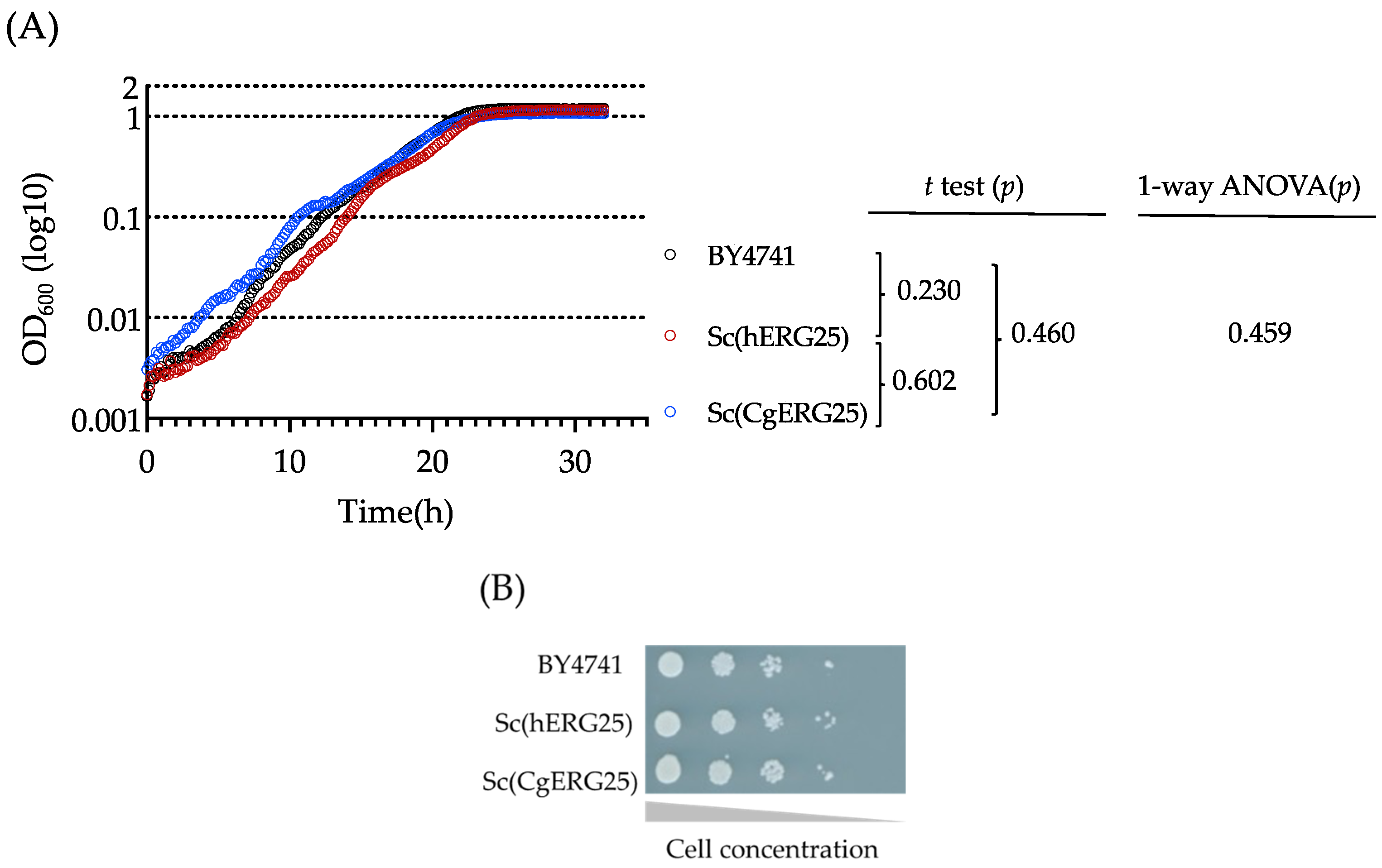
3.1. Complementarity of hERG25 and CgERG25 in S. cerevisiae

We knocked in ERG25 of C. glabrata or its human ortholog, SC4MOL, into S. cerevisiae and isolated clones from tetrads that lacked the S. cerevisiae endogenous ERG25 (Figure S2). Consequently, we replaced the S. cerevisiae ERG25 with either CgERG25 or hERG25(SC4MOL), resulting in the creation of Sc(CgERG25) and Sc(hERG25) strains, respectively (Figure S3), and their transcription levels showed no relatively significant differences (Figure S4). Given the role of ERG25 as an essential gene in S. cerevisiae, the growth of both Sc(hERG25) and Sc(CgERG25) demonstrated functional complementation. To evaluate the completeness of complementation by hERG25 and CgERG25, we conducted an analysis of the growth curves of Sc(hERG25), Sc(CgERG25), and the control strain BY4741. As a result, their growth curves exhibited nearly identical slopes, and statistical analyses employing t-tests and one-way ANOVA indicated no significant differences (Figure 1A). Additionally, a spot assay showed no different colony formation characteristics among them (Figure 1B). Furthermore, we subjected the knock-in strains to incubation in the presence or absence of fluconazole or amphotericin B, observing no significant differences in susceptibility among the strains (Figure S5). These results collectively suggest that both hERG25 and CgERG25 effectively complement the function of ERG25 in S. cerevisiae.

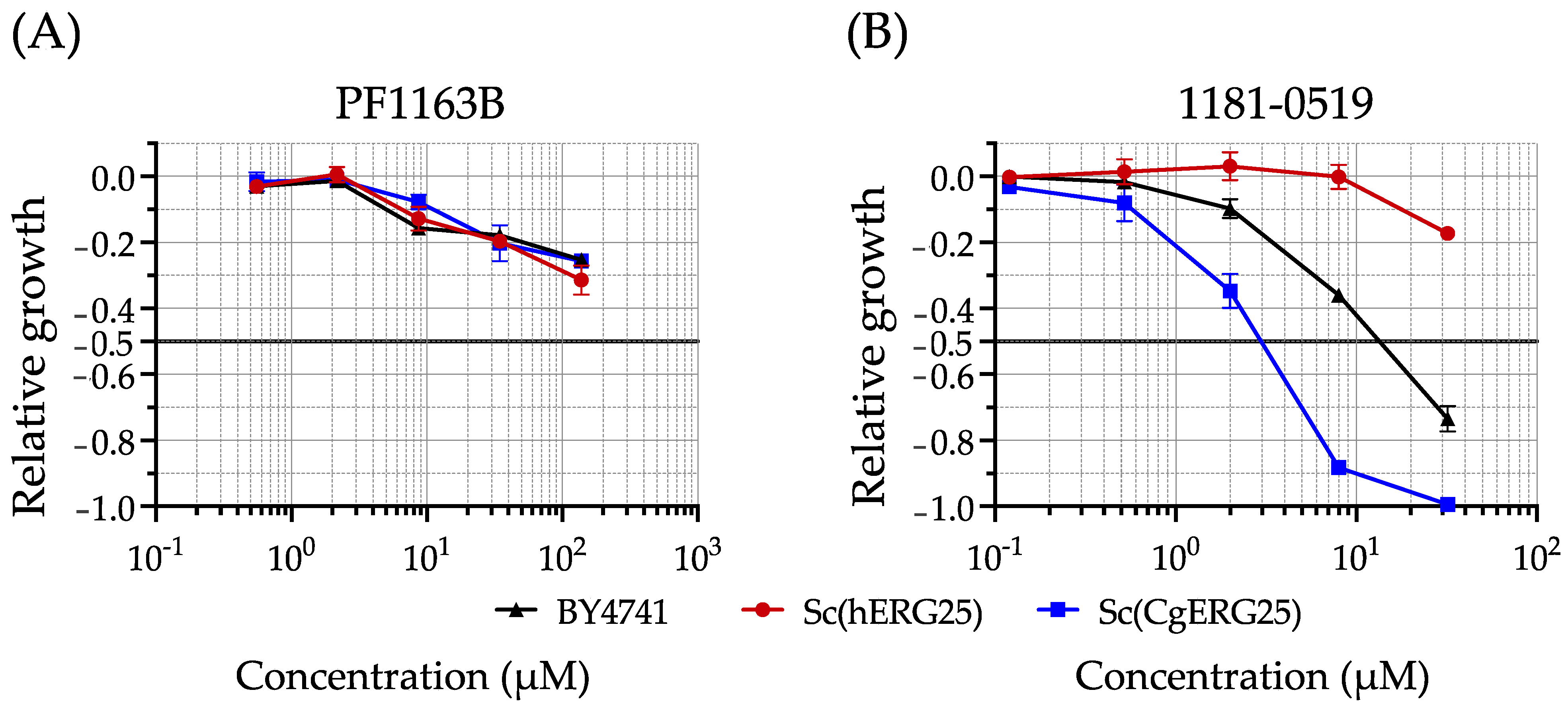
3.2. Drug Susceptibility of Knock-In Strains with Liquid Growth Assays

PF1163B, a known inhibitor of C. albicans Erg25p, distinguishes itself from PF1163A by lacking an additional hydroxyl group on its side chain (Figure S6A). PF1163B exhibits a broader spectrum of activity compared to PF1163A and has been reported to have slight inhibitory effects on C. glabrata [40]. To confirm the usefulness of PF1163B in our study, we conducted a growth inhibition comparison involving BY4741, Sc(hERG25), and Sc(CgERG25) in the presence of PF1163B (Figure 2A). The results indicated slight inhibitory activity against all three strains, although the IC50 was not reached even at the highest concentration of 138 μM (Table 2). Another compound, 1181-0519 (N-[(2E)-2-[(4-nitrophenyl) hydrazinylidene] propyl] acetamide) (Figure S6B), has been reported to possess inhibitory activity against Erg25p in S. cerevisiae [43,44]. Thus, we compared its growth inhibitory effects on BY4741, Sc(hERG25), and Sc(CgERG25) (Figure 2B). The IC50 values for BY4741 and Sc(CgERG25) were 13 µM and 3 µM, respectively, whereas for Sc(hERG25), the IC50 was greater than 32 µM (Table 2). Consequently, we observed growth inhibition of 1181-0519 against Sc(CgERG25) but not against Sc(hERG25).

3.3. Evaluation of 1181-0519 Spectrum

To assess the potential of 1181-0519 as an antifungal agent, we conducted an evaluation of its activity against Candida species. We employed the Clinical Laboratory Standards Institute (CLSI) M27A3 method, which is a standard approach for assessing antifungal drugs. The Candida strains chosen as subjects included C. albicans SC5314, C. glabrata CBS 138, C. auris CBS 10913, C. tropicalis CBS 94, C. parapsilosis CBS 604, and C. krusei CBS 573. The results indicated that 1181-0519 exhibited strong inhibitory activity against C. albicans, C. glabrata, C. auris, C. parapsilosis, and C. krusei, with a minimum inhibitory concentration (MIC) value of less than 2 μM, while relatively weak inhibitory activity was observed against C. tropicalis, with an MIC of 16 µM (Table 3).

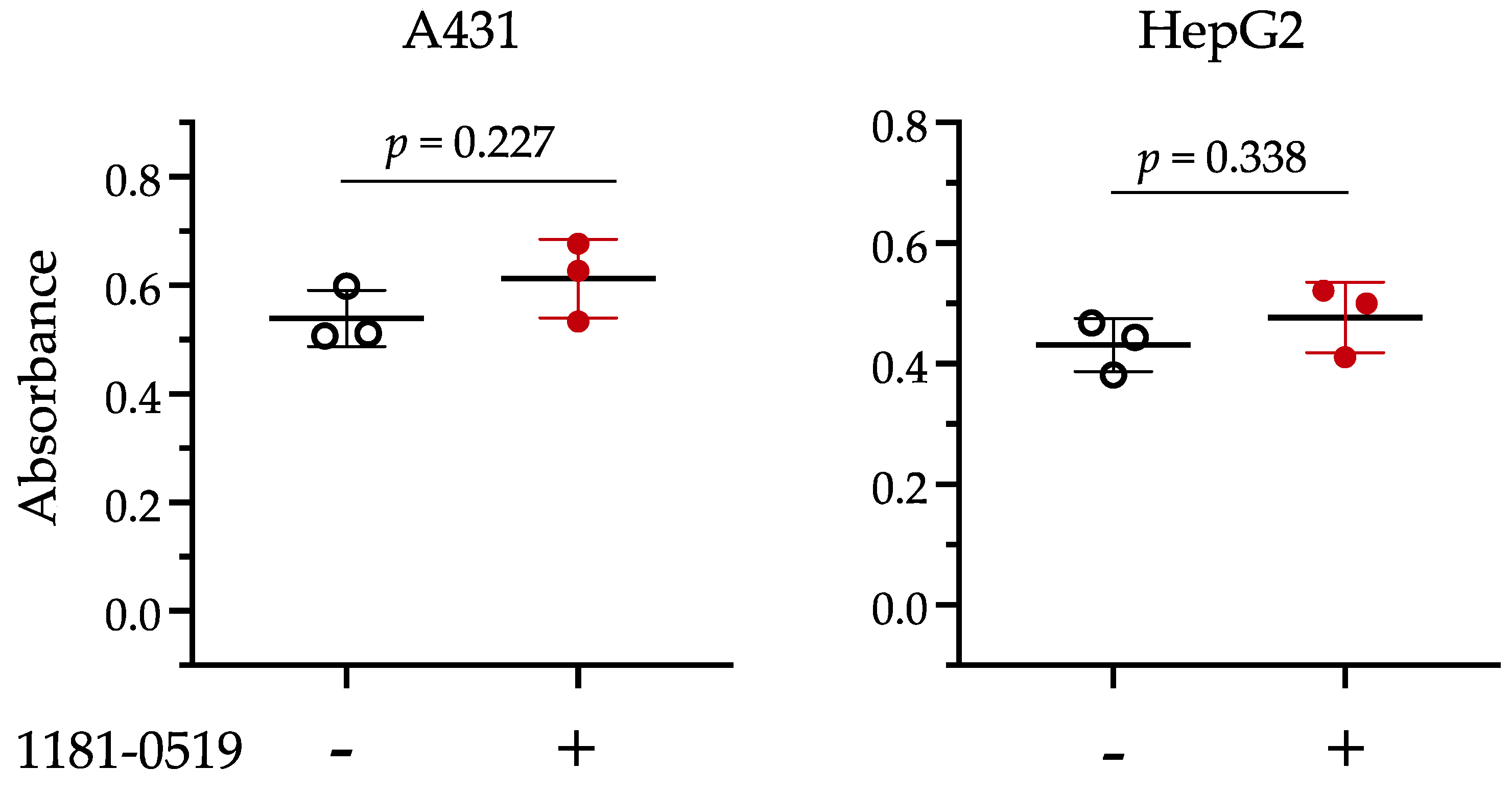
3.4. Cytotoxicity of 1181-0519

To evaluate the cytotoxicity of 1181-0519, we incubated A431 and HepG2 cell lines in medium with or without 100 µM 1181-0519, and then measured mitochondrial dehydrogenase activity using the WST-1 assay with a formazan dye concentration. The absorbance of the formazan dye, serving as an indicator of cell viability, is shown in Figure 3. The results of the t-test indicated that, for both the A431 and HepG2 cell lines, the p-values were 0.227 and 0.338, respectively, suggesting no significant differences between cultures with and without 1181-0519 in the medium. Therefore, no cytotoxicity was detected with 1181-0519 even at a concentration of 100 µM.

3.5. Homology Analysis of Erg25p

Erg25p is a non-heme iron-requiring enzyme and is characterized by three histidine motifs that are conserved throughout eukaryotes [49]. These three histidine motifs, namely HX3 H, HX2 HH, and HX2 HH, are iron-binding sites (Figure S7) and are presumed to be important for enzymatic function [49]. The amino acid homology between human and C. glabrata, human and S. cerevisiae, and C. glabrata and S. cerevisiae was 37.5%, 34.5%, and 89.0%, respectively (EMBOSS Water < Pairwise Sequence Alignment < EMBL − EBI), and the three histidine motifs were conserved in all species (Figure S7). Therefore, they do not account for differences in susceptibility between Sc(hERG25) and Sc(CgERG25) to 1181-0519. Consequently, we can infer that the binding site of Erg25p in 1181-0519 is located outside of those motifs.

4. Discussion

C4-methyl sterol monooxygenase (Erg25p), which is essential for the growth of S. cerevisiae [50], C. albicans [51], and C. glabrata [16], represents a promising target for antifungal drugs with higher efficacy compared to Erg11p, the target molecule of azoles [16]. However, the presence of an orthologous gene for ERG25 in humans necessitates the verification of inhibitor selectivity against fungi. We knocked in C. glabrata ERG25 or the human orthologous gene SC4MOL into S. cerevisiae lacking ERG25, confirming their functional complementation (Figure 1). Using these knock-in strains, we demonstrated that 1181-0519, an inhibitor of Erg25p in S. cerevisiae, exhibited no inhibitory activity against Sc(hERG25), while it did against Sc(CgERG25) (Figure 2). Furthermore, 1181-0519 displayed broad-spectrum activity against Candida species (Table 3) while exhibiting no toxicity to cultured human cells A431 and HepG2 (Figure 3). These findings suggest that 1181-0519 holds potential as an antifungal candidate. Although the growth inhibitory activity of 1181-0519 against S. cerevisiae has been previously reported [43,44], to the best of our knowledge, this is the first report evaluating its efficacy against pathogenic fungi and human genes.
Selective toxicity against fungal cells is a crucial consideration in the development of antifungal drugs, and various methods are available for its assessment. One such method is a molecular-level assay system employing a cell-free system. However, due to the formation of a complex involving Erg25p on the endoplasmic reticulum membrane [36,37], establishing an in vitro cell-free assay system has proven to be challenging. In this study, we demonstrated that simple culture experiments can effectively evaluate the selective inhibitory activity of enzymes with identical functions in different species. As a result, this study highlights the utility of a knock-in strain system as a valuable alternative experimental approach, particularly for proteins that form complexes, where constructing a cell-free assay system poses difficulties.
Once an inhibitor has been identified, attempts should be made to modify its molecular structure to increase its potency, specificity, and broader activity in pathogens. This approach is essential not only for advancing the development of improved drugs but also for pre-empting the emergence of derivative drugs that may follow. Identifying the binding domain of the target streamlines compound development. In ERG25, the amino acid sequences within the three histidine motifs of the active domain were conserved between human and fungal ERG25 (Figure S5), suggesting that the binding domain of 1181-0519 is not those motifs. Notably, C. tropicalis exhibited lower sensitivity compared to other Candida species (Table 3). These variations in susceptibility could provide insights for estimating binding sites. However, since these experiments were conducted on different species using their whole cells, they may have been influenced by intracellular environments other than Erg25p. To accurately assess Erg25p-specific sensitivity to 1181-0519, it would be advantageous to standardize conditions other than Erg25p by constructing ERG25 knock-in strains of these species in S. cerevisiae. This approach would enable the precise evaluation of Erg25p-specific sensitivity to 1181-0519. Subsequently, this information could inform various applications, such as docking simulations aimed at identifying the binding site of 1181-0519 to Erg25p and enhancing its spectrum of action. Therefore, further studies should be conducted on them.
Considering potential side effects is important for the development of antifungal drugs. Although SC4MOL, the human ortholog of ERG25, is not an essential gene for growth in human cells, diseases caused by mutations in this gene have been reported [52]. These diseases result in impaired cholesterol biosynthesis, leading to microcephaly, bilateral congenital cataracts, growth retardation, psoriasiform dermatitis, immune dysfunction, and intellectual disability [53,54,55,56]. Additionally, 4,4-dimethylzymosterol, a substrate of SC4MOL that accumulates upon its inhibition, has been reported to exhibit testicular meiosis activity in mammals and plays a crucial role in regulating cumulus oophorus expansion and oocyte maturation [57,58,59]. Since these diseases and abnormalities manifest during ontogenetic processes, which are challenging to reproduce in cultured cells, using cultured cells for toxicity evaluation is not appropriate. Therefore, in the process of developing antifungal drugs, side effects are typically evaluated through animal experiments, which necessitate an evaluation system that includes pathological models. Furthermore, removing a candidate compound from the development process at this stage can result in significant losses, as previous investments would be wasted. Therefore, it is important to assess selective inhibitory activity at the molecular level in advance using a knock-in strain.
The two knock-in strains produced here can also be utilized in the search for new Erg25p inhibitors other than 1181-0519. Compounds that inhibit the growth of the Sc(CgERG25) strain, but not Sc(hERG25), are likely to act specifically against CgErg25p. Thus, the difference in susceptibility between the two strains can be employed in differentially high-throughput screening for specific inhibitors of Erg25p. In contrast, a compound that inhibits growth against the Sc(hERG25) strain, but not against Sc(CgERG25), is likely to act specifically against SC4MOL. This inhibitor could contribute to the study of SC4MOL deficiency.
Some clinical isolates of C. glabrata have been reported to lose the ability to synthesize ergosterol and do not grow on standard laboratory medium. However, they can grow on media containing serum or bile [16,31,32,33,34]. These strains compensate for the lack of ergosterol by uptake of host cholesterol and subsequently become resistant to azoles and polyene [33,34]. The primary issue lies in the inability to isolate these sterol-requiring strains using the standard media employed in clinical diagnostic tests, as these media lack sterols [26,30], consequently leading to stealth infections. Similar risk may arise when targeting proteins encoded by ERG1, ERG11, or ERG7 [16] which allow cholesterol uptake, enabling cells to continue growing in a host. Conversely, the disfunction of ERG25 or ERG26 prevent the uptake of host cholesterol [16], making inhibitors of Erg25p or Erg26p effective in repressing growth, even in an environment with cholesterol. In addition, the amino acid sequence of Erg25p is more fungal-specific than that of Erg26p. Therefore, Erg25p represents an excellent target molecule in the ergosterol synthetic pathway that does not promote stealth infection by C. glabrata.

5. Conclusions

While Erg25p represents a promising antifungal drug target, its inhibitors may induce side effects in humans if they interfere with human SC4MOL. However, since SC4MOL is non-essential for human cells, and these side effects manifest during the ontogeny process, toxicity cannot be detected in experiments conducted with cultured cells. For inhibitors whose toxicity cannot be assessed in cultured cells due to similar reasons, the experimental system we have demonstrated will serve as a crucial complement to the selective toxicity evaluation system, enabling the assessment of new test compounds targeting both fungi and humans. This system provides valuable insights into the direct impact of selective toxicity for these inhibitors. In fact, the implementation of this evaluation system has revealed that 1181-0519 exhibits specific efficacy against Candida species.

Supplementary Materials

The following supporting information can be downloaded at: https://www.mdpi.com/article/10.3390/jof9101035/s1, Figure S1. DNA alignment (Codon Conversion). Figure S2. Summary of the mutational screen for hERG25 variants complementing the loss of the yeast ortholog ERG25. Figure S3. PCR for confirmation of S. cerevisiae ERG25 deletion. Figure S4. Relative amount of RNA. Figre S5. Antifungal susceptibility of knock-in strains. Figure S6. Molecular structure of two different types of Erg25p inhibitor. Figure S7. Erg25p Alignment. Table S1. List of primers used in this study. Reference [43] is cited in the supplementary materials.

Author Contributions

Conceptualization, K.N. and H.C. Formal analysis: K.N. and H.C. Funding acquisition: H.C. Investigation: K.N., M.O., A.T.-N., K.S. and M.Y. Writing—original draft, K.N. Writing—review and editing, H.C. All authors have read and agreed to the published version of the manuscript.

Funding

This research was supported by AMED under Grant Number JP23fk0108679 and JP22nk0101553, and partly by the National Bio-Resource Project Japan (https://nbrp.jp/en/).

Acknowledgments

PF1163B was provided by Meiji Seika Pharma Co. Ltd.

Conflicts of Interest

The authors declare no conflict of interest.

References

  1. Rehman, S. A Parallel and Silent Emerging Pandemic: Antimicrobial Resistance (AMR) amid COVID-19 Pandemic. J. Infect. Public Health 2023, 16, 611–617. [Google Scholar] [CrossRef] [PubMed]
  2. Lai, C.-C.; Chen, S.-Y.; Ko, W.-C.; Hsueh, P.-R. Increased Antimicrobial Resistance during the COVID-19 Pandemic. Int. J. Antimicrob. Agents 2021, 57, 106324. [Google Scholar] [CrossRef]
  3. Kariyawasam, R.M.; Julien, D.A.; Jelinski, D.C.; Larose, S.L.; Rennert-May, E.; Conly, J.M.; Dingle, T.C.; Chen, J.Z.; Tyrrell, G.J.; Ronksley, P.E.; et al. Antimicrobial Resistance (AMR) in COVID-19 Patients: A Systematic Review and Meta-Analysis (November 2019–June 2021). Antimicrob. Resist. Infect. Control 2022, 11, 45. [Google Scholar] [CrossRef] [PubMed]
  4. Giacobbe, D.R.; Maraolo, A.E.; Simeon, V.; Magnè, F.; Pace, M.C.; Gentile, I.; Chiodini, P.; Viscoli, C.; Sanguinetti, M.; Mikulska, M.; et al. Changes in the Relative Prevalence of Candidaemia Due to Non-albicans Candida Species in Adult In-patients: A Systematic Review, Meta-analysis and Meta-regression. Mycoses 2020, 63, 334–342. [Google Scholar] [CrossRef] [PubMed]
  5. Campoy, S.; Adrio, J.L. Antifungals. Biochem. Pharmacol. 2017, 133, 86–96. [Google Scholar] [CrossRef] [PubMed]
  6. Silva, L.N.; De Mello, T.P.; De Souza Ramos, L.; Branquinha, M.H.; Dos Santos, A.L.S. New and Promising Chemotherapeutics for Emerging Infections Involving Drug-Resistant Non-Albicans Candida Species. Curr. Top. Med. Chem. 2019, 19, 2527–2553. [Google Scholar] [CrossRef]
  7. Houšť, J.; Spížek, J.; Havlíček, V. Antifungal Drugs. Metabolites 2020, 10, 106. [Google Scholar] [CrossRef] [PubMed]
  8. Gray, K.C.; Palacios, D.S.; Dailey, I.; Endo, M.M.; Uno, B.E.; Wilcock, B.C.; Burke, M.D. Amphotericin Primarily Kills Yeast by Simply Binding Ergosterol. Proc. Natl. Acad. Sci. USA 2012, 109, 2234–2239. [Google Scholar] [CrossRef] [PubMed]
  9. Saravolatz, L.D.; Ostrosky-Zeichner, L.; Marr, K.A.; Rex, J.H.; Cohen, S.H. Amphotericin B: Time for a New “Gold Standard”. Clin. Infect. Dis. 2003, 37, 415–425. [Google Scholar] [CrossRef]
  10. Singh, A.; Singh, K.; Sharma, A.; Kaur, K.; Chadha, R.; Bedi, P.M.S. Recent Advances in Antifungal Drug Development Targeting Lanosterol 14α-demethylase ( CYP51 ): A Comprehensive Review with Structural and Molecular Insights. Chem. Biol. Drug Des. 2023, 102, 606–639. [Google Scholar] [CrossRef]
  11. Bossche, H.V.; Marichal, P.; Gorrens, J.; Bellens, D.; Moereels, H.; Janssen, P.A.J. Mutation in Cytochrome P-450-Dependent 14α-Demethylase Results in Decreased Affinity for Azole Antifungals. Biochem. Soc. Trans. 1990, 18, 56–59. [Google Scholar] [CrossRef] [PubMed]
  12. Hitchcock, C.A. Cytochrome P-450-Dependent 14α-Sterol Demethylase of Candida albicans and Its Interaction with Azole Antifungals. Biochem. Soc. Trans. 1991, 19, 782–787. [Google Scholar] [CrossRef] [PubMed]
  13. Hitchcock, C.A.; Adams, D.J. Interaction of Azole Antifungal Antibiotics with Cytochrome P-450-Dependent 14a-Sterol Demethylase Purified from Candida albicans. Biochem. J. 1990, 266, 475–480. [Google Scholar] [CrossRef] [PubMed]
  14. Jamzivar, F.; Shams-Ghahfarokhi, M.; Khoramizadeh, M.; Yousefi, N.; Gholami-Shabani, M. Unraveling the Importance of Molecules of Natural Origin in Antifungal Drug Development through Targeting Ergosterol Biosynthesis Pathway. Iran. J. Microbiol. 2020, 11, 448. [Google Scholar] [CrossRef]
  15. Bhattacharya, S.; Esquivel, B.D.; White, T.C. Overexpression or Deletion of Ergosterol Biosynthesis Genes Alters Doubling Time, Response to Stress Agents, and Drug Susceptibility in Saccharomyces cerevisiae. mBio 2018, 9, e01291-18. [Google Scholar] [CrossRef]
  16. Okamoto, M.; Takahashi-Nakaguchi, A.; Tejima, K.; Sasamoto, K.; Yamaguchi, M.; Aoyama, T.; Nagi, M.; Tanabe, K.; Miyazaki, Y.; Nakayama, H.; et al. Erg25 Controls Host-Cholesterol Uptake Mediated by Aus1p-Associated Sterol-Rich Membrane Domains in Candida glabrata. Front. Cell Dev. Biol. 2022, 10, 820675. [Google Scholar] [CrossRef]
  17. Sanglard, D.; Kuchler, K.; Ischer, F.; Pagani, J.L.; Monod, M.; Bille, J. Mechanisms of Resistance to Azole Antifungal Agents in Candida albicans Isolates from AIDS Patients Involve Specific Multidrug Transporters. Antimicrob. Agents Chemother. 1995, 39, 2378–2386. [Google Scholar] [CrossRef]
  18. Sanglard, D.; Ischer, F.; Monod, M.; Bille, J. Cloning of Candida albicans Genes Conferring Resistance to Azole Antifungal Agents: Characterization of CDR2, a New Multidrug ABC Transporter Gene. Microbiology 1997, 143, 405–416. [Google Scholar] [CrossRef]
  19. Miyazaki, H.; Miyazaki, Y.; Geber, A.; Parkinson, T.; Hitchcock, C.; Falconer, D.J.; Ward, D.J.; Marsden, K.; Bennett, J.E. Fluconazole Resistance Associated with Drug Efflux and Increased Transcription of a Drug Transporter Gene, PDH1, in Candida glabrata. Antimicrob. Agents Chemother. 1998, 42, 1695–1701. [Google Scholar] [CrossRef]
  20. Redding, S.W.; Kirkpatrick, W.R.; Saville, S.; Coco, B.J.; White, W.; Fothergill, A.; Rinaldi, M.; Eng, T.; Patterson, T.F.; Lopez-Ribot, J. Multiple Patterns of Resistance to Fluconazole in Candida glabrata Isolates from a Patient with Oropharyngeal Candidiasis Receiving Head and Neck Radiation. J. Clin. Microbiol. 2003, 41, 619–622. [Google Scholar] [CrossRef]
  21. Bennett, J.E.; Izumikawa, K.; Marr, K.A. Mechanism of Increased Fluconazole Resistance in Candida glabrata during Prophylaxis. Antimicrob. Agents Chemother. 2004, 48, 1773–1777. [Google Scholar] [CrossRef] [PubMed]
  22. Vermitsky, J.-P.; Edlind, T.D. Azole Resistance in Candida glabrata: Coordinate Upregulation of Multidrug Transporters and Evidence for a Pdr1-Like Transcription Factor. Antimicrob. Agents Chemother. 2004, 48, 3773–3781. [Google Scholar] [CrossRef]
  23. Torelli, R.; Posteraro, B.; Ferrari, S.; La Sorda, M.; Fadda, G.; Sanglard, D.; Sanguinetti, M. The ATP-Binding Cassette Transporter–Encoding Gene CgSNQ2 Is Contributing to the CgPDR1-Dependent Azole Resistance of Candida glabrata. Mol. Microbiol. 2008, 68, 186–201. [Google Scholar] [CrossRef] [PubMed]
  24. Sanguinetti, M.; Posteraro, B.; Fiori, B.; Ranno, S.; Torelli, R.; Fadda, G. Mechanisms of Azole Resistance in Clinical Isolates of Candida glabrata Collected during a Hospital Survey of Antifungal Resistance. Antimicrob. Agents Chemother. 2005, 49, 668–679. [Google Scholar] [CrossRef] [PubMed]
  25. Sanglard, D.; Ischer, F.; Bille, J. Role of ATP-Binding-Cassette Transporter Genes in High-Frequency Acquisition of Resistance to Azole Antifungals in Candida glabrata. Antimicrob. Agents Chemother. 2001, 45, 1174–1183. [Google Scholar] [CrossRef] [PubMed]
  26. Badrane, H.; Cheng, S.; Dupont, C.; Hao, B.; Driscoll, E.; Morder, K.; Liu, G.; Newbrough, A.; Fleres, G.; Kaul, D.; et al. Genotypic Diversity and Unrecognized Antifungal Resistance among Populations of Candida glabrata from Positive Blood Cultures. Nat. Commun. 2023, 14, 5918. [Google Scholar] [CrossRef]
  27. Okamoto, M.; Nakano, K.; Takahashi-Nakaguchi, A.; Sasamoto, K.; Yamaguchi, M.; Teixeira, M.C.; Chibana, H. In Candida glabrata, ERMES Component GEM1 Controls Mitochondrial Morphology, mtROS, and Drug Efflux Pump Expression, Resulting in Azole Susceptibility. J. Fungi 2023, 9, 240. [Google Scholar] [CrossRef]
  28. Brun, S.; Aubry, C.; Lima, O.; Filmon, R.; Bergès, T.; Chabasse, D.; Bouchara, J.-P. Relationships between Respiration and Susceptibility to Azole Antifungals in Candida glabrata. Antimicrob. Agents Chemother. 2003, 47, 847–853. [Google Scholar] [CrossRef]
  29. Vandeputte, P.; Tronchin, G.; Rocher, F.; Renier, G.; Bergès, T.; Chabasse, D.; Bouchara, J.-P. Hypersusceptibility to Azole Antifungals in a Clinical Isolate of Candida glabrata with Reduced Aerobic Growth. Antimicrob. Agents Chemother. 2009, 53, 3034–3041. [Google Scholar] [CrossRef]
  30. Peng, Y.; Dong, D.; Jiang, C.; Yu, B.; Wang, X.; Ji, Y. Relationship between Respiration Deficiency and Azole Resistance in Clinical Candida glabrata. FEMS Yeast Res. 2012, 12, 719–727. [Google Scholar] [CrossRef]
  31. Bard, M.; Sturm, A.M.; Pierson, C.A.; Brown, S.; Rogers, K.M.; Nabinger, S.; Eckstein, J.; Barbuch, R.; Lees, N.D.; Howell, S.A.; et al. Sterol Uptake in Candida glabrata: Rescue of Sterol Auxotrophic Strains. Diagn. Microbiol. Infect. Dis. 2005, 52, 285–293. [Google Scholar] [CrossRef] [PubMed]
  32. Hazen, K.C.; Stei, J.; Darracott, C.; Breathnach, A.; May, J.; Howell, S.A. Isolation of Cholesterol-Dependent Candida glabrata from Clinical Specimens. Diagn. Microbiol. Infect. Dis. 2005, 52, 35–37. [Google Scholar] [CrossRef] [PubMed]
  33. Khan, Z.; Ahmad, S.; Joseph, L.; Al-Obaid, K. Isolation of Cholesterol-Dependent, Multidrug-Resistant Candida glabrata strains from Blood Cultures of a Candidemia Patient in Kuwait. BMC Infect. Dis. 2014, 14, 188. [Google Scholar] [CrossRef] [PubMed]
  34. Nagi, M.; Tanabe, K.; Tanaka, K.; Ueno, K.; Nakayama, H.; Ishikawa, J.; Abe, M.; Yamagoe, S.; Umeyama, T.; Nakamura, S.; et al. Exhibition of Antifungal Resistance by Sterol-Auxotrophic Strains of Candida glabrata with Intact Virulence. JAC-Antimicrob. Resist. 2022, 4, dlac018. [Google Scholar] [CrossRef] [PubMed]
  35. Langer, G. Implementation and Use of State-of-the-Art, Cell-Based In Vitro Assays. In New Approaches to Drug Discovery; Nielsch, U., Fuhrmann, U., Jaroch, S., Eds.; Handbook of Experimental Pharmacology; Springer International Publishing: Cham, Switzerland, 2015; Volume 232, pp. 171–190. ISBN 978-3-319-28912-0. [Google Scholar]
  36. Mo, C.; Valachovic, M.; Randall, S.K.; Nickels, J.T.; Bard, M. Protein–Protein Interactions among C-4 Demethylation Enzymes Involved in Yeast Sterol Biosynthesis. Proc. Natl. Acad. Sci. USA 2002, 99, 9739–9744. [Google Scholar] [CrossRef]
  37. Mo, C.; Bard, M. Erg28p is a key protein in the yeast sterol biosynthetic enzyme complex. J. Lipid Res. 2005, 46, 1991–1998. [Google Scholar] [CrossRef]
  38. Kachroo, A.H.; Laurent, J.M.; Yellman, C.M.; Meyer, A.G.; Wilke, C.O.; Marcotte, E.M. Systematic Humanization of Yeast Genes Reveals Conserved Functions and Genetic Modularity. Science 2015, 348, 921–925. [Google Scholar] [CrossRef]
  39. Kuchta, T.; Barková, K.; Kubinec, R. Ergosterol Depletion and 4-Methyl Sterols Accumulation in the Yeast Saccharomyces cerevisiae Treated with an Antifungal, 6-Amino-2-n-Pentylthiobenzothiazole. Biochem. Biophys. Res. Commun. 1992, 189, 85–91. [Google Scholar] [CrossRef]
  40. Nose, H.; Seki, A.; Yaguchi, T.; Hosoya, A.; Sasaki, T.; Hoshiko, S.; Shomura, T. PF1163A and B, New Antifungal Antibiotics Produced by Penicillium Sp. I. Taxonomy of Producing Strain, Fermentation, Isolation and Biological Activities. J. Antibiot. 2000, 53, 33–37. [Google Scholar] [CrossRef]
  41. Nose, H.; Fushimi, H.; Seki, A.; Sasaki, T.; Watabe, H.; Hoshiko, S. PF1163A, a Novel Antifungal Agent, Inhibit Ergosterol Biosynthesis at C-4 Sterol Methyl Oxidase. J. Antibiot. 2002, 55, 969–974. [Google Scholar] [CrossRef]
  42. Kim, S.H.; Steere, L.; Zhang, Y.-K.; McGregor, C.; Hahne, C.; Zhou, Y.; Liu, C.; Cai, Y.; Zhou, H.; Chen, X.; et al. Inhibiting C-4 Methyl Sterol Oxidase with Novel Diazaborines to Target Fungal Plant Pathogens. ACS Chem. Biol. 2022, 17, 1343–1350. [Google Scholar] [CrossRef] [PubMed]
  43. Lee, A.Y.; St. Onge, R.P.; Proctor, M.J.; Wallace, I.M.; Nile, A.H.; Spagnuolo, P.A.; Jitkova, Y.; Gronda, M.; Wu, Y.; Kim, M.K.; et al. Mapping the Cellular Response to Small Molecules Using Chemogenomic Fitness Signatures. Science 2014, 344, 208–211. [Google Scholar] [CrossRef] [PubMed]
  44. Smith, J.D.; Suresh, S.; Schlecht, U.; Wu, M.; Wagih, O.; Peltz, G.; Davis, R.W.; Steinmetz, L.M.; Parts, L.; St. Onge, R.P. Quantitative CRISPR Interference Screens in Yeast Identify Chemical-Genetic Interactions and New Rules for Guide RNA Design. Genome Biol. 2016, 17, 45. [Google Scholar] [CrossRef] [PubMed]
  45. Abe, H. In Vitro Oligosaccharide Synthesis Using Intact Yeast Cells That Display Glycosyltransferases at the Cell Surface through Cell Wall-Anchored Protein Pir. Glycobiology 2003, 13, 87–95. [Google Scholar] [CrossRef]
  46. Nozaki, S.; Niki, H. Exonuclease III (XthA) Enforces In Vivo DNA Cloning of Escherichia coli to Create Cohesive Ends. J. Bacteriol. 2019, 201, 10–1128. [Google Scholar] [CrossRef]
  47. Ueno, K.; Uno, J.; Nakayama, H.; Sasamoto, K.; Mikami, Y.; Chibana, H. Development of a Highly Efficient Gene Targeting System Induced by Transient Repression of YKU80 Expression in Candida glabrata. Eukaryot. Cell 2007, 6, 1239–1247. [Google Scholar] [CrossRef]
  48. Schlecht, U.; Miranda, M.; Suresh, S.; Davis, R.W.; St. Onge, R.P. Multiplex Assay for Condition-Dependent Changes in Protein–Protein Interactions. Proc. Natl. Acad. Sci. USA 2012, 109, 9213–9218. [Google Scholar] [CrossRef]
  49. Shanklin, J.; Whittle, E.; Fox, B.G. Eight Histidine Residues Are Catalytically Essential in a Membrane-Associated Iron Enzyme, Stearoyl-CoA Desaturase, and Are Conserved in Alkane Hydroxylase and Xylene Monooxygenase. Biochemistry 1994, 33, 12787–12794. [Google Scholar] [CrossRef]
  50. Li, L.; Kaplan, J. Characterization of Yeast Methyl Sterol Oxidase (ERG25) and Identification of a Human Homologue. J. Biol. Chem. 1996, 271, 16927–16933. [Google Scholar] [CrossRef] [PubMed]
  51. Pierson, C.A.; Eckstein, J.; Barbuch, R.; Bard, M. Ergosterol Gene Expression in Wild-Type and Ergosterol-Deficient Mutants of Candida albicans. Med. Mycol. 2004, 42, 385–389. [Google Scholar] [CrossRef]
  52. Sukhanova, A.; Gorin, A.; Serebriiskii, I.G.; Gabitova, L.; Zheng, H.; Restifo, D.; Egleston, B.L.; Cunningham, D.; Bagnyukova, T.; Liu, H.; et al. Targeting C4-Demethylating Genes in the Cholesterol Pathway Sensitizes Cancer Cells to EGF Receptor Inhibitors via Increased EGF Receptor Degradation. Cancer Discov. 2013, 3, 96–111. [Google Scholar] [CrossRef] [PubMed]
  53. He, M.; Smith, L.D.; Chang, R.; Li, X.; Vockley, J. The Role of Sterol-C4-Methyl Oxidase in Epidermal Biology. Biochim. Biophys. Acta BBA-Mol. Cell Biol. Lipids 2014, 1841, 331–335. [Google Scholar] [CrossRef] [PubMed]
  54. He, M.; Kratz, L.E.; Michel, J.J.; Vallejo, A.N.; Ferris, L.; Kelley, R.I.; Hoover, J.J.; Jukic, D.; Gibson, K.M.; Wolfe, L.A.; et al. Mutations in the Human SC4MOL Gene Encoding a Methyl Sterol Oxidase Cause Psoriasiform Dermatitis, Microcephaly, and Developmental Delay. J. Clin. Investig. 2011, 121, 976–984. [Google Scholar] [CrossRef] [PubMed]
  55. Frisso, G.; Gelzo, M.; Procopio, E.; Sica, C.; Lenza, M.P.; Dello Russo, A.; Donati, M.A.; Salvatore, F.; Corso, G. A Rare Case of Sterol-C4-Methyl Oxidase Deficiency in a Young Italian Male: Biochemical and Molecular Characterization. Mol. Genet. Metab. 2017, 121, 329–335. [Google Scholar] [CrossRef] [PubMed]
  56. Kalay Yildizhan, I.; Gökpınar İli, E.; Onoufriadis, A.; Kocyigit, P.; Kesidou, E.; Simpson, M.A.; McGrath, J.A.; Kutlay, N.Y.; Kundakci, N. New Homozygous Missense MSMO1 Mutation in Two Siblings with SC4MOL Deficiency Presenting with Psoriasiform Dermatitis. Cytogenet. Genome Res. 2020, 160, 523–530. [Google Scholar] [CrossRef] [PubMed]
  57. Byskov, A.G.; Andersen, C.Y.; Nordholm, L.; Thogersen, H.; Guoliang, X.; Wassmann, O.; Andersen, J.V.; Guddal, E.; Roed, T. Chemical Structure of Sterols That Activate Oocyte Meiosis. Nature 1995, 374, 559–562. [Google Scholar] [CrossRef]
  58. Baltsen, M.; Byskov, A.G. Quantitation of Meiosis Activating Sterols in Human Follicular Fluid Using HPLC and Photodiode Array Detection. Biomed. Chromatogr. 1999, 13, 382–388. [Google Scholar] [CrossRef]
  59. Tsafriri, A.; Cao, X.; Vaknin, K.M.; Popliker, M. Is Meiosis Activating Sterol (MAS) an Obligatory Mediator of Meiotic Resumption in Mammals. Mol. Cell. Endocrinol. 2002, 187, 197–204. [Google Scholar] [CrossRef]
Figure 1. Complementation with human SC4MOL and C. glabrata ERG25 in S. cerevisiae. (A) The growth curve in liquid culture of S. cerevisiae knock-in strains. Cells (5 × 10⁴/well) of three strains were grown in SD medium containing uracil and leucine at 28 °C, with turbidity measurements taken at OD600 every 10 min. The data represent the average of three replicates, and the vertical axis is presented in log10 notation. Statistical analyses, including t-tests and 1-way ANOVA (both two-tailed and unpaired), were performed. (B) Complementation assay by spotting. Starting with approximately 104 cells, cells were diluted to 1/10 and spotted onto SD agar medium. The plates were subsequently placed in an incubator at 28 °C for two days.
Jof 09 01035 g001
Figure 2. Evaluation of growth inhibition by compounds targeting Erg25p. Relative growth inhibition against knock-in strains (A) with PF1163B and (B) with 1181-0519. Sc(hERG25) and Sc(CgERG25) strains were grown in the two drugs. In both plots, the x-axis denotes the concentration of the respective drugs, while the y-axis represents the “Relative growth”, calculated as the area under the curve (AUC) relative to the absence of the drug. The data are based on the average of three replicates, with error bars indicating the standard deviation.
Jof 09 01035 g002
Figure 3. WST-1 assay for evaluation of cytotoxicity of 1181-0519. The A431 (human cell line derived from epidermoid carcinoma) and HepG2 (human hepatoma) cell lines were cultured in RPMI-1640 medium supplemented with 10% (v/v) fetal bovine serum, 0.1% (v/v) penicillin–streptomycin, and 0.1% (v/v) DMSO at 37 °C for 24 h in a 5% CO2 incubator. Absorbance (A450 − A600) value represents the difference between the absorbance at 450 nm minus the absorbance at 600 nm. Cells treated with 100 μM of 1181-0519 are indicated by “−” or “+”, respectively. The bars in the graph represent the average and standard deviation.
Jof 09 01035 g003
Table 1. Strains used in this study.
Strain Parental StrainGenotype and (Plasmid)Resource
Sc(erg25Δ/ERG25)BY4743MATa/α his3Δ1/his3Δ1 leu2Δ0/leu2Δ0 LYS2/lys2Δ0 met15Δ0/MET15 ura3Δ0/ura3Δ0 erg25Δ::KanMX/ERG25In this study
BY4741S. cerevisiaeMATa his3Δ1 leu2Δ0 met15Δ0 ura3Δ0Euroscarf
Sc(hERG25)Sc(erg25Δ/ERG25)erg25Δ::KanMX(YEp352-GAPII-hERG25)In this study
Sc(CgERG25)Sc(hERG25)erg25Δ::KanMX(YEp351-GAPII-CgERG25)In this study
C. albicans SC5314 WTNBRP, Chiba, Japan
C. glabrata CBS 138 WTNBRP, Chiba, Japan
C. auris CBS 10913 WTNBRP, Chiba, Japan
C. tropcalis CBS 94 WTNBRP, Chiba, Japan
C. parapsilosis CBS 604 WTNBRP, Chiba, Japan
C. krusei CBS 573 WTNBRP, Chiba, Japan
Table 2. Susceptibility of the strains against Erg25p inhibitors.
StrainIC50 (µM)
PF1163B1181-0519
BY4741>13813
Sc(hERG25)>138>32
Sc(CgERG25)>1383
The IC50 value is the concentration at which the relative growth is −0.5 in Figure 2
Table 3. MIC of 1181-0519 against Candida species.
StrainMIC (µM)
C. albicans SC53142
C. glabrata CBS 1382
C. auris CBS 109131
C. tropicalis CBS 9416
C. parapsilosis CBS 6041
C. krusei CBS 5732
MIC determination was carried out and described according to the CLSI-recommended method (CLSI M27-A3). Cell cultures at the exponential phase were diluted in RPMI-1640 medium (Sigma) to approximately 1 × 10³ CFU/mL and then treated with 1181-0519 at 28 °C for two days in 96-well plates; 1 µM = 0.25 µg/mL.
Disclaimer/Publisher’s Note: The statements, opinions and data contained in all publications are solely those of the individual author(s) and contributor(s) and not of MDPI and/or the editor(s). MDPI and/or the editor(s) disclaim responsibility for any injury to people or property resulting from any ideas, methods, instructions or products referred to in the content.

